Hepatology
Abstract
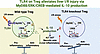
Surgical stress, such as hepatic ischemia-reperfusion (I/R) injury, induces excessive inflammation and adversely affects liver surgery outcomes. Regulatory T cells (Tregs) are crucial for immune homeostasis, yet their protective mechanisms against liver I/R injury remain unclear. In this study, we demonstrated that decreased hepatic Treg abundance correlates with increased liver injury in patients undergoing hepatic hemangioma resections. In murine models, Treg depletion worsened liver I/R injury. Bulk RNA-seq of hepatic Tregs showed enrichment of Toll-like receptor (TLR) signaling pathways, with flow cytometry identifying TLR4 as the most increased TLR after I/R. Treg-specific Tlr4 knockout mice (Treg-Tlr4–/– mice) exhibited exacerbated liver injury following I/R. Adoptive transfer of WT Tregs, but not Tlr4-deficient Tregs, alleviated liver injury in both Treg-depleted and Treg-Tlr4–/– mice. Transcriptomic analysis revealed that IL-10 production was impaired in Tlr4-deficient Tregs. Mechanistically, Tlr4-deficient Tregs showed reduced activation of the MyD88/ERK/CREB pathway, resulting in diminished IL-10 production. Myd88–/– and IL-10–/– Tregs failed to confer protection against liver I/R injury, whereas exogenous IL-10 administration rescued the hepatic dysfunction in Treg-Tlr4–/– mice. Our findings implicate the vital role of TLR4 in Tregs to mitigate liver I/R injury and offer a potential therapeutic option to reduce postoperative complications following liver surgery.
Authors
Hongji Zhang, Yunwei Zhang, Tianxing Ren, Carolyn Tsung, Peng Song, Peng Xu, Guoliang Wang, Chunyan Cao, Changyan Wang, Ping Sun, Qi Zhang, Yanhong Zhu, Xin Zhong, Yong Guan, Xiaofei Zhang, Han Wang, Jinxiang Zhang, Hui Wang
Abstract
Metabolic syndrome and excessive alcohol consumption (MetALD) result in liver injury and fibrosis, which is driven by increased collagen production by activated hepatic stellate cells (HSCs). Our previous studies demonstrated that LARP6, an RNA-binding protein, may facilitate collagen production. However, the expression and function of LARP6 as a regulator of fibrosis development in a disease-relevant model remain poorly understood. We demonstrated that LARP6 was upregulated in human activated HSCs in metabolic dysfunction-associated steatohepatitis (MASH) and MetALD. By using snRNA/ATAC-sequencing, we showed that JUNB upregulated LARP6 expression in activated HSCs. Moreover, LARP6 knockdown in human HSCs suppressed fibrogenic gene expression. By integrating eCLIP analysis and ribosome profiling in HSCs, we showed that LARP6 interacted with mature mRNAs comprising over 300 genes, including RNA structural elements within COL1A1, COL1A2, and COL3A1 to regulate mRNA expression and translation. IP-MS analysis demonstrated LARP6 protein–protein interactions with mRNA translation components and the actin cytoskeleton. Furthermore, dsiRNA-based HSC-specific gene knockdown or pharmacological inhibition of LARP6 attenuated fibrosis development in human MASH and MetALD liver spheroids. Our results suggest LARP6 plays a key role in fibrogenic gene regulation and that targeting LARP6 in human HSCs may represent a therapeutic approach for liver fibrosis.
Authors
Hyun Young Kim, Orel Mizrahi, Wonseok Lee, Sara B. Rosenthal, Cuijuan Han, Brian A. Yee, Steven M. Blue, Jesiel Diaz, Jyotiprakash P. Jonnalagadda, Lena A. Street, Kanani Hokutan, Haeum Jang, Charlene Miciano, Chen-Ting Ma, Andrey A. Bobkov, Eduard Sergienko, Michael R. Jackson, Marko Jovanovic, Branko Stefanovic, Tatiana Kisseleva, Gene W. Yeo, David A. Brenner
Abstract
BACKGROUND. MASLD has a substantial inherited component. Rare variants in Apolipoprotein B gene (APOB) have been implicated in susceptibility to liver steatosis, but their role in disease progression and outcomes is unclear. METHODS. We investigated APOB rare variants in a case-control cohort of people with advanced MASLD vs. healthy controls (n = 510/261), a family-based study (n = 43 and literature meta-analysis), the Million Veteran Program cohort (MVP, n = 94,885) and the UK Biobank (UKBB, n = 417,657). RESULTS. In the clinical cohort, APOB variants were enriched in people with advanced MASLD (OR 13.8, 95% c.i. 2.7-70.7, P = 0.002) and associated with lower circulating lipids, but higher MASLD activity and fibrosis (P < 0.05). In the family study, APOB variants segregated with hepatic steatosis and fibrosis (P < 0.05). Cross-ancestry meta-analysis of the study cohorts yielded pooled ORs for cirrhosis and hepatocellular carcinoma of 1.82, 95% c.i. 1.33-2.49 and 3.53, 95% c.i. 2.09-5.98, respectively. Variants affecting specifically ApoB100 had a three-fold greater impact on hepatic lipid metabolism compared to those impairing also ApoB48 and were specifically protective against coronary artery disease (P < 0.05). Variants affected cirrhosis risk similarly, but ApoB48/100 had a larger impact on hepatocellular carcinoma (P < 0.05). CONCLUSIONS. Rare APOB variants predispose to advanced MASLD and HCC, with distinct contributions from disrupted VLDL and chylomicrons secretion. These findings highlight the interplay between hepatic and intestinal lipid handling, suggesting that APOB genotyping may enhance MASLD risk stratification and case identification. FUNDING. European Union, Italian Ministry of Health, Swedish Research Council, Veteran health administration, NIH.
Authors
Matteo Mureddu, Serena Pelusi, Oveis Jamialahmadi, Marijana Vujkovic, Lorenzo Miano, Hadi Eidgah Torghabehei, Luisa Ronzoni, Francesco Malvestiti, Marco Saracino, Giulia Periti, Vittoria Moretti, Craig C. Teerlink, Julie A. Lynch, Philip S. Tsao, Josephine P. Johnson, Vincenzo La Mura, Robertino Dilena, Saleh A. Alqahtani, Alessandro Cherubini, Francesco Paolo Russo, Roberta D'Ambrosio, Mirella Fraquelli, Salvatore Petta, Luca Miele, Umberto Vespasiani-Gentilucci, Elisabetta Bugianesi, Rosellina M. Mancina, Paolo Parini, Daniele Prati, Kyong-Mi Chang, Carolin V. Schneider, Stefano Romeo, Luca V.C. Valenti
Abstract
Vessels encapsulating tumor clusters (VETC), a distinct vascular pattern in hepatocellular carcinoma (HCC), facilitates non-invasive metastasis in whole cluster. The interaction between VETC and tumor microenvironment requires exploration. Here, we found that compared to human Non-VETC-HCCs, VETC-tumors exhibited more PD1+CD8+ T cells and Tregs, especially TNFRSF4+Tregs and Ki67+Tregs which showed increased immunosuppressive and proliferative activity. Such immunosuppressive status was also detected in tumor emboli of VETC-HCCs, and Treg density in emboli was positively associated with metastatic cell proliferation. VETC-HCCs revealed abundance correlation, closer spatial proximity, and stronger immunosuppressive ligand-receptor interactions between TNFRSF4+Tregs/Ki67+Tregs and PD1+CD8+ T cells. Depleting Tregs in mice reduced PD1+CD8+ T cells in primary lesions, tumor emboli and metastatic foci of VETC-allografts, and attenuated allograft metastasis. TGF-β1 levels were upregulated in endothelial cells of VETC-HCCs and associated with TNFRSF4+Tregs/Ki67+Tregs enrichment. Disrupting VETC formation decreased endothelial TGF-β1 expression, and reduced TNFRSF4+Tregs/Ki67+Tregs, PD1+CD8+ T cells, Treg/CD8+ T cells ratio. Collectively, VETC may enhance Tregs’ activity via TGF-β1, while Tregs promote and sustain CD8+ T cell exhaustion through immune inhibitory ligand-receptor interaction, thereby shaping immunosuppressive microenvironment and enabling tumor cluster to carry such niche to disseminate. These findings disclose mechanisms of tumor immune microenvironment formation and provide rationales for precision medicine.
Authors
Bi-Yu Huang, Zheng-Qi Mi, Xiao-Yu Zhang, Yu-Chen Ji, Meng-Zhi Wu, Zi-Feng Cheng, Chen Xie, Shuai He, Jing Zhu, Jian-Hong Fang, Chong Wu, Bin-Kui Li, Yun-Fei YUAN, Limin Zheng, Shi-Mei Zhuang

Abstract
Calorie restriction (CR) extends maximal lifespan and maintains cellular homeostasis in various animal models. We have previously shown that CR induces a global reduction of protein fractional synthesis rates (FSRs) across the hepatic proteome in mice, but the timing and regulatory mechanisms remain unclear. Nitric oxide (NO), a bioactive molecule upregulated during CR, is a potential regulator of protein synthesis. To explore the role of NO in hepatic proteome fluxes during CR, we used in vivo deuterium labeling from heavy water and liquid chromatography/mass spectrometry–based (LC/MS-based) flux proteomics in WT and NO-deficient (NO–) mice. We observed a transition to reduced global protein FSRs that occurred rapidly between days 25 and 30 of CR. NO deficiency, whether genetic or pharmacological, disrupted the slowing of proteome-wide fluxes and the beneficial effects on body composition and physiology. Administering the NO donor molsidomine restored the reduction in hepatic FSRs in NO– mice. Furthermore, inhibiting NO pharmacologically, whether starting on day 1, day 14, or day 24 of CR, mitigated the reduction in hepatic protein FSRs at day 32, highlighting NO’s critical role during the transition period. These results underscore the importance of NO in CR-induced changes in proteostasis and suggest NO as a potential CR-mimetic target, while offering a specific time window for identifying other signals and testing therapeutic interventions.
Authors
Hector H. Palacios, Edward Cao, Adelaide Cahill, Hussein Mohamad, Marc K. Hellerstein
Abstract
Mitochondrial fission is mediated by dynamin-related protein 1 (gene name DNM1L) and fusion by mitofusins (MFN1 and MFN2) and optic atrophy 1 (OPA1). The role of mitochondrial dynamics in liver disease and cancer remains poorly understood. We used single, double, and triple liver-specific knockout (KO) mice lacking mitochondrial fission and fusion proteins, along with systematic analyses of mitochondrial morphology, untargeted metabolomics, RNA sequencing, hydrodynamic tail vein injection of oncogenes, and human hepatocellular carcinoma samples. Liver-specific Dnm1l KO (L-Dnm1l) mice showed increased ALT levels and hepatic fibrosis, with spontaneous liver tumors developing by 12 to 18 months of age. L-Mfn1 KO and L-Mfn2 KO mice showed no significant liver damage or tumor development, although a small percentage of double knockout (DKO) mice developed tumors. Triple knockout of Dnm1l, Mfn1, and Mfn2 (TKO) mice experienced significantly reduced liver injury and fibrosis, along with decreased spontaneous and oncogene-induced tumorigenesis. L-Dnm1l KO mice showed increased activation of the cGAS-STING-interferon pathway and pyrimidine metabolism, which were significantly normalized in TKO mice. Deletion of hepatic cGas reduced both basal and oncogene-induced liver injury and tumor development in L-Dnm1l KO mice. These findings indicate that mitochondrial dynamics are crucial for maintaining hepatic pyrimidine metabolism and regulating the cGAS-STING-mediated immune response to prevent liver tumorigenesis.
Authors
Xiaowen Ma, Xiaoli Wei, Mengwei Niu, Chen Zhang, Zheyun Peng, Wanqing Liu, Junrong Yan, Xiaoyang Su, Lichun Ma, Shaolei Lu, Wei Cui, Hiromi Sesaki, Wei-Xing Zong, Hong-Min Ni, Wen-Xing Ding

Abstract
Metabolic dysfunction–associated steatotic liver disease–induced (MASLD-induced) hepatocellular carcinoma (HCC) is an emerging malignancy linked to excessive accumulation of adipose tissue and hepatic fat. Understanding the role of adipocytes in the development of MASLD-induced HCC is crucial. In an in vitro coculture system, differentiated adipocytes were found to enhance cancer stemness and drug resistance in HCC through paracrine signaling. Fatty acid–binding protein 4 (FABP4) was preferentially secreted by adipocytes, and recombinant FABP4 further augmented the cancer stem cell (CSC) properties of HCC cells. Notably, Fabp4–/– mice exhibited a marked delay in the progression of MASLD-HCC, which correlated with the increased HCC risk observed in MASLD patients with elevated FABP4 expression. Mass spectrometry analysis identified integrin β 1 (ITGB1) as a binding partner of FABP4. These data, together with a substantial downregulation of the Wnt/β-catenin pathway in Fabp4–/– mouse tumors, revealed that FABP4 augmented liver CSC functions by activating PI3K/AKT/β-catenin signaling via ITGB1. We developed an anti-FABP4 neutralizing antibody that successfully inhibited FABP4-driven CSC functions and suppressed MASLD-induced HCC. In conclusion, our findings indicate that adipocyte-derived FABP4 plays a critical role in the development of MASLD-induced HCC and targeting the ITGB1/PI3K/AKT/β-catenin signaling cascade may offer a promising approach to combat this aggressive disease.
Authors
Carmen Oi Ning Leung, Shilpa Gurung, Katherine Po Sin Chung, Rainbow Wing Hei Leung, Martina Mang Leng Lei, Mandy Sze Man Chan, Gregory Kenneth Muliawan, Shakeel Ahmad Khan, Xue Qian Wu, Jun Yu, Hui Lian Zhu, Yin Ying Lu, Stephanie Ma, Xiaoping Wu, Ruby Lai Chong Hoo, Terence Kin Wah Lee
Abstract
Emerging evidence demonstrates that chronic stress alters immunological, neurochemical and endocrinological functions, thereby promoting tumor progression. However, the underlying metabolic mechanism of chronic stress in tumor progression is still elusive. Using multi-omics analysis, we found that aminopeptidase N (ANPEP) was upregulated in tumors with chronic restraint, associating with the reprogramming of amino acid metabolism. Functional assays revealed that ANPEP promoted liver cancer growth and metastasis. Knockdown of ANPEP blocked chronic stress-induced liver cancer progression. Chronic stress-induced glucocorticoids promoted nuclear receptor subfamily 3 group C member 1 (NR3C1) nuclear translocation to activate ANPEP transcription by directly binding to its promoter. Furthermore, ANPEP promotes glutathione synthesis, subsequently inhibiting reactive oxygen species (ROS)-induced ferroptosis. Mechanistically, ANPEP interacted with solute carrier family 3 member 2 (SLC3A2) to block membrane associated ring-CH-type finger 8-mediated (MARCH8-mediated) lysosome-dependent degradation of SLC3A2, promoting intracellular L-cystine transport, thereby increasing glutathione synthesis. The combination of ANPEP silencing and sorafenib treatment showed a synergistic effect in inhibiting liver cancer progression. Finally, clinical data and mouse models demonstrated that chronic stress drove liver tumor progression via ANPEP-regulated SLC3A2. These findings reveal unanticipated communication between chronic stress and metabolic reprogramming during liver cancer progression, providing potential therapeutic implications for liver cancer.
Authors
Yongkang Wu, Yankun Zhang, Xiaojia Shi, Mengting Wu, Min Sun, Ying Feng, Wenmeng Ma, Xiule Jiang, Dingqi Fei, Mingjian Zhao, Zhuanchang Wu, Chunyang Li, Xiaohong Liang, Lifen Gao, Chunhong Ma, Xuetian Yue
Abstract
Chronic inflammation leads to tissue fibrosis which can disrupt the function of the parenchyma of the organ and ultimately lead to organ failure. The most prevalent form of this occurs in chronic hepatitis which leads to liver fibrosis and, ultimately, cirrhosis and hepatic failure. Although there is no specific treatment for fibrosis, the phosphodiesterase 4 (PDE4) competitive inhibitors have been shown to ameliorate fibrosis in rodent models. However, competitive inhibitors of PDE4 have shown significantly reduced effectiveness due to severe gastrointestinal side effects. The PDE4 family is composed of four genes (PDE4A–D) with each having up to 9 differentially spliced isoforms. Here, we report that PDE4D expression is specifically elevated during the hepatic fibrosis stage of liver disease progression. Furthermore, the expression of the long isoforms of PDE4D is selectively elevated in activated hepatic stellate cells, leading to the enhanced accumulation of extracellular matrix components. In a mouse model of liver fibrosis, genetic ablation of PDE4D or pharmacological inhibition using D159687, a selective allosteric inhibitor targeting the long isoforms of PDE4D, suppresses the expression of inflammatory and profibrogenic genes. These findings establish the long isoforms of PDE4D as key drivers of liver fibrosis and highlight their potential as therapeutic targets to ameliorate liver fibrosis.
Authors
Jeonghan Kim, Heeeun Yoon, Seoung Chan Joe, Antoine Smith, Jinsung Park, Geunhye Hong, Ji Myeong Ha, Eun Bae Kim, Ekihiro Seki, Myung K. Kim, Hae-Ock Lee, Ho-Shik Kim, Jay H. Chung

Abstract
Both adipocytes and hepatocytes have the capacity to store fat, but the factor(s) that determine fat distribution between these cell types remain unknown. In mice fed a high-fat diet, fat initially accumulates predominantly in adipocytes, while hepatic fat accumulation mainly emerges after the onset of epididymal adipocyte death that results in elevated free fatty acids to promote lipid accumulation in hepatocytes. However, it remains unclear whether other signals after adipocyte death are required to direct and/or promote hepatocytes to store fat and subsequently trigger metabolic dysfunction–associated steatotic liver disease (MASLD, formerly known as nonalcoholic fatty liver disease). Using genetically modified mouse models combined with bulk and single-cell RNA-Seq analysis, we demonstrated that visceral adipocyte death induced an accumulation of S100A8+ macrophages in the liver, which was partially induced by fatty acids and apoptotic adipocyte–derived extracellular vesicles. Macrophage-specific deletion of the S100a8 gene reduced hepatic fat accumulation and MASLD severity in mice. Mechanistically, S100A8+ macrophages suppressed cellular communication network factor 3 (CCN3), a negative regulator of CD36, thereby enhancing CD36 expression in hepatocytes. In conclusion, adipocyte death promotes hepatic infiltration of S100A8+ macrophages, which drive hepatocyte lipid storage and subsequently promote MASLD progression through CD36 upregulation, partially mediated by CCN3 suppression.
Authors
Yukun Guan, Yeonsoo Kim, Yang Wang, Ye Eun Cho, Xiaogang Xiang, Seung-Jin Kim, Tiantian Yao, Dechun Feng, Seonghwan Hwang, Bin Gao


Copyright © 2026 American Society for Clinical Investigation
ISSN: 0021-9738 (print), 1558-8238 (online)