Review

Abstract
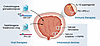
Nearly two-thirds of patients with Alzheimer disease (AD) are women, most of them postmenopausal. While sex differences in AD have historically been attributed to women’s relative longevity, accumulating evidence challenges that view, pointing to female sex–specific biological underpinnings. In particular, neuroendocrine aging and the hormonal shifts that accompany the menopause transition have emerged as potentially modifiable AD risk factors in women. Yet, key neuroendocrine aging-related factors linked to increased AD and dementia risk, such as early menopause, premenopausal bilateral oophorectomy, frequent vasomotor symptoms, and midlife cognitive and mood disturbances, remain underinvestigated. Additionally, although a growing evidence base highlights the potential of menopause hormone therapy for AD prevention, particularly in women undergoing oophorectomy, progress remains hindered by a lack of clinical trials and biomarker-driven studies. This Review calls for a paradigm shift: from viewing AD risk as a byproduct of generalized aging to validating midlife neuroendocrine aging as a distinct window of vulnerability, and an opportunity for prevention. By 2050, over 1.2 billion women worldwide will be in or approaching menopause. The stakes are global, and the opportunity is urgent: to redefine AD prevention through sex-specific, time-sensitive, and biologically informed strategies that translate science into scalable, actionable care.
Authors
Lisa Mosconi

Abstract
Amyotrophic lateral sclerosis (ALS) and frontotemporal dementia (FTD) are progressive neurodegenerative diseases characterized by the nuclear clearance and cytoplasmic aggregation of transactive response DNA/RNA-binding protein of 43 kDa (TDP43). Alternative splicing of TARDBP, the gene encoding TDP43, leads to a surprising diversity of RNA and protein isoforms with unique functions and potential implications for disease pathogenesis. Here, we review the production, properties, and functional consequences of alternative splicing in the development of ALS and FTD, focusing primarily on TDP43 due to its integral connection with the pathogenesis of sporadic as well as familial forms of these diseases. We synthesize current evidence on the biology of alternative TARDBP splicing, highlight key questions regarding its role in TDP43 proteinopathies such as ALS and FTD, and touch on the larger phenomenon of alternative splicing and its relationship to disease.
Authors
Morgan R. Miller, Megan Dykstra, Sami Barmada

Abstract
Neurodegenerative diseases are characterized by protein misfolding and the selective vulnerability of specific neuronal subtypes. This selective vulnerability presents a paradox; most neurodegenerative disease genes are expressed broadly throughout the brain, and some ubiquitously, but only certain types of neurons are lost while others are resistant. The molecular basis for selective neuronal vulnerability has remained a mystery, but recent genomics technological innovations are starting to provide mechanistic insights. Here, we review how single-cell genomics techniques — single-cell transcriptomics, single-cell epigenomics, and spatial transcriptomics — advance our molecular understanding of selective vulnerability and neurodegeneration across Alzheimer disease, Parkinson disease, amyotrophic lateral sclerosis, frontotemporal dementia, and Huntington disease. Together, these approaches reveal the cell types affected in disease, define disease-associated molecular states, nominate candidate determinants of vulnerability and degeneration, and situate degenerating neurons within their local tissue context. Continued development and application of these techniques, including single-cell perturbation screens, will expand descriptive atlases of relevant cell types in health and disease and identify causal mechanisms, revealing the molecular basis of vulnerability and degeneration and informing therapeutic development.
Authors
Olivia Gautier, Thao P. Nguyen, Aaron D. Gitler
Abstract
Urothelial cancers of the urinary tract are the fourth most common malignancy in men, with a shifting demographic affecting younger patients and an increasing incidence in females. In this Review, we discuss recent discoveries and paradigm-shifting clinical trials that impact all stages of urothelial cancer. New therapeutics and drug-delivery devices have led to multiple approvals for treatments of non-muscle invasive bladder cancer. The addition of chemotherapy, immunotherapy, and antibody-drug conjugates is transforming perioperative treatment for patients with muscle-invasive bladder cancer. The use of liquid biomarkers, circulating tumor DNA, and urinary tumor DNA is aiding the identification of patients at risk for local recurrence and possibly those who can avoid systemic therapy. Finally, integrating biomarkers and systemic treatments is creating a paradigm that could lead to the successful treatment of bladder cancer without requiring bladder removal. Overall, these advancements in biomarkers and novel therapeutics are likely to dramatically improve survival for bladder cancer.
Authors
Joshua J. Meeks
Abstract
Diabetic retinopathy (DR), the most common microvascular complication in patients with diabetes mellitus (DM), is a leading cause of vision loss worldwide. Sustained hyperglycemia plays a central role in promoting DR. However, tight glycemic control does not prevent — and indeed sometimes worsens — DR, highlighting the importance of ongoing studies aimed at improving our understanding of this complex disease. Over the last few decades, the dogma that DR is a vascular disease that results in secondary neuronal injury has evolved, as emerging evidence suggests that neurodegeneration occurs in parallel with or prior to vascular cell injury in the retina of patients with DM. This has led to appreciation of DR as a neurovascular disease, characterized by microvascular injury and neurodegeneration, both of which contribute to vision loss. Here, we explore how molecular stress (i.e., glucose dysregulation, dysmetabolism, oxidative stress, and inflammation) promote retinal vascular cell and neuronal injury in patients with DM. We focus on how these processes influence, and are influenced by, genes regulated by the HIF family of transcription factors in glial, vascular, neuronal, and inflammatory cells, with the goal of identifying new therapeutic avenues for the prevention or early treatment of patients with this vision-threating disease.
Authors
Chuanyu Guo, Akrit Sodhi

Abstract
GLP-1 receptor agonist (GLP-1RA) medications have transformed the treatment of type 2 diabetes (T2D) and obesity, with robust evidence for cardiovascular and renal benefits. Nevertheless, GLP-1RA therapy is associated with a pattern of adverse events affecting their safety and tolerability. Here, we delineate mechanisms potentially leading to adverse responses to GLP-1RAs, describe the impact of side effects on treatment persistence, discuss potential mitigation strategies, and identify areas requiring further studies. Concerns that GLP-1RAs raise the risk for acute pancreatitis and pancreatic cancer have been dispelled by long-term clinical trials. However, GLP-1RAs may confer an increased risk for thyroid cancer. Sight-threatening eye complications resulting from rapid reductions in glycemia may be avoided by retinal screening and ophthalmologic treatment before GLP-1RA initiation. The slowing of gastric emptying with GLP-1RA treatment increases the propensity for retained gastric contents, which could increase the risk of aspiration during upper gastrointestinal endoscopy or general anesthesia. These risks may, however, be elevated in individuals with long-standing T2D even in the absence of GLP-1RA treatment. Improved pharmacovigilance and a more standardized, quantitative assessment of adverse events in clinical trials, particularly in the assessment of gastrointestinal symptoms, would facilitate definition of the benefit-risk relationship for individual medications and indications.
Authors
Ryan J. Jalleh, Nicholas J. Talley, Michael Horowitz, Michael A. Nauck

Abstract
The incretin hormone glucagon-like peptide-1 (GLP-1) exerts potent effects on glucose metabolism, prompting the development of therapeutic strategies that enhance activity of the GLP-1 receptor (GLP-1R) pathway. Inhibitors of dipeptidyl peptidase 4 (DPP-4) prolong the half-life of endogenous GLP-1 and typically achieve reductions in HbA1c of 0.5%–0.8%. However, large-scale cardiovascular (CV) outcomes trials (CVOTs) with DPP-4 inhibitors demonstrated CV safety but did not show a reduction in CV events. A second incretin-based therapeutic approach was the development of GLP-1R agonists (GLP-1RAs). Various GLP-1RAs, including liraglutide, semaglutide, and dulaglutide, demonstrated a reduction in CV outcomes in large CVOTs. Initially, these medications were only available as injectable agents for subcutaneous administration, but recent technological advancements have enabled the development of orally available GLP-1RAs. A third incretin-based approach is tirzepatide, a dual agonist of GLP-1R and glucose-dependent insulinotropic polypeptide receptor (GIPR), which achieves greater HbA1c reduction and weight loss compared with GLP-1RAs alone. Ongoing large-scale CVOTs will determine its effects on hard cardiovascular endpoints. This Review summarizes the effects of GLP-1 and GLP-1RAs in the CV system as well as clinical data of GLP-1RAs in individuals with CV disease or high CV risk.
Authors
Florian Kahles, Andreas L. Birkenfeld, Nikolaus Marx

Abstract
Glucagon-like peptide-1 receptor agonists (GLP-1RAs), established therapies for type 2 diabetes and obesity, are increasingly recognized for their potential in neurodegenerative diseases. Preclinical studies across diverse neurodegenerative conditions consistently demonstrate neuroprotective effects of GLP-1RAs, including reduced protein aggregation, enhanced autophagy, improved mitochondrial function, suppression of neuroinflammation, and preservation of synaptic integrity. Epidemiological analyses further suggest reduced incidence of dementia, Parkinson disease, and multiple sclerosis among long-term GLP-1RA users. Early human trials provide signals of target engagement, such as preserved cerebral glucose metabolism, altered inflammatory biomarkers, and slowed brain atrophy, although clinical outcomes to date remain mixed and trials in rarer disorders are sparse. Translation is constrained by uncertainty around optimal molecule choice, CNS penetrance, tolerability, adherence, and heterogeneity of response. Furthermore, next-generation dual and triple agonists may offer enhanced efficacy but remain untested in neurodegeneration. Conceptually, GLP-1RAs share pleiotropic effects with exercise — one of the few interventions with proven disease-modifying potential — by enhancing insulin signaling, stabilizing mitochondria, reducing inflammation, and promoting synaptic plasticity. This overlap highlights their promise as “pharmacological analogues of exercise,” and underscores the need for biomarker-driven, disease-specific trials to establish whether GLP-1RAs can deliver durable disease modification across the spectrum of neurodegenerative diseases.
Authors
Dilan Athauda, Nigel H. Greig, Wassilios G. Meissner, Thomas Foltynie, Sonia Gandhi

Abstract
Circadian clocks govern daily rhythms in cellular and physiological processes, including cell cycle, DNA repair, metabolism, and immune function, that influence cancer development and treatment response. Disruption of circadian regulators either promotes or suppresses malignancy depending on tumor type and biological context. This duality likely reflects systemic rewiring of circadian physiology and direct interactions between clock components and oncogenic pathways. These insights hold clinical relevance for the field of chronotherapy, which seeks to enhance therapeutic efficacy and minimize toxicity by aligning drug administration with circadian rhythms or by targeting elements of the molecular clock. In this Review, we highlight the promise of integrating circadian biology into precision oncology and underscore the importance of cancer type–specific investigations to harness the full therapeutic potential of chronotherapy in cancer.
Authors
Rebecca M. Mello, Selma Masri, Katja A. Lamia

Abstract
Metastatic hormone-sensitive prostate cancer (mHSPC) is a clinically and molecularly heterogeneous disease. Recent insights into the biology underlying disease presentation, volume of disease, and response to therapies are starting to point toward biomarkers to improve selection for intensified and deintensified treatment strategies. In addition, the therapeutic landscape is rapidly changing, with new biomarker-driven studies targeting genotype (e.g., BRCA or PTEN mutant) and phenotype (e.g., prostate-specific membrane antigen status) in development for mHSPC. A better understanding of tumor heterogeneity, clonal evolution, and metastatic homing in prostate cancer will hopefully inform future strategies for local and systemic disease control, personalized monitoring strategies, and improved patient outcomes.
Authors
Alice Bernard-Tessier, Himisha Beltran
No posts were found with this tag.


Copyright © 2026 American Society for Clinical Investigation
ISSN: 0021-9738 (print), 1558-8238 (online)