
Advertisement
Commentary
Open Access |  10.1172/JCI210439
Insulin sensitivity and mitochondrial homeostasis connect lactation to maternal metabolic health
Laura L. Hernandez
Department of Animal and Dairy Sciences, Affiliate Department of Obstetrics and Gynecology, University of Wisconsin-Madison, Madison, Wisconsin, USA.
Address correspondence to: Laura L. Hernandez, 1675 Observatory Dr., Madison, Wisconsin, 53706, USA. Email: llhernandez@wisc.edu.
Find articles by
Hernandez, L.
in:
PubMed
|
Google Scholar
|

Published October 15, 2026 - More info
J Clin Invest. 2026;136(20):e210439. https://doi.org/10.1172/JCI210439.
© 2026 Hernandez This work is licensed under the Creative Commons Attribution 4.0 International License. To view a copy of this license, visit http://creativecommons.org/licenses/by/4.0/.

Related article:
Abstract
Insulin resistance (IR) has emerged as a risk factor for lactation insufficiency and delays the onset of milk secretion after childbirth, termed secretory activation (SA). This may cause inadequate infant weight gain and early breastfeeding cessation. However, the mechanisms underlying delayed SA in insulin-resistant women are unknown. To investigate this, we characterized the mammary transcriptomes and IR-related hormones of 75 breastfeeding women with healthy term infants during postpartum days 1–5. Participants were divided into IR tertiles based on plasma leptin-to-adiponectin ratio measurements. Those in the highest tertile had later SA onset with greater neonatal weight loss during postpartum days 1–5. Transcriptomic analysis on postpartum day 2 (n = 4 high IR vs. n = 8 low IR participants) showed transient suppression of mammary insulin and prolactin signaling genes, increased pro-inflammatory gene expression, and altered expression of more than 200 mammary mitochondrial genes. These alterations were absent on postpartum days 3–5. Cultured mammary epithelial cells treated with insulin showed upregulation of prolactin signaling and oxidative phosphorylation (OXPHOS) genes, with imaging and bioenergetic studies demonstrating that insulin promotes mitochondrial biogenesis and OXPHOS. Thus, our findings delineate roles for insulin in mammary bioenergetics and highlight mitochondrial dysfunction as a mechanism for delayed SA in insulin-resistant women.
Authors
Xin Meng, Taha Elajnaf, Hussam Rostom, Michelle Ma, Annalee Furst, Bryony R. Davies, Isabella R. Honess, Gaurav Pandey, Isadora C. Furigo, Craig L. Doig, Jayne F. Martin Carli, Rajesh V. Thakker, Lars Bode, Kelsey E. Johnson, Fadil M. Hannan
-
Insulin resistance and milk production
Meng et al. provided compelling evidence that insulin signaling is a critical regulator of human milk production and identified mitochondrial dysfunction as a potential mechanistic link between maternal insulin resistance and impaired lactation (14). Because fasting blood samples are difficult to obtain during late pregnancy, human participants were categorized based on their plasma leptin/adiponectin ratio (LAR) at 36 weeks of gestation as a measure of insulin resistance. Women with higher LAR exhibited delayed onset of lactogenesis as measured by increased milk sodium and decreased lactose concentrations. Integrating analyses of human milk samples with mechanistic studies in human and rodent mammary epithelial cell models, the authors demonstrated that insulin regulates mammary epithelial cell function through the AKT signaling pathway and identified widespread alterations in mitochondrial metabolism that were associated with insulin resistance (Figure 1A).
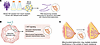 Figure 1
Investigating the association between insulin resistance and insufficient glandular tissue and low milk supply. (A) Meng et al. (14) used RNA extraction of the milk fat globule to look at gene expression in mammary epithelial cells in pregnant women who were categorized as insulin resistant at 36 weeks’ gestation. Women with increased insulin resistance exhibited increased expression of pathways related to mitochondrial dysfunction compared with insulin-sensitive participants. (B) In human and mouse mammary epithelial cells, treatment with high concentrations of insulin increased AKT signaling. The authors also observed increases in oxidative phosphorylation, extracellular acidification, glycolytic capacity, and oxygen consumption consistent with disruptions in mitochondrial homeostasis as well as a reduction in mitochondrial volume and increased mitochondrial fragmentation. (C) Together, the findings indicate that mitochondrial dysfunction links insulin resistance to lactation insufficiency.
A notable strength of the study is its use of bulk RNA sequencing of milk fat globules (MFGs), which are a source for studying gene expression in mammary epithelial cells (15, 16). This approach enabled the authors to define transcriptional signatures associated with maternal insulin resistance and subsequently leverage these data to conduct mechanistic studies in mammary epithelial cell cultures. While this approach is informative and practical, MFG data are not always fully representative of the functional lactating mammary gland, which comprises a complex set of cell types, as observed in bovine, hamster, and mouse models (17, 18). To date, studies have not confirmed the transcriptomic signatures of MFGs and breast tissue during lactation in women; the possible interactions of other cell types on LMS and IGT have yet to be explored.
To establish causality, the authors complemented their human MFG studies with experiments in mammary epithelial cell lines (14). Using human MCF-12A cells, an immortalized cell line representative of luminal epithelial cells, they demonstrated that AKT signaling is a major regulator of oxidative phosphorylation, supporting a central role for insulin in maintaining mitochondrial function in mammary tissue. While these findings suggest that AKT is critical for regulating insulin’s action, a limitation of the experiments is that they do not examine these findings under differentiated, lactogenic conditions, which are more representative of the biological time frame of interest. The authors extended their RNA-level observations in the MCF12A cell line and milk MFG samples by varying insulin concentrations in the mouse mammary epithelial cell line HC11. They observed increases in extracellular acidification, glycolytic capacity, and oxygen consumption, as measured by cell respiration assays. Notably, exposure to high insulin concentrations induced mitochondrial fragmentation and reduced mitochondrial volume, providing additional evidence that chronic insulin exposure alters mitochondrial homeostasis in mammary cells (Figure 1B). Future work should examine these findings in a human mammary epithelial cell model and possibly even primary human mammary epithelial cells to ensure repeatability of the findings from the mouse cell line.
Taken together, Meng et al.’s work identifies insulin signaling and mitochondrial metabolism as central regulators of mammary epithelial cell function and provides an important mechanistic framework linking maternal metabolic health and lactation capacity (Figure 1C). Caution is warranted when applying the hypothesis that insulin resistance is the primary cause of LMS and/or IGT, as the mammary gland comprises a heterogeneous cell population and numerous other cell types that may play crucial roles. Future studies incorporating in vivo models, primary human tissues, and emerging spatial and single-cell approaches will be essential to define how insulin signaling integrates with the broader cellular ecosystem of the mammary gland to regulate successful lactation.


Copyright © 2026 American Society for Clinical Investigation
ISSN: 0021-9738 (print), 1558-8238 (online)