Research Article
Citation Information: J Clin Invest. 2025;135(15):e186065. https://doi.org/10.1172/JCI186065.
Abstract
Platelets play a dual role in hemostasis and inflammation-associated thrombosis and hemorrhage. Although the mechanisms linking inflammation to platelet dysfunction remain poorly understood, our previous work demonstrated that TNF-α alters mitochondrial mass, platelet activation, and autophagy-related pathways in megakaryocytes. Here, we hypothesized that TNF-α impairs platelet function by disrupting autophagy, a process critical for mitochondrial health and cellular metabolism. Using human and murine models of TNF-α–driven diseases, including myeloproliferative neoplasms and rheumatoid arthritis, we found that TNF-α downregulates syntaxin 17 (STX17), a key mediator of autophagosome-lysosome fusion. This disruption inhibited autophagy, leading to the accumulation of dysfunctional mitochondria and reduced mitochondrial respiration. These metabolic alterations compromised platelet-driven clot contraction, a process linked to thrombotic and hemorrhagic complications. Our findings reveal a mechanism by which TNF-α disrupts hemostasis through autophagy inhibition, highlighting TNF-α as a critical regulator of platelet metabolism and function. This study provides potentially new insights into inflammation-associated pathologies and suggests autophagy-targeting strategies as potential therapeutic avenues to restore hemostatic balance.
Authors
Guadalupe Rojas-Sanchez, Jorge Calzada-Martinez, Brandon McMahon, Aaron C. Petrey, Gabriela Dveksler, Gerardo P. Espino-Solis, Orlando Esparza, Giovanny Hernandez, Dennis Le, Eric P. Wartchow, Ken Jones, Lucas H. Ting, Catherine Jankowski, Marguerite R. Kelher, Marilyn Manco-Johnson, Marie L. Feser, Kevin D. Deane, Travis Nemkov, Angelo D’Alessandro, Andrew Thorburn, Paola Maycotte, José A. López, Pavel Davizon-Castillo
Citation Information: J Clin Invest. 2025;135(15):e186135. https://doi.org/10.1172/JCI186135.
Abstract
Psoriatic arthritis (PsA) is a multifaceted, chronic inflammatory disease affecting the skin, joints, and entheses, and it is a major comorbidity of psoriasis. Most patients with PsA present with psoriasis before articular involvement; however, the molecular and cellular mechanisms underlying the link between cutaneous psoriasis and PsA are poorly understood. Here, we found that epidermis-specific SPRY1-deficient mice spontaneously developed PsA-like inflammation involving both the skin and joints. Excessive CXCL10 was secreted by SPRY1-deficient epidermal keratinocytes through enhanced activation of JAK1/2/STAT1 signaling, and CXCL10 blockade attenuated PsA-like inflammation. Of note, CXCL10 was found to bind to CD14, but not CXCR3, to promote the TNF-α production of periarticular CD14hi macrophages via PI3K/AKT and NF-κB signaling pathways. Collectively, this study reveals that SPRY1 deficiency in the epidermis is sufficient to drive both skin and joint inflammation, and it identifies keratinocyte-derived CXCL10 and periarticular CD14hi macrophages as critical links in the skin-joint crosstalk leading to PsA. This keratinocyte SPRY1/CXCL10/periarticular CD14hi macrophage/TNF-α axis provides valuable insights into the mechanisms underlying the transition from psoriasis to PsA and suggests potential therapeutic targets for preventing this progression.
Authors
Fan Xu, Ying-Zhe Cui, Xing-Yu Yang, Yu-Xin Zheng, Xi-Bei Chen, Hao Zhou, Zhao-Yuan Wang, Yuan Zhou, Yi Lu, Ying-Ying Li, Li-Ran Ye, Ni-Chang Fu, Si-Qi Chen, Xue-Yan Chen, Min Zheng, Yong Yang, Xiao-Yong Man
Citation Information: J Clin Invest. 2025;135(15):e188495. https://doi.org/10.1172/JCI188495.
Abstract
Enterovirus D68 (EV-D68) is associated with acute flaccid myelitis (AFM), a poliomyelitis-like illness causing paralysis in young children. However, the mechanisms of paralysis are unclear, and antiviral therapies are lacking. To better understand EV-D68 disease, we inoculated newborn mice intracranially to assess viral tropism, virulence, and immune responses. WT mice inoculated intracranially with a neurovirulent strain of EV-D68 showed infection of spinal cord neurons and developed paralysis. Spinal tissue from infected mice revealed increased levels of chemokines, inflammatory monocytes, macrophages, and T cells relative to those in controls, suggesting that immune cell infiltration influences pathogenesis. To define the contribution of cytokine-mediated immune cell recruitment to disease, we inoculated mice lacking CCR2, a receptor for several EV-D68–upregulated cytokines, or RAG1, which is required for lymphocyte maturation. WT, Ccr2–/–, and Rag1–/– mice had comparable viral titers in spinal tissue. However, Ccr2–/– and Rag1–/– mice were significantly less likely to be paralyzed relative to WT mice. Consistent with impaired T cell recruitment to sites of infection in Ccr2–/– and Rag1–/– mice, antibody-mediated depletion of CD4+ or CD8+ T cells from WT mice diminished paralysis. These results indicate that immune cell recruitment to the spinal cord promotes EV-D68–associated paralysis and illuminate potential new targets for therapeutic intervention.
Authors
Mikal A. Woods Acevedo, Jie Lan, Sarah Maya, Jennifer E. Jones, Isabella E. Bosco, John V. Williams, Megan Culler Freeman, Terence S. Dermody
Citation Information: J Clin Invest. 2025;135(15):e186599. https://doi.org/10.1172/JCI186599.
Abstract
Metastatic prostate cancer (mPC) is a clinically and molecularly heterogeneous disease. While there is increasing recognition of diverse tumor phenotypes across patients, less is known about the molecular and phenotypic heterogeneity present within an individual. In this study, we aimed to define the patterns, extent, and consequences of inter- and intratumoral heterogeneity in lethal prostate cancer. By combining and integrating in situ tissue-based and sequencing approaches, we analyzed over 630 tumor samples from 52 patients with mPC. Our efforts revealed phenotypic heterogeneity at the patient, metastasis, and cellular levels. We observed that intrapatient intertumoral molecular subtype heterogeneity was common in mPC and showed associations with genomic and clinical features. Additionally, cellular proliferation rates varied within a given patient across molecular subtypes and anatomic sites. Single-cell sequencing studies revealed features of morphologically and molecularly divergent tumor cell populations within a single metastatic site. These data provide a deeper insight into the complex patterns of tumoral heterogeneity in mPC with implications for clinical management and the future development of diagnostic and therapeutic approaches.
Authors
Martine P. Roudier, Roman Gulati, Erolcan Sayar, Radhika A. Patel, Micah Tratt, Helen M. Richards, Paloma Cejas, Miguel Munoz Gomez, Xintao Qiu, Yingtian Xie, Brian Hanratty, Samir Zaidi, Jimmy L. Zhao, Mohamed Adil, Chitvan Mittal, Yibai Zhao, Ruth Dumpit, Ilsa Coleman, Jin-Yih Low, Thomas Persse, Patricia Galipeau, John K. Lee, Maria Tretiakova, Meagan Chambers, Funda Vakar-Lopez, Lawrence D. True, Marie Perrone, Hung-Ming Lam, Lori A. Kollath, Chien-Kuang Cornelia Ding, Stephanie Harmon, Heather H. Cheng, Evan Y. Yu, Robert B. Montgomery, Jessica E. Hawley, Daniel W. Lin, Eva Corey, Michael T. Schweizer, Manu Setty, Gavin Ha, Charles L. Sawyers, Colm Morrissey, Henry Long, Peter S. Nelson, Michael C. Haffner
Citation Information: J Clin Invest. 2025;135(15):e173308. https://doi.org/10.1172/JCI173308.
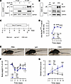
MuSK cysteine-rich domain antibodies are pathogenic in a mouse model of autoimmune myasthenia gravis
Abstract
The neuromuscular junction (NMJ), a synapse between the motor neuron terminal and a skeletal muscle fiber, is crucial throughout life in maintaining the reliable neurotransmission required for functional motricity. Disruption of this system leads to neuromuscular disorders, such as autoimmune myasthenia gravis (MG), the most common form of NMJ disease. MG is caused by autoantibodies directed mostly against the acetylcholine receptor (AChR) or the muscle-specific kinase MuSK. Several studies report immunoreactivity to the Frizzled-like cysteine-rich Wnt-binding domain of MuSK (CRD) in patients, although the pathogenicity of the antibodies involved remains unknown. We showed here that the immunoreactivity to MuSK CRD induced by the passive transfer of anti-MuSKCRD antibodies in mice led to typical MG symptoms, characterized by a loss of body weight and a locomotor deficit. The functional and morphological integrity of the NMJ was compromised with a progressive decay of neurotransmission and disruption of the structure of presynaptic and postsynaptic compartments. We found that anti-MuSKCRD antibodies completely abolished Agrin-mediated AChR clustering by decreasing the Lrp4-MuSK interaction. These results demonstrate the role of the MuSK CRD in MG pathogenesis and improve our understanding of the underlying pathophysiological mechanisms.
Authors
Marius Halliez, Steve Cottin, Axel You, Céline Buon, Antony Grondin, Léa S. Lippens, Mégane Lemaitre, Jérome Ezan, Charlotte Isch, Yann Rufin, Mireille Montcouquiol, Nathalie Sans, Bertrand Fontaine, Julien Messéant, Rozen Le Panse, Laure Strochlic
Citation Information: J Clin Invest. 2025;135(15):e188819. https://doi.org/10.1172/JCI188819.
Abstract
Idiopathic pulmonary fibrosis (IPF) is a fatal fibrotic lung disease characterized by impaired fibroblast clearance and excessive extracellular matrix (ECM) protein production. Wilms tumor 1 (WT1), a transcription factor, is selectively upregulated in IPF fibroblasts. However, the mechanisms by which WT1 contributes to fibroblast accumulation and ECM production remain unknown. Here, we investigated the heterogeneity of WT1-expressing mesenchymal cells using single-nucleus RNA-Seq of distal lung tissues from patients with IPF and control donors. WT1 was selectively upregulated in a subset of IPF fibroblasts that coexpressed several prosurvival and ECM genes. The results of both loss-of-function and gain-of-function studies were consistent with a role for WT1 as a positive regulator of prosurvival genes to impair apoptotic clearance and promote ECM production. Fibroblast-specific overexpression of WT1 augmented fibroproliferation, myofibroblast accumulation, and ECM production during bleomycin-induced pulmonary fibrosis in young and aged mice. Together, these findings suggest that targeting WT1 is a promising strategy for attenuating fibroblast expansion and ECM production during fibrogenesis.
Authors
Harshavardhana H. Ediga, Chanukya P. Vemulapalli, Vishwaraj Sontake, Pradeep K. Patel, Hikaru Miyazaki, Dimitry Popov, Martin B. Jensen, Anil G. Jegga, Steven K. Huang, Christoph Englert, Andreas Schedl, Nishant Gupta, Francis X. McCormack, Satish K. Madala
Citation Information: J Clin Invest. 2025;135(15):e188792. https://doi.org/10.1172/JCI188792.
Abstract
To maintain potassium homeostasis, the kidney’s distal convoluted tubule (DCT) evolved to convert small changes in blood [K+] into robust effects on salt reabsorption. This process requires NaCl cotransporter (NCC) activation by the with-no-lysine (WNK) kinases. During hypokalemia, the kidney-specific WNK1 isoform (KS-WNK1) scaffolds the DCT-expressed WNK signaling pathway within biomolecular condensates of unknown function termed WNK bodies. Here, we show that KS-WNK1 amplified kidney tubule reactivity to blood [K+], in part via WNK bodies. In genetically modified mice, targeted condensate disruption trapped the WNK pathway, causing renal salt wasting that was more pronounced in females. In humans, WNK bodies accumulated as plasma potassium fell below 4.0 mmol/L, suggesting that the human DCT experiences the stress of potassium deficiency, even when [K+] is in the low-to-normal range. These data identify WNK bodies as kinase signal amplifiers that mediate tubular [K+] responsiveness, nephron sexual dimorphism, and BP salt sensitivity. Our results illustrate how biomolecular condensate specialization can optimize a mammalian physiologic stress response that impacts human health.
Authors
Cary R. Boyd-Shiwarski, Rebecca T. Beacham, Jared A. Lashway, Katherine E. Querry, Shawn E. Griffiths, Daniel J. Shiwarski, Sophia A. Knoell, Nga H. Nguyen, Lubika J. Nkashama, Melissa N. Valladares, Anagha Bandaru, Allison L. Marciszyn, Jonathan Franks, Mara Sullivan, Simon C. Watkins, Aylin R. Rodan, Chou-Long Huang, Sean D. Stocker, Ossama B. Kashlan, Arohan R. Subramanya
Citation Information: J Clin Invest. 2025;135(15):e187992. https://doi.org/10.1172/JCI187992.
Abstract
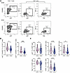
We leveraged specimens from the RV217 prospective study that enrolled participants at high risk of HIV-1 acquisition to investigate how NK cells, conventional T cells, and unconventional T cells influence HIV-1 acquisition. We observed low levels of α4β7 expression on memory CD4+ T cells and invariant NK T (iNKT) cells, 2 cell types highly susceptible to HIV-1 infection, in highly exposed seronegative (HESN) compared with highly exposed seroconverter (HESC) participants. NK cells from HESN individuals had higher levels of α4β7 than did those from HESC individuals, presented a quiescent phenotype, and had a higher capacity to respond to opsonized target cells. We also measured translocated microbial products in plasma and found differences in phylum distribution between HESN and HESC participants that were associated with the immune phenotypes affecting the risk of HIV-1 acquisition. Finally, a logistic regression model combining features of NK cell activation, α4β7 expression on memory CD4+ T cells, and T-box expressed in T cells (Tbet) expression by iNKT cells achieved the highest accuracy in identifying HESN and HESC participants. This immune signature, consisting of increased α4β7 on cells susceptible to HIV infection combined with higher NK cell activation and lower gut-homing potential, could affect the efficacy of HIV-1 prevention strategies such as vaccines.
Authors
Kawthar Machmach, Kombo F. N’guessan, Rohit Farmer, Sucheta Godbole, Dohoon Kim, Lauren McCormick, Noemia S. Lima, Amy R. Henry, Farida Laboune, Isabella Swafford, Sydney K. Mika, Bonnie M. Slike, Jeffrey R. Currier, Leigh Anne Eller, Julie A. Ake, Sandhya Vasan, Merlin L. Robb, Shelly J. Krebs, Daniel C. Douek, Dominic Paquin-Proulx, for the RV217 Study Group
Citation Information: J Clin Invest. 2025;135(15):e177601. https://doi.org/10.1172/JCI177601.
Abstract
Pancreatic islet microvasculature is essential for optimal islet function and glucose homeostasis. However, islet vessel pathogenesis in obesity and its role in the manifestation of metabolic disorders remain understudied. Here, we depict the time-resolved decline of intra-islet endothelial cell responsiveness to VEGF-A and islet vessel function in a mouse model of diet-induced obesity. Longitudinal imaging of sentinel islets transplanted into mouse eyes revealed substantial vascular remodeling and diminished VEGF-A response in islet endothelial cells after 12 weeks of Western diet (WD) feeding. This led to islet vessel barrier dysfunction and hemodynamic dysregulation, delaying transportation of secreted insulin into the blood. Notably, islet vessels exhibited a metabolic memory of previous WD feeding. Neither VEGF-A sensitivity nor the other vascular alterations was fully restored by control diet refeeding, resulting in modest yet significant impairment in glucose clearance despite normalized insulin sensitivity. Mechanistic analysis implicated hyperactivation of atypical PKC under both WD and recovery conditions, which inhibited VEGFR2 internalization and blunted VEGF-A–triggered signal transduction in endothelial cells. In summary, prolonged WD feeding causes irreversible islet endothelial cell desensitization to VEGF-A and islet vessel dysfunction, directly undermining glucose homeostasis.
Authors
Yan Xiong, Andrea Dicker, Montse Visa, Erwin Ilegems, Per-Olof Berggren
Citation Information: J Clin Invest. 2025;135(15):e189075. https://doi.org/10.1172/JCI189075.
Abstract
Gene replacement therapies mediated by adeno-associated viral (AAV) vectors represent a promising approach for treating genetic diseases. However, their modest packaging capacity (~4.7 kb) remains an important constraint and significantly limits their application for genetic disorders involving large genes. A prominent example is Duchenne muscular dystrophy (DMD), whose protein product dystrophin is generated from a 11.2 kb segment of the DMD mRNA. Here, we explored methods that enable efficient expression of full-length dystrophin via triple AAV codelivery. This method exploits the protein trans-splicing mechanism mediated by split inteins. We identified a combination of efficient and specific split intein pairs that enabled the reconstitution of full-length dystrophin from 3 dystrophin fragments. We show that systemic delivery of low doses of the myotropic AAVMYO1 in mdx4cv mice led to efficient expression of full-length dystrophin in the hind limb, diaphragm, and heart muscles. Notably, muscle morphology and physiology were significantly improved in triple-AAV–treated mdx4cv mice versus saline-treated controls. This method shows the feasibility of expressing large proteins from several fragments that were delivered using low doses of myotropic AAV vectors. It can be adapted to other large genes involved in disorders for which gene replacement remains challenged by the modest AAV cargo capacity.
Authors
Hichem Tasfaout, Timothy S. McMillen, Theodore R. Reyes, Christine L. Halbert, Rong Tian, Michael Regnier, Jeffrey S. Chamberlain
No posts were found with this tag.


Copyright © 2025 American Society for Clinical Investigation
ISSN: 0021-9738 (print), 1558-8238 (online)


