
Advertisement
Research ArticleCell biologyPulmonology
Open Access |  10.1172/JCI193367
Loss of RPGR disrupts motile cilia and causes primary ciliary dyskinesia by affecting F-actin dynamics
Yang Wu,1 Erika Tavares,2 Binrun Liang,1 Wallace B. Wee,3 Vito Mennella,4 Han-Chao Feng,1 Jiaying Cao,1 Pui Yee Wong,1 Jiayi Zheng,5 Mu He,5 Kirk AJ Stephenson,6 Liran Hanan Hochma,6 Janice Min Li,2 Nan-Peng Chen,7 Sharon D. Dell,3,8 Elise Heon,2,6 and Zhen Liu1
1Department of Life Science, The Hong Kong University of Science and Technology, Hong Kong SAR, China.
2Genetics & Genome Biology Program, and
3Child Health Evaluative Sciences, The Hospital for Sick Children, Toronto, Canada.
4Department of Biochemistry, School of Biological and Behavioural Sciences, Queen Mary University of London, London, United Kingdom.
5School of Biomedical Sciences, The University of Hong Kong, Hong Kong SAR, China.
6Department of Ophthalmology and Vision Sciences, The Hospital for Sick Children, University of Toronto, Toronto, Canada.
7Institute of Systems and Physical Biology, Shenzhen Bay Laboratory, Shenzhen, China.
8Division of Respiratory Medicine, BC Children’s Hospital, The University of British Columbia, Vancouver, Canada.
Address correspondence to: Sharon D. Dell, 4480 Oak Street, Vancouver, British Columbia, V6H3V4, Canada. Phone: 604.875.2119; Email: sharon.dell@bcchr.ca. Or to: Elise Heon, 170 Elizabeth St., Toronto, Ontario, M5G 1E8, Canada. Phone: 416.813.6525; Email: elise.heon@sickkids.ca. Or to: Zhen Liu, 1 University Road, Clear Water Bay, Kowloon, Hong Kong SAR, China. Phone: 85234692553; Email: zhenliu@ust.hk.
Find articles by Wu, Y. in: PubMed | Google Scholar
1Department of Life Science, The Hong Kong University of Science and Technology, Hong Kong SAR, China.
2Genetics & Genome Biology Program, and
3Child Health Evaluative Sciences, The Hospital for Sick Children, Toronto, Canada.
4Department of Biochemistry, School of Biological and Behavioural Sciences, Queen Mary University of London, London, United Kingdom.
5School of Biomedical Sciences, The University of Hong Kong, Hong Kong SAR, China.
6Department of Ophthalmology and Vision Sciences, The Hospital for Sick Children, University of Toronto, Toronto, Canada.
7Institute of Systems and Physical Biology, Shenzhen Bay Laboratory, Shenzhen, China.
8Division of Respiratory Medicine, BC Children’s Hospital, The University of British Columbia, Vancouver, Canada.
Address correspondence to: Sharon D. Dell, 4480 Oak Street, Vancouver, British Columbia, V6H3V4, Canada. Phone: 604.875.2119; Email: sharon.dell@bcchr.ca. Or to: Elise Heon, 170 Elizabeth St., Toronto, Ontario, M5G 1E8, Canada. Phone: 416.813.6525; Email: elise.heon@sickkids.ca. Or to: Zhen Liu, 1 University Road, Clear Water Bay, Kowloon, Hong Kong SAR, China. Phone: 85234692553; Email: zhenliu@ust.hk.
Find articles by
Tavares, E.
in:
PubMed
|
Google Scholar
|

1Department of Life Science, The Hong Kong University of Science and Technology, Hong Kong SAR, China.
2Genetics & Genome Biology Program, and
3Child Health Evaluative Sciences, The Hospital for Sick Children, Toronto, Canada.
4Department of Biochemistry, School of Biological and Behavioural Sciences, Queen Mary University of London, London, United Kingdom.
5School of Biomedical Sciences, The University of Hong Kong, Hong Kong SAR, China.
6Department of Ophthalmology and Vision Sciences, The Hospital for Sick Children, University of Toronto, Toronto, Canada.
7Institute of Systems and Physical Biology, Shenzhen Bay Laboratory, Shenzhen, China.
8Division of Respiratory Medicine, BC Children’s Hospital, The University of British Columbia, Vancouver, Canada.
Address correspondence to: Sharon D. Dell, 4480 Oak Street, Vancouver, British Columbia, V6H3V4, Canada. Phone: 604.875.2119; Email: sharon.dell@bcchr.ca. Or to: Elise Heon, 170 Elizabeth St., Toronto, Ontario, M5G 1E8, Canada. Phone: 416.813.6525; Email: elise.heon@sickkids.ca. Or to: Zhen Liu, 1 University Road, Clear Water Bay, Kowloon, Hong Kong SAR, China. Phone: 85234692553; Email: zhenliu@ust.hk.
Find articles by Liang, B. in: PubMed | Google Scholar
1Department of Life Science, The Hong Kong University of Science and Technology, Hong Kong SAR, China.
2Genetics & Genome Biology Program, and
3Child Health Evaluative Sciences, The Hospital for Sick Children, Toronto, Canada.
4Department of Biochemistry, School of Biological and Behavioural Sciences, Queen Mary University of London, London, United Kingdom.
5School of Biomedical Sciences, The University of Hong Kong, Hong Kong SAR, China.
6Department of Ophthalmology and Vision Sciences, The Hospital for Sick Children, University of Toronto, Toronto, Canada.
7Institute of Systems and Physical Biology, Shenzhen Bay Laboratory, Shenzhen, China.
8Division of Respiratory Medicine, BC Children’s Hospital, The University of British Columbia, Vancouver, Canada.
Address correspondence to: Sharon D. Dell, 4480 Oak Street, Vancouver, British Columbia, V6H3V4, Canada. Phone: 604.875.2119; Email: sharon.dell@bcchr.ca. Or to: Elise Heon, 170 Elizabeth St., Toronto, Ontario, M5G 1E8, Canada. Phone: 416.813.6525; Email: elise.heon@sickkids.ca. Or to: Zhen Liu, 1 University Road, Clear Water Bay, Kowloon, Hong Kong SAR, China. Phone: 85234692553; Email: zhenliu@ust.hk.
Find articles by Wee, W. in: PubMed | Google Scholar
1Department of Life Science, The Hong Kong University of Science and Technology, Hong Kong SAR, China.
2Genetics & Genome Biology Program, and
3Child Health Evaluative Sciences, The Hospital for Sick Children, Toronto, Canada.
4Department of Biochemistry, School of Biological and Behavioural Sciences, Queen Mary University of London, London, United Kingdom.
5School of Biomedical Sciences, The University of Hong Kong, Hong Kong SAR, China.
6Department of Ophthalmology and Vision Sciences, The Hospital for Sick Children, University of Toronto, Toronto, Canada.
7Institute of Systems and Physical Biology, Shenzhen Bay Laboratory, Shenzhen, China.
8Division of Respiratory Medicine, BC Children’s Hospital, The University of British Columbia, Vancouver, Canada.
Address correspondence to: Sharon D. Dell, 4480 Oak Street, Vancouver, British Columbia, V6H3V4, Canada. Phone: 604.875.2119; Email: sharon.dell@bcchr.ca. Or to: Elise Heon, 170 Elizabeth St., Toronto, Ontario, M5G 1E8, Canada. Phone: 416.813.6525; Email: elise.heon@sickkids.ca. Or to: Zhen Liu, 1 University Road, Clear Water Bay, Kowloon, Hong Kong SAR, China. Phone: 85234692553; Email: zhenliu@ust.hk.
Find articles by Mennella, V. in: PubMed | Google Scholar
1Department of Life Science, The Hong Kong University of Science and Technology, Hong Kong SAR, China.
2Genetics & Genome Biology Program, and
3Child Health Evaluative Sciences, The Hospital for Sick Children, Toronto, Canada.
4Department of Biochemistry, School of Biological and Behavioural Sciences, Queen Mary University of London, London, United Kingdom.
5School of Biomedical Sciences, The University of Hong Kong, Hong Kong SAR, China.
6Department of Ophthalmology and Vision Sciences, The Hospital for Sick Children, University of Toronto, Toronto, Canada.
7Institute of Systems and Physical Biology, Shenzhen Bay Laboratory, Shenzhen, China.
8Division of Respiratory Medicine, BC Children’s Hospital, The University of British Columbia, Vancouver, Canada.
Address correspondence to: Sharon D. Dell, 4480 Oak Street, Vancouver, British Columbia, V6H3V4, Canada. Phone: 604.875.2119; Email: sharon.dell@bcchr.ca. Or to: Elise Heon, 170 Elizabeth St., Toronto, Ontario, M5G 1E8, Canada. Phone: 416.813.6525; Email: elise.heon@sickkids.ca. Or to: Zhen Liu, 1 University Road, Clear Water Bay, Kowloon, Hong Kong SAR, China. Phone: 85234692553; Email: zhenliu@ust.hk.
Find articles by Feng, H. in: PubMed | Google Scholar
1Department of Life Science, The Hong Kong University of Science and Technology, Hong Kong SAR, China.
2Genetics & Genome Biology Program, and
3Child Health Evaluative Sciences, The Hospital for Sick Children, Toronto, Canada.
4Department of Biochemistry, School of Biological and Behavioural Sciences, Queen Mary University of London, London, United Kingdom.
5School of Biomedical Sciences, The University of Hong Kong, Hong Kong SAR, China.
6Department of Ophthalmology and Vision Sciences, The Hospital for Sick Children, University of Toronto, Toronto, Canada.
7Institute of Systems and Physical Biology, Shenzhen Bay Laboratory, Shenzhen, China.
8Division of Respiratory Medicine, BC Children’s Hospital, The University of British Columbia, Vancouver, Canada.
Address correspondence to: Sharon D. Dell, 4480 Oak Street, Vancouver, British Columbia, V6H3V4, Canada. Phone: 604.875.2119; Email: sharon.dell@bcchr.ca. Or to: Elise Heon, 170 Elizabeth St., Toronto, Ontario, M5G 1E8, Canada. Phone: 416.813.6525; Email: elise.heon@sickkids.ca. Or to: Zhen Liu, 1 University Road, Clear Water Bay, Kowloon, Hong Kong SAR, China. Phone: 85234692553; Email: zhenliu@ust.hk.
Find articles by Cao, J. in: PubMed | Google Scholar
1Department of Life Science, The Hong Kong University of Science and Technology, Hong Kong SAR, China.
2Genetics & Genome Biology Program, and
3Child Health Evaluative Sciences, The Hospital for Sick Children, Toronto, Canada.
4Department of Biochemistry, School of Biological and Behavioural Sciences, Queen Mary University of London, London, United Kingdom.
5School of Biomedical Sciences, The University of Hong Kong, Hong Kong SAR, China.
6Department of Ophthalmology and Vision Sciences, The Hospital for Sick Children, University of Toronto, Toronto, Canada.
7Institute of Systems and Physical Biology, Shenzhen Bay Laboratory, Shenzhen, China.
8Division of Respiratory Medicine, BC Children’s Hospital, The University of British Columbia, Vancouver, Canada.
Address correspondence to: Sharon D. Dell, 4480 Oak Street, Vancouver, British Columbia, V6H3V4, Canada. Phone: 604.875.2119; Email: sharon.dell@bcchr.ca. Or to: Elise Heon, 170 Elizabeth St., Toronto, Ontario, M5G 1E8, Canada. Phone: 416.813.6525; Email: elise.heon@sickkids.ca. Or to: Zhen Liu, 1 University Road, Clear Water Bay, Kowloon, Hong Kong SAR, China. Phone: 85234692553; Email: zhenliu@ust.hk.
Find articles by Wong, P. in: PubMed | Google Scholar
1Department of Life Science, The Hong Kong University of Science and Technology, Hong Kong SAR, China.
2Genetics & Genome Biology Program, and
3Child Health Evaluative Sciences, The Hospital for Sick Children, Toronto, Canada.
4Department of Biochemistry, School of Biological and Behavioural Sciences, Queen Mary University of London, London, United Kingdom.
5School of Biomedical Sciences, The University of Hong Kong, Hong Kong SAR, China.
6Department of Ophthalmology and Vision Sciences, The Hospital for Sick Children, University of Toronto, Toronto, Canada.
7Institute of Systems and Physical Biology, Shenzhen Bay Laboratory, Shenzhen, China.
8Division of Respiratory Medicine, BC Children’s Hospital, The University of British Columbia, Vancouver, Canada.
Address correspondence to: Sharon D. Dell, 4480 Oak Street, Vancouver, British Columbia, V6H3V4, Canada. Phone: 604.875.2119; Email: sharon.dell@bcchr.ca. Or to: Elise Heon, 170 Elizabeth St., Toronto, Ontario, M5G 1E8, Canada. Phone: 416.813.6525; Email: elise.heon@sickkids.ca. Or to: Zhen Liu, 1 University Road, Clear Water Bay, Kowloon, Hong Kong SAR, China. Phone: 85234692553; Email: zhenliu@ust.hk.
Find articles by Zheng, J. in: PubMed | Google Scholar
1Department of Life Science, The Hong Kong University of Science and Technology, Hong Kong SAR, China.
2Genetics & Genome Biology Program, and
3Child Health Evaluative Sciences, The Hospital for Sick Children, Toronto, Canada.
4Department of Biochemistry, School of Biological and Behavioural Sciences, Queen Mary University of London, London, United Kingdom.
5School of Biomedical Sciences, The University of Hong Kong, Hong Kong SAR, China.
6Department of Ophthalmology and Vision Sciences, The Hospital for Sick Children, University of Toronto, Toronto, Canada.
7Institute of Systems and Physical Biology, Shenzhen Bay Laboratory, Shenzhen, China.
8Division of Respiratory Medicine, BC Children’s Hospital, The University of British Columbia, Vancouver, Canada.
Address correspondence to: Sharon D. Dell, 4480 Oak Street, Vancouver, British Columbia, V6H3V4, Canada. Phone: 604.875.2119; Email: sharon.dell@bcchr.ca. Or to: Elise Heon, 170 Elizabeth St., Toronto, Ontario, M5G 1E8, Canada. Phone: 416.813.6525; Email: elise.heon@sickkids.ca. Or to: Zhen Liu, 1 University Road, Clear Water Bay, Kowloon, Hong Kong SAR, China. Phone: 85234692553; Email: zhenliu@ust.hk.
Find articles by He, M. in: PubMed | Google Scholar
1Department of Life Science, The Hong Kong University of Science and Technology, Hong Kong SAR, China.
2Genetics & Genome Biology Program, and
3Child Health Evaluative Sciences, The Hospital for Sick Children, Toronto, Canada.
4Department of Biochemistry, School of Biological and Behavioural Sciences, Queen Mary University of London, London, United Kingdom.
5School of Biomedical Sciences, The University of Hong Kong, Hong Kong SAR, China.
6Department of Ophthalmology and Vision Sciences, The Hospital for Sick Children, University of Toronto, Toronto, Canada.
7Institute of Systems and Physical Biology, Shenzhen Bay Laboratory, Shenzhen, China.
8Division of Respiratory Medicine, BC Children’s Hospital, The University of British Columbia, Vancouver, Canada.
Address correspondence to: Sharon D. Dell, 4480 Oak Street, Vancouver, British Columbia, V6H3V4, Canada. Phone: 604.875.2119; Email: sharon.dell@bcchr.ca. Or to: Elise Heon, 170 Elizabeth St., Toronto, Ontario, M5G 1E8, Canada. Phone: 416.813.6525; Email: elise.heon@sickkids.ca. Or to: Zhen Liu, 1 University Road, Clear Water Bay, Kowloon, Hong Kong SAR, China. Phone: 85234692553; Email: zhenliu@ust.hk.
Find articles by Stephenson, K. in: PubMed | Google Scholar
1Department of Life Science, The Hong Kong University of Science and Technology, Hong Kong SAR, China.
2Genetics & Genome Biology Program, and
3Child Health Evaluative Sciences, The Hospital for Sick Children, Toronto, Canada.
4Department of Biochemistry, School of Biological and Behavioural Sciences, Queen Mary University of London, London, United Kingdom.
5School of Biomedical Sciences, The University of Hong Kong, Hong Kong SAR, China.
6Department of Ophthalmology and Vision Sciences, The Hospital for Sick Children, University of Toronto, Toronto, Canada.
7Institute of Systems and Physical Biology, Shenzhen Bay Laboratory, Shenzhen, China.
8Division of Respiratory Medicine, BC Children’s Hospital, The University of British Columbia, Vancouver, Canada.
Address correspondence to: Sharon D. Dell, 4480 Oak Street, Vancouver, British Columbia, V6H3V4, Canada. Phone: 604.875.2119; Email: sharon.dell@bcchr.ca. Or to: Elise Heon, 170 Elizabeth St., Toronto, Ontario, M5G 1E8, Canada. Phone: 416.813.6525; Email: elise.heon@sickkids.ca. Or to: Zhen Liu, 1 University Road, Clear Water Bay, Kowloon, Hong Kong SAR, China. Phone: 85234692553; Email: zhenliu@ust.hk.
Find articles by Hanan Hochma, L. in: PubMed | Google Scholar
1Department of Life Science, The Hong Kong University of Science and Technology, Hong Kong SAR, China.
2Genetics & Genome Biology Program, and
3Child Health Evaluative Sciences, The Hospital for Sick Children, Toronto, Canada.
4Department of Biochemistry, School of Biological and Behavioural Sciences, Queen Mary University of London, London, United Kingdom.
5School of Biomedical Sciences, The University of Hong Kong, Hong Kong SAR, China.
6Department of Ophthalmology and Vision Sciences, The Hospital for Sick Children, University of Toronto, Toronto, Canada.
7Institute of Systems and Physical Biology, Shenzhen Bay Laboratory, Shenzhen, China.
8Division of Respiratory Medicine, BC Children’s Hospital, The University of British Columbia, Vancouver, Canada.
Address correspondence to: Sharon D. Dell, 4480 Oak Street, Vancouver, British Columbia, V6H3V4, Canada. Phone: 604.875.2119; Email: sharon.dell@bcchr.ca. Or to: Elise Heon, 170 Elizabeth St., Toronto, Ontario, M5G 1E8, Canada. Phone: 416.813.6525; Email: elise.heon@sickkids.ca. Or to: Zhen Liu, 1 University Road, Clear Water Bay, Kowloon, Hong Kong SAR, China. Phone: 85234692553; Email: zhenliu@ust.hk.
Find articles by Li, J. in: PubMed | Google Scholar
1Department of Life Science, The Hong Kong University of Science and Technology, Hong Kong SAR, China.
2Genetics & Genome Biology Program, and
3Child Health Evaluative Sciences, The Hospital for Sick Children, Toronto, Canada.
4Department of Biochemistry, School of Biological and Behavioural Sciences, Queen Mary University of London, London, United Kingdom.
5School of Biomedical Sciences, The University of Hong Kong, Hong Kong SAR, China.
6Department of Ophthalmology and Vision Sciences, The Hospital for Sick Children, University of Toronto, Toronto, Canada.
7Institute of Systems and Physical Biology, Shenzhen Bay Laboratory, Shenzhen, China.
8Division of Respiratory Medicine, BC Children’s Hospital, The University of British Columbia, Vancouver, Canada.
Address correspondence to: Sharon D. Dell, 4480 Oak Street, Vancouver, British Columbia, V6H3V4, Canada. Phone: 604.875.2119; Email: sharon.dell@bcchr.ca. Or to: Elise Heon, 170 Elizabeth St., Toronto, Ontario, M5G 1E8, Canada. Phone: 416.813.6525; Email: elise.heon@sickkids.ca. Or to: Zhen Liu, 1 University Road, Clear Water Bay, Kowloon, Hong Kong SAR, China. Phone: 85234692553; Email: zhenliu@ust.hk.
Find articles by Chen, N. in: PubMed | Google Scholar
1Department of Life Science, The Hong Kong University of Science and Technology, Hong Kong SAR, China.
2Genetics & Genome Biology Program, and
3Child Health Evaluative Sciences, The Hospital for Sick Children, Toronto, Canada.
4Department of Biochemistry, School of Biological and Behavioural Sciences, Queen Mary University of London, London, United Kingdom.
5School of Biomedical Sciences, The University of Hong Kong, Hong Kong SAR, China.
6Department of Ophthalmology and Vision Sciences, The Hospital for Sick Children, University of Toronto, Toronto, Canada.
7Institute of Systems and Physical Biology, Shenzhen Bay Laboratory, Shenzhen, China.
8Division of Respiratory Medicine, BC Children’s Hospital, The University of British Columbia, Vancouver, Canada.
Address correspondence to: Sharon D. Dell, 4480 Oak Street, Vancouver, British Columbia, V6H3V4, Canada. Phone: 604.875.2119; Email: sharon.dell@bcchr.ca. Or to: Elise Heon, 170 Elizabeth St., Toronto, Ontario, M5G 1E8, Canada. Phone: 416.813.6525; Email: elise.heon@sickkids.ca. Or to: Zhen Liu, 1 University Road, Clear Water Bay, Kowloon, Hong Kong SAR, China. Phone: 85234692553; Email: zhenliu@ust.hk.
Find articles by Dell, S. in: PubMed | Google Scholar
1Department of Life Science, The Hong Kong University of Science and Technology, Hong Kong SAR, China.
2Genetics & Genome Biology Program, and
3Child Health Evaluative Sciences, The Hospital for Sick Children, Toronto, Canada.
4Department of Biochemistry, School of Biological and Behavioural Sciences, Queen Mary University of London, London, United Kingdom.
5School of Biomedical Sciences, The University of Hong Kong, Hong Kong SAR, China.
6Department of Ophthalmology and Vision Sciences, The Hospital for Sick Children, University of Toronto, Toronto, Canada.
7Institute of Systems and Physical Biology, Shenzhen Bay Laboratory, Shenzhen, China.
8Division of Respiratory Medicine, BC Children’s Hospital, The University of British Columbia, Vancouver, Canada.
Address correspondence to: Sharon D. Dell, 4480 Oak Street, Vancouver, British Columbia, V6H3V4, Canada. Phone: 604.875.2119; Email: sharon.dell@bcchr.ca. Or to: Elise Heon, 170 Elizabeth St., Toronto, Ontario, M5G 1E8, Canada. Phone: 416.813.6525; Email: elise.heon@sickkids.ca. Or to: Zhen Liu, 1 University Road, Clear Water Bay, Kowloon, Hong Kong SAR, China. Phone: 85234692553; Email: zhenliu@ust.hk.
Find articles by
Heon, E.
in:
PubMed
|
Google Scholar
|
1Department of Life Science, The Hong Kong University of Science and Technology, Hong Kong SAR, China.
2Genetics & Genome Biology Program, and
3Child Health Evaluative Sciences, The Hospital for Sick Children, Toronto, Canada.
4Department of Biochemistry, School of Biological and Behavioural Sciences, Queen Mary University of London, London, United Kingdom.
5School of Biomedical Sciences, The University of Hong Kong, Hong Kong SAR, China.
6Department of Ophthalmology and Vision Sciences, The Hospital for Sick Children, University of Toronto, Toronto, Canada.
7Institute of Systems and Physical Biology, Shenzhen Bay Laboratory, Shenzhen, China.
8Division of Respiratory Medicine, BC Children’s Hospital, The University of British Columbia, Vancouver, Canada.
Address correspondence to: Sharon D. Dell, 4480 Oak Street, Vancouver, British Columbia, V6H3V4, Canada. Phone: 604.875.2119; Email: sharon.dell@bcchr.ca. Or to: Elise Heon, 170 Elizabeth St., Toronto, Ontario, M5G 1E8, Canada. Phone: 416.813.6525; Email: elise.heon@sickkids.ca. Or to: Zhen Liu, 1 University Road, Clear Water Bay, Kowloon, Hong Kong SAR, China. Phone: 85234692553; Email: zhenliu@ust.hk.
Find articles by Liu, Z. in: PubMed | Google Scholar
Published March 31, 2026 - More info
J Clin Invest. 2026;136(10):e193367. https://doi.org/10.1172/JCI193367.
© 2026 Wu et al. This work is licensed under the Creative Commons Attribution 4.0 International License. To view a copy of this license, visit http://creativecommons.org/licenses/by/4.0/.
Received: March 13, 2025; Accepted: March 17, 2026

-
Introduction
Cilia are highly conserved organelles that protrude from the cell surface and are essential for cellular sensing, signaling, or motility (1, 2). Cilia can generally be classified into motile cilia or sensory (primary) cilia (3), based on their structure and whether they beat (Figure 1A). Motile cilia align along the surface of the respiratory tract, ependyma, and fallopian tubes; beat in synchrony; and function in airway mucociliary clearance, cerebral fluid circulation, and egg delivery (1). The sensory cilia are solitary and appear on nearly all cells, receiving environmental stimuli and eliciting signaling pathways, such as Hedgehog signaling (4). The photoreceptor outer segment is a specialized sensory cilium dedicated to phototransduction, a process key to vision (5).
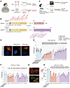 Figure 1
MCCs with pathological RPGR variants presented with short cilia and decreased ciliation. (A) Cartoon showing RPGR as the causative gene for RP and PCD, as well as the focus of this study. (B) Distributions of variants of the 32 patients participating in this study. Red indicates variants that affect both isoforms and correspond to LoF or splicing; pink corresponds to variants with missense effect; and purple corresponds to variants affecting only RPGRORF15. Variants in patients 9 and 15 are predicted to be missense and affect splicing. (C) The proposed workflow for this study. (D) Immunostaining of human nasal cells of control individuals and patients with RPGR variants. Green represents RPGR and red represents α-tubulin. MIP, maximum intensity projection. Scale bar: 10 μm. (E) Cilia length measurements for the nasal MCCs from control participants and patients with RP. Individuals with PCD are distinguished with a red asterisk. (F) Cilia length measurements for the MCCs from control participants and patients with RP cultured at the ALI for 8 weeks. (G) Immunostaining of basal body (POC1B) and cilia (acetylated tubulin) showed reduced ciliation in the MCCs of a patient. Scale bar: 5 μm. (H) Ciliation was reduced for the MCCs bearing pathological RPGRex1–19 or RPGRORF15 variants. Data represent mean ± SEM. *P < 0.05, **P < 0.01, ***P < 0.001, ****P < 0.0001 by 2-tailed t test (E, F, and H). For (E), we performed 1 experiment in a subset of the cohort that included 6 healthy control participants and 28 patients. For (F–H), we performed 3 independent replicates for a subset of the cohort that included 3 healthy control participants and 18 patients.
Conditions due to defective function, development, or maintenance of cilia are collectively referred to as ciliopathies. Primary ciliary dyskinesia (PCD) is an important motile ciliopathy (6, 7), affecting approximately 1 in 7,500 people worldwide (8). Major features of PCD include oto-sino-pulmonary diseases, which can be life-threatening because of the progressive deterioration of lung function. Retinitis pigmentosa (RP) is a genetically heterogeneous disorder characterized by degeneration of photoreceptors, leading to night blindness and sight loss (9). The incidence rate of RP ranges between 1 in 3,000 to 4,000 people (10). RP is linked to various gene variants related to the visual pathway, and variants associated with sensory cilia are known to contribute to RP (11).
Deleterious variants involving the retinitis pigmentosa GTPase regulator (RPGR) gene account for approximately 70% of X-linked RP (12–14) and are also a known cause of X-linked PCD (15). RPGR is expressed in sensory and motile cilia (16, 17). As a result, patients with RPGR deleterious variants could present with both motile and sensory ciliopathy phenotypes (Figure 1A). RPGR is expressed in 2 major isoforms (Figure 1B) (18–21): one is constitutively expressed in multiple organs and cell types (NM_000328.3; RPGRex1–19), and the other has a retina-specific expression pattern (NM_001034853.2; RPGRORF15). RPGRex1–19 has 19 exons, of which RPGRORF15 shares exons 1–14 (18, 22–25). RPGRORF15 is an X-linked RP mutational hotspot, whereas RPGRex1–19 is ubiquitously expressed and was initially regarded as the sole isoform in airway multiciliated cells (MCCs) (17, 18). In the retina, RPGR contributes to outer-segment membrane disk renewal, and variants involving either RPGRex1–19 or RPGRORF15 cause early-onset RP by disrupting actin dynamics (26).
RPGR’s role in motile cilia remains unclear. Whereas most PCD causative genes encode axoneme components, dynein arm docking or assembly factors, or transcription factors that regulate multiciliogenesis, RPGR is distinct and may play a role in ciliary transport (27–30). The gold standard for confirming PCD diagnosis is either ciliary structural defects detected by transmission electron microscopy (TEM) and/or biallelic variants in known PCD genes detected by DNA sequencing. However, these standards cannot diagnose all PCD cases, including RPGR variants, as some RPGR variants could be variants of unknown significance or may not lead to classic TEM changes (31, 32). Previous clinical reports showed that variants in RPGR associated with PCD can result in static cilia, impaired cilia beat, or disrupted beat coordination (15, 19, 33). However, these reports were based on a limited number of patients. Which variants predispose patients to PCD and how RPGR regulates motile cilia properties are still unclear.
Using super-resolution microscopy and live-cell imaging of patient samples, we observed that patients with pathological variants affecting both isoforms (RPGRex1–19 variants) had sparse, shorter, and mostly static motile cilia. For RPGRORF15 variants, shorter cilia were observed for a portion of patients, and the ciliation level was decreased for all patients. Most MCC cilia with RPGRORF15 variants either exhibited restricted motility or were static. These phenotypes were recapitulated in CRISPR-mediated RPGR KO MCCs.
Investigating the mechanisms underlying how RPGR regulates motile cilia revealed its role in regulating F-actin dynamics. With RPGR variants, a condensed F-actin meshwork accumulates and persists at the apical surface of MCCs, preventing some cilia from extruding from the surface and restricting their movement on the cell surface. This persistence of F-actin meshwork is associated with a temporary reduction in gelsolin levels at the apical surface. To examine whether the phenotype was specific to F-actin changes, we treated RPGR KO MCCs and patient MCCs with pathological RPGR variants with either the actin polymerization inhibitor latrunculin A (LatA) or the ROCK inhibitor Y27632, and we found that cilia length and ciliation level were ameliorated, and cilia beat properties were partially restored.
To summarize, working with a cohort of 32 patients with different RPGR variants, we provide the first mechanistic study, to our knowledge, of RPGR’s function in the respiratory system. Although PCD was more prevalent in patients with RPGRex1–19 variants, cilia anomalies were seen in patients with both RPGRex1–19 and RPGRORF15 variants, and patients with RPGRORF15 variants had frequent respiratory findings, though these were not characteristic of PCD. This work also reveals a distinct role of RPGR compared with that of other genes associated with PCD: RPGR modulates F-actin dynamics at the apical surface to control multiciliogenesis and regulate ciliary beat machinery. The methods reported here can be translated clinically to identify individuals at risk of mucociliary clearance defects and improve patient outcomes, with appropriate management.
-
Results
Patient characterization. This study was approved by the ethics board of participating institutions, and recruitment respected the tenets of the Declaration of Helsinki. We recruited a cohort of 32 patients with X-linked RP with a wide range of RPGRex1–19 and RPGRORF15 variants, including 1 female patient with retinal degeneration (Figure 1, B and C, and Supplemental Table 1; supplemental material available online with this article; https://doi.org/10.1172/JCI193367DS1), and 12 healthy volunteers. All patients were recruited through eye clinics and had a disease-associated RPGR variant and retinal degeneration. The predicted impact of each identified variant was validated using literature, pathogenicity and conservation predictive algorithms, allele frequency databases, and splicing assays (34–39) (Supplemental Table 1 and Supplemental Figure 1). RPGRex1–19 variants were found in 16 patients: 8 were considered loss of function (LoF), 5 were missense resulting in an in-frame deletion, 2 had missense variants with loss of transcript due to incomplete splicing (missense splicing), and 1 had a missense. LoF variants affecting the RPGRORF15 isoform were present in the remaining 16 patients (Supplemental Table 1).
Our assessment of the 32 patients with either RPGRex1–19 or RPGRORF15 variants revealed a range of respiratory manifestations: 19 of 31 (61%) had sinus diseases, 15 of 30 (50%) had lower airway diseases, and 19 of 31 patients (61%) had hearing issues, including hearing loss and/or otitis media (Supplemental Table 2). Of the 16 patients carrying RPGRORF15 variants, 5 (31%) had sinus disease, 4 (25%) had lower airway disease, and 8 (50%) had hearing issues.
Additional clinical assessments were performed, and these indicated 9 of 28 patients (32%) had abnormal lung function with obstructive ventilation defects (n = 7 mild, 1 moderate, and 1 severe defect). Lung-imaging airway abnormalities were present on either chest radiographs or computed tomography scan in 11 of 27 patients (41%), of whom 6 had bronchiectasis. Of the 16 patients carrying RPGRORF15 variants, 2 (13%) had abnormal lung function, and 3 (19%) had abnormal chest radiographs.
Combining the results from all assessments, 6 patients (19%) met criteria for a clinical diagnosis of PCD. All exhibited symptoms of severe oto-sino-pulmonary disease with symptom onset in early childhood, with reduced nasal nitric oxide levels and airway abnormalities on chest imaging, and all of whom had RPGRex1–19 variants. Only 2 of the 6 patients were diagnosed with PCD prior to being recruited into our study.
Patients with pathological RPGRex1–19 or RPGRORF15 variants had cilia defects. Fresh nasal cilia were available from 29 patients. The mean age of patients at the time of nasal biopsy was 30 years (range 8–63 years). Confocal microscopy of fresh MCCs immunostained with RPGR antibodies and antibodies targeting cilia showed that RPGR was absent in most patients with RPGRex1–19 variants but was properly localized in the cells bearing RPGRORF15 variants (Figure 1D). Cilia length measurements showed that 13 of 14 patients (93%) with RPGRex1–19 variants, and 7 of 14 patients (50%) with RPGRORF15 variants had decreased cilia length (Figure 1E). This suggests that overall RPGR is important for motile cilia growth or length regulation.
Analysis of MCCs from a female patient (case 21) with severe RP due to an RPGRex1–19 variant [c.122C>A; p.(Ser41*)] showed that half of her MCCs did not express RPGR and they had shorter cilia because of unfavorable X-inactivation (Supplemental Figure 2A). Because fresh nasal cells might be subject to infection and other environmental stress, we redifferentiated the isolated basal cells at the air-liquid interface (ALI) for further assessment. Differentiated cells were available from 18 patients. Cilia length of the differentiated MCCs at 4 and 8 weeks was measured by analyzing the signal from acetylated tubulin (Figure 1F and Supplemental Figure 2B). Of 18 patients, 5 with RPGRex1–19 variants and 3 with RPGRORF15 variants had cells with significantly shorter motile cilia than did control individuals (Figure 1F). Shorter cilia were more prevalent for 4-week MCCs in 8 of 9 patients with RPGRex1–19 variants and in 8 of 9 patients with RPGRORF15 variants (Supplemental Figure 2B). The shorter cilia in MCCs bearing RPGRORF15 variants suggest that RPGRORF15 also contributes to the growth of the motile cilia. ALI cell culture also enabled us to quantify the ciliation level, defined as the number of cilia divided by the number of basal bodies per MCC (Figure 1, G and H, and Supplemental Figure 2C). Patients with either RPGRex1–19 (n = 9 of 9) or RPGRORF15 (n = 9 of 9) variants had significantly decreased ciliation level compared with healthy control individuals (Figure 1H).
Altogether, our characterization of the MCCs from patients with a wide variety of pathogenic and likely pathogenic RPGRex1–19 and RPGRORF15 variants shows that disruption of RPGR function may lead to reduced ciliation and shortened cilia length.
Damaging RPGRex1–19 and RPGRORF15 variants affect cilia beat. To investigate how RPGR regulates cilia motility, cilia beating was measured for MCCs redifferentiated at ALI for 8 weeks. We stained the whole filter with wheat germ agglutinin–Alexa Fluor 488 to mark the highly glycosylated motile cilia (40) and imaged the MCCs with an inverted fluorescence microscope (Figure 2A). The frequency of pixel signal fluctuations in Supplemental Video 1 was initially used to represent the overall cilia motility within the field of interest. This method clearly distinguishes 2 patients from a healthy control individual in the frequency map (Figure 2B). To quantify the differences, we further measured the cilia beat frequency in individual cells from patients with different variants (Supplemental Figure 3). Our analysis showed that most motile cilia from patients with RPGRex1–19 variants were static or had a restricted beat (n = 7 of 9; 78%), whereas for patients 2 and 15, a mixture of slow and fast beat cilia was observed (Figure 2C). All patients with variants in RPGRORF15 (n = 9 of 9) had decreased cilia beat frequency (Figure 2C).
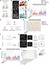 Figure 2
MCCs with RPGR pathological variants had an impaired and uncoordinated cilia beat. (A) A cartoon showing labeling motile cilia with wheat germ agglutinin–Alexa 488 (WGA-Alexa 488). (B) The pixel frequency map shows cilia beat impairment in the MCCs with pathological RPGR variants. MIP, maximum intensity projection. CBF, cilia beat frequency. Scale bar: 10 μm. (C) Characterization of CBF in cells from 18 patients with RP in this study. (D) Different cilia beat modes identified in RPGR LoF MCCs. (E) Characterization of cilia beat waveform distribution for 18 patients with RP in this study. (F) A cartoon showing labeling POC1B and centriolin to assess rotational polarity and its disruption in MCC bearing a pathological RPGR variant. (G) Immunostaining shows rotational polarity in healthy control cells and its disruption in 1 patient with a pathological RPGRex1–19 variant. Scale bars: 2 μm; insert, 2 μm. (H) Characterization of rotational polarity in cells from 18 patients with RP in this study. (I) Single-cell RNA-Seq revealed the expression of RPGR mRNA and its different isoforms in airway epithelial cells. ECM, extracellular matrix. (J) RT-PCR showed the expression of the RPGRORF15 isoform in human airway epithelial cells (HNCs cultured at ALI for >8 weeks). Data represent mean ± SEM. The box shows the 25th–75th percentile, with the middle line showing the median (I). ***P < 0.001, ****P < 0.0001 by 2-tailed t test (C, E, and H). For (C, E, and H), we performed 3 technical replicates for a subset of the cohort that included cells from 3 healthy control individuals and 18 patients.
The motile cilia beating of healthy control cells had a distinguishable power and recovery stroke, as well as synchronization of the metachronal wave (Supplemental Video 1). On the contrary, MCCs from patients with RP showed noticeable heterogeneity in those features, between patients as well as among cells from the same patient (Supplemental Video 1). Regarding the cilia beat waveform, besides the normal cilia beat, 4 different types of beat phenotypes could be identified: (a) static cilia, (b) restricted cilia beat (low amplitude), (c) rigid cilia beat with a loss of waveform, and (d) cilia beat in a rotational and/or uncoordinated manner (Figure 2D). We quantified the percentages of cells exhibiting different cilia beat modes and found that most cells with either RPGRex1–19 or RPGRORF15 variants displayed static cilia or a restricted cilia beat (n = 8 of 9 [89%] for RPGRex1–19 and 6 of 9 (67%) for RPGRORF15 variants), whereas the rest (n = 1 of 9 [11%] for RPGRex1–19 and n = 3 of 9 [33%] for RPGRORF15 variants) had a rigid or uncoordinated beat (Figure 2E).
To quantitatively compare beat coordination, we used a 3D structured illumination microscopy–based (3D-SIM–based) method we previously developed (41) to examine the rotational polarity of the basal foot (Figure 2, F and G), an appendage structure localized at the base of each cilium (41, 42). In healthy MCCs, all basal feet point toward the direction of cilia beat, a phenomenon called rotational polarity (42–44) (Figure 2F), which is disrupted in patients with PCD, because of defective cilia beat (41). We performed this analysis and found that the rotational polarity was disrupted in cells with either RPGRex1–19 (n = 9 of 9) or RPGRORF15 (n = 9 of 9) variants (Figure 2H).
Considering all results together, damaging variants in RPGRex1–19 and RPGRORF15 affect airway MCC cilia beat frequency, waveform, and coordination.
Patients with RPGR-RP with severe PCD symptoms had severe cilia defects. Patients with severe clinical respiratory PCD symptoms had RPGRex1–19 LoF variants or in-frame deletions and had severe cilia defects. All 6 patients whose condition met the clinical diagnostic criteria of PCD had shortened nasal cilia length (Figure 1E). We successfully redifferentiated cells from 3 patients (patients 11, 26, and 9); 2 had cells with reduced cilia length and ciliation, and the third had cells with slowed cilia growth, supporting our findings from fresh cells (Figure 1, F and H, and Supplemental Figure 2B). Cilia beat analysis of MCCs from those same 3 patients with PCD showed that most MCCs presented with static cilia or a restricted cilia beat, and the rotational polarity was disrupted in all cases (Figure 2, C, E, and H, and Supplemental Figure 4A).
Four patients with RP bearing the same RPGRex1–19 variant [c.934G>T; p.(Glu260_Thr311del)] presented with different clinical and cellular phenotypes: 2 patients presented with a clinical PCD phenotype and shorter nasal cilia length, and patients 23 and 24 only had milder respiratory symptoms and intermediate nasal cilia lengths (Figure 1E, Supplemental Figure 4B, and Supplemental Table 2). This suggests multifactorial influences in the development of PCD.
While most patients with RPGR-RP had motile cilia defects, only a subpopulation presented with a distinctly abnormal respiratory phenotype, with 6 patients showing a strong PCD phenotype and bearing variants affecting both isoforms. However, in cases of patients with RPGRORF15 variants, abnormal cilia and respiratory phenotypes were also observed, suggesting these patients may be affected by respiratory illnesses caused by cilia dysfunction (Figures 1 and 2, Supplemental Figure 4C, and Supplemental Table 2).
RPGRORF15 isoform was expressed in airway MCCs. Although the RPGRex1–19 isoform was once regarded as the sole isoform in airway MCCs (17), we showed that cells from patients with RPGRORF15 variants presented with cilia defects (Figure 1, E, F, and H), albeit with less severe respiratory phenotypes (Supplemental Table 2). To validate if the RPGRORF15 isoform is expressed in MCCs, we first examined the single-cell sequencing data of a fetus’s trachea generated in a previous study by He et al. (45). All RPGR isoforms were first treated as a single gene. At the mRNA level, compared with other cell types, RPGR was mainly expressed in the MCC progenitors (Foxn4 expressing) and mature MCCs (Figure 2I). We next separated different isoforms and performed isoform-specific expression analysis. The result showed that the expression levels of the RPGRORF15 isoform were comparable to the predominantly expressed RPGRex1–19 isoform, confirming that RPGRORF15 is expressed in airway cells. The expression of the RPGRORF15 isoform was upregulated during the early stage of multiciliogenesis (Figure 2I), indicating its role in cilia formation. To further examine the expression of RPGRORF15 isoform in MCCs, we performed RT-PCR of the total mRNA transcripts extracted from healthy control cells and RPGR KO MCCs. The result further supports that RPGRORF15 is expressed in airway cells (Figure 2J and Supplemental Figure 5), consolidating the single-cell sequencing data.
RPGR KO MCCs presented with sparse and short motile cilia. To determine the RPGR specificity of our findings, we generated RPGR KO MCCs by CRISPR-mediated genomic perturbation of healthy human nasal or bronchial basal cells. By using an optimized protocol that includes medium adjustments and adaptation of 2 sgRNAs simultaneously (Figure 3, A and B), we successfully produced RPGR KO MCCs, as confirmed by DNA gel electrophoresis and Sanger sequencing (Figure 3C). The percentage of cells in which both gRNAs were effective reached 92.3%, resulting in an overall KO efficiency of approximately 99% (Figure 3C). Immunostaining showed a complete absence of RPGR compared with the control (Figure 3D). We further characterized the cilia properties of 5 biological replicates, including 3 human bronchial epithelial cell (HBEC) and 2 human nasal cell (HNC) samples, showing that cilia length was affected in all (Figure 3, E and F). Quantification of the ciliation level of MCCs showed that, although basal body number was unaffected, ciliation was significantly decreased (Figure 3, E and G, and Supplemental Figure 2D), which suggests that RPGR regulates ciliation but not basal body amplification or docking. To rule out the possibility that the phenotype was due to a secondary defect in MCC differentiation, we showed the expression level of FOXJ1 and CCNO, as assessed by qRT-PCR, was unchanged in RPGR KO MCCs (n = 3) (Figure 3H).
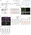 Figure 3
RPGR KO MCCs presented with sparse and short motile cilia. (A) Workflow of generating RPGR KO MCCs. Indel, insertion-deletion. HSVM, high-speed video microscopy. (B) The location of gRNAs and the expected genome edits. (C) DNA gel and Sanger sequencing showed high efficiency of RPGR KO in MCCs. (D) Immunostaining of MCCs with RPGR antibody showed complete RPGR removal in RPGR KO MCCs. Scale bars: 10 μm; insert, 5 μm. (E) Immunostaining with POC1B and acetylated-tubulin antibodies show the reduction of ciliation length and ciliation for RPGR KO HBEC/HNC cells. Scale bars: 10 μm; insert, 5 μm. (F) Cilia length was significantly disrupted in all 5 biological replicates. (G) Ciliation was severely affected for all 5 RPGR KO biological replicates. (H) FOXJ1 and CCNO expressions were unaffected in RPGR KO MCCs. For each gene, 2 sets of primers were used. Data represent mean ± SEM. ***P < 0.001, ****P < 0.0001 by 2-way repeated ANOVA followed by Šídák’s post hoc test (F) or 2-tailed t test (G and H). For (F and G), we performed 5 experiments using cells from 5 control individuals and corresponding RPGR KO MCCs. For (H), we performed 3 experiments using cells from 3 control individuals and corresponding KO MCCs.
Taken together, removing RPGR by CRISPR perturbation from airway MCCs led to shortened cilia and reduced ciliation levels, recapitulating the cellular phenotypes observed in the patients.
RPGR KO MCCs presented with static cilia and/or abnormal cilia motility. Live-cell imaging of RPGR KO MCCs (Figure 4, A–D) showed that most cells (68%; n = 4 of 5 biological replicates) had static cilia (Figure 4, B, upper and middle, and Supplemental Video 2), leading to disrupted fluorescent bead clearance in vitro (Supplemental Video 3). There was large heterogeneity within and among different biological replicates (Figure 4C). A quantification of cilia beat frequency of the RPGR KO MCCs showed results consistent with patient data. Analysis of the cilia beat waveform, in 4 of 5 replicates, showed static cilia were predominant; for the fifth replicate, although most cilia could beat with a normal frequency compared with the control (NS; n = 2 technical replicates), both the waveform and beat coordination were abnormal (Figure 4D).
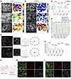 Figure 4
RPGR KO MCCs presented with limited motility and defective rotational and planar polarity. (A and B) The pixel frequency map shows control MCC cilia beat and its impairment in RPGR KO MCCs. CBF, cilia beat frequency; MIP, maximum intensity projection. Scale bar: 10 μm. (C and D) Characterization of CBF and beat mode changes for 5 biological replicates in this study. (E) Immunostaining showed rotational polarity in control cells and its disruption in RPGR KO MCCs. Scale bars: 2 μm; insert, 1 μm. (F) Rotational polarity was disrupted for all RPGR KO samples in this study. (G–I) Vangl1 is mislocalized in the MCCs from an HBEC RPGR KO sample and from a sample from a patient with RP, suggesting loss of planar polarity. Scale bar: 5 μm. Data represent mean ± SEM. The center, upper, and lower lines represent the median, upper, and lower quartiles, respectively (C and F). ***P < 0.001, ****P < 0.0001 by 2-tailed t test (C and F) or Fisher’s exact test (D). For (C, D, F, and H), we performed 5 experiments using cells from 5 control individuals and corresponding KO MCCs. (I) Results from 9 of 10 patients (see Supplemental Figure 2F for details).
The rotational polarity assessment of RPGR KO cells showed a drastic disruption of beat coordination for all biological replicates (n = 5) (Figure 4, E and F). Previous studies suggested the disruption of planar cell polarity (PCP) for patient cells bearing RPGR variants (46, 47); therefore, we stained the ALI filters with antibodies targeting the planar polarity protein Vangl1 (48, 49). For control cells, Vangl1 was specifically enriched on 1 side of the MCCs. In both RPGR KO HNCs and HBECs, as well as in patient cells with RPGRex1–19 or RPGRORF15 variants, the asymmetrical distribution of Vangl1 was largely lost, suggesting a disruption in planar polarity (Figure 4, G–I, and Supplemental Figure 2, E and F).
Furthermore, we designed a gRNA that specifically targets the RPGR ORF15 region and achieved a KO efficiency of 70% in HBECs and 59% in HNCs (Supplemental Figure 6A). Characterization of the MCCs generated with the ORF15-specific gRNA revealed that cilia length, ciliation, cilia beat frequency, waveform, and coordination were all affected, as was the distribution of Vangl1 (Supplemental Figure 6, B–H). These changes impaired the clearance of fluorescent beads and recapitulated the phenotypes of the patient MCCs bearing RPGRORF15 variants (Supplemental Figure 6I).
To summarize, RPGR defects caused by CRISPR perturbation led to either static cilia or cilia with altered motility, disrupting both planar and rotational polarity.
RPGR localizes to the transition zone and cilia membrane; its loss does not affect the integrity of the transition zone or the localization of axoneme dyneins and central pair components. To inspect how RPGR regulates motile cilia, we first examined RPGR subcellular localization by 3D-SIM. We stained airway MCCs from healthy control individuals with a validated RPGR antibody. 3D-SIM provides approximately 120 nm resolution (50) and showed a clear transition zone (TZ) distribution of RPGR by colocalization with the TZ marker RPGRIP1L, as well as a weak signal in cilia (Figure 5A). We also used stochastic optical reconstruction microscopy (STORM) (51), which provides approximately 20 nm resolution, to examine the distribution of RPGR and found that it formed a dotted ring pattern at the TZ for each cilium as well as cilia localized puncta (Figure 5B). Additionally, we examined the distribution of RPGR at different multiciliogenesis stages (2–8 weeks) by stimulated emission depletion (STED) microscopy (approximately 40 nm resolution; ref. 52) and found that RPGR consistently localized to the TZ and motile cilia (Figure 5C and Supplemental Figure 7).
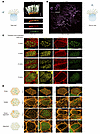 Figure 5
RPGR mainly localized to the TZ and cilia membrane; the relative distribution of RPGR and F-actin is shown. (A) 3D-SIM shows RPGR mainly locates to the TZ of motile cilia. Scale bar: 5 μm. (B) STORM showed RPGR presents with a dotted ring structure at the TZ (rectangle) and puncta along cilia. Scale bars: 5 μm; insert, 500 nm. (C) RPGR locates to the TZ throughout the differentiation of MCCs. Phalloidin labels the apical F-actin, and no colocalization was observed between RPGR and F-actin. Their distributions are largely exclusive. Scale bar: 5 μm. (D) The dynamics of apical F-actin during the differentiation of MCCs. Scale bar: 5 μm. Results here represent 4 biological replicates with similar results.
The subcellular localization of RPGR and its interaction with TZ components suggest it might be involved in ciliary transportation and maintenance of cilia composition. Because most cilia of RPGR LoF cells were static, we wondered if the outer dynein arms, inner dynein arms, and central pair components were intact. We assessed 13 samples from patients with RPGRex1–19 or RPGRORF15 variants and found that outer dynein arm marker DNAH5, inner dynein arm marker DNALI1, and central pair component SPEF2 all showed proper localization (Supplemental Figure 8, A–D), consistent with the normal TEM results (Supplemental Figure 8, E and F) (53). RPGR interacts with the TZ components RPGRIP1L, CEP290, and NPHP4 (54, 55). Immunostaining showed the loss of RPGR did not affect the distribution of these TZ components (Supplemental Figure 9A), and other TZ components, such as AHI1, MKS1, MKS3, and CC2D2A, were all properly localized (Supplemental Figure 9B), suggesting that RPGR is dispensable for the integrity of the TZ.
RPGR KO MCCs maintained an abnormal, condensed, apical F-actin meshwork. RPGR has been reported to regulate F-actin polymerization (56, 57), and loss of RPGR in hTERT-RPE1 cells induces the formation of actin bundles (56). Using an Rpgr KO mouse model, Megaw et al. found that RPGR regulates photoreceptor ciliary-tip actin dynamics, and its dysregulation affects outer segment membrane turnover (26, 58). To investigate if RPGR also regulates F-actin organization in MCCs, we first looked at the relative distribution of apical F-actin and RPGR. Notably, we found no obvious colocalization between the 2 (Figure 5C). We next stained centrin-GFP–labeled MCCs cultured for different weeks with phalloidin-STAR RED and imaged the dynamics of F-actin using STED microscopy. During the early stage of multiciliogenesis, the apical surface was mostly covered with F-actin meshwork, where the gaps were decorated with centrin-GFP florets (Figure 5D). The F-actin meshwork is critical for basal body docking (59, 60). As MCCs mature, the F-actin meshwork gradually dissolves, allowing more scattered or aligned centrin-GFP–labeled basal bodies to emerge (Figure 5D). The F-actin pattern now condenses into bright puncta or bundles (Figure 5D). The filamentous F-actin is supposed to align basal bodies to synchronize cilia beat (60, 61).
We next characterized the apical F-actin in RPGR KO MCCs. In 4-week RPGR KO MCCs, F-actin was comparable between control and RPGR KO cells (Figure 6A). However, after maturation, RPGR KO MCCs preserved the meshwork and failed to form F-actin puncta or bundles (Figure 6B), which affected the extrusion of the cilia. These results were confirmed using STORM imaging of phalloidin-Alexa Fluor 647–labeled MCCs (Figure 6C). We further quantified the F-actin/G-actin ratio in MCCs by immunoblotting, and the result showed an obvious increase in F-actin for both 4-week and 8-week RPGR KO samples (Figure 6, D–G), aligning with super-resolution images.
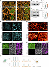 Figure 6
Mature RPGR LoF MCCs presented with condensed apical F-actin meshwork that did not dissolve. (A) Distribution of F-actin in 4-week control and RPGR KO MCCs. Scale bar: 5 μm. (B) Distribution of F-actin in 8-week control cells and RPGR KO MCCs. Scale bar: 5 μm. (C) STORM imaging of the apical F-actin in both RPGR KO cells and healthy control cells. Scale bars: 5 μm; insert, 1 μm. (A–C) Findings from 4 biological replicates are represented. (D and E) Immunoblotting showed increased F-actin in 4-week RPGR KO MCCs. Results were summarized from 3 biological replicates. Mr, relative molecular mass. (F and G) Immunoblotting showed increased F-actin in 8-week RPGR KO MCCs. Results were summarized from 3 biological replicates. (H) STED imaging of the apical F-actin in MCCs from patients with RP and healthy control cells. Scale bars: 5 μm; insert, 1 μm. (I and J) Apical gelsolin was diminished in 4-week RPGR LoF MCCs. (I) RPGR KO HBEC MCCs. (J) MCCs from 1 patient with RP. Scale bar: 2 μm. Data represent mean ± SEM. **P < 0.01, ***P < 0.001 by 2-tailed t test (E and G). (H) represents results from 8 patients with RP (see Supplemental Figure 11, C and D, for details). (I) represents results from 5 biological replicates. (J) represents similar results from 17 patients (see Supplemental Figure 13, B and C, for details). (K) The proposed model of this study: without RPGR, F-actin meshwork persists at the apical surface, preventing ciliation and cilia elongation; cilia beat is severely impaired.
Analysis of MCCs from patients with RP showed results mostly consistent with those observed in RPGR KO cells (Supplemental Figure 10, A–D, and Supplemental Figure 11, A–D). For most patients with variants in RPGRex1–19 (n = 6 of 9) or RPGRORF15 (n = 7 of 9), the apical F-actin meshwork in 8-week MCCs could still be readily discerned by either STED or 3D-SIM (Figure 6H; Supplemental Figure 10, C and D; and Supplemental Figure 11, C and D). However, in some patients, the F-actin accumulation seemed to be milder or normal (n = 3 of 9 for RPGRex1–19 variants and n = 2 of 9 for RPGRORF15 variants) (Supplemental Figure 10, C and D, and Supplemental Figure 11E).
To further validate the involvement of F-actin in muticiliogenesis, we treated healthy control MCCs with Rho activator II, a known compound to stabilize F-actin (62, 63), and observed cilia properties at ALI 8 weeks. F-actin stabilization substantially affected cilia length and ciliation, reduced cilia beat frequency, changed cilia beat waveform, and most MCCs lost cilia beat coordination (Supplemental Figure 12, A–I). We also found that bead clearance was paralyzed in Rho activator II–treated MCCs (Supplemental Figure 12J). This suggested that F-actin accumulation could account for the observed motile cilia phenotypes in RPGR LoF MCCs.
In photoreceptor and hTERT-RPE1 cells, RPGR regulates actin dynamics through interaction with gelsolin and cofilin (26, 58). Staining gelsolin for RPGR KO MCCs showed that in both RPGR KO HNCs (n = 2) and HBECs (n = 3), the cell surface, the layer where F-actin mainly locates, had temporarily diminished the gelsolin signal, suggesting that RPGR might regulate F-actin dynamics through locating gelsolin (Figure 6I and Supplemental Figure 13A). Gelsolin exists in active and inactive forms, and it is the active gelsolin that binds to F-actin (64, 65). Reduction of gelsolin binding to F-actin in RPGR KO MCCs suggests that RPGR might be involved in gelsolin activation. We further examined the distribution of gelsolin in MCCs bearing different RPGR variants and found a consistent decrease in the gelsolin that colocalized with the apical F-actin (n = 8 of 8 for RPGRex1–19 variants and n = 7 of 9 for RPGRORF15 variants) (Figure 6J and Supplemental Figure 13, B and C).
In summary, RPGR regulates the dynamics of F-actin, and RPGR KO MCCs and patient MCCs with pathological RPGR variants had an F-actin meshwork that failed to dissolve.
Motile cilia anomalies caused by RPGR LoF were improved with latrunculin A or Y27632 treatment. If RPGR regulates motile cilia through F-actin, treatment with actin polymerization inhibitors (64, 66) should rescue or ameliorate the phenotype. Previous studies showed that overexpression of active gelsolin can rescue the ciliation issue in RPGR KD hTERT-RPE1 cells (58). We first worked on hTERT-RPE1, due to its quick turnaround time. From our RPGR KO hTERT-RPE1 cell pool, we found reduced ciliation and cilia length defects after RPGR removal, consistent with findings from previous studies (67) and our motile cilia data (Supplemental Figure 14, A–C). Treatment of hTERT-RPE1 control and RPGR KO cells with 0.2 μM latrunculin A (LatA) for 24 hours restored ciliation and cilia length of RPGR KO cells (Supplemental Figure 14, D and E). This suggests that LatA can restore the cilia phenotype caused by loss of RPGR and that RPGR functions upstream of actin in cilia regulation. We also applied LatA on the fibroblast cells from 2 patients with RPGR-RP with cilia defects (Supplemental Figure 14, F and H) and found that LatA treatment partially rescued the cilia phenotypes (Supplemental Figure 14, G and I).
To investigate whether LatA treatment could rescue motile cilia, we applied LatA to ALI-cultured RPGR KO MCCs. Previous studies indicated an increase of RhoA activity in RPGR KD hTERT-RPE1 cells (47) or Rpgr KO mouse models (68); therefore, we also used a ROCK inhibitor, Y27632, to modulate actin dynamics. We added LatA or Y27632 starting from the ALI day 0 and observed cilia properties at the ALI week 4, because RPGR is strongly expressed during the early stage of multiciliogenesis (Figure 2I and Supplemental Figure 15).
After treatment, although the cilia length could be fully restored, ciliation was partially rescued for both LatA and Y27632 treatment in 2 biological replicates (Figure 7, A–C). Additionally, we examined the distribution of Vangl1 and gelsolin. Although LatA treatment did not rescue their distributions, Y27632 showed obvious rescue of these 2 phenotypes (Figure 7, D and E).
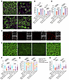 Figure 7
LatA and Y27632 treatment ameliorated the motile cilia defect in RPGR KO MCCs. (A) Immunostaining showed the reduction in ciliation and cilia length in RPGR KO MCCs, along with the rescued phenotypes following LatA and Y27632 treatment. Scale bar: 10 μm. (B and C) Image quantification shows improvements in cilia length and ciliation for 2 RPGR KO biological replicates. (D) Y27632, but not LatA, restored the polarized distribution of Vangl1 at the apical surface in the 4-week HBEC sample. Scale bars: 5 μm; insert, 5 μm. (E) Y27632, but not LatA, restored the distribution of gelsolin at the apical surface in one 4-week HBEC sample. Scale bar: 5 μm. (F–H) The cilia beat anomalies in RPGR LoF MCCs were partially rescued by LatA or Y27632 treatment. (F) Both treatments improved cilia beat frequency (CBF). (G) Both treatments partially rescued the waveform, with more cells displaying a normal beating pattern. (H) The disrupted cilia beat coordination was partially rescued by LatA or Y27632 treatment. Results were from 2 biological replicates (B, C, and F–H). Data represent mean ± SEM. The center, upper, and lower lines represent the median, upper, and lower quartiles, respectively (F and H). **P < 0.01, ***P < 0.001, ****P < 0.0001 by 2-tailed t test (B, C, F, and H) or Fisher’s exact test (G).
Treatment of RPGR KO MCCs with LatA or Y27632 led to significant improvements in cilia beat frequency, waveform, and coordination (Figure 7, F–H, and Supplemental Video 4). We also found that, after treatment with LatA and Y27632, the directional movement of the beads could be readily observed (n = 2; Supplemental Figure 16 and Supplemental Video 5), suggesting that F-actin depolymerization can, indeed, ameliorate the motile cilia phenotypes caused by RPGR defect.
We further treated the MCCs from 17 patients with either RPGRex1–19 or RPGRORF15 variants with LatA and Y27632 and found results consistent with those we observed in the RPGR KO MCCs (Figure 8 and Supplemental Figure 17). Of the MCCs from the 17 patients, treatment with either LatA or Y27632 resulted in the restoration of Vangl1 in cells of 11 patients and restoration of gelsolin in 12 by at least 1 method. Improvements in cilia length, beat frequency, waveform, and coordination were observed in MCCs of 12 patients, and MCCs of 13 patients had an increase in ciliation levels. This led to enhanced mucociliary clearance for 12 cases. Notably, no rescue effect was observed in 4 patients. No obvious difference was found between RPGRex1–19 and RPGRORF15 variants.
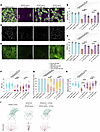 Figure 8
LatA and Y27632 treatment ameliorated the motile cilia defect in patient MCCs with RPGR variants. (A) Immunostaining showed that LatA and Y27632 can partially rescue cilia length and ciliation defects in the MCCs from a patient with RP (patient 9). Scale bar: 10 μm. (B and C) The cilia length and ciliation level in MCCs from patients with pathological variants (patients 9 and 18) were improved by LatA and Y27632 treatment. (D and E) The distribution of Vangl1 and gelsolin in patient cells with RPGR defects can be rescued by Y27632 but not LatA. Scale bar: 10 μm. (F–H) The cilia beat anomalies in MCCs from patients with RPGRex1–19 or RPGRORF15 pathological variants were alleviated by either LatA or Y27632 treatment. (F) Both treatments improved cilia beat frequency (CBF). (G) Both treatments partially rescued the waveform, with more cells displaying a normal beating pattern. (H) The disrupted cilia beat coordination was partially rescued by LatA or Y27632 treatment. (B, C, F–H) Data represent more than 11 of 17 patients with RPGRex1–19 or RPGRORF15 variants. The detailed summary information can be found in Supplemental Figure 17. Data represent mean ± SEM. The center, upper, and lower lines represent the median, upper, and lower quartiles, respectively (F and H). **P < 0.01, ****P < 0.0001 by 2-tailed t test (B, C, F, and H) or Fisher’s exact test (G). (I) A summary cartoon shows that the cilia defects in RPGR KO MCCs or RPGR-deficient patient MCCs can be alleviated by Y27632 treatments.
Interestingly, Y27632 outperformed LatA in rescuing the distribution of Vangl1 and gelsolin and in improving the cilia beat waveform (Figures 7 and 8 and Supplemental Figure 17). This result suggests that RPGR may not directly interact with actin but rather regulates actin dynamics by suppressing the RhoA/ROCK activity.
Moreover, we tested treatment starting at a later stage (from ALI week 4 and observed cilia properties at ALI week 8) and also found improvements in bead clearance in 2 of 2 RPGR KO MCCs and 5 of 9 patient MCCs (Supplemental Figures 18 and 19). Our study suggests that early rescue has a better performance, which is consistent with the increased expression of RPGR during the early stage of multiciliogenesis. Additionally, after 1 month of treatment withdrawal, there was a slight deterioration of cilia properties in RPGR KO or patient MCCs; however, most parameters were still improved compared with the cells without any treatment, and mucociliary clearance was still preserved after drug withdrawal (n = 8 of 9 patient MCCs and n = 2 of 2 RPGR KO MCCs) (Supplemental Figures 20 and 21).
In summary, the structural anomalies and dysfunctions of motile cilia caused by RPGR LoF can be partially rescued by either LatA or Y27632 treatment.


Copyright © 2026 American Society for Clinical Investigation
ISSN: 0021-9738 (print), 1558-8238 (online)