
Advertisement
Commentary
Open Access |  10.1172/JCI208384
Therapy-resistant lymphomas may want nucleotides, but they just need to grow
Carlos Carmona-Fontaine
Center for Genomics & Systems Biology, Department of Biology, New York University, New York, New York, USA.
Address correspondence to: Carlos Carmona-Fontaine, New York University, Brown Building Of 760, 31 Washington Pl., New York, New York 10003, USA. Email: cf97@nyu.edu.
Find articles by Carmona-Fontaine, C. in: PubMed | Google Scholar
Published July 15, 2026 - More info
J Clin Invest. 2026;136(14):e208384. https://doi.org/10.1172/JCI208384.
© 2026 Carmona-Fontaine This work is licensed under the Creative Commons Attribution 4.0 International License. To view a copy of this license, visit http://creativecommons.org/licenses/by/4.0/.

Related article:
Abstract
Antimetabolites, chemotherapy targeting nucleotide biosynthesis, are among the oldest and most widely used cancer treatments, yet resistance remains a daunting barrier, especially in the fight against B cell lymphomas. However, the underlying mechanisms of this resistance have long remained elusive. Using an innovative, integrated omics approach, we unexpectedly identified that the accumulation of dipeptides and upregulation of the dipeptide transporter SLC15A3 underlie resistance to nucleotide deficiency in a Myc-driven large B cell lymphoma mouse model. A similar mechanism occurred after long treatment of human B cell lymphoma cells with the chemotherapeutic purine synthesis inhibitor 6-mercaptopurine (6MP). Mechanistically, we demonstrated that dipeptides containing essential amino acids activated the growth and survival mTOR complex 1 (mTORC1) signaling pathway. Notably, SLC15A3 specifically interacted with mTOR on the lysosome, boosting mTORC1 activity selectively in resistant lymphoma cells but not in parental cancer cells. Silencing SLC15A3 diminished mTORC1 activity and restored resistant lymphoma sensitivity to 6MP. Strikingly, resistant lymphomas, but not primary tumors, exhibited heightened sensitivity to the clinical mTOR inhibitor, rapamycin, in culture and in vivo. We extended these findings in human lymphoma biopsies, which revealed increased SLC15A3 expression following antimetabolite therapy. Together, our study uncovered a metabolic adaptation that fuels cancer resistance to nucleotide deficiency and positions the mTORC1 inhibitor, rapamycin, as a potential therapeutic strategy for transforming the management of chemotherapy-resistant lymphomas.
Authors
Haojun Yang, Vincenzo Andrea Zingaro, Kevin Boardman, Ashish Noronha, Ekin Guney, Lingru Xue, Saishma Hoigebazar, Isabelle Liu, Sohit Miglani, Siyu Chen, Hieu Vu, Kwun Wah Wen, Hao G. Nguyen, Hani Goodarzi, Ralph J. DeBerardinis, Davide Ruggero
-
Nucleotide deficiency–resistant lymphomas accumulate dipeptides by upregulating SLC15A3
When Jagger and Richards wrote the lines “You can’t always get what you want, but if you try sometime…you get what you need” in the Rolling Stones’ hit song “You Can’t Always Get What You Want (Satisfaction),” surely they were not thinking about cancer biology. But according to an article by Yang et al. in this issue of the JCI, these song lyrics neatly describe how malignant lymphomas become resistant to antimetabolite therapy (1). Antimetabolite therapies, including 6-mercaptopurine (6MP) and methotrexate, are both a long-standing treatment for malignant lymphomas and a possible therapeutic approach for cancers with limited treatment options, such as malignant rhabdoid tumors (2). Unfortunately, many lymphomas become resistant to these drugs (3), and the molecular adaptations enabling this resistance were not well understood.
MYC amplification is a prominent oncogenic driver in B cell malignancies (4). The Ruggero lab had previously developed a MYC-driven large B cell lymphoma mouse model where the deletion of phosphoribosyl pyrophosphate synthetase 2 (PRPS2), a rate-limiting enzyme in nucleic acid biosynthesis, mimics the effects of antimetabolite therapies (5). As expected, the absence of PRPS2 drastically reduces tumorigenesis, but about 40% of these mice eventually develop tumors, making this an elegant genetic model to study resistance to nucleotide deficiency.
In the present work, Yang et al. extended prior findings in the MYC-driven large B cell lymphoma model. They isolated MYC-driven lymphoma cells with or without the loss of Prps2 and compared their metabolic steady states using untargeted metabolomics. They observed that Prps2-null lymphomas significantly accumulated dipeptides, most of which contained a glutamine or glycerol residue paired with an essential amino acid. Interestingly, the levels of these dipeptides were higher than in lymphomas with functional PRPS2 but were comparable to premalignant or wild-type B cells. In future work, it may be interesting to investigate the function of elevated peptides in normal B cells.
In parallel to their metabolomic comparisons, Yang et al. looked at transcriptional and translational changes and singled out the significant increase in Slc15a3 mRNA levels in MYC-driven Prps2-null lymphomas. SLC15A3 is a proton-coupled oligopeptide and histidine transporter localized in endosomal and lysosomal vesicles. The authors then linked observations of dipeptide accumulation with enhanced Slc15a3 expression, showing that SLC15A3 was responsible for the increase in dipeptide levels observed in therapy-resistant cells. In turn, the upregulation of SLC15A3 was essential for resistance to nucleotide deficiency in their MYC-driven lymphoma mouse model as well as essential for the emergence of 6MP resistance in human B cell lymphoma cells in culture. Providing clinical support to their model, biopsies from patients obtained before and after antimetabolite therapy showed a significant increase in SLC15A3 levels after treatment. One of the aspects that makes this work interesting is that the increases in SLC15A3 and dipeptides did not resolve the strained nucleotide biosynthesis induced by antimetabolite therapy. Instead, these increases counterbalanced the antiproliferative effects of nucleotide scarcity, with growth stimulation via the activation of mTOR signaling (Figure 1A).
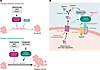 Figure 1
Antimetabolite-resistant lymphomas circumvent the need for nucleotides by activating mTOR. (A) Antimetabolite therapies like methotrexate and 6MP inhibit cell proliferation by reducing nucleotide biosynthesis, but many malignant lymphomas become resistant to these therapies. Yang et al. (1) used a Prps2–/– model of MYC-driven large B cell lymphoma to model resistance to nucleotide deficiency. They found that therapy-resistant lymphomas did not develop an ability to fix nucleotide scarcity. Instead, they overrode the growth inhibition by antimetabolite therapies through mTOR activation, forcing cells to proliferate. mTORC1, mTOR complex 1. (B) Mechanistically, they showed that increased expression of SLC15A3 led to accumulation of dipeptides, including glycyl-leucine, in therapy-resistant lymphomas, driving mTORC1 activation. SLC15A3 may directly or indirectly activate this complex, and Yang et al.’s metabolomic analyses suggested that vitamin D3 may negatively regulate Slc15a3 expression. Potential targets for therapeutic interventions are shown in red.
Article tools
- Download citation information
- Send a comment
- Terms of use
- Standard abbreviations
- Need help? Email the journal
Metrics
Go to
- Top
- Abstract
- Nucleotide deficiency–resistant lymphomas accumulate dipeptides by upregulating SLC15A3
- All you need is growth
- Identifying therapeutic opportunities for patients with chemoresistant tumors
- But what about the nucleotides that cells want?
- Conflict of interest
- Funding support
- Footnotes
- References
- Version history


Copyright © 2026 American Society for Clinical Investigation
ISSN: 0021-9738 (print), 1558-8238 (online)