Go to JCI Insight
  • About
  • Editors
  • Consulting Editors
  • For authors
  • Publication ethics
  • Publication alerts by email
  • Advertising
  • Job board
  • Contact
  • Clinical Research and Public Health
  • Current issue
  • Past issues
  • By specialty
    • COVID-19
    • Cardiology
    • Gastroenterology
    • Immunology
    • Metabolism
    • Nephrology
    • Neuroscience
    • Oncology
    • Pulmonology
    • Vascular biology
    • All ...
  • Videos
    • Conversations with Giants in Medicine
    • Video Abstracts
  • Reviews
    • View all reviews ...
    • Complement Biology and Therapeutics (May 2025)
    • Evolving insights into MASLD and MASH pathogenesis and treatment (Apr 2025)
    • Microbiome in Health and Disease (Feb 2025)
    • Substance Use Disorders (Oct 2024)
    • Clonal Hematopoiesis (Oct 2024)
    • Sex Differences in Medicine (Sep 2024)
    • Vascular Malformations (Apr 2024)
    • View all review series ...
  • Viewpoint
  • Collections
    • In-Press Preview
    • Clinical Research and Public Health
    • Research Letters
    • Letters to the Editor
    • Editorials
    • Commentaries
    • Editor's notes
    • Reviews
    • Viewpoints
    • 100th anniversary
    • Top read articles

  • Current issue
  • Past issues
  • Specialties
  • Reviews
  • Review series
  • Conversations with Giants in Medicine
  • Video Abstracts
  • In-Press Preview
  • Clinical Research and Public Health
  • Research Letters
  • Letters to the Editor
  • Editorials
  • Commentaries
  • Editor's notes
  • Reviews
  • Viewpoints
  • 100th anniversary
  • Top read articles
  • About
  • Editors
  • Consulting Editors
  • For authors
  • Publication ethics
  • Publication alerts by email
  • Advertising
  • Job board
  • Contact

Neuroscience

  • 636 Articles
  • 7 Posts
  • ← Previous
  • 1
  • 2
  • …
  • 36
  • 37
  • 38
  • …
  • 63
  • 64
  • Next →
Alternatively spliced mu opioid receptor C termini impact the diverse actions of morphine
Jin Xu, … , Gavril W. Pasternak, Ying-Xian Pan
Published March 20, 2017
Citation Information: J Clin Invest. 2017. https://doi.org/10.1172/JCI88760.
View: Text | PDF

Alternatively spliced mu opioid receptor C termini impact the diverse actions of morphine

  • Text
  • PDF
Abstract

Extensive 3′ alternative splicing of the mu opioid receptor gene OPRM1 creates multiple C-terminal splice variants. However, their behavioral relevance remains unknown. The present study generated 3 mutant mouse models with truncated C termini in 2 different mouse strains, C57BL/6J (B6) and 129/SvEv (129). One mouse truncated all C termini downstream of Oprm1 exon 3 (mE3M mice), while the other two selectively truncated C-terminal tails encoded by either exon 4 (mE4M mice) or exon 7 (mE7M mice). Studies of these mice revealed divergent roles for the C termini in morphine-induced behaviors, highlighting the importance of C-terminal variants in complex morphine actions. In mE7M-B6 mice, the exon 7–associated truncation diminished morphine tolerance and reward without altering physical dependence, whereas the exon 4–associated truncation in mE4M-B6 mice facilitated morphine tolerance and reduced morphine dependence without affecting morphine reward. mE7M-B6 mutant mice lost morphine-induced receptor desensitization in the brain stem and hypothalamus, consistent with exon 7 involvement in morphine tolerance. In cell-based studies, exon 7–associated variants shifted the bias of several mu opioids toward β-arrestin 2 over G protein activation compared with the exon 4–associated variant, suggesting an interaction of exon 7–associated C-terminal tails with β-arrestin 2 in morphine-induced desensitization and tolerance. Together, the differential effects of C-terminal truncation illustrate the pharmacological importance of OPRM1 3′ alternative splicing.

Authors

Jin Xu, Zhigang Lu, Ankita Narayan, Valerie P. Le Rouzic, Mingming Xu, Amanda Hunkele, Taylor G. Brown, William F. Hoefer, Grace C. Rossi, Richard C. Rice, Arlene Martínez-Rivera, Anjali M. Rajadhyaksha, Luca Cartegni, Daniel L. Bassoni, Gavril W. Pasternak, Ying-Xian Pan

×

Prostaglandin-mediated inhibition of serotonin signaling controls the affective component of inflammatory pain
Anand Kumar Singh, … , Michael Fritz, David Engblom
Published March 13, 2017
Citation Information: J Clin Invest. 2017. https://doi.org/10.1172/JCI90678.
View: Text | PDF

Prostaglandin-mediated inhibition of serotonin signaling controls the affective component of inflammatory pain

  • Text
  • PDF
Abstract

Pain is fundamentally unpleasant and induces a negative affective state. The affective component of pain is mediated by circuits that are distinct from those mediating the sensory-discriminative component. Here, we have investigated the role of prostaglandins in the affective dimension of pain using a rodent pain assay based on conditioned place aversion to formalin injection, an inflammatory noxious stimulus. We found that place aversion induced by inflammatory pain depends on prostaglandin E2 that is synthesized by cyclooxygenase 2 in neural cells. Further, mice lacking the prostaglandin E2 receptor EP3 selectively on serotonergic cells or selectively in the area of the dorsal raphe nucleus failed to form an aversion to formalin-induced pain, as did mice lacking the serotonin transporter. Chemogenetic manipulations revealed that EP3 receptor activation elicited conditioned place aversion to pain via inhibition of serotonergic neurons. In contrast to their role in inflammatory pain aversion, EP3 receptors on serotonergic cells were dispensable for acute nociceptive behaviors and for aversion induced by thermal pain or a κ opioid receptor agonist. Collectively, our findings show that prostaglandin-mediated modulation of serotonergic transmission controls the affective component of inflammatory pain.

Authors

Anand Kumar Singh, Joanna Zajdel, Elahe Mirrasekhian, Nader Almoosawi, Isabell Frisch, Anna M. Klawonn, Maarit Jaarola, Michael Fritz, David Engblom

×

Angiotensin AT1A receptors on leptin receptor–expressing cells control resting metabolism
Kristin E. Claflin, … , Kamal Rahmouni, Justin L. Grobe
Published March 6, 2017
Citation Information: J Clin Invest. 2017. https://doi.org/10.1172/JCI88641.
View: Text | PDF

Angiotensin AT1A receptors on leptin receptor–expressing cells control resting metabolism

  • Text
  • PDF
Abstract

Leptin contributes to the control of resting metabolic rate (RMR) and blood pressure (BP) through its actions in the arcuate nucleus (ARC). The renin-angiotensin system (RAS) and angiotensin AT1 receptors within the brain are also involved in the control of RMR and BP, but whether this regulation overlaps with leptin’s actions is unclear. Here, we have demonstrated the selective requirement of the AT1A receptor in leptin-mediated control of RMR. We observed that AT1A receptors colocalized with leptin receptors (LEPRs) in the ARC. Cellular coexpression of AT1A and LEPR was almost exclusive to the ARC and occurred primarily within neurons expressing agouti-related peptide (AgRP). Mice lacking the AT1A receptor specifically in LEPR-expressing cells failed to show an increase in RMR in response to a high-fat diet and deoxycorticosterone acetate–salt (DOCA-salt) treatments, but BP control remained intact. Accordingly, loss of RMR control was recapitulated in mice lacking AT1A in AgRP-expressing cells. We conclude that angiotensin activates divergent mechanisms to control BP and RMR and that the brain RAS functions as a major integrator for RMR control through its actions at leptin-sensitive AgRP cells of the ARC.

Authors

Kristin E. Claflin, Jeremy A. Sandgren, Allyn M. Lambertz, Benjamin J. Weidemann, Nicole K. Littlejohn, Colin M.L. Burnett, Nicole A. Pearson, Donald A. Morgan, Katherine N. Gibson-Corley, Kamal Rahmouni, Justin L. Grobe

×

Aggregation of scaffolding protein DISC1 dysregulates phosphodiesterase 4 in Huntington’s disease
Motomasa Tanaka, … , Miles D. Houslay, Akira Sawa
Published March 6, 2017
Citation Information: J Clin Invest. 2017. https://doi.org/10.1172/JCI85594.
View: Text | PDF

Aggregation of scaffolding protein DISC1 dysregulates phosphodiesterase 4 in Huntington’s disease

  • Text
  • PDF
Abstract

Huntington’s disease (HD) is a polyglutamine (polyQ) disease caused by aberrant expansion of the polyQ tract in Huntingtin (HTT). While motor impairment mediated by polyQ-expanded HTT has been intensively studied, molecular mechanisms for nonmotor symptoms in HD, such as psychiatric manifestations, remain elusive. Here we have demonstrated that HTT forms a ternary protein complex with the scaffolding protein DISC1 and cAMP-degrading phosphodiesterase 4 (PDE4) to regulate PDE4 activity. We observed pathological cross-seeding between DISC1 and mutant HTT aggregates in the brains of HD patients as well as in a murine model that recapitulates the polyQ pathology of HD (R6/2 mice). In R6/2 mice, consequent reductions in soluble DISC1 led to dysregulation of DISC1-PDE4 complexes, aberrantly increasing the activity of PDE4. Importantly, exogenous expression of a modified DISC1, which binds to PDE4 but not mutant HTT, normalized PDE4 activity and ameliorated anhedonia in the R6/2 mice. We propose that cross-seeding of mutant HTT and DISC1 and the resultant changes in PDE4 activity may underlie the pathology of a specific subset of mental manifestations of HD, which may provide an insight into molecular signaling in mental illness in general.

Authors

Motomasa Tanaka, Koko Ishizuka, Yoko Nekooki-Machida, Ryo Endo, Noriko Takashima, Hideyuki Sasaki, Yusuke Komi, Amy Gathercole, Elaine Huston, Kazuhiro Ishii, Kelvin Kai-Wan Hui, Masaru Kurosawa, Sun-Hong Kim, Nobuyuki Nukina, Eiki Takimoto, Miles D. Houslay, Akira Sawa

×

Activation of murine pre-proglucagon–producing neurons reduces food intake and body weight
Ronald P. Gaykema, … , Kevin W. Williams, Michael M. Scott
Published February 20, 2017
Citation Information: J Clin Invest. 2017. https://doi.org/10.1172/JCI81335.
View: Text | PDF

Activation of murine pre-proglucagon–producing neurons reduces food intake and body weight

  • Text
  • PDF
Abstract

Peptides derived from pre-proglucagon (GCG peptides) act in both the periphery and the CNS to change food intake, glucose homeostasis, and metabolic rate while playing a role in anxiety behaviors and physiological responses to stress. Although the actions of GCG peptides produced in the gut and pancreas are well described, the role of glutamatergic GGC peptide–secreting hindbrain neurons in regulating metabolic homeostasis has not been investigated. Here, we have shown that chemogenetic stimulation of GCG-producing neurons reduces metabolic rate and food intake in fed and fasted states and suppresses glucose production without an effect on glucose uptake. Stimulation of GCG neurons had no effect on corticosterone secretion, body weight, or conditioned taste aversion. In the diet-induced obese state, the effects of GCG neuronal stimulation on gluconeogenesis were lost, while the food intake–lowering effects remained, resulting in reductions in body weight and adiposity. Our work suggests that GCG peptide–expressing neurons can alter feeding, metabolic rate, and glucose production independent of their effects on hypothalamic pituitary-adrenal (HPA) axis activation, aversive conditioning, or insulin secretion. We conclude that GCG neurons likely stimulate separate populations of downstream cells to produce a change in food intake and glucose homeostasis and that these effects depend on the metabolic state of the animal.

Authors

Ronald P. Gaykema, Brandon A. Newmyer, Matteo Ottolini, Vidisha Raje, Daniel M. Warthen, Philip S. Lambeth, Maria Niccum, Ting Yao, Yiru Huang, Ira G. Schulman, Thurl E. Harris, Manoj K. Patel, Kevin W. Williams, Michael M. Scott

×

Synaptic UNC13A protein variant causes increased neurotransmission and dyskinetic movement disorder
Noa Lipstein, … , Judith J. Jans, Nils Brose
Published February 13, 2017
Citation Information: J Clin Invest. 2017. https://doi.org/10.1172/JCI90259.
View: Text | PDF

Synaptic UNC13A protein variant causes increased neurotransmission and dyskinetic movement disorder

  • Text
  • PDF
Abstract

Munc13 proteins are essential regulators of neurotransmitter release at nerve cell synapses. They mediate the priming step that renders synaptic vesicles fusion-competent, and their genetic elimination causes a complete block of synaptic transmission. Here we have described a patient displaying a disorder characterized by a dyskinetic movement disorder, developmental delay, and autism. Using whole-exome sequencing, we have shown that this condition is associated with a rare, de novo Pro814Leu variant in the major human Munc13 paralog UNC13A (also known as Munc13-1). Electrophysiological studies in murine neuronal cultures and functional analyses in Caenorhabditis elegans revealed that the UNC13A variant causes a distinct dominant gain of function that is characterized by increased fusion propensity of synaptic vesicles, which leads to increased initial synaptic vesicle release probability and abnormal short-term synaptic plasticity. Our study underscores the critical importance of fine-tuned presynaptic control in normal brain function. Further, it adds the neuronal Munc13 proteins and the synaptic vesicle priming process that they control to the known etiological mechanisms of psychiatric and neurological synaptopathies.

Authors

Noa Lipstein, Nanda M. Verhoeven-Duif, Francesco E. Michelassi, Nathaniel Calloway, Peter M. van Hasselt, Katarzyna Pienkowska, Gijs van Haaften, Mieke M. van Haelst, Ron van Empelen, Inge Cuppen, Heleen C. van Teeseling, Annemieke M.V. Evelein, Jacob A. Vorstman, Sven Thoms, Olaf Jahn, Karen J. Duran, Glen R. Monroe, Timothy A. Ryan, Holger Taschenberger, Jeremy S. Dittman, Jeong-Seop Rhee, Gepke Visser, Judith J. Jans, Nils Brose

×

The chromatin remodeling factor CHD7 controls cerebellar development by regulating reelin expression
Danielle E. Whittaker, … , Cathy Fernandes, M. Albert Basson
Published February 6, 2017
Citation Information: J Clin Invest. 2017. https://doi.org/10.1172/JCI83408.
View: Text | PDF

The chromatin remodeling factor CHD7 controls cerebellar development by regulating reelin expression

  • Text
  • PDF
Abstract

The mechanisms underlying the neurodevelopmental deficits associated with CHARGE syndrome, which include cerebellar hypoplasia, developmental delay, coordination problems, and autistic features, have not been identified. CHARGE syndrome has been associated with mutations in the gene encoding the ATP-dependent chromatin remodeler CHD7. CHD7 is expressed in neural stem and progenitor cells, but its role in neurogenesis during brain development remains unknown. Here we have shown that deletion of Chd7 from cerebellar granule cell progenitors (GCps) results in reduced GCp proliferation, cerebellar hypoplasia, developmental delay, and motor deficits in mice. Genome-wide expression profiling revealed downregulated expression of the gene encoding the glycoprotein reelin (Reln) in Chd7-deficient GCps. Recessive RELN mutations have been associated with severe cerebellar hypoplasia in humans. We found molecular and genetic evidence that reductions in Reln expression contribute to GCp proliferative defects and cerebellar hypoplasia in GCp-specific Chd7 mouse mutants. Finally, we showed that CHD7 is necessary for maintaining an open, accessible chromatin state at the Reln locus. Taken together, this study shows that Reln gene expression is regulated by chromatin remodeling, identifies CHD7 as a previously unrecognized upstream regulator of Reln, and provides direct in vivo evidence that a mammalian CHD protein can control brain development by modulating chromatin accessibility in neuronal progenitors.

Authors

Danielle E. Whittaker, Kimberley L.H. Riegman, Sahrunizam Kasah, Conor Mohan, Tian Yu, Blanca Pijuan Sala, Husam Hebaishi, Angela Caruso, Ana Claudia Marques, Caterina Michetti, María Eugenia Sanz Smachetti, Apar Shah, Mara Sabbioni, Omer Kulhanci, Wee-Wei Tee, Danny Reinberg, Maria Luisa Scattoni, Holger Volk, Imelda McGonnell, Fiona C. Wardle, Cathy Fernandes, M. Albert Basson

×

Kisspeptin modulates sexual and emotional brain processing in humans
Alexander N. Comninos, … , Stephen R. Bloom, Waljit S. Dhillo
Published January 23, 2017
Citation Information: J Clin Invest. 2017. https://doi.org/10.1172/JCI89519.
View: Text | PDF

Kisspeptin modulates sexual and emotional brain processing in humans

  • Text
  • PDF
Abstract

BACKGROUND. Sex, emotion, and reproduction are fundamental and tightly entwined aspects of human behavior. At a population level in humans, both the desire for sexual stimulation and the desire to bond with a partner are important precursors to reproduction. However, the relationships between these processes are incompletely understood. The limbic brain system has key roles in sexual and emotional behaviors, and is a likely candidate system for the integration of behavior with the hormonal reproductive axis. We investigated the effects of kisspeptin, a recently identified key reproductive hormone, on limbic brain activity and behavior.

METHODS. Using a combination of functional neuroimaging and hormonal and psychometric analyses, we compared the effects of kisspeptin versus vehicle administration in 29 healthy heterosexual young men.

RESULTS. We demonstrated that kisspeptin administration enhanced limbic brain activity specifically in response to sexual and couple-bonding stimuli. Furthermore, kisspeptin’s enhancement of limbic brain structures correlated with psychometric measures of reward, drive, mood, and sexual aversion, providing functional significance. In addition, kisspeptin administration attenuated negative mood.

CONCLUSIONS. Collectively, our data provide evidence of an undescribed role for kisspeptin in integrating sexual and emotional brain processing with reproduction in humans. These results have important implications for our understanding of reproductive biology and are highly relevant to the current pharmacological development of kisspeptin as a potential therapeutic agent for patients with common disorders of reproductive function.

FUNDING. National Institute for Health Research (NIHR), Wellcome Trust (Ref 080268), and the Medical Research Council (MRC).

Authors

Alexander N. Comninos, Matthew B. Wall, Lysia Demetriou, Amar J. Shah, Sophie A. Clarke, Shakunthala Narayanaswamy, Alexander Nesbitt, Chioma Izzi-Engbeaya, Julia K. Prague, Ali Abbara, Risheka Ratnasabapathy, Victoria Salem, Gurjinder M. Nijher, Channa N. Jayasena, Mark Tanner, Paul Bassett, Amrish Mehta, Eugenii A. Rabiner, Christoph Hönigsperger, Meire Ribeiro Silva, Ole Kristian Brandtzaeg, Elsa Lundanes, Steven Ray Wilson, Rachel C. Brown, Sarah A. Thomas, Stephen R. Bloom, Waljit S. Dhillo

×

Nasal neuron PET imaging quantifies neuron generation and degeneration
Genevieve C. Van de Bittner, … , Mark W. Albers, Jacob M. Hooker
Published January 23, 2017
Citation Information: J Clin Invest. 2017. https://doi.org/10.1172/JCI89162.
View: Text | PDF

Nasal neuron PET imaging quantifies neuron generation and degeneration

  • Text
  • PDF
Abstract

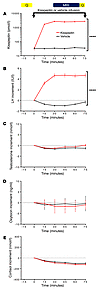
Olfactory dysfunction is broadly associated with neurodevelopmental and neurodegenerative diseases and predicts increased mortality rates in healthy individuals. Conventional measurements of olfactory health assess odor processing pathways within the brain and provide a limited understanding of primary odor detection. Quantification of the olfactory sensory neurons (OSNs), which detect odors within the nasal cavity, would provide insight into the etiology of olfactory dysfunction associated with disease and mortality. Notably, OSNs are continually replenished by adult neurogenesis in mammals, including humans, so OSN measurements are primed to provide specialized insights into neurological disease. Here, we have evaluated a PET radiotracer, [11C]GV1-57, that specifically binds mature OSNs and quantifies the mature OSN population in vivo. [11C]GV1-57 monitored native OSN population dynamics in rodents, detecting OSN generation during postnatal development and aging-associated neurodegeneration. [11C]GV1-57 additionally measured rates of neuron regeneration after acute injury and early-stage OSN deficits in a rodent tauopathy model of neurodegenerative disease. Preliminary assessment in nonhuman primates suggested maintained uptake and saturable binding of [18F]GV1-57 in primate nasal epithelium, supporting its translational potential. Future applications for GV1-57 include monitoring additional diseases or conditions associated with olfactory dysregulation, including cognitive decline, as well as monitoring effects of neuroregenerative or neuroprotective therapeutics.

Authors

Genevieve C. Van de Bittner, Misha M. Riley, Luxiang Cao, Janina Ehses, Scott P. Herrick, Emily L. Ricq, Hsiao-Ying Wey, Michael J. O’Neill, Zeshan Ahmed, Tracey K. Murray, Jaclyn E. Smith, Changning Wang, Frederick A. Schroeder, Mark W. Albers, Jacob M. Hooker

×

Chemogenetic stimulation of striatal projection neurons modulates responses to Parkinson’s disease therapy
Cristina Alcacer, … , Tim Fieblinger, Maria Angela Cenci
Published January 23, 2017
Citation Information: J Clin Invest. 2017. https://doi.org/10.1172/JCI90132.
View: Text | PDF

Chemogenetic stimulation of striatal projection neurons modulates responses to Parkinson’s disease therapy

  • Text
  • PDF
Abstract

Parkinson’s disease (PD) patients experience loss of normal motor function (hypokinesia), but can develop uncontrollable movements known as dyskinesia upon treatment with L-DOPA. Poverty or excess of movement in PD has been attributed to overactivity of striatal projection neurons forming either the indirect (iSPNs) or the direct (dSPNs) pathway, respectively. Here, we investigated the two pathways’ contribution to different motor features using SPN type–specific chemogenetic stimulation in rodent models of PD (PD mice) and L-DOPA–induced dyskinesia (LID mice). Using the activatory Gq-coupled human M3 muscarinic receptor (hM3Dq), we found that chemogenetic stimulation of dSPNs mimicked, while stimulation of iSPNs abolished the therapeutic action of L-DOPA in PD mice. In LID mice, hM3Dq stimulation of dSPNs exacerbated dyskinetic responses to L-DOPA, while stimulation of iSPNs inhibited these responses. In the absence of L-DOPA, only chemogenetic stimulation of dSPNs mediated through the Gs-coupled modified rat muscarinic M3 receptor (rM3Ds) induced appreciable dyskinesia in PD mice. Combining D2 receptor agonist treatment with rM3Ds-dSPN stimulation reproduced all symptoms of LID. These results demonstrate that dSPNs and iSPNs oppositely modulate both therapeutic and dyskinetic responses to dopamine replacement therapy in PD. We also show that chemogenetic stimulation of different signaling pathways in dSPNs leads to markedly different motor outcomes. Our findings have important implications for the design of effective antiparkinsonian and antidyskinetic drug therapies.

Authors

Cristina Alcacer, Laura Andreoli, Irene Sebastianutto, Johan Jakobsson, Tim Fieblinger, Maria Angela Cenci

×
  • ← Previous
  • 1
  • 2
  • …
  • 36
  • 37
  • 38
  • …
  • 63
  • 64
  • Next →
DREAM suppression in Huntington’s disease
José Naranjo and colleagues reveal that downregulation of DREAM mediates derepression of ATF6, and this elevation of ATF6 plays an early neuroprotective role in Huntington’s disease…
Published January 11, 2016
Scientific Show StopperNeuroscience

Extra-cerebellar motor symptoms in Angelman’s syndrome
Caroline Bruinsma and colleagues evaluated cerebellar involvement in Angelman’s Syndrome motor deficits…
Published October 20, 2015
Scientific Show StopperNeuroscience

An epigenetic intervention for neurodegenerative diseases
Eva Benito and colleagues demonstrate that SAHA, a histone-deacetylase inhibitor, improves spatial memory and selectively regulates the neuronal epigenome in a mouse model of neurodegeneration…
Published August 17, 2015
Scientific Show StopperNeuroscience

Genetic and environmental interactions in Parkinson’s disease
Alevtina Zharikov and colleagues reveal that interplay between α-synuclein and environmental toxin exposure influences parkinsonian neurodegeneration…
Published June 15, 2015
Scientific Show StopperNeuroscience

TREM2 keeps myelinated axons under wraps
Pietro Poliani, Yaming Wang, and colleagues demonstrate that TREM2 deficiency reduces age-associated expansion of microglia and microglia-dependent remyelination…
Published April 20, 2015
Scientific Show StopperNeuroscience

Synergy among Parkinson’s disease-associated genes
Durga Meka and colleagues demonstrate that crosstalk between parkin and RET maintains mitochondrial integrity and protects dopaminergic neurons…
Published March 30, 2015
Scientific Show StopperNeuroscience

A model of periventricular leukomalacia
Tamar Licht, Talia Dor-Wollman and colleagues demonstrate that specific vulnerability of immature blood vessels surrounding ventricles predisposes to hypoxia-induced periventricular leukomalacia…
Published February 17, 2015
Scientific Show StopperNeuroscience
Advertisement

Copyright © 2025 American Society for Clinical Investigation
ISSN: 0021-9738 (print), 1558-8238 (online)

Sign up for email alerts