Advertisement
Rapalogs and mTOR inhibitors as anti-aging therapeutics
Dudley W. Lamming,1,2 Lan Ye,3 David M. Sabatini,1,2 and Joseph A. Baur3
1Whitehead Institute for Biomedical Research, Department of Biology, Howard Hughes Medical Institute, and The David H. Koch Institute for Integrative Cancer Research, Massachusetts Institute of Technology, Cambridge, Massachusetts, USA. 2Broad Institute of MIT and Harvard, Cambridge, Massachusetts, USA. 3Institute for Diabetes, Obesity, and Metabolism, and Department of Physiology, Perelman School of Medicine, University of Pennsylvania, Philadelphia, Pennsylvania, USA.
Address correspondence to: Joseph A. Baur, Institute for Diabetes, Obesity, and Metabolism, and Department of Physiology, Perelman School of Medicine, University of Pennsylvania, 12-114 Translational Research Center, 3400 Civic Center Blvd, Philadelphia, Pennsylvania 19104, USA. Phone: 215.573.6543; Fax: 215.898.5408; E-mail: Baur@mail.med.upenn.edu.
Find articles by Lamming, D. in: PubMed | Google Scholar
1Whitehead Institute for Biomedical Research, Department of Biology, Howard Hughes Medical Institute, and The David H. Koch Institute for Integrative Cancer Research, Massachusetts Institute of Technology, Cambridge, Massachusetts, USA. 2Broad Institute of MIT and Harvard, Cambridge, Massachusetts, USA. 3Institute for Diabetes, Obesity, and Metabolism, and Department of Physiology, Perelman School of Medicine, University of Pennsylvania, Philadelphia, Pennsylvania, USA.
Address correspondence to: Joseph A. Baur, Institute for Diabetes, Obesity, and Metabolism, and Department of Physiology, Perelman School of Medicine, University of Pennsylvania, 12-114 Translational Research Center, 3400 Civic Center Blvd, Philadelphia, Pennsylvania 19104, USA. Phone: 215.573.6543; Fax: 215.898.5408; E-mail: Baur@mail.med.upenn.edu.
Find articles by Ye, L. in: PubMed | Google Scholar
1Whitehead Institute for Biomedical Research, Department of Biology, Howard Hughes Medical Institute, and The David H. Koch Institute for Integrative Cancer Research, Massachusetts Institute of Technology, Cambridge, Massachusetts, USA. 2Broad Institute of MIT and Harvard, Cambridge, Massachusetts, USA. 3Institute for Diabetes, Obesity, and Metabolism, and Department of Physiology, Perelman School of Medicine, University of Pennsylvania, Philadelphia, Pennsylvania, USA.
Address correspondence to: Joseph A. Baur, Institute for Diabetes, Obesity, and Metabolism, and Department of Physiology, Perelman School of Medicine, University of Pennsylvania, 12-114 Translational Research Center, 3400 Civic Center Blvd, Philadelphia, Pennsylvania 19104, USA. Phone: 215.573.6543; Fax: 215.898.5408; E-mail: Baur@mail.med.upenn.edu.
Find articles by Sabatini, D. in: PubMed | Google Scholar
1Whitehead Institute for Biomedical Research, Department of Biology, Howard Hughes Medical Institute, and The David H. Koch Institute for Integrative Cancer Research, Massachusetts Institute of Technology, Cambridge, Massachusetts, USA. 2Broad Institute of MIT and Harvard, Cambridge, Massachusetts, USA. 3Institute for Diabetes, Obesity, and Metabolism, and Department of Physiology, Perelman School of Medicine, University of Pennsylvania, Philadelphia, Pennsylvania, USA.
Address correspondence to: Joseph A. Baur, Institute for Diabetes, Obesity, and Metabolism, and Department of Physiology, Perelman School of Medicine, University of Pennsylvania, 12-114 Translational Research Center, 3400 Civic Center Blvd, Philadelphia, Pennsylvania 19104, USA. Phone: 215.573.6543; Fax: 215.898.5408; E-mail: Baur@mail.med.upenn.edu.
Find articles by Baur, J. in: PubMed | Google Scholar
Published March 1, 2013 - More info
J Clin Invest. 2013;123(3):980–989. https://doi.org/10.1172/JCI64099.
© 2013 The American Society for Clinical Investigation
-
A brief history of rapamycin and mechanistic target of rapamycin
Rapamycin was discovered in the soil of Easter Island as a compound produced by Streptomyces hygroscopicus that was capable of inhibiting the proliferation of the yeast Candida albicans but did not affect the growth of bacteria (1). In mammals, rapamycin was found to inhibit the immune response and was subsequently adopted as a standard therapy to prevent graft rejection in transplant recipients and to treat autoimmune disorders (2, 3). Rapamycin also broadly inhibits the growth and proliferation of mammalian cells, spurring more recent interest in its use as a cancer therapy (4).
Mechanistically, rapamycin binds FKBP12, an immunophilin with prolyl isomerase activity. Two additional proteins required for its effects in yeast were identified in a genetic screen in 1991 and termed target of rapamycin 1 (TOR1) and TOR2 (5). During 1994 and 1995, three separate groups isolated a 289-kDa kinase that is bound and inhibited by the rapamycin-FKBP12 complex in mammalian cells (6–8). This kinase is now known as the mechanistic target of rapamycin (mTOR) and is approximately 40% homologous to Saccharomyces cerevisiae TOR proteins and highly conserved among eukaryotes.
mTOR is found in two complexes that have distinct functions and different sensitivities to the action of rapamycin. mTOR complex 1 (mTORC1; consisting of mTOR, raptor, mLST8/GβL, PRAS40, DEPTOR) plays a key role in the regulation of translation and cell growth via phosphorylation of substrates that include S6 kinase (S6K) and eukaryotic initiation factor eIF4E binding protein (4E-BP), and is potently inhibited by rapamycin. In contrast, mTORC2 (consisting of mTOR, rictor, mLST8/GβL, mSIN1, protor, DEPTOR) regulates a diverse set of substrates, including AKT S473, serum/glucocorticoid regulated kinase, and PKC-α, and is acutely resistant to rapamycin, although it can become physically disrupted during chronic exposure. mTORCs receive inputs through a wide variety of signaling mechanisms and have roles in many aspects of physiology, which have been reviewed in depth (9). Briefly, mTORC1 responds to signals that include amino acids, glucose, WNT ligands, oxygen, cAMP, and insulin/IGF-1. The regulation of mTORC2 activity is less clear but may involve interaction with ribosomes (10). Insulin/IGF-1 signaling to mTORC1 is mediated in part by mTORC2 via AKT phosphorylation. In turn, mTORC1 activation feeds back to attenuate insulin/IGF-1 signaling via S6K1 and GRB10 (Figure 1 and ref. 11).
 Figure 1
mTOR signaling. mTOR is found in two complexes, mTORC1 and mTORC2. mTORC1 is regulated in part via the TSC complex, which normally act as a GTPase-activating protein for Rheb to suppress mTORC1 signaling. mTORC1 is also regulated by amino acids via the Ras-related GTP binding (Rag) family of small GTPases. The Rag proteins activate mTORC1 by localizing mTORC1 to the lysosome via interaction with the ragulator complex (110). mTORC1 promotes growth by enhancing ribosomal biogenesis, translation, and other anabolic processes, while inhibiting autophagy. mTORC1 suppresses insulin/IGF-1 signaling via direct regulation of Grb10 and S6K, which subsequently reduces signaling to mTORC2. AKT, an inhibitor of TSC1/2, is one of several direct substrates of mTORC2. Processes that are upregulated by mTOR signaling are shown in red; those that are downregulated by mTOR signaling are shown in blue.

-
Connecting mTOR signaling to aging
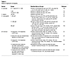
A role for TOR signaling in aging was first revealed in 2003, when Vellai and colleagues showed that RNAi against let-363/CeTor significantly extended the life span of Caenorhabditis elegans and functioned independently from daf-16, a FOXO homolog that had previously been shown to influence life span (12). This was rapidly followed by the demonstration that genetic inhibition of TOR signaling extends life span in Drosophila melanogaster and the budding yeast S. cerevisiae (13, 14). Genetic inhibition of mTOR signaling in mammals is a delicate matter, as the mTOR protein kinase, raptor, rictor, and mLST8 are all essential for development (15). Recently, we demonstrated that female Mtor+/–Mlst8+/– mice have reduced mTORC1 activity and increased longevity, similar to the phenotype reported by Selman and colleagues for mice that lack S6K1, one of the principal substrates of mTORC1 (16, 17). Therefore, the link between mTOR signaling and longevity appears to be conserved from yeast to mammals (Table 1).

-
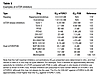
Effects of rapamycin on longevity
Rapamycin extends life span in yeast, worms, and flies (Table 2 and refs. 18–21). In 2009, rapamycin was shown to extend both mean and maximum life spans of male and female genetically heterogeneous mice (offspring of a four-way cross between long-lived, inbred strains) (22). Remarkably, the treatment was not initiated until the mice had reached an advanced age (20 months), roughly equivalent to a human age of 60 years. In a follow-up study beginning at 9 months of age, rapamycin extended median life span in males and females by 10% and 18%, respectively, and maximum life span by 16% and 13% (23). Rapamycin was microencapsulated in an enteric coating that enabled delivery in the food during these studies, and the blood level achieved was approximately three-fold higher than the typical therapeutic range for immunosuppression in humans (24).
Other studies have also found a positive effect of rapamycin on life span. Chen et al. found that rapamycin decreased mortality rate in aged male C57BL/6 mice (25). Anisimov et al. showed that rapamycin extends the maximum life span (mean life span of the last 10% surviving) in a short-lived, tumor-prone strain of mice (FVB/N HER-2/neu transgenic) (26). While this study provides strong evidence that rapamycin can be beneficial in the setting of cancer, the choice of strain makes it hard to separate anticancer effects from aging per se. However, rapamycin also extends life span in 129/Sv mice, an inbred strain with a more typical life span and tumor incidence (27). Impressively, 22.9% of the treated mice remained alive at the death of the last control animal.
Taken together, these observations make rapamycin the best-supported candidate for a mammalian longevity drug. Understanding its mechanism of action has the potential to offer insight into the nature of the underlying aging process and may lead to new therapeutic approaches to alleviate the burden of age-related diseases. However, the mechanism accounting for the anti-aging effects of rapamycin is not yet clear (Table 2).
-
Potential mechanisms of life span extension by rapamycin
Anticancer effects. Cancer is the most common cause of death for laboratory mice, and rapamycin is an anticancer drug. Therefore, it remains possible that life span extension by rapamycin is secondary to tumor suppression and unrelated to the underlying aging process. There are several reasons why we do not favor this model. First, the initial experiments linking rapamycin and mTOR inhibition to longevity were performed in organisms that are mainly postmitotic (worms and flies) or single celled (yeast) and therefore do not experience cancer. Second, rapamycin increases maximum longevity, providing support for the idea that it slows multiple age-related pathologies. Targeting a single disease should not substantially increase the life spans of the longest-lived individuals in a group, as the oldest individuals will be at very high risk for most or all causes of death unless the underlying aging process has been postponed. Third, rapamycin has been shown to delay multiple age-related changes in mice, including loss of stem cell function (25), cognitive decline (28), retinopathy (29), accumulation of subcellular alterations in the myocardium, liver degeneration, endometrial hyperplasia, tendon stiffening, and decline in physical activity (30). Moreover, rapamycin is therapeutic in rodent models of cardiac hypertrophy (31, 32) and neurodegenerative diseases (33–35), conditions that affect aging humans. While cancer prevention clearly plays a major role in the survival benefit conferred by rapamycin, it is important to understand that cancer is an age-related disease, and its prevention is an expected consequence of any therapy that slows aging.
Translation. mTORC1, via S6K and 4E-BP, plays a central role in the regulation of translation, and it is worth considering whether reduced protein synthesis per se might mediate the effects of rapamycin on longevity. For example, decreasing the overall rate of translation might allow better fidelity during synthesis and/or relieve stress on the mechanisms that degrade erroneous, misfolded, or damaged proteins (36). Indeed, experiments in S. cerevisiae, C. elegans, and D. melanogaster have demonstrated that deletion or siRNA-mediated knockdown of ribosomal subunits, S6K, or translation initiation factors results in increased life span and S6K1 deletion extends life span in female mice, whereas 4E-BP deletion blocks the life-extending effects of caloric restriction (CR) in flies (13, 37–40).
Recent findings challenge the view that translation per se is the key to the benefits of TOR/mTOR inhibition. While female mice lacking S6K1 have extended life spans, there is no discernible effect on overall translation, at least in skeletal muscle (41). In addition, the long life spans of worms that lack a key translation initiation factor can still be further increased by TOR deletion, implying that distinct mechanisms are at play (38). Moreover, life span extension due to deletion of translation initiation factors is dependent on daf-16, whereas life span extension by depletion of TOR, S6K, or ribosomal subunits is not, again pointing to the involvement of multiple distinct mechanisms (12, 37). Interestingly, reducing TOR using RNAi fails to further extend the life spans of eat-2 mutant worms, a model for CR, despite suppressing the already low rate of protein synthesis by an additional 49% (37). Furthermore, inactivation of the worm homolog of AMPK is sufficient to suppress life span extension in animals lacking S6K, ostensibly without affecting translation (16). Clearly the relationship between translation and longevity is more complex than initially supposed.
Translation of specific mRNAs may influence life span. While complete loss of mTOR function has a major effect on general translation, rapamycin has a more subtle effect, most likely because a subset of the functions of 4E-BP is rapamycin resistant (42, 43). Both rapamycin and complete mTOR inhibition preferentially suppress translation of mRNAs with 5′ terminal oligopyrimidine motifs, suggesting a potential role for these genes in longevity (43, 44). Despite decreasing overall protein synthesis, CR in flies specifically enhances the translation of a subset of mRNAs that have short and less structured 5′ UTRs, including nuclear-encoded mitochondrial genes (40). The TOR substrate 4E-BP is required for this effect and for life span extension. In yeast lacking ribosomal subunits or TOR, full life span extension requires increased translation of a specific transcript, GCN4 (45). Expression of GCN4 is limited by multiple upstream ORFs that normally sequester ribosomes that bind to the mRNA. Under conditions of decreased TOR activity or large ribosomal subunit abundance, the upstream ORFs are more frequently bypassed to initiate translation of the GCN4 ORF. These examples highlight subtleties in the regulation of translation that we are only beginning to appreciate.
Autophagy. Another effect of mTOR inhibition that has been linked to longevity is the induction of autophagy, a process by which cells recycle their proteins and organelles. Autophagy allows cells to survive nutrient-limited conditions and is a central mechanism by which damaged components are removed. Under conditions of nutrient sufficiency, mTOR phosphorylates and inhibits the autophagy-initiating kinase ULK1 (46). Inactivation of genes involved in autophagy decreases life span in yeast (chronological), C. elegans, and Drosophila, and promotion of autophagy in the fly nervous system extends life span (47–49). Furthermore, autophagy is required for the extension by rapamycin of yeast chronological life span (47) and for life span extension by CR or genetic inhibition of mTOR signaling in worms (50).
In mammals autophagy also appears to play a significant role in the aging process. Most dramatically, the induction of autophagy is sufficient to rejuvenate the liver histology and function of aged mice (51). Furthermore, autophagy seems to be upregulated in CR mice, and to mediate some of the beneficial effects of a CR diet on the heart, liver, and kidneys (52–54). Cells from long-lived Snell dwarf mice also show evidence of increased autophagy (55). Cardiomyocytes isolated from aged mice have lower autophagy and exhibit defects in calcium handling, both of which are corrected by exposure to rapamycin ex vivo (56). However, increased autophagy may not always be beneficial, and indeed may contribute to the pro-aging phenotype of progeroid mice (57).
Interestingly, rapamycin ameliorates nuclear blebbing and premature senescence in cells derived from patients with Hutchinson-Gilford progeria, a rare premature aging syndrome (58). The disease results from a misspliced variant of lamin A, termed progerin, that accumulates to a large degree in patients and is also detected in smaller amounts during normal cellular aging (59, 60). Rapamycin appears to stimulate clearance of progerin from diseased cells via autophagy, and thus may limit the normal age-related accumulation of progerin as well. Overall, the appropriate regulation of autophagy is likely to be a critical determinant of healthy aging.
Stem cell maintenance. Rapamycin has a number of interesting effects on stem cell function. Hyperactive signaling upstream of mTORC1 due to deletion of Pten, deletion of tuberous sclerosis 1 (Tsc1), or constitutive activation of AKT reduces the number and functional capacity of HSCs (61–63). Rapamycin treatment can restore normal self-renewal capacity in a subpopulation of mouse HSCs that have spontaneously high oxidative stress and reduced functional capacity (64). More recently, Chen et al. noted that mTORC1 activity is elevated in HSCs derived from aged mice, which display functional deficits reminiscent of those caused by Tsc1 deletion (25). Rapamycin restored functional capacity in HSCs from aged mice and boosted the immune response to influenza virus. Rapamycin also increases intestinal stem cell self-renewal via inhibition of mTORC1 in the adjacent Paneth cells, similar to effects that have been in observed in CR animals (65). In addition, rapamycin enhances the reprogramming of somatic cells to generate induced pluripotent stem cells, suggesting a general promotion of stem cell function (66). On the other hand, rapamycin impairs pluripotency, reduces proliferation, and promotes differentiation in human embryonic stem cells (67, 68). In mouse embryonic stem cells, expression of pluripotency markers is more resistant to rapamycin treatment, yet cell size and proliferation are still reduced and differentiation is enhanced (67, 69). Intriguingly, rapamycin depletes leukemia-initiating cells and inhibits both the self-renewal and differentiation capacities of stem cells derived from infantile hemangioma, suggesting a protective effect against cancer stem cells (61, 70). Taken together, these results suggest that rapamycin modulates the behavior of stem cells and generally favors the retention of “stemness” and a more youthful phenotype in the adult stem cells types that have been studied.
Antiinflammatory mechanisms. The original clinical use of rapamycin as an immunosuppressant should not be overlooked when it comes to longevity. Chronic, low-grade inflammation is a feature of aging, and almost every chronic disease has an inflammatory component (71). A complete discussion of the immunological effects of rapamycin is beyond the scope of this Review, and the topic has been covered elsewhere (72). Importantly, the drug has both positive and negative effects on innate and adaptive immunity, with a net outcome that is more complex than simple immunosuppression, as exemplified by its ability to enhance the immunization of aged mice against influenza virus (25).
mTORC2-dependent mechanisms. Despite the high specificity of rapamycin for mTORC1 during acute treatment, chronic exposure can also inhibit mTORC2. This effect was first observed in certain cultured cell lines (73), and we have recently shown that it also occurs in vivo in multiple tissues including liver, muscle, and adipose (see Figure 2). It is currently unclear whether inhibition of mTORC2 plays a role in the pro-longevity effects of rapamycin. Female mice lacking S6K1 and female Mtor+/–Mlst8+/– mice are ostensibly long lived due to impairments in mTORC1-dependent signaling, but data from C. elegans suggest that inhibition of mTORC2 can also promote longevity (21, 74). Interestingly, life span extension by disruption of mTORC1 in worms requires skn-1 (the homolog of mammalian NRF1/2) and daf-16 (the homolog of mammalian FOXOs), both transcription factors that control genes involved in stress defenses. Life span extension by rapamycin or mTORC2 disruption, however, requires only SKN-1. Consistent with a role for general stress defenses in the benefits of rapamycin, both worms and flies with impaired TOR function are stress resistant, and induction of NRF1/2 and FOXO target genes has been detected in the livers of mice treated with rapamycin (2 mg/kg daily for two weeks) (20, 21).
 Figure 2
Chronic rapamycin treatment disrupts mTORC2. (A) In vivo, nutrients and growth factors drive the activity of mTORC1 and mTORC2, which promote growth, aging, and insulin sensitivity. (B) Acute treatment with rapamycin inhibits mTORC1 signaling, restricting growth and promoting longevity without reducing insulin sensitivity. (C) Chronic treatment with rapamycin inhibits both mTORC1 and mTORC2, restricting growth and impairing insulin signaling, but promoting longevity.

-
Prospects for safer mTOR inhibitors
Direct inhibition of mTOR. Based on its ability to inhibit cell proliferation, there has been significant interest in treating cancers with rapamycin. Several derivatives of rapamycin (rapalogs) with improved pharmacokinetics have been developed, including temsirolimus, everolimus, ridaforolimus, 32-deoxo-rapamycin, and zotarolimus. Despite intense interest and promising results in animal models of cancer, rapalogs have generally disappointed in human trials and are currently approved only for the treatment of renal cell carcinoma (temsirolimus and everolimus) and for patients with specific types of pancreatic cancer or tuberous sclerosis (reviewed in ref. 84).
One possible explanation for the disappointing results to date is that in human cancer, rapalogs predominately inhibit mTORC1, leading to increased PI3K and AKT signaling by preventing negative feedback through S6K and GRB10 (Figure 1). AKT activity may be attenuated by subsequent mTORC2 disruption during chronic treatment but, if not sufficiently controlled, can promote cancer growth. Pharmaceutical interest has therefore focused on two new classes of compound: mTOR kinase inhibitors that inhibit both mTORC1 and mTORC2, and dual PI3K/mTOR kinase inhibitors (Table 3). mTOR kinase inhibitors such as torin 1 and WYE-125132 in particular have revealed important but previously unknown biological mechanisms, including rapamycin-resistant functions of mTORC1 (85, 86). However, as these compounds strongly inhibit both mTORC1 and mTORC2, it is unlikely that they will prove to have fewer undesirable side effects than rapamycin. One interesting possible exception is caffeine, which is a weak inhibitor of TOR. TOR inhibition mediates life span extension in yeast exposed to caffeine, and it is possible that the dose received from coffee might be sufficient to have a mild effect on mTOR in humans (87).
Indirect inhibitors. Studies showing that S6K1–/– mice and Mtor+/–Mlst8+/– mice have extended longevity (16, 17) suggest that specific inhibition of mTORC1, or perhaps of S6K1, may provide many of the same benefits for age-related diseases as rapamycin. S6K1 inhibitors are now being developed (88), but even if sufficient selectivity is achieved, these compounds will require many years of development before FDA approval.
Fortunately, a number of FDA-approved compounds reduce mTORC1 activity. The most widely used by far is aspirin, which has been shown to decrease S6K phosphorylation in response to TNF-α signaling (89). Aspirin may act in part by inhibiting the phosphorylation of TSC1 by IKKβ (90), but it was recently demonstrated that aspirin can also activate AMPK (91). AMPK inhibits mTORC1 activity through two independent mechanisms, the activating phosphorylation of TSC2 and the inhibitory phosphorylation of raptor, an essential component of mTORC1 (92, 93). We might therefore expect other compounds that activate AMPK to specifically inhibit mTORC1 activity. In fact, this is the case: activation of AMPK by 5-aminoimidazole-4-carboxamide-1β-d-ribonucleoside (AICAR) results in decreased mTORC1 activity (94). Interestingly, aspirin influences longevity in rodent models, extending the average but not maximum life span of male mice (95), and has been found to decrease cancer-related and all-cause mortality in humans (96).
A screen of FDA-approved compounds for regulators of autophagy identified four compounds that reduce mTORC1 activity without affecting mTORC2: perhexiline, niclosamide, rottlerin, and amiodarone (97). Rottlerin regulates mTORC1 in a TSC-dependent fashion, but the mechanisms of action for perhexiline, niclosamide, and amiodarone are TSC independent (97). At least one natural product, phenethyl isothiocyanate, has also been shown to inhibit mTORC1 activity in a TSC-dependent manner (98). Given the wide variety of factors that can influence signaling through the mTOR complexes, many drugs are likely to have downstream effects on these pathways, particularly those that target insulin/IGF-1 signaling.
Is metformin a safer mTOR inhibitor? A widely used FDA-approved AMPK activator is metformin, the first-line drug for the treatment of type 2 diabetes (99). Treatment with metformin lowers blood glucose levels, inhibits lipolysis, and decreases circulating free fatty acids, while producing few undesired side effects (100). The exact mechanism by which metformin acts is uncertain, but much attention has been focused on its ability to activate AMPK (101). Metformin inhibits phosphorylation of the mTORC1 substrates S6K1 and 4E-BP1 and decreases translation (102). While these effects were originally believed to result solely from the action of AMPK, it was recently demonstrated that metformin also regulates mTORC1 directly via inhibition of the Ras-related GTP binding (Rag) GTPases (see Figure 3 and ref. 103) and indirectly via upregulation of REDD1, which promotes TSC2 activity (104).
 Figure 3
Metformin regulates mTORC1 signaling. Metformin activates AMPK by inhibiting oxidative phosphorylation, which in turn negatively regulates mTORC1 signaling via activation of TSC2 and inhibitory phosphorylation of raptor. In parallel, metformin inhibits mTORC1 signaling by suppressing the activity of the Rag GTPases and upregulating REDD1.
Substantial evidence suggests that metformin functions to promote longevity in worms, rodents, and humans. Metformin extends both the life span and health span of the nematode C. elegans (105). These effects are independent of the insulin signaling pathway but are dependent on AMPK, as well as the oxidative stress transcription factor SKN-1/NRF2 (105). Metformin extends the life span of short-lived, tumor-prone HER2/neu mice and female SHR mice (106, 107). The National Institute on Aging Intervention Testing Program is currently treating genetically heterogeneous mice with metformin in order to definitively test its effect on life span. Interestingly, a long-term study in human patients found that treatment with metformin in patients with diabetes decreased mortality from all causes, including diabetes-related mortality, cancer, and myocardial infarction (108, 109). Importantly, an effect on maximum life span in humans or long-lived rodents has yet to be demonstrated.

Article tools
- Download citation information
- Send a comment
- Terms of use
- Standard abbreviations
- Need help? Email the journal
Review Series
Aging
-
The sirtuin family’s role in aging and age-associated pathologies
-
The role of aging upon β cell turnover
-
Causes, consequences, and reversal of immune system aging
-
Cellular senescence and the senescent secretory phenotype: therapeutic opportunities
-
Rapalogs and mTOR inhibitors as anti-aging therapeutics
-
The role of mitochondria in aging
-
Telomeres and age-related disease: how telomere biology informs clinical paradigms
-
Coming of age: molecular drivers of aging and therapeutic opportunities
Metrics
Go to
- Top
- Abstract
- A brief history of rapamycin and mechanistic target of rapamycin
- Connecting mTOR signaling to aging
- Effects of rapamycin on longevity
- Potential mechanisms of life span extension by rapamycin
- mTOR-independent mechanisms
- Rapamycin side effects
- Prospects for safer mTOR inhibitors
- Conclusion
- Acknowledgments
- Footnotes
- References
- Version history


Copyright © 2026 American Society for Clinical Investigation
ISSN: 0021-9738 (print), 1558-8238 (online)