Review Series
Abstract
Therapies based on glucagon-like peptide-1 (GLP-1) reduce rates of cardiovascular and chronic kidney disease in people with type 2 diabetes and/or obesity, with ongoing clinical trials investigating their effects in people with metabolic liver disease, arthritis, and both substance use and neurodegenerative disorders. Acute and chronic activation of GLP-1 receptor signaling also reduces systemic and tissue inflammation in mice and humans, through weight loss–dependent and –independent mechanisms, actions that may contribute to the expanding spectrum of clinical benefits ascribed to GLP-1 medicines. In this Review, we highlight current understanding of the direct and indirect antiinflammatory effects and mechanisms of GLP-1 medicines in both preclinical and clinical studies, covering emerging concepts, clinical relevance, and areas of uncertainty that require further investigation.
Authors
Chi Kin Wong, Daniel J. Drucker
Abstract
Glucagon-like peptide-1 (GLP-1) was initially considered to be a hormone with a predominant role in regulating glucose metabolism by inducing insulin secretion, reducing glucagon secretion, and ameliorating insulin resistance, with the last effect being largely dependent on the induction of weight loss. In more recent years, the role of this peptide beyond metabolism has progressively been explored, including its impact on kidney physiology and kidney clinical outcomes in people with obesity with or without diabetes. Indeed, despite only modest expression of the GLP-1 receptor in the kidney, the renoprotective actions of GLP-1 and its receptor agonists have become an area of intensive investigation. This Review appraises the current status of GLP-1 peptide and its receptor agonists and focuses on the preclinical as well as recent seminal clinical findings defining the kidney benefits conferred by GLP-1 receptor agonist treatment in people living with type 2 diabetes and obesity.
Authors
Mark E. Cooper, Daniël H. van Raalte
Abstract
Cancer diagnoses are prevalent in people with obesity and type 2 diabetes, and abundant clinical evidence supports the protective effects of weight loss for cancer prevention. Glucagon-like peptide-1 (GLP-1) receptor agonists have revolutionized obesity and type 2 diabetes medicine and alleviate many comorbidities of these metabolic diseases. In this Review, we summarize the current clinical evidence for GLP-1 receptor agonists and cancer risk, including thyroid, pancreatic, gastrointestinal, and hormone-dependent malignancies. With few exceptions, recent meta-analyses report that GLP-1 receptor therapies do not increase cancer incidence and may lower risk in some cases. Preclinical studies reinforce the anticancer effects of GLP-1 receptor therapies, even in non-obese models. However, there are still many opportunities for translational insight as the field grows. Immune-modulating effects of GLP-1 receptor agonists are reported in several preclinical cancer studies, which may reflect direct action on immune cells or result from improved metabolic function. We highlight ongoing clinical trials for GLP-1 receptor therapies in cancer patients, and offer considerations for preclinical studies, including perspectives on the timing and duration of GLP-1 receptor agonist treatment, concurrent use of standard anticancer therapies, and interpretation of models of cancer risk versus progression.
Authors
Estefania Valencia-Rincón, Rajani Rai, Vishal Chandra, Elizabeth A. Wellberg
Abstract
Pancreatic cancer (PC) is a devastating disease, due in part to its diagnosis frequently being made at an advanced stage. Ongoing efforts are aimed at identifying early-stage PC in high-risk individuals, as early detection leads to downstaging of PC and improvements in survival. However, there are a myriad of challenges that arise when trying to optimize PC early detection strategies, including selection of the appropriate high-risk individuals and selection of the test or combination of tests that should be performed. Here, we discuss the populations that are the strongest candidates for PC screening and review professional PC screening guidelines. We also summarize the current state of imaging techniques for early detection of PC and further review many studied biomarkers — ranging from nucleic acid targets, proteins, and the microbiome — to highlight the current state of the field and the challenges that remain in the years to come.
Authors
Michael J. Shen, Arsia Jamali, Bryson W. Katona
Abstract
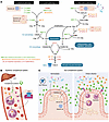
The gut microbiota plays a crucial role in maintaining intestinal homeostasis and influencing various aspects of host physiology, including immune function. Recent advances have highlighted the emerging importance of the complement system, particularly the C3 protein, as a key player in microbiota-host interactions. Traditionally known for its role in innate immunity, the complement system is now recognized for its interactions with microbial communities within the gut, where it promotes immune tolerance and protects against enteric infections. This Review explores the gut complement system as a possibly novel frontier in microbiota-host communication and examines its role in shaping microbial diversity, modulating inflammatory responses, and contributing to intestinal health. We discuss the dynamic interplay between microbiota-derived signals and complement activation, with a focus on the C3 protein and its effect on both the gut microbiome and host immune responses. Furthermore, we highlight the therapeutic potential of targeting complement pathways to restore microbial balance and treat diseases such as inflammatory bowel disease and colorectal cancer. By elucidating the functions of the gut complement system, we offer insights into its potential as a target for microbiota-based interventions aimed at restoring intestinal homeostasis and preventing disease.
Authors
Xianbin Tian, Lan Zhang, Xinyang Qian, Yangqing Peng, Fengyixin Chen, Sarah Bengtson, Zhiqing Wang, Meng Wu
Abstract
The complement system has emerged as a critical regulator of intestinal homeostasis, inflammation, and cancer. In this Review, we explore the multifaceted roles of complement in the gastrointestinal tract, highlighting its canonical and noncanonical functions across intestinal epithelial and immune cells. Under homeostatic conditions, intestinal cells produce complement that maintains barrier integrity and modulates local immune responses, but complement dysregulation contributes to intestinal inflammation and promotes colon cancer. We discuss recent clinical and preclinical studies to provide a cohesive overview of how complement-mediated modulation of immune and nonimmune cell functions can protect or exacerbate inflammation and colon cancer development. The complement system plays a dual role in the intestine, with certain components supporting tissue protection and repair and others exacerbating inflammation. Intriguingly, distinct complement pathways modulate colon cancer progression and response to therapy, with novel findings suggesting that the C3a/C3aR axis constrains early tumor development but may limit antitumor immunity. The recent discovery of intracellular complement activation and tissue-specific complement remains vastly underexplored in the context of intestinal inflammation and colon cancer. Collectively, complement functions are context- and cell-type-dependent, acting both as a shield and a sword in intestinal diseases. Future studies dissecting the temporal and spatial dynamics of complement are essential for leveraging its potential as a biomarker and therapeutic in colon cancer.
Authors
Carsten Krieg, Silvia Guglietta
Abstract
The complement system is an evolutionarily conserved host defense system that has evolved from invertebrates to mammals. Over time, this system has become increasingly appreciated as having effects beyond purely bacterial clearance, with clinically relevant implications in transplantation, particularly lung transplantation. For many years, complement activation in lung transplantation was largely focused on antibody-mediated injuries. However, recent studies have highlighted the importance of both canonical and noncanonical complement activation in shaping adaptive immune responses, which influence alloimmunity. These studies, together with the emergence of FDA-approved complement therapeutics and other drugs in the pipeline that function at different points of the cascade, have led to an increased interest in regulating the complement system to improve donor organ availability as well as improving both short- and long-term outcomes after lung transplantation. In this Review, we provide an overview of the when, what, and how of complement in lung transplantation, posing the questions of: when does complement activation occur, what components of the complement system are activated, and how can this activation be controlled? We conclude that complement activation occurs at multiple stages of the transplant process and that randomized controlled trials will be necessary to realize the therapeutic potential of neutralizing this activation to improve outcomes after lung transplantation.
Authors
Hrishikesh S. Kulkarni, John A. Belperio, Carl Atkinson
Abstract
Metabolic dysfunction–associated steatohepatitis (MASH) is a progressive form of liver disease characterized by hepatocyte injury, inflammation, and fibrosis. The transition from metabolic dysfunction–associated steatotic liver disease (MASLD) to MASH is driven by the accumulation of toxic lipid and metabolic intermediates resulting from increased hepatic uptake of fatty acids, elevated de novo lipogenesis, and impaired mitochondrial oxidation. These changes promote hepatocyte stress and cell death, activate macrophages, and induce a fibrogenic phenotype in hepatic stellate cells (HSCs). Key metabolites, including saturated fatty acids, free cholesterol, ceramides, lactate, and succinate, act as paracrine signals that reinforce inflammatory and fibrotic responses across multiple liver cell types. Crosstalk between hepatocytes, macrophages, and HSCs, along with spatial shifts in mitochondrial activity, creates a feed-forward cycle of immune activation and tissue remodeling. Systemic inputs, such as insulin-resistant adipose tissue and impaired clearance of dietary lipids and branched-chain amino acids, further contribute to liver injury. Together, these pathways establish a metabolically driven network linking nutrient excess to chronic liver inflammation and fibrosis. This Review outlines how coordinated disruptions in lipid metabolism and intercellular signaling drive MASH pathogenesis and provides a framework for understanding disease progression across tissue and cellular compartments.
Authors
Gregory R. Steinberg, Andre C. Carpentier, Dongdong Wang
Abstract
Metabolic dysfunction–associated steatotic liver disease (MASLD), now the most common cause of chronic liver disease, is estimated to affect around 30% of the global population. In MASLD, chronic liver injury can result in scarring or fibrosis, with the degree of fibrosis being the best-known predictor of adverse clinical outcomes. Hence, there is huge interest in developing new therapies to inhibit or reverse fibrosis in MASLD. However, this has been challenging to achieve, as the biology of fibrosis and candidate antifibrotic therapeutic targets have remained poorly described in patient samples. In recent years, the advent of single-cell and spatial omics approaches that can be applied to human samples have started to transform our understanding of fibrosis biology in MASLD. In this Review, we describe these technological advances and discuss the new insights such studies have provided, focusing on the role of epithelial cell plasticity, mesenchymal cell activation, scar-associated macrophage accumulation, and inflammatory cell stimulation as regulators of liver fibrosis. We also consider how omics techniques can enhance our understanding of evolving concepts in the field, such as hot versus cold fibrosis and the mechanisms of liver fibrosis regression. Finally, we touch on future developments and how they are likely to inform a more mechanistic understanding about how fibrosis might differ between patients and how this could influence optimal therapeutic approaches.
Authors
Fabio Colella, Neil C. Henderson, Prakash Ramachandran
Abstract
Inflammatory bowel diseases (IBDs) are complex immune disorders that arise at the intersection of genetic susceptibility, environmental exposures, and dysbiosis of the gut microbiota. Our understanding of the role of the microbiome in IBD has greatly expanded over the past few decades, although efforts to translate this knowledge into precision microbiome-based interventions for the prevention and management of disease have thus far met limited success. Here we survey and synthesize recent primary research in order to propose an updated conceptual framework for the role of the microbiome in IBD. We argue that accounting for gut microbiome context — elements such disease regionality, phase of disease, diet, medication use, and patient lifestyle — is essential for the development of a clear and mechanistic understanding of the microbiome’s contribution to pathogenesis or health. Armed with better mechanistic and contextual understanding, we will be better prepared to translate this knowledge into effective and precise strategies for microbiome restitution.
Authors
Megan S. Kennedy, Eugene B. Chang
No posts were found with this tag.


Copyright © 2025 American Society for Clinical Investigation
ISSN: 0021-9738 (print), 1558-8238 (online)


