
Advertisement
Commentary
Open Access |  10.1172/JCI208767
Good cop, bad cop? Rethinking the roles of cardiac macrophages in injury
Janmes Karunamurthy,1 Ziyi Li,1 and Ajitha Thanabalasuriar1,2
1Department of Pharmacology and Therapeutics and
2Department of Microbiology and Immunology, McGill University, Montreal, Quebec, Canada.
Address correspondence to: Ajitha Thanabalasuriar, McIntyre Medical Sciences Building, 3655 Promenade Sir William Osler, Lab 1320, Montreal, Quebec, H3G 1Y6, Canada. Email: ajitha.thanabalasuriar@mcgill.ca.
Find articles by Karunamurthy, J. in: PubMed | Google Scholar
1Department of Pharmacology and Therapeutics and
2Department of Microbiology and Immunology, McGill University, Montreal, Quebec, Canada.
Address correspondence to: Ajitha Thanabalasuriar, McIntyre Medical Sciences Building, 3655 Promenade Sir William Osler, Lab 1320, Montreal, Quebec, H3G 1Y6, Canada. Email: ajitha.thanabalasuriar@mcgill.ca.
Find articles by Li, Z. in: PubMed | Google Scholar
1Department of Pharmacology and Therapeutics and
2Department of Microbiology and Immunology, McGill University, Montreal, Quebec, Canada.
Address correspondence to: Ajitha Thanabalasuriar, McIntyre Medical Sciences Building, 3655 Promenade Sir William Osler, Lab 1320, Montreal, Quebec, H3G 1Y6, Canada. Email: ajitha.thanabalasuriar@mcgill.ca.
Find articles by Thanabalasuriar, A. in: PubMed | Google Scholar
Published August 3, 2026 - More info
J Clin Invest. 2026;136(15):e208767. https://doi.org/10.1172/JCI208767.
© 2026 Karunamurthy et al. This work is licensed under the Creative Commons Attribution 4.0 International License. To view a copy of this license, visit http://creativecommons.org/licenses/by/4.0/.

Related article:
Abstract
Cardiac macrophages are broadly studied as 2 subtypes, tissue-resident CX3C chemokine motif receptor 1 positive (CX3CR1+) that are also CC motif chemokine receptor 2 negative (CCR2–) and monocyte-derived CCR2+. Previous systemic loss-of-function approaches suggested unique roles for each subtype in the heart, with CCR2+ being inflammatory and CX3CR1+ being prohealing. Here, we employed a cardiac-specific gain-of-function approach to selectively enhance either macrophage subtype. A robust increase in basal CCR2+ macrophages in the heart by targeted CC chemokine ligand 2 (Ccl2) expression did not induce inflammation, cause fibroblast activation, or impair cardiac function. However, increased CCR2+ macrophages reciprocally diminished self-renewing tissue-resident macrophages and worsened cardiac fibrosis due to pressure overload stimulation. Conversely, augmented expression of colony-stimulating factor-1 (Csf1) in the heart promoted selective expansion of resident CX3CR1+ macrophages, which exerted no pathophysiological consequences at steady state. However, pressure overload in these mice with expanded CX3CR1+ macrophages showed a CCR2+ macrophage–dependent inflammation leading to exacerbated cardiac dysfunction, simultaneously protecting from adverse remodeling and cardiac fibrosis. In conclusion, cardiac-specific selective enrichment of macrophage subtypes shows their intricate interplay and unique functional roles in regulating myocardial inflammation and fibrosis during hypertrophy and at homeostasis.
Authors
Rajesh K. Kasam, Ronald J. Vagnozzi, Yasuhide Kuwabara, Anne Katrine Z. Johansen, N. Scott Blair, Vikram Prasad, Suh-Chin J. Lin, Akanksha Rajput, Michelle Nieman, Jeffery D. Molkentin
-
Breaking the dogma
While earlier studies have revealed many details of CMs and their lineages, one major caveat of their results is the use of systemic models (5, 8, 10). Whole-body genetic knockouts and broad pharmacological inhibition are useful research tools, but it is difficult to conclude the results are specific to the cells of interest. Thus, understanding the organ-specific consequences of targeting CMs is required to establish their needed medicinal potential. In this issue of the JCI, Kasam et al. bring clarity to this knowledge gap by utilizing a cardiac-specific gain-of-function approach (11).
Using viral vectors encoding CC chemokine ligand 2 (Ccl2) or colony-stimulating factor-1 (Csf1) under the control of the cardiac troponin promoter, the authors elevated cardiac levels of CCL2 and CSF1 in mice. This led to heart-specific expansion of CCR2+ mo-CMs and CX3CR1+ em-CM populations, respectively. With this approach, Kasam et al. showed that increased numbers of CCR2+ or CX3CR1+ CMs in the heart did not alter baseline cardiac function, which was evaluated for over 1 year after initial stimulation in young and aged animals. Curiously, in aged animals, elevated CSF1 levels promoted proliferation of em-CMs, challenging the prevailing view that these cells lose their replicative capacity with age (19). Additionally, expansion of CCR2+ macrophages in adult mice resulted in a corresponding reduction in a subset of CX3CR1+ macrophages, suggesting competition for shared niches between these populations.
To understand the role of each CM lineage in heart failure, the authors examined the responses of treated mice to TAC-induced pressure overload. Elevated cardiac CCL2 levels amplified inflammatory cytokine production and exacerbated fibrosis 8 weeks after TAC, consistent with the established pathogenic role of CCR2+ mo-CMs (Figure 1A). However, no hypertrophy or decline of systolic function was observed. Yet, TAC in animals with expanded CX3CR1+ em-CMs did cause severe reductions in systolic pumping ability despite no increase in fibrosis relative to the control (Figure 1B). Interestingly, when TAC was performed prior to elevation of CX3CR1+ CMs, no significant changes in cardiac function or fibrosis were observed (Figure 1C). These findings suggest that CX3CR1+ CMs can play both protective and detrimental roles in early heart failure, depending on the timing of their expansion.
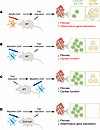 Figure 1
Cardiac macrophages alter cardiac stress responses. Kasam et al. used cardiac-specific strategies to selectively expand either mo-CM or em-CM populations in the context of experimental TAC (11). (A) When treated with AAV9-Ccl2, expansion of CCR2+ mo-CMs increased both fibrosis and inflammatory genes after TAC induction. (B) Mice given MyoAAV-Csf1 before TAC was induced had elevated CX3CR1+ em-CMs and faced a significant decline in heart function alongside a reduced fibrotic response. (C) Wild-type mice challenged with TAC prior to MyoAAV-Csf1 treatment had preserved cardiac function compared with B, with similar decrease in fibrosis observed. (D) Ccr2-null mice were administered MyoAAV-Csf1, producing high numbers of only em-CMs in the heart. After TAC stimulation, these mice showed reduced fibrosis and reduced inflammatory gene expression and no decline in heart function compared with controls or the MyoAAV-Csf1–treated mice in B.
Kasam et al. went on to show compelling mechanistic insights by examining the potential pathological role of CCR2+ CMs in em-CM expanded hearts after TAC induction. In animals lacking CCR2+ CMs, elevated cardiac Csf1 expression followed by TAC no longer produced the previously observed decline in cardiac function. Instead, these mice exhibited a reduced inflammatory profile by 8 weeks post-TAC while maintaining the low-fibrosis phenotype as seen in mice with high cardiac Csf1 alone (Figure 1D). These results suggest that although CCR2+ mo-CMs are key drivers of adverse outcomes in heart failure, the CX3CR1+ CM population may play a role in coordinating the pro-inflammatory effects through communications with CCR2+ CMs.


Copyright © 2026 American Society for Clinical Investigation
ISSN: 0021-9738 (print), 1558-8238 (online)