
Advertisement
Commentary
Open Access |  10.1172/JCI207921
Ammonia in the crosshairs: microbial targets for metabolic dysfunction–associated steatohepatitis prevention
Vanessa A. Leone1 and Arion Kennedy2
1Department of Animal & Dairy Sciences, University of Wisconsin-Madison, Madison, Wisconsin, USA.
2Department of Molecular and Structural Biochemistry, College of Agriculture and Life Sciences, North Carolina State University, Raleigh, North Carolina, USA.
Address correspondence to: Arion Kennedy, North Carolina State University, Department of Molecular and Structural Biochemistry, 355 Polk Hall, Raleigh, North Carolina 27695, USA. Phone: 919.513.4347; Email: akmidget@ncsu.edu. Or to: Vanessa A. Leone, University of Wisconsin-Madison, Department of Animal & Dairy Sciences, Meat Science & Animal Biologics Discovery Building, Room 2118, 1933 Observatory Dr., Madison, Wisconsin 53706, USA. Phone: 608.262.5551; Email: valeone@wisc.edu.
Find articles by
Leone, V.
in:
PubMed
|
Google Scholar
|

1Department of Animal & Dairy Sciences, University of Wisconsin-Madison, Madison, Wisconsin, USA.
2Department of Molecular and Structural Biochemistry, College of Agriculture and Life Sciences, North Carolina State University, Raleigh, North Carolina, USA.
Address correspondence to: Arion Kennedy, North Carolina State University, Department of Molecular and Structural Biochemistry, 355 Polk Hall, Raleigh, North Carolina 27695, USA. Phone: 919.513.4347; Email: akmidget@ncsu.edu. Or to: Vanessa A. Leone, University of Wisconsin-Madison, Department of Animal & Dairy Sciences, Meat Science & Animal Biologics Discovery Building, Room 2118, 1933 Observatory Dr., Madison, Wisconsin 53706, USA. Phone: 608.262.5551; Email: valeone@wisc.edu.
Find articles by Kennedy, A. in: PubMed | Google Scholar
Published July 1, 2026 - More info
J Clin Invest. 2026;136(13):e207921. https://doi.org/10.1172/JCI207921.
© 2026 Leone et al. This work is licensed under the Creative Commons Attribution 4.0 International License. To view a copy of this license, visit http://creativecommons.org/licenses/by/4.0/.

Related article:
Abstract
The global prevalence of metabolic dysfunction–associated steatohepatitis (MASH) is rising, driven by a complex interplay of metabolic disturbances, inflammation, and fibrosis, yet effective treatment options remain limited. This study examined the relationships among intestinal microbial dysbiosis, ammonia production, and hepatic CD8+ T cell activity in MASH, then assessed the therapeutic potential of DT-109, a glycine-based tripeptide. We investigated the gut/liver axis across human cohorts and both nonhuman primate and mouse MASH models. Multiomics approaches were used to characterize ileal microbiota, ammonia levels, and hepatic immune and metabolic pathways. Causality was verified through microbiota transplantation, C. perfringens NirA-knockout mutants, and functional validation in vitro and in vivo. The efficacy of DT-109 was evaluated in nonhuman primates and mice. Our results revealed a significant increase in the ammonia-producing gut bacterium C. perfringens, which led to elevated intestinal ammonia and disruption of the intestinal barrier in MASH. Elevated ammonia levels triggered FosB-mediated upregulation of CCL5 in CD8+ T cells, which in turn drove T cell cytotoxicity in the liver. Notably, DT-109 effectively lowered C. perfringens abundance, reduced intestinal ammonia, restored intestinal barrier integrity, and alleviated CD8+ T cell dysregulation in MASH. These results identify a distinct mechanism in which gut-derived ammonia drives CD8+ T cell–mediated MASH and demonstrate that DT-109 effectively targets this axis by inhibiting C. perfringens and reducing ammonia, ultimately ameliorating MASH.
Authors
Pengxiang Qu, Shusi Ding, Yanru Zhang, Yang Zhao, Erfei Song, Liangshuo Hu, Ruike Ding, Wenbin Cao, Yiting Hou, Jia Qi, Juan Zhao, Chenjing Duan, Shuangqing Liu, Chong Shen, Ying Zhao, Yanhong Guo, Zuowen Zheng, Shiwei Luo, Huizhong Hu, Liang Bai, Sihai Zhao, Bo Wang, Shuixiang He, Yi Wu, Xuelian Xiong, Qiutong Wu, Weiwang Gu, Oren Rom, Aimin Xu, Lemin Zheng, Jifeng Zhang, Enqi Liu, Y. Eugene Chen
-
Barrier integrity and CD8+ T cell polarization in MASH
A key advance of the study by Qu et al. is the identification of a spatially resolved mechanistic axis linking microbial ammonia production in the ileum to epithelial cell dysfunction and hepatic immune remodeling (Figure 1). Elevated ammonia has previously been shown to impair epithelial cell integrity through mitochondrial depolarization, oxidative stress, and inflammatory signaling via NF-κB, thereby promoting cytokine production and cellular injury (12). In animal models, elevated ammonia is further associated with loss of tight junction proteins, goblet cell depletion, mucus disruption, and recruitment of inflammatory cells that amplify tissue damage (13, 14).
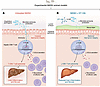 Figure 1
DT-109 inhibits microbial ammonia-driven CD8+ T cell activation in experimental MASH. (A) Qu et al. showed that ammonia production by nitrite reductase A (NirA)–expressing C. perfringens disrupts gut barrier function in diet-induced MASH models, which leads to increased exposure of hepatic CD8+ T cells to ammonia. Increased ammonia levels drive CCL5 expression mediated through FosB in hepatic CD8+ T cells, enhancing recruitment and cytotoxicity of effector cells. This resulted in severe liver inflammation and fibrosis. (B) In experimental MASH animal models, treatment with DT-109, a glycine-based tripeptide, inhibited NirA-mediated ammonia production in C. perfringens, resulting in downregulation of FosB-mediated CCL5 expression and reduced CD8+ T cell activation, effectively reducing liver injury and fibrosis (5).
Consistent with these observations, Qu et al. further demonstrated that microbial ammonia production by C. perfringens disrupted ileal barrier integrity, reducing expression of the tight junction protein occludin and perturbing the mucus layer by reducing mucin 2 protein expression in experimental MASH. Barrier dysfunction may facilitate microbial translocation, cytokine and damage-associated molecular pattern production, and antigen localization to regional lymph nodes, thereby promoting inflammatory immune activation.
Hepatic CD8+ T cells are established drivers of liver inflammation, immune cell infiltration, and fibrosis in MASH (15). Under MASH conditions, hepatic CD8+ T cell activation is shaped by cytokine signaling such as IL-15, antigen presentation, microbial metabolites such as acetate, and the gut microbiota (15, 16). Using heat-inactivated and nitrite reductase A (NirA–deficient) C. perfringens, Qu et al. demonstrated that bacterial ammonia production promoted upregulation of the chemokine CCL5 in hepatic CD8+ T cells, enhancing T cell cytotoxicity in experimental MASH. Mechanistically, ammonia directly induced FosB-mediated CCL5 expression in CD8+ T cells in vitro, linking this microbial metabolite directly to pathogenic hepatic CD8+ T cell polarization. Together with its effects on ileal barrier integrity, these findings position ammonia as a central mediator of gut-immune communication in MASH.
Beyond its effects on immunity through disruptions in barrier integrity, ammonia may also directly alter immune cell metabolism. Ammonia is detoxified primarily through glutamine synthetase–mediated conversion to glutamine, which can serve as a carbon and nitrogen donor or be hydrolyzed back to glutamate and ammonia by glutaminase (9). In immune cells, altered glutamine flux influences mTOR signaling, redox balance, and glycolytic programming (17). Elevated ammonia has been shown to induce lysosomal damage, mitochondrial dysfunction, and ROS production in CD8+ T cells, resulting in impaired metabolic fitness and reduced effector responses (18). In human CD8+ T cells, ammonia exposure reduces proliferation, IFN-γ and TNF-α production, degranulation, and antigen-specific cytotoxic responses (18). In contrast, Qu et al.’s findings suggest ammonia can also promote pathogenic CD8+ T cell activation in MASH, enhancing effector recruitment and cytotoxicity. Together, these results point to an important unresolved question regarding the extent to which ammonia directly reprograms hepatic resident CD8+ T cell subsets versus indirectly modulating peripheral CD8+ T cells that traffic to the liver. Qu et al. provided initial insight through single-cell RNA-sequencing analysis, demonstrating that DT-109 treatment altered transcriptional programs within hepatic T cell subsets in experimental MASH. Understanding these bidirectional interactions between barrier integrity, cytokine signaling, and local metabolites will be essential for the development of MASLD/MASH therapies that restore barrier function while reprogramming CD8+ T cell responses toward protective, rather than pathogenic, states.
Article tools
- Download citation information
- Send a comment
- Terms of use
- Standard abbreviations
- Need help? Email the journal
Metrics
Go to


Copyright © 2026 American Society for Clinical Investigation
ISSN: 0021-9738 (print), 1558-8238 (online)