Go to JCI Insight
  • About
  • Editors
  • Consulting Editors
  • For authors
  • Publication ethics
  • Publication alerts by email
  • Advertising
  • Job board
  • Contact
  • Clinical Research and Public Health
  • Current issue
  • Past issues
  • By specialty
    • COVID-19
    • Cardiology
    • Gastroenterology
    • Immunology
    • Metabolism
    • Nephrology
    • Neuroscience
    • Oncology
    • Pulmonology
    • Vascular biology
    • All ...
  • Videos
    • Conversations with Giants in Medicine
    • Video Abstracts
  • Reviews
    • View all reviews ...
    • Complement Biology and Therapeutics (May 2025)
    • Evolving insights into MASLD and MASH pathogenesis and treatment (Apr 2025)
    • Microbiome in Health and Disease (Feb 2025)
    • Substance Use Disorders (Oct 2024)
    • Clonal Hematopoiesis (Oct 2024)
    • Sex Differences in Medicine (Sep 2024)
    • Vascular Malformations (Apr 2024)
    • View all review series ...
  • Viewpoint
  • Collections
    • In-Press Preview
    • Clinical Research and Public Health
    • Research Letters
    • Letters to the Editor
    • Editorials
    • Commentaries
    • Editor's notes
    • Reviews
    • Viewpoints
    • 100th anniversary
    • Top read articles

  • Current issue
  • Past issues
  • Specialties
  • Reviews
  • Review series
  • Conversations with Giants in Medicine
  • Video Abstracts
  • In-Press Preview
  • Clinical Research and Public Health
  • Research Letters
  • Letters to the Editor
  • Editorials
  • Commentaries
  • Editor's notes
  • Reviews
  • Viewpoints
  • 100th anniversary
  • Top read articles
  • About
  • Editors
  • Consulting Editors
  • For authors
  • Publication ethics
  • Publication alerts by email
  • Advertising
  • Job board
  • Contact

Concise Communications

  • 148 Articles
  • 0 Posts
  • ← Previous
  • 1
  • 2
  • …
  • 13
  • 14
  • 15
  • Next →
Activation of tumor suppressor protein PP2A inhibits KRAS-driven tumor growth
Jaya Sangodkar, … , Michael Ohlmeyer, Goutham Narla
Published May 15, 2017
Citation Information: J Clin Invest. 2017;127(6):2081-2090. https://doi.org/10.1172/JCI89548.
View: Text | PDF

Activation of tumor suppressor protein PP2A inhibits KRAS-driven tumor growth

  • Text
  • PDF
Abstract

Targeted cancer therapies, which act on specific cancer-associated molecular targets, are predominantly inhibitors of oncogenic kinases. While these drugs have achieved some clinical success, the inactivation of kinase signaling via stimulation of endogenous phosphatases has received minimal attention as an alternative targeted approach. Here, we have demonstrated that activation of the tumor suppressor protein phosphatase 2A (PP2A), a negative regulator of multiple oncogenic signaling proteins, is a promising therapeutic approach for the treatment of cancers. Our group previously developed a series of orally bioavailable small molecule activators of PP2A, termed SMAPs. We now report that SMAP treatment inhibited the growth of KRAS-mutant lung cancers in mouse xenografts and transgenic models. Mechanistically, we found that SMAPs act by binding to the PP2A Aα scaffold subunit to drive conformational changes in PP2A. These results show that PP2A can be activated in cancer cells to inhibit proliferation. Our strategy of reactivating endogenous PP2A may be applicable to the treatment of other diseases and represents an advancement toward the development of small molecule activators of tumor suppressor proteins.

Authors

Jaya Sangodkar, Abbey Perl, Rita Tohme, Janna Kiselar, David B. Kastrinsky, Nilesh Zaware, Sudeh Izadmehr, Sahar Mazhar, Danica D. Wiredja, Caitlin M. O’Connor, Divya Hoon, Neil S. Dhawan, Daniela Schlatzer, Shen Yao, Daniel Leonard, Alain C. Borczuk, Giridharan Gokulrangan, Lifu Wang, Elena Svenson, Caroline C. Farrington, Eric Yuan, Rita A. Avelar, Agnes Stachnik, Blake Smith, Vickram Gidwani, Heather M. Giannini, Daniel McQuaid, Kimberly McClinch, Zhizhi Wang, Alice C. Levine, Rosalie C. Sears, Edward Y. Chen, Qiaonan Duan, Manish Datt, Shozeb Haider, Avi Ma’ayan, Analisa DiFeo, Neelesh Sharma, Matthew D. Galsky, David L. Brautigan, Yiannis A. Ioannou, Wenqing Xu, Mark R. Chance, Michael Ohlmeyer, Goutham Narla

×

Reducing expression of synapse-restricting protein Ephexin5 ameliorates Alzheimer’s-like impairment in mice
Gabrielle L. Sell, … , Thomas B. Schaffer, Seth S. Margolis
Published March 27, 2017
Citation Information: J Clin Invest. 2017;127(5):1646-1650. https://doi.org/10.1172/JCI85504.
View: Text | PDF

Reducing expression of synapse-restricting protein Ephexin5 ameliorates Alzheimer’s-like impairment in mice

  • Text
  • PDF
Abstract

Accumulation of amyloid-β (Aβ) protein may cause synapse degeneration and cognitive impairment in Alzheimer’s disease (AD) by reactivating expression of the developmental synapse repressor protein Ephexin5 (also known as ARHGEF15). Here, we have reported that Aβ is sufficient to acutely promote the production of Ephexin5 in mature hippocampal neurons and in mice expressing human amyloid precursor protein (hAPP mice), a model for familial AD that produces high brain levels of Aβ. Ephexin5 expression was highly elevated in the hippocampi of human AD patients, indicating its potential relevance to AD. We also observed elevated Ephexin5 expression in the hippocampi of hAPP mice. Removal of Ephexin5 expression eliminated hippocampal dendritic spine loss and rescued AD-associated behavioral deficits in the hAPP mice. Furthermore, selective reduction of Ephexin5 expression using shRNA in the dentate gyrus of presymptomatic adolescent hAPP mice was sufficient to protect these mice from developing cognitive impairment. Thus, pathological elevation of Ephexin5 expression critically drives Aβ-induced memory impairment, and strategies aimed at reducing Ephexin5 levels may represent an effective approach to treating AD.

Authors

Gabrielle L. Sell, Thomas B. Schaffer, Seth S. Margolis

×

Kisspeptin modulates sexual and emotional brain processing in humans
Alexander N. Comninos, … , Stephen R. Bloom, Waljit S. Dhillo
Published January 23, 2017
Citation Information: J Clin Invest. 2017;127(2):709-719. https://doi.org/10.1172/JCI89519.
View: Text | PDF

Kisspeptin modulates sexual and emotional brain processing in humans

  • Text
  • PDF
Abstract

BACKGROUND. Sex, emotion, and reproduction are fundamental and tightly entwined aspects of human behavior. At a population level in humans, both the desire for sexual stimulation and the desire to bond with a partner are important precursors to reproduction. However, the relationships between these processes are incompletely understood. The limbic brain system has key roles in sexual and emotional behaviors, and is a likely candidate system for the integration of behavior with the hormonal reproductive axis. We investigated the effects of kisspeptin, a recently identified key reproductive hormone, on limbic brain activity and behavior.

METHODS. Using a combination of functional neuroimaging and hormonal and psychometric analyses, we compared the effects of kisspeptin versus vehicle administration in 29 healthy heterosexual young men.

RESULTS. We demonstrated that kisspeptin administration enhanced limbic brain activity specifically in response to sexual and couple-bonding stimuli. Furthermore, kisspeptin’s enhancement of limbic brain structures correlated with psychometric measures of reward, drive, mood, and sexual aversion, providing functional significance. In addition, kisspeptin administration attenuated negative mood.

CONCLUSIONS. Collectively, our data provide evidence of an undescribed role for kisspeptin in integrating sexual and emotional brain processing with reproduction in humans. These results have important implications for our understanding of reproductive biology and are highly relevant to the current pharmacological development of kisspeptin as a potential therapeutic agent for patients with common disorders of reproductive function.

FUNDING. National Institute for Health Research (NIHR), Wellcome Trust (Ref 080268), and the Medical Research Council (MRC).

Authors

Alexander N. Comninos, Matthew B. Wall, Lysia Demetriou, Amar J. Shah, Sophie A. Clarke, Shakunthala Narayanaswamy, Alexander Nesbitt, Chioma Izzi-Engbeaya, Julia K. Prague, Ali Abbara, Risheka Ratnasabapathy, Victoria Salem, Gurjinder M. Nijher, Channa N. Jayasena, Mark Tanner, Paul Bassett, Amrish Mehta, Eugenii A. Rabiner, Christoph Hönigsperger, Meire Ribeiro Silva, Ole Kristian Brandtzaeg, Elsa Lundanes, Steven Ray Wilson, Rachel C. Brown, Sarah A. Thomas, Stephen R. Bloom, Waljit S. Dhillo

×

Mechanism for leptin’s acute insulin-independent effect to reverse diabetic ketoacidosis
Rachel J. Perry, … , Gary W. Cline, Gerald I. Shulman
Published January 23, 2017
Citation Information: J Clin Invest. 2017;127(2):657-669. https://doi.org/10.1172/JCI88477.
View: Text | PDF

Mechanism for leptin’s acute insulin-independent effect to reverse diabetic ketoacidosis

  • Text
  • PDF
Abstract

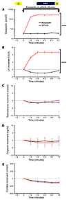
The mechanism by which leptin reverses diabetic ketoacidosis (DKA) is unknown. We examined the acute insulin-independent effects of leptin replacement therapy in a streptozotocin-induced rat model of DKA. Leptin infusion reduced rates of lipolysis, hepatic glucose production (HGP), and hepatic ketogenesis by 50% within 6 hours and were independent of any changes in plasma glucagon concentrations; these effects were abrogated by coinfusion of corticosterone. Treating leptin- and corticosterone-infused rats with an adipose triglyceride lipase inhibitor blocked corticosterone-induced increases in plasma glucose concentrations and rates of HGP and ketogenesis. Similarly, adrenalectomized type 1 diabetic (T1D) rats exhibited decreased rates of lipolysis, HGP, and ketogenesis; these effects were reversed by corticosterone infusion. Leptin-induced decreases in lipolysis, HGP, and ketogenesis in DKA were also nullified by relatively small increases (15 to 70 pM) in plasma insulin concentrations. In contrast, the chronic glucose-lowering effect of leptin in a STZ-induced mouse model of poorly controlled T1D was associated with decreased food intake, reduced plasma glucagon and corticosterone concentrations, and decreased ectopic lipid (triacylglycerol/diacylglycerol) content in liver and muscle. Collectively, these studies demonstrate marked differences in the acute insulin-independent effects by which leptin reverses fasting hyperglycemia and ketoacidosis in a rodent model of DKA versus the chronic pleotropic effects by which leptin reverses hyperglycemia in a non-DKA rodent model of T1D.

Authors

Rachel J. Perry, Liang Peng, Abudukadier Abulizi, Lynn Kennedy, Gary W. Cline, Gerald I. Shulman

×

Blocking type I interferon signaling enhances T cell recovery and reduces HIV-1 reservoirs
Liang Cheng, … , Lishan Su, Liguo Zhang
Published December 12, 2016
Citation Information: J Clin Invest. 2017;127(1):269-279. https://doi.org/10.1172/JCI90745.
View: Text | PDF

Blocking type I interferon signaling enhances T cell recovery and reduces HIV-1 reservoirs

  • Text
  • PDF
Abstract

Despite the efficient suppression of HIV-1 replication that can be achieved with combined antiretroviral therapy (cART), low levels of type I interferon (IFN-I) signaling persist in some individuals. This sustained signaling may impede immune recovery and foster viral persistence. Here we report studies using a monoclonal antibody to block IFN-α/β receptor (IFNAR) signaling in humanized mice (hu-mice) that were persistently infected with HIV-1. We discovered that effective cART restored the number of human immune cells in HIV-1–infected hu-mice but did not rescue their immune hyperactivation and dysfunction. IFNAR blockade fully reversed HIV-1–induced immune hyperactivation and rescued anti–HIV-1 immune responses in T cells from HIV-1–infected hu-mice. Finally, we found that IFNAR blockade in the presence of cART reduced the size of HIV-1 reservoirs in lymphoid tissues and delayed HIV-1 rebound after cART cessation in the HIV-1–infected hu-mice. We conclude that low levels of IFN-I signaling contribute to HIV-1–associated immune dysfunction and foster HIV-1 persistence in cART-treated hosts. Our results suggest that blocking IFNAR may provide a potential strategy to enhance immune recovery and reduce HIV-1 reservoirs in individuals with sustained elevations in IFN-I signaling during suppressive cART.

Authors

Liang Cheng, Jianping Ma, Jingyun Li, Dan Li, Guangming Li, Feng Li, Qing Zhang, Haisheng Yu, Fumihiko Yasui, Chaobaihui Ye, Li-Chung Tsao, Zhiyuan Hu, Lishan Su, Liguo Zhang

×

Targeting CAG repeat RNAs reduces Huntington’s disease phenotype independently of huntingtin levels
Laura Rué, … , Xavier Estivill, Eulàlia Martí
Published October 10, 2016
Citation Information: J Clin Invest. 2016;126(11):4319-4330. https://doi.org/10.1172/JCI83185.
View: Text | PDF

Targeting CAG repeat RNAs reduces Huntington’s disease phenotype independently of huntingtin levels

  • Text
  • PDF
Abstract

Huntington’s disease (HD) is a polyglutamine disorder caused by a CAG expansion in the Huntingtin (HTT) gene exon 1. This expansion encodes a mutant protein whose abnormal function is traditionally associated with HD pathogenesis; however, recent evidence has also linked HD pathogenesis to RNA stable hairpins formed by the mutant HTT expansion. Here, we have shown that a locked nucleic acid–modified antisense oligonucleotide complementary to the CAG repeat (LNA-CTG) preferentially binds to mutant HTT without affecting HTT mRNA or protein levels. LNA-CTGs produced rapid and sustained improvement of motor deficits in an R6/2 mouse HD model that was paralleled by persistent binding of LNA-CTG to the expanded HTT exon 1 transgene. Motor improvement was accompanied by a pronounced recovery in the levels of several striatal neuronal markers severely impaired in R6/2 mice. Furthermore, in R6/2 mice, LNA-CTG blocked several pathogenic mechanisms caused by expanded CAG RNA, including small RNA toxicity and decreased Rn45s expression levels. These results suggest that LNA-CTGs promote neuroprotection by blocking the detrimental activity of CAG repeats within HTT mRNA. The present data emphasize the relevance of expanded CAG RNA to HD pathogenesis, indicate that inhibition of HTT expression is not required to reverse motor deficits, and further suggest a therapeutic potential for LNA-CTG in polyglutamine disorders.

Authors

Laura Rué, Mónica Bañez-Coronel, Jordi Creus-Muncunill, Albert Giralt, Rafael Alcalá-Vida, Gartze Mentxaka, Birgit Kagerbauer, M. Teresa Zomeño-Abellán, Zeus Aranda, Veronica Venturi, Esther Pérez-Navarro, Xavier Estivill, Eulàlia Martí

×

Adipocyte-specific deletion of Ip6k1 reduces diet-induced obesity by enhancing AMPK-mediated thermogenesis
Qingzhang Zhu, … , James C. Barrow, Anutosh Chakraborty
Published October 4, 2016
Citation Information: J Clin Invest. 2016;126(11):4273-4288. https://doi.org/10.1172/JCI85510.
View: Text | PDF

Adipocyte-specific deletion of Ip6k1 reduces diet-induced obesity by enhancing AMPK-mediated thermogenesis

  • Text
  • PDF
Abstract

Enhancing energy expenditure (EE) is an attractive strategy to combat obesity and diabetes. Global deletion of Ip6k1 protects mice from diet-induced obesity (DIO) and insulin resistance, but the tissue-specific mechanism by which IP6K1 regulates body weight is unknown. Here, we have demonstrated that IP6K1 regulates fat accumulation by modulating AMPK-mediated adipocyte energy metabolism. Cold exposure led to downregulation of Ip6k1 in murine inguinal and retroperitoneal white adipose tissue (IWAT and RWAT) depots. Adipocyte-specific deletion of Ip6k1 (AdKO) enhanced thermogenic EE, which protected mice from high-fat diet–induced weight gain at ambient temperature (23°C), but not at thermoneutral temperature (30°C). AdKO-induced increases in thermogenesis also protected mice from cold-induced decreases in body temperature. UCP1, PGC1α, and other markers of browning and thermogenesis were elevated in IWAT and RWAT of AdKO mice. Cold-induced activation of sympathetic signaling was unaltered, whereas AMPK was enhanced, in AdKO IWAT. Moreover, beige adipocytes from AdKO IWAT displayed enhanced browning, which was diminished by AMPK depletion. Furthermore, we determined that IP6 and IP6K1 differentially regulate upstream kinase-mediated AMPK stimulatory phosphorylation in vitro. Finally, treating mildly obese mice with the IP6K inhibitor TNP enhanced thermogenesis and inhibited progression of DIO. Thus, IP6K1 regulates energy metabolism via a mechanism that could potentially be targeted in obesity.

Authors

Qingzhang Zhu, Sarbani Ghoshal, Ana Rodrigues, Su Gao, Alice Asterian, Theodore M. Kamenecka, James C. Barrow, Anutosh Chakraborty

×

Tumor immune profiling predicts response to anti–PD-1 therapy in human melanoma
Adil I. Daud, … , Matthew F. Krummel, Michael D. Rosenblum
Published August 15, 2016
Citation Information: J Clin Invest. 2016;126(9):3447-3452. https://doi.org/10.1172/JCI87324.
View: Text | PDF

Tumor immune profiling predicts response to anti–PD-1 therapy in human melanoma

  • Text
  • PDF
Abstract

BACKGROUND. Immune checkpoint blockade is revolutionizing therapy for advanced cancer, but many patients do not respond to treatment. The identification of robust biomarkers that predict clinical response to specific checkpoint inhibitors is critical in order to stratify patients and to rationally select combinations in the context of an expanding array of therapeutic options.

METHODS. We performed multiparameter flow cytometry on freshly isolated metastatic melanoma samples from 2 cohorts of 20 patients each prior to treatment and correlated the subsequent clinical response with the tumor immune phenotype.

RESULTS. Increasing fractions of programmed cell death 1 high/cytotoxic T lymphocyte–associated protein 4 high (PD-1hiCTLA-4hi) cells within the tumor-infiltrating CD8+ T cell subset strongly correlated with response to therapy (RR) and progression-free survival (PFS). Functional analysis of these cells revealed a partially exhausted T cell phenotype. Assessment of metastatic lesions during anti–PD-1 therapy demonstrated a release of T cell exhaustion, as measured by an accumulation of highly activated CD8+ T cells within tumors, with no effect on Tregs.

CONCLUSIONS. Our data suggest that the relative abundance of partially exhausted tumor-infiltrating CD8+ T cells predicts response to anti–PD-1 therapy. This information can be used to appropriately select patients with a high likelihood of achieving a clinical response to PD-1 pathway inhibition.

FUNDING. This work was funded by a generous gift provided by Inga-Lill and David Amoroso as well as a generous gift provided by Stephen Juelsgaard and Lori Cook.

Authors

Adil I. Daud, Kimberly Loo, Mariela L. Pauli, Robert Sanchez-Rodriguez, Priscila Munoz Sandoval, Keyon Taravati, Katy Tsai, Adi Nosrati, Lorenzo Nardo, Michael D. Alvarado, Alain P. Algazi, Miguel H. Pampaloni, Iryna V. Lobach, Jimmy Hwang, Robert H. Pierce, Iris K. Gratz, Matthew F. Krummel, Michael D. Rosenblum

×

Recurrent EZH1 mutations are a second hit in autonomous thyroid adenomas
Davide Calebiro, … , Luca Persani, Ralf Paschke
Published August 8, 2016
Citation Information: J Clin Invest. 2016;126(9):3383-3388. https://doi.org/10.1172/JCI84894.
View: Text | PDF

Recurrent EZH1 mutations are a second hit in autonomous thyroid adenomas

  • Text
  • PDF
Abstract

Autonomous thyroid adenomas (ATAs) are a frequent cause of hyperthyroidism. Mutations in the genes encoding the TSH receptor (TSHR) or the Gs protein α subunit (GNAS) are found in approximately 70% of ATAs. The involvement of other genes and the pathogenesis of the remaining cases are presently unknown. Here, we performed whole-exome sequencing in 19 ATAs that were paired with normal DNA samples and identified a recurrent hot-spot mutation (c.1712A>G; p.Gln571Arg) in the enhancer of zeste homolog 1 (EZH1) gene, which codes for a catalytic subunit of the polycomb complex. Targeted screening in an independent cohort confirmed that this mutation occurs with high frequency (27%) in ATAs. EZH1 mutations were strongly associated with known (TSHR, GNAS) or presumed (adenylate cyclase 9 [ADCY9]) alterations in cAMP pathway genes. Furthermore, functional studies revealed that the p.Gln571Arg EZH1 mutation caused increased histone H3 trimethylation and increased proliferation of thyroid cells. In summary, this study revealed that a hot-spot mutation in EZH1 is the second most frequent genetic alteration in ATAs. The association between EZH1 and TSHR mutations suggests a 2-hit model for the pathogenesis of these tumors, whereby constitutive activation of the cAMP pathway and EZH1 mutations cooperate to induce the hyperproliferation of thyroid cells.

Authors

Davide Calebiro, Elisa S. Grassi, Markus Eszlinger, Cristina L. Ronchi, Amod Godbole, Kerstin Bathon, Fabiana Guizzardi, Tiziana de Filippis, Knut Krohn, Holger Jaeschke, Thomas Schwarzmayr, Rifat Bircan, Hulya Iliksu Gozu, Seda Sancak, Marek Niedziela, Tim M. Strom, Martin Fassnacht, Luca Persani, Ralf Paschke

×

Destabilized SMC5/6 complex leads to chromosome breakage syndrome with severe lung disease
Saskia N. van der Crabben, … , Johanne M. Murray, Gijs van Haaften
Published July 18, 2016
Citation Information: J Clin Invest. 2016;126(8):2881-2892. https://doi.org/10.1172/JCI82890.
View: Text | PDF

Destabilized SMC5/6 complex leads to chromosome breakage syndrome with severe lung disease

  • Text
  • PDF
Abstract

The structural maintenance of chromosomes (SMC) family of proteins supports mitotic proliferation, meiosis, and DNA repair to control genomic stability. Impairments in chromosome maintenance are linked to rare chromosome breakage disorders. Here, we have identified a chromosome breakage syndrome associated with severe lung disease in early childhood. Four children from two unrelated kindreds died of severe pulmonary disease during infancy following viral pneumonia with evidence of combined T and B cell immunodeficiency. Whole exome sequencing revealed biallelic missense mutations in the NSMCE3 (also known as NDNL2) gene, which encodes a subunit of the SMC5/6 complex that is essential for DNA damage response and chromosome segregation. The NSMCE3 mutations disrupted interactions within the SMC5/6 complex, leading to destabilization of the complex. Patient cells showed chromosome rearrangements, micronuclei, sensitivity to replication stress and DNA damage, and defective homologous recombination. This work associates missense mutations in NSMCE3 with an autosomal recessive chromosome breakage syndrome that leads to defective T and B cell function and acute respiratory distress syndrome in early childhood.

Authors

Saskia N. van der Crabben, Marije P. Hennus, Grant A. McGregor, Deborah I. Ritter, Sandesh C.S. Nagamani, Owen S. Wells, Magdalena Harakalova, Ivan K. Chinn, Aaron Alt, Lucie Vondrova, Ron Hochstenbach, Joris M. van Montfrans, Suzanne W. Terheggen-Lagro, Stef van Lieshout, Markus J. van Roosmalen, Ivo Renkens, Karen Duran, Isaac J. Nijman, Wigard P. Kloosterman, Eric Hennekam, Jordan S. Orange, Peter M. van Hasselt, David A. Wheeler, Jan J. Palecek, Alan R. Lehmann, Antony W. Oliver, Laurence H. Pearl, Sharon E. Plon, Johanne M. Murray, Gijs van Haaften

×
  • ← Previous
  • 1
  • 2
  • …
  • 13
  • 14
  • 15
  • Next →

No posts were found with this tag.

Advertisement

Copyright © 2025 American Society for Clinical Investigation
ISSN: 0021-9738 (print), 1558-8238 (online)

Sign up for email alerts