
Submit a comment
Abstract
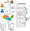
Effector CD8+ T cells are key drivers of type 1 diabetes (T1D) pathogenesis, yet questions remain regarding the molecular defects leading to altered cytotoxicity, peripheral tissue phenotype, and receptor specificity. We analyzed human pancreatic lymph nodes (pLNs) using mass cytometry and single-cell RNA-seq (scRNA-seq) with combined T cell receptor (TCR) profiling. Cytometric analysis revealed enrichment of T stem cell memory–like (TSCM-like) cells (CD8+CD45RA+CD27+CD28+CCR7+CXCR3+) in T1D pLNs. scRNA-seq indicated an elevated inflammatory cytokine gene signature (IFITM3, LTB) along with regulators of terminal differentiation (BCL6, BCL3), coupled with downregulation of exhaustion-associated genes (DUSP2, NR4A2, TSC22D3) in CD8+ T cells in T1D pLNs. Immune response enrichment analysis (IREA) indicated IL-15 signaling as a significant driver of these phenotypes. Integrated TCR and transcriptomics analysis revealed a cluster of diverse naive-like CD8+ T cell clones in T1D pLNs. Comparison of pLNs and pancreatic tissue slice isolates indicated sharing of effector CD8+ T cells, with enhanced terminal effector signatures within the pancreas relative to paired pLNs. Multiplex imaging revealed differential localization of T cell factor 1 (TCF1)- and thymocyte selection-associated high mobility group box protein (TOX)-expressing T cells in the pancreas, with islet-proximal TCF1+TOX+ cells displaying a mixture of activation and exhaustion-associated phenotypes. Thus, we provide multimodal cellular profiles enriched in T1D tissues for consideration in therapeutic targeting.
Authors
Leeana D. Peters, Howard R. Seay, Justin A. Smith, Amanda L. Posgai, Reed L. Berkowitz, Clive H. Wasserfall, Mark A. Atkinson, Rhonda Bacher, Maigan A. Brusko, Todd M. Brusko
Guidelines
The Editorial Board will only consider comments that are deemed relevant and of interest to readers. The Journal will not post data that have not been subjected to peer review; or a comment that is essentially a reiteration of another comment.
- Comments appear on the Journal’s website and are linked from the original article’s web page.
- Authors are notified by email if their comments are posted.
- The Journal reserves the right to edit comments for length and clarity.
- No appeals will be considered.
- Comments are not indexed in PubMed.
Specific requirements
- Maximum length, 400 words
- Entered as plain text or HTML
- Author’s name and email address, to be posted with the comment
- Declaration of all potential conflicts of interest (even if these are not ultimately posted); see the Journal’s conflict-of-interest policy
- Comments may not include figures


Copyright © 2026 American Society for Clinical Investigation
ISSN: 0021-9738 (print), 1558-8238 (online)