
Advertisement
Research ArticleCell biologyInflammationNeuroscience
Open Access |  10.1172/JCI209546
Edited HSPCs rescue frataxin deficiency and inflammation-driven multisystemic pathology in Friedreich’s ataxia mice
Anusha Sivakumar,1 Alexis N. Corl,1 Keneth Jordan Muñoz,1 Subhashini Arumugam,1 Betul B. Gunes,1 Angelyn B. Solis,1 Rishika Murthy,1 Lilas Rony El-Hachem,1 Donald P. Pizzo,2 Ian A. Blair,3,4 Teerapat Rojsajjakul,3,4 Clementina Mesaros,3,4 Xinlong Wan,1 Joseph N. Rainaldi,1 Rafael A. Badell-Grau,1 Veenita Khare,1 Kevin Eric Thai,1 Kavya Biederman,1 and Stephanie Cherqui1
1Department of Pediatrics, Division of Genetics, and
2Department of Pathology, University of California, San Diego, La Jolla, California, USA.
3Penn/CHOP, Friedreich’s Ataxia Center of Excellence, Philadelphia, Pennsylvania, USA.
4Center of Excellence in Environmental Toxicology, Department of Systems Pharmacology and Translational Therapeutics, Perelman School of Medicine, University of Pennsylvania, Philadelphia, Pennsylvania, USA.
Address correspondence to: Stephanie Cherqui, Department of Pediatrics, Division of Genetics, University of California, San Diego, 9500 Gilman Drive, MC 0734, La Jolla, California 92093-0734, USA. Phone: 858.822.1023; Email: scherqui@ucsd.edu.
Find articles by Sivakumar, A. in: PubMed | Google Scholar
1Department of Pediatrics, Division of Genetics, and
2Department of Pathology, University of California, San Diego, La Jolla, California, USA.
3Penn/CHOP, Friedreich’s Ataxia Center of Excellence, Philadelphia, Pennsylvania, USA.
4Center of Excellence in Environmental Toxicology, Department of Systems Pharmacology and Translational Therapeutics, Perelman School of Medicine, University of Pennsylvania, Philadelphia, Pennsylvania, USA.
Address correspondence to: Stephanie Cherqui, Department of Pediatrics, Division of Genetics, University of California, San Diego, 9500 Gilman Drive, MC 0734, La Jolla, California 92093-0734, USA. Phone: 858.822.1023; Email: scherqui@ucsd.edu.
Find articles by Corl, A. in: PubMed | Google Scholar
1Department of Pediatrics, Division of Genetics, and
2Department of Pathology, University of California, San Diego, La Jolla, California, USA.
3Penn/CHOP, Friedreich’s Ataxia Center of Excellence, Philadelphia, Pennsylvania, USA.
4Center of Excellence in Environmental Toxicology, Department of Systems Pharmacology and Translational Therapeutics, Perelman School of Medicine, University of Pennsylvania, Philadelphia, Pennsylvania, USA.
Address correspondence to: Stephanie Cherqui, Department of Pediatrics, Division of Genetics, University of California, San Diego, 9500 Gilman Drive, MC 0734, La Jolla, California 92093-0734, USA. Phone: 858.822.1023; Email: scherqui@ucsd.edu.
Find articles by Muñoz, K. in: PubMed | Google Scholar
1Department of Pediatrics, Division of Genetics, and
2Department of Pathology, University of California, San Diego, La Jolla, California, USA.
3Penn/CHOP, Friedreich’s Ataxia Center of Excellence, Philadelphia, Pennsylvania, USA.
4Center of Excellence in Environmental Toxicology, Department of Systems Pharmacology and Translational Therapeutics, Perelman School of Medicine, University of Pennsylvania, Philadelphia, Pennsylvania, USA.
Address correspondence to: Stephanie Cherqui, Department of Pediatrics, Division of Genetics, University of California, San Diego, 9500 Gilman Drive, MC 0734, La Jolla, California 92093-0734, USA. Phone: 858.822.1023; Email: scherqui@ucsd.edu.
Find articles by Arumugam, S. in: PubMed | Google Scholar
1Department of Pediatrics, Division of Genetics, and
2Department of Pathology, University of California, San Diego, La Jolla, California, USA.
3Penn/CHOP, Friedreich’s Ataxia Center of Excellence, Philadelphia, Pennsylvania, USA.
4Center of Excellence in Environmental Toxicology, Department of Systems Pharmacology and Translational Therapeutics, Perelman School of Medicine, University of Pennsylvania, Philadelphia, Pennsylvania, USA.
Address correspondence to: Stephanie Cherqui, Department of Pediatrics, Division of Genetics, University of California, San Diego, 9500 Gilman Drive, MC 0734, La Jolla, California 92093-0734, USA. Phone: 858.822.1023; Email: scherqui@ucsd.edu.
Find articles by Gunes, B. in: PubMed | Google Scholar
1Department of Pediatrics, Division of Genetics, and
2Department of Pathology, University of California, San Diego, La Jolla, California, USA.
3Penn/CHOP, Friedreich’s Ataxia Center of Excellence, Philadelphia, Pennsylvania, USA.
4Center of Excellence in Environmental Toxicology, Department of Systems Pharmacology and Translational Therapeutics, Perelman School of Medicine, University of Pennsylvania, Philadelphia, Pennsylvania, USA.
Address correspondence to: Stephanie Cherqui, Department of Pediatrics, Division of Genetics, University of California, San Diego, 9500 Gilman Drive, MC 0734, La Jolla, California 92093-0734, USA. Phone: 858.822.1023; Email: scherqui@ucsd.edu.
Find articles by Solis, A. in: PubMed | Google Scholar
1Department of Pediatrics, Division of Genetics, and
2Department of Pathology, University of California, San Diego, La Jolla, California, USA.
3Penn/CHOP, Friedreich’s Ataxia Center of Excellence, Philadelphia, Pennsylvania, USA.
4Center of Excellence in Environmental Toxicology, Department of Systems Pharmacology and Translational Therapeutics, Perelman School of Medicine, University of Pennsylvania, Philadelphia, Pennsylvania, USA.
Address correspondence to: Stephanie Cherqui, Department of Pediatrics, Division of Genetics, University of California, San Diego, 9500 Gilman Drive, MC 0734, La Jolla, California 92093-0734, USA. Phone: 858.822.1023; Email: scherqui@ucsd.edu.
Find articles by Murthy, R. in: PubMed | Google Scholar
1Department of Pediatrics, Division of Genetics, and
2Department of Pathology, University of California, San Diego, La Jolla, California, USA.
3Penn/CHOP, Friedreich’s Ataxia Center of Excellence, Philadelphia, Pennsylvania, USA.
4Center of Excellence in Environmental Toxicology, Department of Systems Pharmacology and Translational Therapeutics, Perelman School of Medicine, University of Pennsylvania, Philadelphia, Pennsylvania, USA.
Address correspondence to: Stephanie Cherqui, Department of Pediatrics, Division of Genetics, University of California, San Diego, 9500 Gilman Drive, MC 0734, La Jolla, California 92093-0734, USA. Phone: 858.822.1023; Email: scherqui@ucsd.edu.
Find articles by El-Hachem, L. in: PubMed | Google Scholar
1Department of Pediatrics, Division of Genetics, and
2Department of Pathology, University of California, San Diego, La Jolla, California, USA.
3Penn/CHOP, Friedreich’s Ataxia Center of Excellence, Philadelphia, Pennsylvania, USA.
4Center of Excellence in Environmental Toxicology, Department of Systems Pharmacology and Translational Therapeutics, Perelman School of Medicine, University of Pennsylvania, Philadelphia, Pennsylvania, USA.
Address correspondence to: Stephanie Cherqui, Department of Pediatrics, Division of Genetics, University of California, San Diego, 9500 Gilman Drive, MC 0734, La Jolla, California 92093-0734, USA. Phone: 858.822.1023; Email: scherqui@ucsd.edu.
Find articles by Pizzo, D. in: PubMed | Google Scholar
1Department of Pediatrics, Division of Genetics, and
2Department of Pathology, University of California, San Diego, La Jolla, California, USA.
3Penn/CHOP, Friedreich’s Ataxia Center of Excellence, Philadelphia, Pennsylvania, USA.
4Center of Excellence in Environmental Toxicology, Department of Systems Pharmacology and Translational Therapeutics, Perelman School of Medicine, University of Pennsylvania, Philadelphia, Pennsylvania, USA.
Address correspondence to: Stephanie Cherqui, Department of Pediatrics, Division of Genetics, University of California, San Diego, 9500 Gilman Drive, MC 0734, La Jolla, California 92093-0734, USA. Phone: 858.822.1023; Email: scherqui@ucsd.edu.
Find articles by Blair, I. in: PubMed | Google Scholar
1Department of Pediatrics, Division of Genetics, and
2Department of Pathology, University of California, San Diego, La Jolla, California, USA.
3Penn/CHOP, Friedreich’s Ataxia Center of Excellence, Philadelphia, Pennsylvania, USA.
4Center of Excellence in Environmental Toxicology, Department of Systems Pharmacology and Translational Therapeutics, Perelman School of Medicine, University of Pennsylvania, Philadelphia, Pennsylvania, USA.
Address correspondence to: Stephanie Cherqui, Department of Pediatrics, Division of Genetics, University of California, San Diego, 9500 Gilman Drive, MC 0734, La Jolla, California 92093-0734, USA. Phone: 858.822.1023; Email: scherqui@ucsd.edu.
Find articles by Rojsajjakul, T. in: PubMed | Google Scholar
1Department of Pediatrics, Division of Genetics, and
2Department of Pathology, University of California, San Diego, La Jolla, California, USA.
3Penn/CHOP, Friedreich’s Ataxia Center of Excellence, Philadelphia, Pennsylvania, USA.
4Center of Excellence in Environmental Toxicology, Department of Systems Pharmacology and Translational Therapeutics, Perelman School of Medicine, University of Pennsylvania, Philadelphia, Pennsylvania, USA.
Address correspondence to: Stephanie Cherqui, Department of Pediatrics, Division of Genetics, University of California, San Diego, 9500 Gilman Drive, MC 0734, La Jolla, California 92093-0734, USA. Phone: 858.822.1023; Email: scherqui@ucsd.edu.
Find articles by Mesaros, C. in: PubMed | Google Scholar
1Department of Pediatrics, Division of Genetics, and
2Department of Pathology, University of California, San Diego, La Jolla, California, USA.
3Penn/CHOP, Friedreich’s Ataxia Center of Excellence, Philadelphia, Pennsylvania, USA.
4Center of Excellence in Environmental Toxicology, Department of Systems Pharmacology and Translational Therapeutics, Perelman School of Medicine, University of Pennsylvania, Philadelphia, Pennsylvania, USA.
Address correspondence to: Stephanie Cherqui, Department of Pediatrics, Division of Genetics, University of California, San Diego, 9500 Gilman Drive, MC 0734, La Jolla, California 92093-0734, USA. Phone: 858.822.1023; Email: scherqui@ucsd.edu.
Find articles by Wan, X. in: PubMed | Google Scholar
1Department of Pediatrics, Division of Genetics, and
2Department of Pathology, University of California, San Diego, La Jolla, California, USA.
3Penn/CHOP, Friedreich’s Ataxia Center of Excellence, Philadelphia, Pennsylvania, USA.
4Center of Excellence in Environmental Toxicology, Department of Systems Pharmacology and Translational Therapeutics, Perelman School of Medicine, University of Pennsylvania, Philadelphia, Pennsylvania, USA.
Address correspondence to: Stephanie Cherqui, Department of Pediatrics, Division of Genetics, University of California, San Diego, 9500 Gilman Drive, MC 0734, La Jolla, California 92093-0734, USA. Phone: 858.822.1023; Email: scherqui@ucsd.edu.
Find articles by Rainaldi, J. in: PubMed | Google Scholar
1Department of Pediatrics, Division of Genetics, and
2Department of Pathology, University of California, San Diego, La Jolla, California, USA.
3Penn/CHOP, Friedreich’s Ataxia Center of Excellence, Philadelphia, Pennsylvania, USA.
4Center of Excellence in Environmental Toxicology, Department of Systems Pharmacology and Translational Therapeutics, Perelman School of Medicine, University of Pennsylvania, Philadelphia, Pennsylvania, USA.
Address correspondence to: Stephanie Cherqui, Department of Pediatrics, Division of Genetics, University of California, San Diego, 9500 Gilman Drive, MC 0734, La Jolla, California 92093-0734, USA. Phone: 858.822.1023; Email: scherqui@ucsd.edu.
Find articles by Badell-Grau, R. in: PubMed | Google Scholar
1Department of Pediatrics, Division of Genetics, and
2Department of Pathology, University of California, San Diego, La Jolla, California, USA.
3Penn/CHOP, Friedreich’s Ataxia Center of Excellence, Philadelphia, Pennsylvania, USA.
4Center of Excellence in Environmental Toxicology, Department of Systems Pharmacology and Translational Therapeutics, Perelman School of Medicine, University of Pennsylvania, Philadelphia, Pennsylvania, USA.
Address correspondence to: Stephanie Cherqui, Department of Pediatrics, Division of Genetics, University of California, San Diego, 9500 Gilman Drive, MC 0734, La Jolla, California 92093-0734, USA. Phone: 858.822.1023; Email: scherqui@ucsd.edu.
Find articles by Khare, V. in: PubMed | Google Scholar
1Department of Pediatrics, Division of Genetics, and
2Department of Pathology, University of California, San Diego, La Jolla, California, USA.
3Penn/CHOP, Friedreich’s Ataxia Center of Excellence, Philadelphia, Pennsylvania, USA.
4Center of Excellence in Environmental Toxicology, Department of Systems Pharmacology and Translational Therapeutics, Perelman School of Medicine, University of Pennsylvania, Philadelphia, Pennsylvania, USA.
Address correspondence to: Stephanie Cherqui, Department of Pediatrics, Division of Genetics, University of California, San Diego, 9500 Gilman Drive, MC 0734, La Jolla, California 92093-0734, USA. Phone: 858.822.1023; Email: scherqui@ucsd.edu.
Find articles by Thai, K. in: PubMed | Google Scholar
1Department of Pediatrics, Division of Genetics, and
2Department of Pathology, University of California, San Diego, La Jolla, California, USA.
3Penn/CHOP, Friedreich’s Ataxia Center of Excellence, Philadelphia, Pennsylvania, USA.
4Center of Excellence in Environmental Toxicology, Department of Systems Pharmacology and Translational Therapeutics, Perelman School of Medicine, University of Pennsylvania, Philadelphia, Pennsylvania, USA.
Address correspondence to: Stephanie Cherqui, Department of Pediatrics, Division of Genetics, University of California, San Diego, 9500 Gilman Drive, MC 0734, La Jolla, California 92093-0734, USA. Phone: 858.822.1023; Email: scherqui@ucsd.edu.
Find articles by Biederman, K. in: PubMed | Google Scholar
1Department of Pediatrics, Division of Genetics, and
2Department of Pathology, University of California, San Diego, La Jolla, California, USA.
3Penn/CHOP, Friedreich’s Ataxia Center of Excellence, Philadelphia, Pennsylvania, USA.
4Center of Excellence in Environmental Toxicology, Department of Systems Pharmacology and Translational Therapeutics, Perelman School of Medicine, University of Pennsylvania, Philadelphia, Pennsylvania, USA.
Address correspondence to: Stephanie Cherqui, Department of Pediatrics, Division of Genetics, University of California, San Diego, 9500 Gilman Drive, MC 0734, La Jolla, California 92093-0734, USA. Phone: 858.822.1023; Email: scherqui@ucsd.edu.
Find articles by Cherqui, S. in: PubMed | Google Scholar
Published October 15, 2026 - More info
J Clin Invest. 2026;136(20):e209546. https://doi.org/10.1172/JCI209546.
© 2026 Sivakumar et al. This work is licensed under the Creative Commons Attribution 4.0 International License. To view a copy of this license, visit http://creativecommons.org/licenses/by/4.0/.
Received: May 28, 2026; Accepted: July 23, 2026

Related article:
Abstract
Friedreich’s ataxia (FRDA) is a triplet repeat expansion disorder with multisystem, progressive symptoms caused by reduced frataxin protein levels and compromised mitochondrial function. Restoration of frataxin protein is a focus of clinical trials involving adeno-associated viral vectors, but substantial barriers exist because of the immune response they elicit. In this issue of the JCI, Sivakumar et al. harnessed CRISPR/Cas9 editing of endogenous hematopoietic stem and progenitor cells (HSPCs) to excise the frataxin gene (FXN) repeat expansion ex vivo, followed by autologous transplantation. A single, systemic infusion of corrected HSPCs resulted in long-term engraftment and sustained multiorgan benefit in a humanized mouse model of FRDA. This improvement in brain, cardiac, and pancreatic symptoms, mediated by myeloid progeny of the engrafted stem cells, provides strong support for the potential effectiveness of this approach in humans and bypasses the need to correct the FXN deficiency in the affected tissue and cells directly.
Authors
Lucy R. Osborne
-
Results
Transplantation of mPPL-001 leads to successful long-term engraftment of mPPL-001–derived cells in hematopoietic compartments and primary organs affected by FRDA. The therapeutic efficacy of mPPL-001 was assessed in the humanized YG8JR mouse model of FRDA, which harbors an hFXN transgene with > 800 GAA repeats on a murine fxn-null background (mfxn–/– hFXN+) (52). Therefore, this model has the key advantage of enabling the use of our previously optimized hFXN gene–editing ribonucleoprotein (RNP) complex (previously called 4RNPEnh) (48), comprising a high-fidelity Cas9 nuclease and 2 sgRNAs (UP4 and DN4) flanking the GAA expansion mutation in hFXN and delivered via electroporation in the presence of an electroporation facilitator (48). Genome-wide off-target activity of UP4 and DN4 sgRNAs was assessed by ONE-seq against the hg38 human reference genome, the Human Genome Diversity Project, and variants from the 1000 Genomes Project (Supplemental Figure 1A; supplemental material available online with this article; https://doi.org/10.1172/JCI209546DS1). This analysis nominated 52,109 potential off-target sites for UP4 and 45,998 for DN4 (Supplemental Figure 1A). Subsequent next-generation sequencing of in vitro cleavage products generated using the RNP complex and off-target DNA libraries identified fewer than 5 tier 1 sites (>1% the rate of on-target editing with low functional annotation) and fewer than 71 tier 2 sites (>1% the rate of on-target editing with low functional annotation) and predominantly within intronic/noncoding regions at low frequency of editing, supporting high specificity and a low off-target risk profile for both guides (Supplemental Figure 1B).
To generate the murine surrogate for evaluating the in vivo therapeutic impact of PPL-001, Sca1+ bone marrow HSPCs (analogous to human CD34+ HSPCs) were isolated from YG8JR mice and edited with RNP complex to produce mPPL-001 or mock electroporated without Cas9 (mock HSPCs) (Figure 1A). Sca1+ HSPCs from multiple YG8JR donors were pooled for the generation mPPL-001 and mock HSPCs. Average on-target hFXN GAA excision efficiency in Sca1+ HSPCs ranged from 7.6% to 30.5%, with a mean of 20.3% (Figure 1B). This variability likely reflects donor-to-donor biological differences of Sca1+ HSPCs prior to electroporation. mPPL-001 retained in vitro colony-forming capacity comparable with that of mock HSPCs (Supplemental Figure 1C).
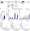 Figure 1
mPPL-001–derived cells engraft in hematopoietic and FRDA-affected tissues in YG8JR mice. (A) Schematic of generation and transplantation of mPPL-001 (created with BioRender). (B) hFXN GAA excision efficiency in mPPL-001 assessed 7 days after electroporation (mock HSPCs, n = 5; mPPL-001, n = 10). (C) Engraftment of mPPL-001–derived cells in hematopoietic tissues and organs affected by FRDA (mock = 2M/2F and mPPL-001= 10M/3F for all tissues except cerebellum [n = 12] and pancreas [n = 10]). (D) Regression analysis of excision efficiency in mPPL-001 versus mPPL-001–derived cells engrafted in bone marrow (P = 0.075; F = 3.879; r2 = 0.26). (E) Regression analysis of mPPL-001–derived cells in bone marrow versus engraftment in spleen (P = 0.03; F = 6.351; r2 = 0.36) and thymus (P = 0.057; F = 4.494; r2 = 0.29). Data are presented as mean ± SEM.
mPPL-001 or mock HSPCs were transplanted into lethally irradiated 2.0- to 2.5-month-old YG8JR recipients (mPPL-001 = 13, 10 male/3 female [10M/3F]; mock HSPC = 11, 6M/5F) and analyzed 6.5–7 months post-transplantation. Age-matched WT (n = 9; 6M/3F) and untreated YG8JR (n = 9; 5M/4F) littermates served as controls. Engraftment and biodistribution analyses revealed a substantial proportion of mPPL-001–derived cells within the hematopoietic compartments, with mean chimerism level of 7.6% in bone marrow, 5.9% in peripheral blood, 26.5% in the spleen, and 19.8% in the thymus (Figure 1C). Additionally, mPPL-001–derived cells were also detected in multiple FRDA-affected tissues, including the cerebellum (0.52%), cerebrum and brain stem (0.55%), spinal cord (1.09%), heart (2.19%), eye (1.38%), pancreas (3.13%), and skeletal muscle (4.14%) (Figure 1C). The ex vivo hFXN GAA excision efficiency in mPPL-001 correlated with the level of mPPL-001–derived cells in the bone marrow (P = 0.075; F = 3.879; r2 = 0.26), which in turn correlated with chimerism in spleen (P = 0.03; F = 6.351; r2 = 0.36) and thymus (P = 0.057; F = 4.494; r2 = 0.29) (Figure 1, D and E). These data demonstrate the durable engraftment of mPPL-001 within the bone marrow niche and its widespread presence across multiple tissues, including those affected by the disease.
mPPL-001 transplantation leads to increased hFXN expression and improves locomotor function and cerebellar integrity in YG8JR mice. The humanized YG8JR mice develop progressive locomotor deficits and cerebellar atrophy, beginning around 9 months of age (53). Catwalk gait analysis revealed that untreated and mock HSPC–transplanted YG8JR mice exhibited significant alterations in gait patterns, including prolonged left hind paw swing duration, reduced duty cycle, coupling of hind paws, and imbalanced frequency of cruciate step sequence, cruciate steps A and B, compared with WT mice (Figure 2A). Transplantation of mPPL-001 significantly improved these deficits (Figure 2A) and demonstrated trends of positive correlation between excision efficiency in mPPL-001 and hematopoietic engraftment of mPPL-001 with improvements in left hind swing duration (Supplemental Figure 2A). Histological analysis of cerebellar architecture using H&E staining revealed significant thinning of both the GCL and molecular layer (ML) in untreated and mock HSPC–transplanted YG8JR mice compared with WT controls (Figure 2B). Consistent with these findings, NeuN immunostaining confirmed marked neuronal loss within the GCL of untreated YG8JR and mock mice compared with WT (Figure 2C). Transplantation of mPPL-001 significantly preserved cerebellar integrity, maintaining GCL neuronal density and ML thickness compared with both YG8JR and mock mice, although values remained significantly lower than WT controls (Figure 2, B and C). Notably, GCL and ML thickness was reduced in pretransplant, 2.0- to 2.5-month-old YG8JR mice (age of transplant) compared with WT mice, and mPPL-001 transplantation preserved this architecture at 8 months of age (Figure 2D). These findings indicate that mPPL-001 effectively stabilizes established cerebellar pathology, preventing further progression, although it does not reverse existing damage.
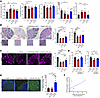 Figure 2
Transplantation of mPPL-001 preserves gait performance and cerebellar architecture in YG8JR mice. (A) Catwalk gait parameters in YG8JR mice transplanted with mPPL-001 (YG8JR/mPPL-001) compared with untreated (YG8JR) and mock HSPC–transplanted (YG8JR/mock HSPC) YG8JR controls and WT littermates (WT). One mock HSPC recipient was excluded from analysis owing to weak paw prints and noncompliant runs (WT = 6M/3F, YG8JR = 5M/4F, mock = 5M/4F, and mPPL-001 = 10M/3F). (B) Representative H&E cerebellar images and quantification of GCL and ML thickness (WT = 6M/3F, YG8JR = 5M/4F, mock = 6M/5F, and mPPL-001 = 10M/3F) (C) Representative NeuN images of cerebellum and quantification of GCL thickness. (D) GCL and ML thickness in 2.0- to 2.5-month-old YG8JR mice (age at transplant) and 8-month-old mPPL-001 recipients. (E) Representative images and quantification of hFXN immunostaining. Scale bars: 50 μm. (F) Regression analysis of bone marrow engraftment versus cerebellar hFXN expression in mPPL-001 recipients. Data are presented as mean ± SEM, analyzed by 1-way ANOVA. *P ≤ 0.05, **P ≤ 0.01, ***P ≤ 0.001, and ****P ≤ 0.0001.
To assess whether improved gait and cerebellar neuroprotection were associated with changes in FXN expression, cerebellar hFXN was evaluated by immunofluorescence. A significant increase in hFXN expression was observed in the cerebellum of mPPL-001 recipients compared with both untreated and mock cohorts (Figure 2E). Cerebellar hFXN expression levels positively correlated with bone marrow engraftment of mPPL-001–derived cells (P = 0.04; F = 5.28; r2 = 0.32) (Figure 2F), indicating that higher GAA excision efficiency in HSPCs is associated with increased tissue FXN expression.
The convergence of these effects highlights the importance of defining the level of GAA excision required in the drug product, mPPL-001, for consistent neurological benefit. Gait impairment is the earliest clinical manifestation of FRDA and is seen in all affected individuals (54). This phenotype is recapitulated in the YG8JR mouse model. Cohort-level comparisons among the mPPL-001 recipient YG8JR mice revealed that mPPL-001 with lower editing efficiency of 7.6% conferred limited benefits, whereas cohorts receiving approximately 14% GAA excision showed partial restoration of FXN expression and gait-related measures (Supplemental Figure 3). In contrast, cohorts receiving mPPL-001 with excision efficiencies > 14% showed greater and consistent improvements across YG8JR recipients, suggesting > 14% GAA excision efficiency in mPPL-001 as a minimum threshold for consistent therapeutic benefit in YG8JR mice.
Altogether, these results demonstrate that mPPL-001 transplantation improves locomotor deficits and preserves cerebellar architecture and neuronal integrity in YG8JR mice, with therapeutic effects associated with GAA excision efficiency in HSPCs and increased cerebellar FXN expression.
mPPL-001 transplantation modulates microglia-mediated cerebellar neuroinflammation in YG8JR mice. To evaluate the neuroprotective impact of mPPL-001 therapy, bulk RNA-seq was performed on cerebellar tissue at 6 months posttransplant. Principal component analysis revealed that untreated and mock HSPC–transplanted YG8JR mice clustered together and distinctly from WT mice (Supplemental Figure 4A), reflecting disease-associated transcriptional dysregulation. In contrast, mPPL-001–treated mice exhibited a transcriptional profile partially overlapping with WT mice (Supplemental Figure 4A). Permutational multivariate analysis of variance (PERMANOVA) demonstrated a significant effect of mPPL-001 on global transcriptomic composition (pseudo-F = 1.74, R² = 0.32, P = 0.049), indicating that experimental condition accounted for 32% of total transcriptional variance (Supplemental Figure 4B).
To assess transcriptional correction by mPPL-001, we integrated differential expression changes across disease and treatment conditions (Supplemental Figure 4, C and D, and Supplemental Table 1). Of the 709 genes significantly altered with disease and treatment response (P < 0.01), 102 were associated with mPPL-001 treatment. This subset included 24 genes that exhibited transcriptional rescue following mPPL-001 compared with disease and 78 genes that were independent of disease signature (Supplemental Figure 4C). Among the rescued genes, 17 were fully restored to WT-like levels, while 7 showed partial recovery. hFXN mRNA sequence was manually added to the GRCm39 mouse genome, and its expression was markedly increased in mPPL-001–treated YG8JR mice compared with both untreated and mock HSPC–transplanted mice, although it remained lower than endogenous murine fxn levels in WT mice (Figure 3, A and B). Analysis of the rescued gene set revealed enrichment of immune and inflammatory pathways, supporting a targeted therapeutic effect of mPPL-001 on microglia-associated neuroinflammatory programs (Supplemental Table 1). This signature included MHC class II antigen presentation genes (H2-Aa, H2-Eb1, and the invariant chain Cd74) together with innate immune response receptors (Tlr3 and Tlr4) (Supplemental Table 1). Leading-edge analysis further identified inflammatory and myeloid-associated programs involving Ikbkg and Il15ra, mononuclear cell recruitment–associated genes (Marcks and Fundc1) and cell death regulators (Bcl6 and Ripk1) (Figure 3, A and B). These immune-associated changes were accompanied by altered expressions of extracellular matrix and tissue-remodeling transcripts (Ddr2, Hspg2, Frem2, and Aebp1) as well as genes associated with membrane signaling, cellular stress response, and metabolic transport (Cav1, Emp3, Rcn3, and Slc38a5). Notably, Xbp1, the ER unfolded protein response transcription factor that drives inflammatory responses (55), is markedly upregulated in the FRDA disease groups compared with WT, and its expression is normalized in mPPL-001–treated mice (Figure 3B). Together, these findings demonstrate that mPPL-001 transplantation attenuates a coordinated neuroinflammatory program, involving microglial activation, antigen presentation, inflammatory cell death signaling, and extracellular matrix remodeling in the FRDA cerebellum.
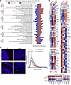 Figure 3
mPPL-001 transplantation partially restores the transcriptomic profile of YG8JR cerebellum, improving microglial stress and neuroinflammation. (A) Pathway enrichment analysis of key drivers of cerebellar pathology in YG8JR mice (WT = 2M/1F, YG8JR = 2M/2F, mock = 2M/2F, and mPPL-001 = 4M/1F). (B) Leading-edge analysis of genes contributing most to pathway enrichment. (C) Representative images of cerebellar Iba1+ microglia and Sholl analysis of ramifications. Statistical analysis compares number of intersections among the YG8JR group, and the asterisk represents significance between mock HSPC and mPPL-001 YG8JR recipients. Scale bar: 50 μm. Data are presented as mean ± SEM, analyzed by 1-way ANOVA. *P ≤ 0.05.
GSEA identified significant suppression of transcriptional regulation and genome maintenance programs, DNA integrity checkpoint, and damage response and repair, with representative genes including Fus, Kmt5c, Cgas, Rnf138, Brca2, Mcm5, and Kdm2a in untreated and mock HSPC–transplanted YG8JR cerebellum (Figure 3B and Supplemental Table 2). Importantly, these pathways showed partial restoration following mPPL-001 transplantation, demonstrating that mPPL-001 not only attenuates inflammatory programs but also promotes recovery of transcriptional and genomic maintenance pathways that are suppressed in the disease state (Figure 3, A and B, and Supplemental Table 2).
To confirm the microglial and neuroinflammatory transcriptional signatures in YG8JR mice, we analyzed morphology and branching complexity of Iba1+ cerebellar microglia by Sholl analysis (Figure 3C). Untreated and mock YG8JR mice displayed amoeboid, activated microglia, whereas mPPL-001 transplantation restored ramified, homeostatic morphology compared with WT mice (Figure 3C).
Together, these findings demonstrate that mPPL-001 transplantation provides cerebellar neuroprotection by modulating the local inflammatory environment, including microglial activation, and normalizing dysregulated transcriptional networks critical for neuronal integrity, providing an additional mechanism for its neuroprotective efficacy in the FRDA disease model.
mPPL-001 transplantation increases FXN expression in the heart and preserves cardiac architecture. Cardiac pathology in FRDA involves a complex interplay of mitochondrial proliferation and dysfunction, progressive loss of contractile fibers, fibrosis, and calcium dysregulation (13), resulting in ventricular wall thickening and diastolic dysfunction, while preserving systolic function until advanced stages of the disease (56, 57). Consistent with previous reports describing mild and late-onset cardiac abnormalities in YG8JR mice (10, 58), M-mode echocardiography of untreated 8- to 9-month-old untreated YG8JR mice revealed no significant differences in cardiac function compared with WT controls (Figure 4A). However, myeloablative irradiation is known to exacerbate cellular and mitochondrial stress (59); accordingly, mock HSPC–transplanted YG8JR mice exhibited a restrictive cardiac phenotype characterized by reduced diastolic and systolic left ventricular internal diameter compared with WT (Figure 4A). In contrast, mPPL-001–treated mice exhibited significant improvement in these dimensional parameters, nearing WT levels, and showed positive correlation with engraftment of mPPL-001–derived cells in the hematopoietic compartments (Figure 4A and Supplemental Figure 5, A and B). Elevated left ventricular mass was observed and remained unchanged in YG8JR mice of all cohorts compared with WT mice and was not significantly different among the YG8JR mice (Figure 4A). Notably, this pathology was present in 2-month-old pretransplant YG8JR controls (Supplemental Figure 5C).
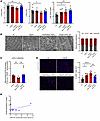 Figure 4
mPPL-001 transplantation improves cardiac architecture and inflammation in YG8JR mice. (A) M-mode echocardiography parameters in mPPL-001–transplanted mice compared with controls (WT = 5M/3F, YG8JR = 5M/4F, mock = 6M/4F, and mPPL-001 = 9M/3F). LVIDd, diastolic left ventricular internal diameter; LVIDs, systolic left ventricular internal diameter; LVM, left ventricular mass. (B) Representative electron microscopy images and quantification of mitochondrial morphology in heart sections. Scale bars: 2 μm. The asterisk represents significance of normal versus fragmented mitochondria distribution in mock HSPC recipients. (C) Mature mitochondrial hFXN expression in YG8JR mice. (D) Regression analysis of mPPL-001–derived cells in bone marrow versus mitochondrial hFXN expression in mPPL-001 recipients. (E) Representative immunofluorescence images and quantification of CD68+ macrophages in the heart. Scale bar: 50 μm. Data are presented as mean ± SEM, analyzed by 1-way (A, C, and E) or 2-way (B) ANOVA. *P ≤ 0.05, **P ≤ 0.01, and ****P ≤ 0.0001.
Ultrastructural analysis of untreated YG8JR hearts by electron microscopy demonstrated that mitochondrial morphology was comparable with WT, while mock HSPC transplantation significantly increased the proportion of fragmented mitochondria (Figure 4B). In contrast, mitochondrial morphology in mPPL-001 recipients closely resembled WT mice (Figure 4B). Given the subtle mitochondrial phenotype changes in the heart, mitochondrially localized hFXN was quantified using a highly sensitive and previously validated triple quadrupole mass spectrometry method (60–62). Mock mice exhibited significantly reduced mitochondrial hFXN expression compared with untreated YG8JR mice (Figure 4C), consistent with observed structural abnormalities limited to the myeloablative cohort. Notably, mPPL-001 transplantation significantly increased mitochondrial hFXN levels relative to mock mice and showed trends of positive correlation with engraftment of mPPL-001–derived cells within the bone marrow (P = 0.1; F = 3.2; r2 = 0.26) (Figure 4D).
In the FRDA patient myocardium, compensatory hypertrophy is associated with chronic, low-grade inflammation, driven by proinflammatory CD68+ macrophages (63). Similarly, untreated and mock HSPC–transplanted YG8JR hearts exhibited a significant increase in CD68+ macrophages, which were ameliorated in mPPL-001–treated mice (Figure 4E).
These results demonstrate that YG8JR mice, despite exhibiting mild cardiac structural abnormalities, recapitulate key inflammatory pathological features observed in FRDA patients. mPPL-001 transplantation mitigates these abnormalities by increasing mitochondrially localized hFXN expression and attenuating myocardial inflammation, thereby preserving cardiac architecture.
mPPL-001 transplantation attenuates pancreatic inflammatory macrophages, protecting β cells of the islets of Langerhans in YG8JR mice. Diabetes affects approximately 10%–30% of FRDA patients and is often associated with impaired glucose tolerance and insulin deficiency (64, 65). In YG8JR mice, fasting blood glucose levels remained comparable with those of WT mice across all groups up to 6 months posttransplantation (Supplemental Figure 6A). Histological analysis of H&E-stained pancreatic sections revealed reduced islet cellularity in untreated and mock HSPC–transplanted YG8JR mice compared with WT controls (Figure 5A). Insulin immunofluorescence confirmed significant loss of β cells in both untreated and mock YG8JR groups compared with WT (Figure 5C). Notably, this islet atrophy was already present in 2-month-old YG8JR mice, which corresponds to the age of mPPL-001 transplant, demonstrating a preexisting condition (Supplemental Figure 6B). Remarkably, mPPL-001 transplantation restored the abundance of insulin-producing β cells to that of healthy controls, demonstrating recovery of physiological architecture (Figure 5, A and B). This restoration of abundance of insulin-positive β cells showed positive correlation engraftment of mPPL-001–derived cells in hematopoietic organs (Supplemental Figure 6C).
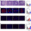 Figure 5
mPPL-001 transplantation preserves insulin-producing β cells of pancreatic islet of Langerhans by attenuating inflammation. (A) Representative H&E images and quantification of islet of Langerhans’ cellularity in pancreas (WT = 6M/3F, YG8JR = 4M/4F, mock = 5M/3F, and mPPL-001 = 9M/1F). 3–7 islets per mouse were quantified (B) Representative immunofluorescence images and quantification of insulin-producing β cells. 3–7 islets per mouse were quantified. (C) Representative immunofluorescence images and quantification of pancreatic inflammatory CD68+ macrophages. (D) Representative immunofluorescence images and quantification of pancreatic antiinflammatory CD206+ cells. Scale bars: 50 μm. Data are presented as mean ± SEM, analyzed by 1-way ANOVA. *P ≤ 0.05, **P ≤ 0.01, ***P ≤ 0.001, and ****P ≤ 0.0001.
Since emerging evidence demonstrates that resolution of CD11b+ and CD68+ macrophage-mediated pancreatic inflammation can promote β cell regeneration and tissue repair (66–70), we investigated the inflammatory status in YG8JR mice across the treatment groups. Untreated and mock YG8JR mice exhibited significantly increased CD68+ inflammatory macrophages in the pancreas compared with WT (Figure 5C). In contrast, CD68+ macrophage abundance was significantly decreased in mPPL-001–transplanted mice (Figure 5C). Additionally, mPPL-001 treatment was associated with enrichment of CD206+ antiinflammatory macrophages compared with untreated and mock HSPC–transplanted YG8JR mice, nearing WT levels (Figure 5D). Taken together, our results identify an inflammatory component in FRDA pancreatic pathology that could contribute to β cell degeneration and demonstrate that mPPL-001 transplantation exerts therapeutic benefits by supporting a shift toward antiinflammatory immunological modulation and tissue repair in the pancreas and preserving insulin-producing β cells.


Copyright © 2026 American Society for Clinical Investigation
ISSN: 0021-9738 (print), 1558-8238 (online)