
Advertisement
Commentary
Open Access |  10.1172/JCI207798
Emerging roles of ATR beyond DNA damage repair: orchestrating transcriptional reprogramming during epithelial-to-mesenchymal transition
Aida Mestre-Farrera,1 Zhimin Hu,1 and Jing Yang1,2
1Department of Pharmacology, Moores Cancer Center, and
2Department of Pediatrics, UCSD, La Jolla, California, USA.
Address correspondence to: Jing Yang, UCSD, 3855 Health Sciences Drive, MC0819, La Jolla, California 92093, USA. Email: jingyang@ucsd.edu.
Find articles by Mestre-Farrera, A. in: PubMed | Google Scholar
1Department of Pharmacology, Moores Cancer Center, and
2Department of Pediatrics, UCSD, La Jolla, California, USA.
Address correspondence to: Jing Yang, UCSD, 3855 Health Sciences Drive, MC0819, La Jolla, California 92093, USA. Email: jingyang@ucsd.edu.
Find articles by Hu, Z. in: PubMed | Google Scholar
1Department of Pharmacology, Moores Cancer Center, and
2Department of Pediatrics, UCSD, La Jolla, California, USA.
Address correspondence to: Jing Yang, UCSD, 3855 Health Sciences Drive, MC0819, La Jolla, California 92093, USA. Email: jingyang@ucsd.edu.
Find articles by Yang, J. in: PubMed | Google Scholar
Published August 17, 2026 - More info
J Clin Invest. 2026;136(16):e207798. https://doi.org/10.1172/JCI207798.
© 2026 Mestre-Farrera et al. This work is licensed under the Creative Commons Attribution 4.0 International License. To view a copy of this license, visit http://creativecommons.org/licenses/by/4.0/.

Related articles:
Abstract
Transitions of cancer cells between distinct cell states, which are typically driven by transcription reprogramming, fuel tumor plasticity, metastasis, and therapeutic resistance. Whether the transitions between cell states can be therapeutically targeted remains unknown. Here, using the epithelial-to-mesenchymal transition (EMT) as a model, we show that the transcription reprogramming during a cell-state transition induces genomic instability through R-loops and transcription-replication conflicts and that the cell-state transition cannot occur without the ATR kinase, a key regulator of the replication stress response. ATR inhibition during EMT not only increased transcription- and replication-dependent genomic instability, but also disrupted transcription reprogramming. Unexpectedly, ATR inhibition elevated R-loop–associated DNA damage at the SNAI1 gene, a key driver of the transcription reprogramming during EMT, triggering ATM- and Polycomb-mediated transcription repression of SNAI1. Beyond SNAI1, ATR also suppressed R-loops and antagonized repressive chromatin at a subset of EMT genes. Importantly, inhibition of ATR in tumors undergoing EMT reduced tumor growth and metastasis, suggesting that ATR inhibition eliminates cancer cells in transition. Thus, during EMT, ATR not only protects genome integrity but also enables transcription reprogramming, revealing that ATR is a safeguard of cell-state transitions and a target to suppress tumor plasticity.
Authors
Parasvi S. Patel, Jacob P. Matson, Xiaojuan Ran, Marcello Stanzione, Ajinkya S. Kawale, Mingchao Wang, Sneha Saxena, Conrad Sander, Jacquelyn Curtis, Jessica L. Hopkins, Edmond Wong, Ryan B. Corcoran, Daniel A. Haber, Nicholas J. Dyson, Shyamala Maheswaran, Lee Zou
Abstract
Our research uncovers a role for ATR in responding to ECM stiffness and promoting epithelial-to-mesenchymal transition (EMT) and metastasis. ATR, when deubiquitinated and upregulated by USP21 under enhanced ECM stiffness conditions, phosphorylates the nuclear protein SUN2, which promotes β-catenin nuclear translocation and EMT. ATM-mediated EMT promotes polymorphonuclear myeloid-derived suppressor cell recruitment and inhibits CD103+ dendritic cells, fostering an immunosuppressive tumor milieu. ATR inhibition disrupts this malignant cascade by promoting mesenchymal-to-epithelial transition to enhance antitumor immunity and mitigate metastases. Consistently, circulating HLA-DR+ dendritic cells were also enhanced following treatment with the ATR inhibitor berzosertib in patients with therapeutically resistant early-stage breast cancer. Our data suggest that ATR-targeted therapy may be optimized by considering both DNA damage–dependent and EMT-inducing effects of ATR
Authors
Xinyi Tu, Xiangyu Zeng, Yaoliang Sun, Yaobin Ouyang, Lingling Zhu, Ping Yin, Kevin Pavelko, Roberto Leon-Ferre, Yanxia Jiang, Haidong Dong, Jodi Carter, Shouhai Zhu, Jann N. Sarkaria, Liewei Wang, Jinzhou Huang, Kuntian Luo, Yiqun Han, Zheming Wu, Zhenkun Lou, Robert W. Mutter
-
Implications and future directions
Collectively, the studies by Patel et al. and Tu et al. propose a comprehensive model for ATR in tumor progression and metastasis, positioning it as a central hub for EMT induction triggered by convergent biochemical and mechanical stimuli from the microenvironment. The model proposes that during tumor progression, activated cancer-associated fibroblasts (CAFs) secrete TGF-β, initiating EMT-related transcriptional reprogramming. This rapid transcription reprogramming activity generates R-loop–associated DNA damage that necessitates ATR-mediated repair. Simultaneously, CAFs modify the ECM to increase stiffness, which cooperates with TGF-β to activate transcription factors like TWIST1 and SNAI1 to further boost EMT transcriptional reprogramming. High ECM stiffness further stabilizes ATR protein, driving β-catenin localization and resolving R-loop–induced damage. These reinforcing positive feedback loops demonstrate how mechanical and biochemical cues converge on ATR to orchestrate the EMT program (Figure 1).
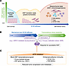 Figure 1
ATR integrates ECM mechanical input and transcriptional stress to regulate EMT plasticity. (A) During EMT, epithelial cells lose polarity and acquire traits of mesenchymal cells that support tumor progression and metastasis, including enhanced motility, stem-like properties, and resistance to antitumor therapies. This gradual transition allows tumor cells to adapt to stressors and limitations of the tumor microenvironment. (B) Patel et al. (14) showed that during tumor development, tumor cells undergoing EMT experienced transcriptional stress and R-loop accumulation, which activated ATR. In parallel, Tu et al. (15) found that increasing ECM stiffness stabilized ATR via USP21-mediated deubiquitination, which further enhanced EMT transition. At high ECM stiffness, tumor cells transitioned toward a mesenchymal state, gained invasiveness, and promoted an immunosuppressive tumor microenvironment, characterized by increased PMN myeloid-derived suppressor cells (PMN-MDSCs) and Tregs and decreased CD103+ dendritic cells (DCs). DDR, DNA damage response. (C) Pharmacological inhibition of ATR in the work of Patel et al. and Tu et al. disrupted EMT-associated transcriptional programs and enhanced antitumor immunity, respectively. Collectively, these effects suppressed tumor progression and metastasis.
These groundbreaking studies prompt critical questions regarding the noncanonical roles of ATR, which extend beyond its established function in DNA damage repair. During the transcriptional reprogramming associated with EMT, it remains to be elucidated how ATR selectively localizes to TGF-β–induced EMT gene promoters instead of other transcriptionally active genes. Although SUN2 is established as a key ATR substrate for β-catenin nuclear translocation, the established role of the LINC complex in modulating heterochromatin in response to mechanical cues suggests a more extensive function beyond β-catenin. This indicates that ATR may orchestrate a broader mechanosensitive EMT program in response to ECM rigidity, extending its influence well beyond canonical DNA damage repair pathways.
The EMT program contributes substantially to tumor metastasis and therapeutic resistance across various human malignancies. Emerging evidence further underscores the critical role of EMT in driving tumor-mediated immune suppression, particularly within the context of ATR inhibition. While several ATR inhibitors are currently undergoing clinical evaluation in combination with chemotherapy and radiotherapy (16, 17), the discovery of ATR as a central hub in EMT regulation suggests a broader therapeutic window. Investigating the impact of ATR inhibitors on diverse forms of therapy resistance may expand their clinical utility beyond canonical DNA damage repair mechanisms.
Article tools
- Download citation information
- Send a comment
- Terms of use
- Standard abbreviations
- Need help? Email the journal
Metrics
Go to
- Top
- Abstract
- ATR signaling in cellular homeostasis and cancer progression
- ATR-dependent R-loop suppression and transcriptional reprogramming promote EMT
- ECM stiffness stabilizes ATR protein to promote EMT
- Implications and future directions
- Conflict of interest
- Funding support
- Footnotes
- References
- Version history


Copyright © 2026 American Society for Clinical Investigation
ISSN: 0021-9738 (print), 1558-8238 (online)