
Advertisement
Research ArticleCell biologyMetabolismOncology
Open Access |  10.1172/JCI205579
Nuclear cGAS governs transcription to control lipid metabolism, tumor growth, and therapy response
Yu Deng,1,2 Matthew J. Niederhuber,3 Zhichuan Zhu,1,2 Austin J. Hepperla,1,3,4,5,6 Le Yu,1,2 Quentin Hahn,2 Joshua Hartsell,7 Tooba Rashid,8 Piotr A. Mieczkowski,1,6 Hemant Kelkar,6 Jeremy M. Simon,1,4,5,6,9 Yanqiong Zhang,1,2 Alessandra Livraghi-Butrico,10 Meredith J. Zeller,10 Joseph G.C. Kennedy,10 Jazmin F. DePriest,10 Jiong Li,11,12,13 Yan Zhang,11,13 Xing Qiu,14 H. Ümit Kaniskan,14 Jian Jin,14 Corbin D. Jones,6,15 Laura E. Herring,16 Douglas H. Phanstiel,1,6,8,17,18 Xiaojing Liu,7 Jesse R. Raab,1,6 Brian D. Strahl,1,2 and Pengda Liu1,2
1Lineberger Comprehensive Cancer Center,
2Department of Biochemistry and Biophysics,
3Bioinformatics and Analytics Research Collaborative (BARC),
4Carolina Institute for Developmental Disabilities,
5University of North Carolina (UNC) Neuroscience Center, and
6Department of Genetics, University of North Carolina at Chapel Hill, Chapel Hill, North Carolina, USA.
7Department of Molecular and Structural Biochemistry, North Carolina State University, Raleigh, North Carolina, USA.
8Curriculum in Bioinformatics and Computational Biology, University of North Carolina at Chapel Hill, Chapel Hill, North Carolina, USA.
9Department of Data Science, Dana-Farber Cancer Institute Department of Biostatistics, Harvard T.H. Chan School of Public Health, Boston, Massachusetts, USA.
10Marsico Lung Institute/UNC Cystic Fibrosis Center, School of Medicine, University of North Carolina at Chapel Hill, Chapel Hill, North Carolina, USA.
11Department of Medicinal Chemistry, School of Pharmacy;
12Philips Institute for Oral and Craniofacial Molecular Biology, School of Dentistry; and
13Massey Comprehensive Cancer Center, Virginia Commonwealth University, Richmond, Virginia, USA.
14Mount Sinai Center for Therapeutics Discovery, Departments of Pharmacological Sciences, Oncological Sciences and Neuroscience, Tisch Cancer Institute, Icahn School of Medicine at Mount Sinai, New York, USA.
15Department of Biology;
16UNC Proteomics Core Facility, Department of Pharmacology;
17Thurston Arthritis Research Center; and
18Department of Cell Biology and Physiology, University of North Carolina at Chapel Hill, Chapel Hill, North Carolina, USA.
Address for correspondence. Pengda Liu, 21-246 Lineberger Building, 450 West Drive, CB#7295, Chapel Hill, North Carolina 27599, USA. Phone: 919.966.3522; Email: pengda_liu@med.unc.edu.
Find articles by Deng, Y. in: PubMed | Google Scholar
1Lineberger Comprehensive Cancer Center,
2Department of Biochemistry and Biophysics,
3Bioinformatics and Analytics Research Collaborative (BARC),
4Carolina Institute for Developmental Disabilities,
5University of North Carolina (UNC) Neuroscience Center, and
6Department of Genetics, University of North Carolina at Chapel Hill, Chapel Hill, North Carolina, USA.
7Department of Molecular and Structural Biochemistry, North Carolina State University, Raleigh, North Carolina, USA.
8Curriculum in Bioinformatics and Computational Biology, University of North Carolina at Chapel Hill, Chapel Hill, North Carolina, USA.
9Department of Data Science, Dana-Farber Cancer Institute Department of Biostatistics, Harvard T.H. Chan School of Public Health, Boston, Massachusetts, USA.
10Marsico Lung Institute/UNC Cystic Fibrosis Center, School of Medicine, University of North Carolina at Chapel Hill, Chapel Hill, North Carolina, USA.
11Department of Medicinal Chemistry, School of Pharmacy;
12Philips Institute for Oral and Craniofacial Molecular Biology, School of Dentistry; and
13Massey Comprehensive Cancer Center, Virginia Commonwealth University, Richmond, Virginia, USA.
14Mount Sinai Center for Therapeutics Discovery, Departments of Pharmacological Sciences, Oncological Sciences and Neuroscience, Tisch Cancer Institute, Icahn School of Medicine at Mount Sinai, New York, USA.
15Department of Biology;
16UNC Proteomics Core Facility, Department of Pharmacology;
17Thurston Arthritis Research Center; and
18Department of Cell Biology and Physiology, University of North Carolina at Chapel Hill, Chapel Hill, North Carolina, USA.
Address for correspondence. Pengda Liu, 21-246 Lineberger Building, 450 West Drive, CB#7295, Chapel Hill, North Carolina 27599, USA. Phone: 919.966.3522; Email: pengda_liu@med.unc.edu.
Find articles by Niederhuber, M. in: PubMed | Google Scholar
1Lineberger Comprehensive Cancer Center,
2Department of Biochemistry and Biophysics,
3Bioinformatics and Analytics Research Collaborative (BARC),
4Carolina Institute for Developmental Disabilities,
5University of North Carolina (UNC) Neuroscience Center, and
6Department of Genetics, University of North Carolina at Chapel Hill, Chapel Hill, North Carolina, USA.
7Department of Molecular and Structural Biochemistry, North Carolina State University, Raleigh, North Carolina, USA.
8Curriculum in Bioinformatics and Computational Biology, University of North Carolina at Chapel Hill, Chapel Hill, North Carolina, USA.
9Department of Data Science, Dana-Farber Cancer Institute Department of Biostatistics, Harvard T.H. Chan School of Public Health, Boston, Massachusetts, USA.
10Marsico Lung Institute/UNC Cystic Fibrosis Center, School of Medicine, University of North Carolina at Chapel Hill, Chapel Hill, North Carolina, USA.
11Department of Medicinal Chemistry, School of Pharmacy;
12Philips Institute for Oral and Craniofacial Molecular Biology, School of Dentistry; and
13Massey Comprehensive Cancer Center, Virginia Commonwealth University, Richmond, Virginia, USA.
14Mount Sinai Center for Therapeutics Discovery, Departments of Pharmacological Sciences, Oncological Sciences and Neuroscience, Tisch Cancer Institute, Icahn School of Medicine at Mount Sinai, New York, USA.
15Department of Biology;
16UNC Proteomics Core Facility, Department of Pharmacology;
17Thurston Arthritis Research Center; and
18Department of Cell Biology and Physiology, University of North Carolina at Chapel Hill, Chapel Hill, North Carolina, USA.
Address for correspondence. Pengda Liu, 21-246 Lineberger Building, 450 West Drive, CB#7295, Chapel Hill, North Carolina 27599, USA. Phone: 919.966.3522; Email: pengda_liu@med.unc.edu.
Find articles by Zhu, Z. in: PubMed | Google Scholar
1Lineberger Comprehensive Cancer Center,
2Department of Biochemistry and Biophysics,
3Bioinformatics and Analytics Research Collaborative (BARC),
4Carolina Institute for Developmental Disabilities,
5University of North Carolina (UNC) Neuroscience Center, and
6Department of Genetics, University of North Carolina at Chapel Hill, Chapel Hill, North Carolina, USA.
7Department of Molecular and Structural Biochemistry, North Carolina State University, Raleigh, North Carolina, USA.
8Curriculum in Bioinformatics and Computational Biology, University of North Carolina at Chapel Hill, Chapel Hill, North Carolina, USA.
9Department of Data Science, Dana-Farber Cancer Institute Department of Biostatistics, Harvard T.H. Chan School of Public Health, Boston, Massachusetts, USA.
10Marsico Lung Institute/UNC Cystic Fibrosis Center, School of Medicine, University of North Carolina at Chapel Hill, Chapel Hill, North Carolina, USA.
11Department of Medicinal Chemistry, School of Pharmacy;
12Philips Institute for Oral and Craniofacial Molecular Biology, School of Dentistry; and
13Massey Comprehensive Cancer Center, Virginia Commonwealth University, Richmond, Virginia, USA.
14Mount Sinai Center for Therapeutics Discovery, Departments of Pharmacological Sciences, Oncological Sciences and Neuroscience, Tisch Cancer Institute, Icahn School of Medicine at Mount Sinai, New York, USA.
15Department of Biology;
16UNC Proteomics Core Facility, Department of Pharmacology;
17Thurston Arthritis Research Center; and
18Department of Cell Biology and Physiology, University of North Carolina at Chapel Hill, Chapel Hill, North Carolina, USA.
Address for correspondence. Pengda Liu, 21-246 Lineberger Building, 450 West Drive, CB#7295, Chapel Hill, North Carolina 27599, USA. Phone: 919.966.3522; Email: pengda_liu@med.unc.edu.
Find articles by Hepperla, A. in: PubMed | Google Scholar
1Lineberger Comprehensive Cancer Center,
2Department of Biochemistry and Biophysics,
3Bioinformatics and Analytics Research Collaborative (BARC),
4Carolina Institute for Developmental Disabilities,
5University of North Carolina (UNC) Neuroscience Center, and
6Department of Genetics, University of North Carolina at Chapel Hill, Chapel Hill, North Carolina, USA.
7Department of Molecular and Structural Biochemistry, North Carolina State University, Raleigh, North Carolina, USA.
8Curriculum in Bioinformatics and Computational Biology, University of North Carolina at Chapel Hill, Chapel Hill, North Carolina, USA.
9Department of Data Science, Dana-Farber Cancer Institute Department of Biostatistics, Harvard T.H. Chan School of Public Health, Boston, Massachusetts, USA.
10Marsico Lung Institute/UNC Cystic Fibrosis Center, School of Medicine, University of North Carolina at Chapel Hill, Chapel Hill, North Carolina, USA.
11Department of Medicinal Chemistry, School of Pharmacy;
12Philips Institute for Oral and Craniofacial Molecular Biology, School of Dentistry; and
13Massey Comprehensive Cancer Center, Virginia Commonwealth University, Richmond, Virginia, USA.
14Mount Sinai Center for Therapeutics Discovery, Departments of Pharmacological Sciences, Oncological Sciences and Neuroscience, Tisch Cancer Institute, Icahn School of Medicine at Mount Sinai, New York, USA.
15Department of Biology;
16UNC Proteomics Core Facility, Department of Pharmacology;
17Thurston Arthritis Research Center; and
18Department of Cell Biology and Physiology, University of North Carolina at Chapel Hill, Chapel Hill, North Carolina, USA.
Address for correspondence. Pengda Liu, 21-246 Lineberger Building, 450 West Drive, CB#7295, Chapel Hill, North Carolina 27599, USA. Phone: 919.966.3522; Email: pengda_liu@med.unc.edu.
Find articles by Yu, L. in: PubMed | Google Scholar
1Lineberger Comprehensive Cancer Center,
2Department of Biochemistry and Biophysics,
3Bioinformatics and Analytics Research Collaborative (BARC),
4Carolina Institute for Developmental Disabilities,
5University of North Carolina (UNC) Neuroscience Center, and
6Department of Genetics, University of North Carolina at Chapel Hill, Chapel Hill, North Carolina, USA.
7Department of Molecular and Structural Biochemistry, North Carolina State University, Raleigh, North Carolina, USA.
8Curriculum in Bioinformatics and Computational Biology, University of North Carolina at Chapel Hill, Chapel Hill, North Carolina, USA.
9Department of Data Science, Dana-Farber Cancer Institute Department of Biostatistics, Harvard T.H. Chan School of Public Health, Boston, Massachusetts, USA.
10Marsico Lung Institute/UNC Cystic Fibrosis Center, School of Medicine, University of North Carolina at Chapel Hill, Chapel Hill, North Carolina, USA.
11Department of Medicinal Chemistry, School of Pharmacy;
12Philips Institute for Oral and Craniofacial Molecular Biology, School of Dentistry; and
13Massey Comprehensive Cancer Center, Virginia Commonwealth University, Richmond, Virginia, USA.
14Mount Sinai Center for Therapeutics Discovery, Departments of Pharmacological Sciences, Oncological Sciences and Neuroscience, Tisch Cancer Institute, Icahn School of Medicine at Mount Sinai, New York, USA.
15Department of Biology;
16UNC Proteomics Core Facility, Department of Pharmacology;
17Thurston Arthritis Research Center; and
18Department of Cell Biology and Physiology, University of North Carolina at Chapel Hill, Chapel Hill, North Carolina, USA.
Address for correspondence. Pengda Liu, 21-246 Lineberger Building, 450 West Drive, CB#7295, Chapel Hill, North Carolina 27599, USA. Phone: 919.966.3522; Email: pengda_liu@med.unc.edu.
Find articles by Hahn, Q. in: PubMed | Google Scholar
1Lineberger Comprehensive Cancer Center,
2Department of Biochemistry and Biophysics,
3Bioinformatics and Analytics Research Collaborative (BARC),
4Carolina Institute for Developmental Disabilities,
5University of North Carolina (UNC) Neuroscience Center, and
6Department of Genetics, University of North Carolina at Chapel Hill, Chapel Hill, North Carolina, USA.
7Department of Molecular and Structural Biochemistry, North Carolina State University, Raleigh, North Carolina, USA.
8Curriculum in Bioinformatics and Computational Biology, University of North Carolina at Chapel Hill, Chapel Hill, North Carolina, USA.
9Department of Data Science, Dana-Farber Cancer Institute Department of Biostatistics, Harvard T.H. Chan School of Public Health, Boston, Massachusetts, USA.
10Marsico Lung Institute/UNC Cystic Fibrosis Center, School of Medicine, University of North Carolina at Chapel Hill, Chapel Hill, North Carolina, USA.
11Department of Medicinal Chemistry, School of Pharmacy;
12Philips Institute for Oral and Craniofacial Molecular Biology, School of Dentistry; and
13Massey Comprehensive Cancer Center, Virginia Commonwealth University, Richmond, Virginia, USA.
14Mount Sinai Center for Therapeutics Discovery, Departments of Pharmacological Sciences, Oncological Sciences and Neuroscience, Tisch Cancer Institute, Icahn School of Medicine at Mount Sinai, New York, USA.
15Department of Biology;
16UNC Proteomics Core Facility, Department of Pharmacology;
17Thurston Arthritis Research Center; and
18Department of Cell Biology and Physiology, University of North Carolina at Chapel Hill, Chapel Hill, North Carolina, USA.
Address for correspondence. Pengda Liu, 21-246 Lineberger Building, 450 West Drive, CB#7295, Chapel Hill, North Carolina 27599, USA. Phone: 919.966.3522; Email: pengda_liu@med.unc.edu.
Find articles by Hartsell, J. in: PubMed | Google Scholar
1Lineberger Comprehensive Cancer Center,
2Department of Biochemistry and Biophysics,
3Bioinformatics and Analytics Research Collaborative (BARC),
4Carolina Institute for Developmental Disabilities,
5University of North Carolina (UNC) Neuroscience Center, and
6Department of Genetics, University of North Carolina at Chapel Hill, Chapel Hill, North Carolina, USA.
7Department of Molecular and Structural Biochemistry, North Carolina State University, Raleigh, North Carolina, USA.
8Curriculum in Bioinformatics and Computational Biology, University of North Carolina at Chapel Hill, Chapel Hill, North Carolina, USA.
9Department of Data Science, Dana-Farber Cancer Institute Department of Biostatistics, Harvard T.H. Chan School of Public Health, Boston, Massachusetts, USA.
10Marsico Lung Institute/UNC Cystic Fibrosis Center, School of Medicine, University of North Carolina at Chapel Hill, Chapel Hill, North Carolina, USA.
11Department of Medicinal Chemistry, School of Pharmacy;
12Philips Institute for Oral and Craniofacial Molecular Biology, School of Dentistry; and
13Massey Comprehensive Cancer Center, Virginia Commonwealth University, Richmond, Virginia, USA.
14Mount Sinai Center for Therapeutics Discovery, Departments of Pharmacological Sciences, Oncological Sciences and Neuroscience, Tisch Cancer Institute, Icahn School of Medicine at Mount Sinai, New York, USA.
15Department of Biology;
16UNC Proteomics Core Facility, Department of Pharmacology;
17Thurston Arthritis Research Center; and
18Department of Cell Biology and Physiology, University of North Carolina at Chapel Hill, Chapel Hill, North Carolina, USA.
Address for correspondence. Pengda Liu, 21-246 Lineberger Building, 450 West Drive, CB#7295, Chapel Hill, North Carolina 27599, USA. Phone: 919.966.3522; Email: pengda_liu@med.unc.edu.
Find articles by Rashid, T. in: PubMed | Google Scholar
1Lineberger Comprehensive Cancer Center,
2Department of Biochemistry and Biophysics,
3Bioinformatics and Analytics Research Collaborative (BARC),
4Carolina Institute for Developmental Disabilities,
5University of North Carolina (UNC) Neuroscience Center, and
6Department of Genetics, University of North Carolina at Chapel Hill, Chapel Hill, North Carolina, USA.
7Department of Molecular and Structural Biochemistry, North Carolina State University, Raleigh, North Carolina, USA.
8Curriculum in Bioinformatics and Computational Biology, University of North Carolina at Chapel Hill, Chapel Hill, North Carolina, USA.
9Department of Data Science, Dana-Farber Cancer Institute Department of Biostatistics, Harvard T.H. Chan School of Public Health, Boston, Massachusetts, USA.
10Marsico Lung Institute/UNC Cystic Fibrosis Center, School of Medicine, University of North Carolina at Chapel Hill, Chapel Hill, North Carolina, USA.
11Department of Medicinal Chemistry, School of Pharmacy;
12Philips Institute for Oral and Craniofacial Molecular Biology, School of Dentistry; and
13Massey Comprehensive Cancer Center, Virginia Commonwealth University, Richmond, Virginia, USA.
14Mount Sinai Center for Therapeutics Discovery, Departments of Pharmacological Sciences, Oncological Sciences and Neuroscience, Tisch Cancer Institute, Icahn School of Medicine at Mount Sinai, New York, USA.
15Department of Biology;
16UNC Proteomics Core Facility, Department of Pharmacology;
17Thurston Arthritis Research Center; and
18Department of Cell Biology and Physiology, University of North Carolina at Chapel Hill, Chapel Hill, North Carolina, USA.
Address for correspondence. Pengda Liu, 21-246 Lineberger Building, 450 West Drive, CB#7295, Chapel Hill, North Carolina 27599, USA. Phone: 919.966.3522; Email: pengda_liu@med.unc.edu.
Find articles by Mieczkowski, P. in: PubMed | Google Scholar
1Lineberger Comprehensive Cancer Center,
2Department of Biochemistry and Biophysics,
3Bioinformatics and Analytics Research Collaborative (BARC),
4Carolina Institute for Developmental Disabilities,
5University of North Carolina (UNC) Neuroscience Center, and
6Department of Genetics, University of North Carolina at Chapel Hill, Chapel Hill, North Carolina, USA.
7Department of Molecular and Structural Biochemistry, North Carolina State University, Raleigh, North Carolina, USA.
8Curriculum in Bioinformatics and Computational Biology, University of North Carolina at Chapel Hill, Chapel Hill, North Carolina, USA.
9Department of Data Science, Dana-Farber Cancer Institute Department of Biostatistics, Harvard T.H. Chan School of Public Health, Boston, Massachusetts, USA.
10Marsico Lung Institute/UNC Cystic Fibrosis Center, School of Medicine, University of North Carolina at Chapel Hill, Chapel Hill, North Carolina, USA.
11Department of Medicinal Chemistry, School of Pharmacy;
12Philips Institute for Oral and Craniofacial Molecular Biology, School of Dentistry; and
13Massey Comprehensive Cancer Center, Virginia Commonwealth University, Richmond, Virginia, USA.
14Mount Sinai Center for Therapeutics Discovery, Departments of Pharmacological Sciences, Oncological Sciences and Neuroscience, Tisch Cancer Institute, Icahn School of Medicine at Mount Sinai, New York, USA.
15Department of Biology;
16UNC Proteomics Core Facility, Department of Pharmacology;
17Thurston Arthritis Research Center; and
18Department of Cell Biology and Physiology, University of North Carolina at Chapel Hill, Chapel Hill, North Carolina, USA.
Address for correspondence. Pengda Liu, 21-246 Lineberger Building, 450 West Drive, CB#7295, Chapel Hill, North Carolina 27599, USA. Phone: 919.966.3522; Email: pengda_liu@med.unc.edu.
Find articles by Kelkar, H. in: PubMed | Google Scholar
1Lineberger Comprehensive Cancer Center,
2Department of Biochemistry and Biophysics,
3Bioinformatics and Analytics Research Collaborative (BARC),
4Carolina Institute for Developmental Disabilities,
5University of North Carolina (UNC) Neuroscience Center, and
6Department of Genetics, University of North Carolina at Chapel Hill, Chapel Hill, North Carolina, USA.
7Department of Molecular and Structural Biochemistry, North Carolina State University, Raleigh, North Carolina, USA.
8Curriculum in Bioinformatics and Computational Biology, University of North Carolina at Chapel Hill, Chapel Hill, North Carolina, USA.
9Department of Data Science, Dana-Farber Cancer Institute Department of Biostatistics, Harvard T.H. Chan School of Public Health, Boston, Massachusetts, USA.
10Marsico Lung Institute/UNC Cystic Fibrosis Center, School of Medicine, University of North Carolina at Chapel Hill, Chapel Hill, North Carolina, USA.
11Department of Medicinal Chemistry, School of Pharmacy;
12Philips Institute for Oral and Craniofacial Molecular Biology, School of Dentistry; and
13Massey Comprehensive Cancer Center, Virginia Commonwealth University, Richmond, Virginia, USA.
14Mount Sinai Center for Therapeutics Discovery, Departments of Pharmacological Sciences, Oncological Sciences and Neuroscience, Tisch Cancer Institute, Icahn School of Medicine at Mount Sinai, New York, USA.
15Department of Biology;
16UNC Proteomics Core Facility, Department of Pharmacology;
17Thurston Arthritis Research Center; and
18Department of Cell Biology and Physiology, University of North Carolina at Chapel Hill, Chapel Hill, North Carolina, USA.
Address for correspondence. Pengda Liu, 21-246 Lineberger Building, 450 West Drive, CB#7295, Chapel Hill, North Carolina 27599, USA. Phone: 919.966.3522; Email: pengda_liu@med.unc.edu.
Find articles by
Simon, J.
in:
PubMed
|
Google Scholar
|

1Lineberger Comprehensive Cancer Center,
2Department of Biochemistry and Biophysics,
3Bioinformatics and Analytics Research Collaborative (BARC),
4Carolina Institute for Developmental Disabilities,
5University of North Carolina (UNC) Neuroscience Center, and
6Department of Genetics, University of North Carolina at Chapel Hill, Chapel Hill, North Carolina, USA.
7Department of Molecular and Structural Biochemistry, North Carolina State University, Raleigh, North Carolina, USA.
8Curriculum in Bioinformatics and Computational Biology, University of North Carolina at Chapel Hill, Chapel Hill, North Carolina, USA.
9Department of Data Science, Dana-Farber Cancer Institute Department of Biostatistics, Harvard T.H. Chan School of Public Health, Boston, Massachusetts, USA.
10Marsico Lung Institute/UNC Cystic Fibrosis Center, School of Medicine, University of North Carolina at Chapel Hill, Chapel Hill, North Carolina, USA.
11Department of Medicinal Chemistry, School of Pharmacy;
12Philips Institute for Oral and Craniofacial Molecular Biology, School of Dentistry; and
13Massey Comprehensive Cancer Center, Virginia Commonwealth University, Richmond, Virginia, USA.
14Mount Sinai Center for Therapeutics Discovery, Departments of Pharmacological Sciences, Oncological Sciences and Neuroscience, Tisch Cancer Institute, Icahn School of Medicine at Mount Sinai, New York, USA.
15Department of Biology;
16UNC Proteomics Core Facility, Department of Pharmacology;
17Thurston Arthritis Research Center; and
18Department of Cell Biology and Physiology, University of North Carolina at Chapel Hill, Chapel Hill, North Carolina, USA.
Address for correspondence. Pengda Liu, 21-246 Lineberger Building, 450 West Drive, CB#7295, Chapel Hill, North Carolina 27599, USA. Phone: 919.966.3522; Email: pengda_liu@med.unc.edu.
Find articles by Zhang, Y. in: PubMed | Google Scholar
1Lineberger Comprehensive Cancer Center,
2Department of Biochemistry and Biophysics,
3Bioinformatics and Analytics Research Collaborative (BARC),
4Carolina Institute for Developmental Disabilities,
5University of North Carolina (UNC) Neuroscience Center, and
6Department of Genetics, University of North Carolina at Chapel Hill, Chapel Hill, North Carolina, USA.
7Department of Molecular and Structural Biochemistry, North Carolina State University, Raleigh, North Carolina, USA.
8Curriculum in Bioinformatics and Computational Biology, University of North Carolina at Chapel Hill, Chapel Hill, North Carolina, USA.
9Department of Data Science, Dana-Farber Cancer Institute Department of Biostatistics, Harvard T.H. Chan School of Public Health, Boston, Massachusetts, USA.
10Marsico Lung Institute/UNC Cystic Fibrosis Center, School of Medicine, University of North Carolina at Chapel Hill, Chapel Hill, North Carolina, USA.
11Department of Medicinal Chemistry, School of Pharmacy;
12Philips Institute for Oral and Craniofacial Molecular Biology, School of Dentistry; and
13Massey Comprehensive Cancer Center, Virginia Commonwealth University, Richmond, Virginia, USA.
14Mount Sinai Center for Therapeutics Discovery, Departments of Pharmacological Sciences, Oncological Sciences and Neuroscience, Tisch Cancer Institute, Icahn School of Medicine at Mount Sinai, New York, USA.
15Department of Biology;
16UNC Proteomics Core Facility, Department of Pharmacology;
17Thurston Arthritis Research Center; and
18Department of Cell Biology and Physiology, University of North Carolina at Chapel Hill, Chapel Hill, North Carolina, USA.
Address for correspondence. Pengda Liu, 21-246 Lineberger Building, 450 West Drive, CB#7295, Chapel Hill, North Carolina 27599, USA. Phone: 919.966.3522; Email: pengda_liu@med.unc.edu.
Find articles by
Livraghi-Butrico, A.
in:
PubMed
|
Google Scholar
|
1Lineberger Comprehensive Cancer Center,
2Department of Biochemistry and Biophysics,
3Bioinformatics and Analytics Research Collaborative (BARC),
4Carolina Institute for Developmental Disabilities,
5University of North Carolina (UNC) Neuroscience Center, and
6Department of Genetics, University of North Carolina at Chapel Hill, Chapel Hill, North Carolina, USA.
7Department of Molecular and Structural Biochemistry, North Carolina State University, Raleigh, North Carolina, USA.
8Curriculum in Bioinformatics and Computational Biology, University of North Carolina at Chapel Hill, Chapel Hill, North Carolina, USA.
9Department of Data Science, Dana-Farber Cancer Institute Department of Biostatistics, Harvard T.H. Chan School of Public Health, Boston, Massachusetts, USA.
10Marsico Lung Institute/UNC Cystic Fibrosis Center, School of Medicine, University of North Carolina at Chapel Hill, Chapel Hill, North Carolina, USA.
11Department of Medicinal Chemistry, School of Pharmacy;
12Philips Institute for Oral and Craniofacial Molecular Biology, School of Dentistry; and
13Massey Comprehensive Cancer Center, Virginia Commonwealth University, Richmond, Virginia, USA.
14Mount Sinai Center for Therapeutics Discovery, Departments of Pharmacological Sciences, Oncological Sciences and Neuroscience, Tisch Cancer Institute, Icahn School of Medicine at Mount Sinai, New York, USA.
15Department of Biology;
16UNC Proteomics Core Facility, Department of Pharmacology;
17Thurston Arthritis Research Center; and
18Department of Cell Biology and Physiology, University of North Carolina at Chapel Hill, Chapel Hill, North Carolina, USA.
Address for correspondence. Pengda Liu, 21-246 Lineberger Building, 450 West Drive, CB#7295, Chapel Hill, North Carolina 27599, USA. Phone: 919.966.3522; Email: pengda_liu@med.unc.edu.
Find articles by Zeller, M. in: PubMed | Google Scholar
1Lineberger Comprehensive Cancer Center,
2Department of Biochemistry and Biophysics,
3Bioinformatics and Analytics Research Collaborative (BARC),
4Carolina Institute for Developmental Disabilities,
5University of North Carolina (UNC) Neuroscience Center, and
6Department of Genetics, University of North Carolina at Chapel Hill, Chapel Hill, North Carolina, USA.
7Department of Molecular and Structural Biochemistry, North Carolina State University, Raleigh, North Carolina, USA.
8Curriculum in Bioinformatics and Computational Biology, University of North Carolina at Chapel Hill, Chapel Hill, North Carolina, USA.
9Department of Data Science, Dana-Farber Cancer Institute Department of Biostatistics, Harvard T.H. Chan School of Public Health, Boston, Massachusetts, USA.
10Marsico Lung Institute/UNC Cystic Fibrosis Center, School of Medicine, University of North Carolina at Chapel Hill, Chapel Hill, North Carolina, USA.
11Department of Medicinal Chemistry, School of Pharmacy;
12Philips Institute for Oral and Craniofacial Molecular Biology, School of Dentistry; and
13Massey Comprehensive Cancer Center, Virginia Commonwealth University, Richmond, Virginia, USA.
14Mount Sinai Center for Therapeutics Discovery, Departments of Pharmacological Sciences, Oncological Sciences and Neuroscience, Tisch Cancer Institute, Icahn School of Medicine at Mount Sinai, New York, USA.
15Department of Biology;
16UNC Proteomics Core Facility, Department of Pharmacology;
17Thurston Arthritis Research Center; and
18Department of Cell Biology and Physiology, University of North Carolina at Chapel Hill, Chapel Hill, North Carolina, USA.
Address for correspondence. Pengda Liu, 21-246 Lineberger Building, 450 West Drive, CB#7295, Chapel Hill, North Carolina 27599, USA. Phone: 919.966.3522; Email: pengda_liu@med.unc.edu.
Find articles by Kennedy, J. in: PubMed | Google Scholar
1Lineberger Comprehensive Cancer Center,
2Department of Biochemistry and Biophysics,
3Bioinformatics and Analytics Research Collaborative (BARC),
4Carolina Institute for Developmental Disabilities,
5University of North Carolina (UNC) Neuroscience Center, and
6Department of Genetics, University of North Carolina at Chapel Hill, Chapel Hill, North Carolina, USA.
7Department of Molecular and Structural Biochemistry, North Carolina State University, Raleigh, North Carolina, USA.
8Curriculum in Bioinformatics and Computational Biology, University of North Carolina at Chapel Hill, Chapel Hill, North Carolina, USA.
9Department of Data Science, Dana-Farber Cancer Institute Department of Biostatistics, Harvard T.H. Chan School of Public Health, Boston, Massachusetts, USA.
10Marsico Lung Institute/UNC Cystic Fibrosis Center, School of Medicine, University of North Carolina at Chapel Hill, Chapel Hill, North Carolina, USA.
11Department of Medicinal Chemistry, School of Pharmacy;
12Philips Institute for Oral and Craniofacial Molecular Biology, School of Dentistry; and
13Massey Comprehensive Cancer Center, Virginia Commonwealth University, Richmond, Virginia, USA.
14Mount Sinai Center for Therapeutics Discovery, Departments of Pharmacological Sciences, Oncological Sciences and Neuroscience, Tisch Cancer Institute, Icahn School of Medicine at Mount Sinai, New York, USA.
15Department of Biology;
16UNC Proteomics Core Facility, Department of Pharmacology;
17Thurston Arthritis Research Center; and
18Department of Cell Biology and Physiology, University of North Carolina at Chapel Hill, Chapel Hill, North Carolina, USA.
Address for correspondence. Pengda Liu, 21-246 Lineberger Building, 450 West Drive, CB#7295, Chapel Hill, North Carolina 27599, USA. Phone: 919.966.3522; Email: pengda_liu@med.unc.edu.
Find articles by DePriest, J. in: PubMed | Google Scholar
1Lineberger Comprehensive Cancer Center,
2Department of Biochemistry and Biophysics,
3Bioinformatics and Analytics Research Collaborative (BARC),
4Carolina Institute for Developmental Disabilities,
5University of North Carolina (UNC) Neuroscience Center, and
6Department of Genetics, University of North Carolina at Chapel Hill, Chapel Hill, North Carolina, USA.
7Department of Molecular and Structural Biochemistry, North Carolina State University, Raleigh, North Carolina, USA.
8Curriculum in Bioinformatics and Computational Biology, University of North Carolina at Chapel Hill, Chapel Hill, North Carolina, USA.
9Department of Data Science, Dana-Farber Cancer Institute Department of Biostatistics, Harvard T.H. Chan School of Public Health, Boston, Massachusetts, USA.
10Marsico Lung Institute/UNC Cystic Fibrosis Center, School of Medicine, University of North Carolina at Chapel Hill, Chapel Hill, North Carolina, USA.
11Department of Medicinal Chemistry, School of Pharmacy;
12Philips Institute for Oral and Craniofacial Molecular Biology, School of Dentistry; and
13Massey Comprehensive Cancer Center, Virginia Commonwealth University, Richmond, Virginia, USA.
14Mount Sinai Center for Therapeutics Discovery, Departments of Pharmacological Sciences, Oncological Sciences and Neuroscience, Tisch Cancer Institute, Icahn School of Medicine at Mount Sinai, New York, USA.
15Department of Biology;
16UNC Proteomics Core Facility, Department of Pharmacology;
17Thurston Arthritis Research Center; and
18Department of Cell Biology and Physiology, University of North Carolina at Chapel Hill, Chapel Hill, North Carolina, USA.
Address for correspondence. Pengda Liu, 21-246 Lineberger Building, 450 West Drive, CB#7295, Chapel Hill, North Carolina 27599, USA. Phone: 919.966.3522; Email: pengda_liu@med.unc.edu.
Find articles by Li, J. in: PubMed | Google Scholar
1Lineberger Comprehensive Cancer Center,
2Department of Biochemistry and Biophysics,
3Bioinformatics and Analytics Research Collaborative (BARC),
4Carolina Institute for Developmental Disabilities,
5University of North Carolina (UNC) Neuroscience Center, and
6Department of Genetics, University of North Carolina at Chapel Hill, Chapel Hill, North Carolina, USA.
7Department of Molecular and Structural Biochemistry, North Carolina State University, Raleigh, North Carolina, USA.
8Curriculum in Bioinformatics and Computational Biology, University of North Carolina at Chapel Hill, Chapel Hill, North Carolina, USA.
9Department of Data Science, Dana-Farber Cancer Institute Department of Biostatistics, Harvard T.H. Chan School of Public Health, Boston, Massachusetts, USA.
10Marsico Lung Institute/UNC Cystic Fibrosis Center, School of Medicine, University of North Carolina at Chapel Hill, Chapel Hill, North Carolina, USA.
11Department of Medicinal Chemistry, School of Pharmacy;
12Philips Institute for Oral and Craniofacial Molecular Biology, School of Dentistry; and
13Massey Comprehensive Cancer Center, Virginia Commonwealth University, Richmond, Virginia, USA.
14Mount Sinai Center for Therapeutics Discovery, Departments of Pharmacological Sciences, Oncological Sciences and Neuroscience, Tisch Cancer Institute, Icahn School of Medicine at Mount Sinai, New York, USA.
15Department of Biology;
16UNC Proteomics Core Facility, Department of Pharmacology;
17Thurston Arthritis Research Center; and
18Department of Cell Biology and Physiology, University of North Carolina at Chapel Hill, Chapel Hill, North Carolina, USA.
Address for correspondence. Pengda Liu, 21-246 Lineberger Building, 450 West Drive, CB#7295, Chapel Hill, North Carolina 27599, USA. Phone: 919.966.3522; Email: pengda_liu@med.unc.edu.
Find articles by Zhang, Y. in: PubMed | Google Scholar
1Lineberger Comprehensive Cancer Center,
2Department of Biochemistry and Biophysics,
3Bioinformatics and Analytics Research Collaborative (BARC),
4Carolina Institute for Developmental Disabilities,
5University of North Carolina (UNC) Neuroscience Center, and
6Department of Genetics, University of North Carolina at Chapel Hill, Chapel Hill, North Carolina, USA.
7Department of Molecular and Structural Biochemistry, North Carolina State University, Raleigh, North Carolina, USA.
8Curriculum in Bioinformatics and Computational Biology, University of North Carolina at Chapel Hill, Chapel Hill, North Carolina, USA.
9Department of Data Science, Dana-Farber Cancer Institute Department of Biostatistics, Harvard T.H. Chan School of Public Health, Boston, Massachusetts, USA.
10Marsico Lung Institute/UNC Cystic Fibrosis Center, School of Medicine, University of North Carolina at Chapel Hill, Chapel Hill, North Carolina, USA.
11Department of Medicinal Chemistry, School of Pharmacy;
12Philips Institute for Oral and Craniofacial Molecular Biology, School of Dentistry; and
13Massey Comprehensive Cancer Center, Virginia Commonwealth University, Richmond, Virginia, USA.
14Mount Sinai Center for Therapeutics Discovery, Departments of Pharmacological Sciences, Oncological Sciences and Neuroscience, Tisch Cancer Institute, Icahn School of Medicine at Mount Sinai, New York, USA.
15Department of Biology;
16UNC Proteomics Core Facility, Department of Pharmacology;
17Thurston Arthritis Research Center; and
18Department of Cell Biology and Physiology, University of North Carolina at Chapel Hill, Chapel Hill, North Carolina, USA.
Address for correspondence. Pengda Liu, 21-246 Lineberger Building, 450 West Drive, CB#7295, Chapel Hill, North Carolina 27599, USA. Phone: 919.966.3522; Email: pengda_liu@med.unc.edu.
Find articles by Qiu, X. in: PubMed | Google Scholar
1Lineberger Comprehensive Cancer Center,
2Department of Biochemistry and Biophysics,
3Bioinformatics and Analytics Research Collaborative (BARC),
4Carolina Institute for Developmental Disabilities,
5University of North Carolina (UNC) Neuroscience Center, and
6Department of Genetics, University of North Carolina at Chapel Hill, Chapel Hill, North Carolina, USA.
7Department of Molecular and Structural Biochemistry, North Carolina State University, Raleigh, North Carolina, USA.
8Curriculum in Bioinformatics and Computational Biology, University of North Carolina at Chapel Hill, Chapel Hill, North Carolina, USA.
9Department of Data Science, Dana-Farber Cancer Institute Department of Biostatistics, Harvard T.H. Chan School of Public Health, Boston, Massachusetts, USA.
10Marsico Lung Institute/UNC Cystic Fibrosis Center, School of Medicine, University of North Carolina at Chapel Hill, Chapel Hill, North Carolina, USA.
11Department of Medicinal Chemistry, School of Pharmacy;
12Philips Institute for Oral and Craniofacial Molecular Biology, School of Dentistry; and
13Massey Comprehensive Cancer Center, Virginia Commonwealth University, Richmond, Virginia, USA.
14Mount Sinai Center for Therapeutics Discovery, Departments of Pharmacological Sciences, Oncological Sciences and Neuroscience, Tisch Cancer Institute, Icahn School of Medicine at Mount Sinai, New York, USA.
15Department of Biology;
16UNC Proteomics Core Facility, Department of Pharmacology;
17Thurston Arthritis Research Center; and
18Department of Cell Biology and Physiology, University of North Carolina at Chapel Hill, Chapel Hill, North Carolina, USA.
Address for correspondence. Pengda Liu, 21-246 Lineberger Building, 450 West Drive, CB#7295, Chapel Hill, North Carolina 27599, USA. Phone: 919.966.3522; Email: pengda_liu@med.unc.edu.
Find articles by Kaniskan, H. in: PubMed | Google Scholar
1Lineberger Comprehensive Cancer Center,
2Department of Biochemistry and Biophysics,
3Bioinformatics and Analytics Research Collaborative (BARC),
4Carolina Institute for Developmental Disabilities,
5University of North Carolina (UNC) Neuroscience Center, and
6Department of Genetics, University of North Carolina at Chapel Hill, Chapel Hill, North Carolina, USA.
7Department of Molecular and Structural Biochemistry, North Carolina State University, Raleigh, North Carolina, USA.
8Curriculum in Bioinformatics and Computational Biology, University of North Carolina at Chapel Hill, Chapel Hill, North Carolina, USA.
9Department of Data Science, Dana-Farber Cancer Institute Department of Biostatistics, Harvard T.H. Chan School of Public Health, Boston, Massachusetts, USA.
10Marsico Lung Institute/UNC Cystic Fibrosis Center, School of Medicine, University of North Carolina at Chapel Hill, Chapel Hill, North Carolina, USA.
11Department of Medicinal Chemistry, School of Pharmacy;
12Philips Institute for Oral and Craniofacial Molecular Biology, School of Dentistry; and
13Massey Comprehensive Cancer Center, Virginia Commonwealth University, Richmond, Virginia, USA.
14Mount Sinai Center for Therapeutics Discovery, Departments of Pharmacological Sciences, Oncological Sciences and Neuroscience, Tisch Cancer Institute, Icahn School of Medicine at Mount Sinai, New York, USA.
15Department of Biology;
16UNC Proteomics Core Facility, Department of Pharmacology;
17Thurston Arthritis Research Center; and
18Department of Cell Biology and Physiology, University of North Carolina at Chapel Hill, Chapel Hill, North Carolina, USA.
Address for correspondence. Pengda Liu, 21-246 Lineberger Building, 450 West Drive, CB#7295, Chapel Hill, North Carolina 27599, USA. Phone: 919.966.3522; Email: pengda_liu@med.unc.edu.
Find articles by
Jin, J.
in:
PubMed
|
Google Scholar
|
1Lineberger Comprehensive Cancer Center,
2Department of Biochemistry and Biophysics,
3Bioinformatics and Analytics Research Collaborative (BARC),
4Carolina Institute for Developmental Disabilities,
5University of North Carolina (UNC) Neuroscience Center, and
6Department of Genetics, University of North Carolina at Chapel Hill, Chapel Hill, North Carolina, USA.
7Department of Molecular and Structural Biochemistry, North Carolina State University, Raleigh, North Carolina, USA.
8Curriculum in Bioinformatics and Computational Biology, University of North Carolina at Chapel Hill, Chapel Hill, North Carolina, USA.
9Department of Data Science, Dana-Farber Cancer Institute Department of Biostatistics, Harvard T.H. Chan School of Public Health, Boston, Massachusetts, USA.
10Marsico Lung Institute/UNC Cystic Fibrosis Center, School of Medicine, University of North Carolina at Chapel Hill, Chapel Hill, North Carolina, USA.
11Department of Medicinal Chemistry, School of Pharmacy;
12Philips Institute for Oral and Craniofacial Molecular Biology, School of Dentistry; and
13Massey Comprehensive Cancer Center, Virginia Commonwealth University, Richmond, Virginia, USA.
14Mount Sinai Center for Therapeutics Discovery, Departments of Pharmacological Sciences, Oncological Sciences and Neuroscience, Tisch Cancer Institute, Icahn School of Medicine at Mount Sinai, New York, USA.
15Department of Biology;
16UNC Proteomics Core Facility, Department of Pharmacology;
17Thurston Arthritis Research Center; and
18Department of Cell Biology and Physiology, University of North Carolina at Chapel Hill, Chapel Hill, North Carolina, USA.
Address for correspondence. Pengda Liu, 21-246 Lineberger Building, 450 West Drive, CB#7295, Chapel Hill, North Carolina 27599, USA. Phone: 919.966.3522; Email: pengda_liu@med.unc.edu.
Find articles by Jones, C. in: PubMed | Google Scholar
1Lineberger Comprehensive Cancer Center,
2Department of Biochemistry and Biophysics,
3Bioinformatics and Analytics Research Collaborative (BARC),
4Carolina Institute for Developmental Disabilities,
5University of North Carolina (UNC) Neuroscience Center, and
6Department of Genetics, University of North Carolina at Chapel Hill, Chapel Hill, North Carolina, USA.
7Department of Molecular and Structural Biochemistry, North Carolina State University, Raleigh, North Carolina, USA.
8Curriculum in Bioinformatics and Computational Biology, University of North Carolina at Chapel Hill, Chapel Hill, North Carolina, USA.
9Department of Data Science, Dana-Farber Cancer Institute Department of Biostatistics, Harvard T.H. Chan School of Public Health, Boston, Massachusetts, USA.
10Marsico Lung Institute/UNC Cystic Fibrosis Center, School of Medicine, University of North Carolina at Chapel Hill, Chapel Hill, North Carolina, USA.
11Department of Medicinal Chemistry, School of Pharmacy;
12Philips Institute for Oral and Craniofacial Molecular Biology, School of Dentistry; and
13Massey Comprehensive Cancer Center, Virginia Commonwealth University, Richmond, Virginia, USA.
14Mount Sinai Center for Therapeutics Discovery, Departments of Pharmacological Sciences, Oncological Sciences and Neuroscience, Tisch Cancer Institute, Icahn School of Medicine at Mount Sinai, New York, USA.
15Department of Biology;
16UNC Proteomics Core Facility, Department of Pharmacology;
17Thurston Arthritis Research Center; and
18Department of Cell Biology and Physiology, University of North Carolina at Chapel Hill, Chapel Hill, North Carolina, USA.
Address for correspondence. Pengda Liu, 21-246 Lineberger Building, 450 West Drive, CB#7295, Chapel Hill, North Carolina 27599, USA. Phone: 919.966.3522; Email: pengda_liu@med.unc.edu.
Find articles by
Herring, L.
in:
PubMed
|
Google Scholar
|
1Lineberger Comprehensive Cancer Center,
2Department of Biochemistry and Biophysics,
3Bioinformatics and Analytics Research Collaborative (BARC),
4Carolina Institute for Developmental Disabilities,
5University of North Carolina (UNC) Neuroscience Center, and
6Department of Genetics, University of North Carolina at Chapel Hill, Chapel Hill, North Carolina, USA.
7Department of Molecular and Structural Biochemistry, North Carolina State University, Raleigh, North Carolina, USA.
8Curriculum in Bioinformatics and Computational Biology, University of North Carolina at Chapel Hill, Chapel Hill, North Carolina, USA.
9Department of Data Science, Dana-Farber Cancer Institute Department of Biostatistics, Harvard T.H. Chan School of Public Health, Boston, Massachusetts, USA.
10Marsico Lung Institute/UNC Cystic Fibrosis Center, School of Medicine, University of North Carolina at Chapel Hill, Chapel Hill, North Carolina, USA.
11Department of Medicinal Chemistry, School of Pharmacy;
12Philips Institute for Oral and Craniofacial Molecular Biology, School of Dentistry; and
13Massey Comprehensive Cancer Center, Virginia Commonwealth University, Richmond, Virginia, USA.
14Mount Sinai Center for Therapeutics Discovery, Departments of Pharmacological Sciences, Oncological Sciences and Neuroscience, Tisch Cancer Institute, Icahn School of Medicine at Mount Sinai, New York, USA.
15Department of Biology;
16UNC Proteomics Core Facility, Department of Pharmacology;
17Thurston Arthritis Research Center; and
18Department of Cell Biology and Physiology, University of North Carolina at Chapel Hill, Chapel Hill, North Carolina, USA.
Address for correspondence. Pengda Liu, 21-246 Lineberger Building, 450 West Drive, CB#7295, Chapel Hill, North Carolina 27599, USA. Phone: 919.966.3522; Email: pengda_liu@med.unc.edu.
Find articles by Phanstiel, D. in: PubMed | Google Scholar
1Lineberger Comprehensive Cancer Center,
2Department of Biochemistry and Biophysics,
3Bioinformatics and Analytics Research Collaborative (BARC),
4Carolina Institute for Developmental Disabilities,
5University of North Carolina (UNC) Neuroscience Center, and
6Department of Genetics, University of North Carolina at Chapel Hill, Chapel Hill, North Carolina, USA.
7Department of Molecular and Structural Biochemistry, North Carolina State University, Raleigh, North Carolina, USA.
8Curriculum in Bioinformatics and Computational Biology, University of North Carolina at Chapel Hill, Chapel Hill, North Carolina, USA.
9Department of Data Science, Dana-Farber Cancer Institute Department of Biostatistics, Harvard T.H. Chan School of Public Health, Boston, Massachusetts, USA.
10Marsico Lung Institute/UNC Cystic Fibrosis Center, School of Medicine, University of North Carolina at Chapel Hill, Chapel Hill, North Carolina, USA.
11Department of Medicinal Chemistry, School of Pharmacy;
12Philips Institute for Oral and Craniofacial Molecular Biology, School of Dentistry; and
13Massey Comprehensive Cancer Center, Virginia Commonwealth University, Richmond, Virginia, USA.
14Mount Sinai Center for Therapeutics Discovery, Departments of Pharmacological Sciences, Oncological Sciences and Neuroscience, Tisch Cancer Institute, Icahn School of Medicine at Mount Sinai, New York, USA.
15Department of Biology;
16UNC Proteomics Core Facility, Department of Pharmacology;
17Thurston Arthritis Research Center; and
18Department of Cell Biology and Physiology, University of North Carolina at Chapel Hill, Chapel Hill, North Carolina, USA.
Address for correspondence. Pengda Liu, 21-246 Lineberger Building, 450 West Drive, CB#7295, Chapel Hill, North Carolina 27599, USA. Phone: 919.966.3522; Email: pengda_liu@med.unc.edu.
Find articles by Liu, X. in: PubMed | Google Scholar
1Lineberger Comprehensive Cancer Center,
2Department of Biochemistry and Biophysics,
3Bioinformatics and Analytics Research Collaborative (BARC),
4Carolina Institute for Developmental Disabilities,
5University of North Carolina (UNC) Neuroscience Center, and
6Department of Genetics, University of North Carolina at Chapel Hill, Chapel Hill, North Carolina, USA.
7Department of Molecular and Structural Biochemistry, North Carolina State University, Raleigh, North Carolina, USA.
8Curriculum in Bioinformatics and Computational Biology, University of North Carolina at Chapel Hill, Chapel Hill, North Carolina, USA.
9Department of Data Science, Dana-Farber Cancer Institute Department of Biostatistics, Harvard T.H. Chan School of Public Health, Boston, Massachusetts, USA.
10Marsico Lung Institute/UNC Cystic Fibrosis Center, School of Medicine, University of North Carolina at Chapel Hill, Chapel Hill, North Carolina, USA.
11Department of Medicinal Chemistry, School of Pharmacy;
12Philips Institute for Oral and Craniofacial Molecular Biology, School of Dentistry; and
13Massey Comprehensive Cancer Center, Virginia Commonwealth University, Richmond, Virginia, USA.
14Mount Sinai Center for Therapeutics Discovery, Departments of Pharmacological Sciences, Oncological Sciences and Neuroscience, Tisch Cancer Institute, Icahn School of Medicine at Mount Sinai, New York, USA.
15Department of Biology;
16UNC Proteomics Core Facility, Department of Pharmacology;
17Thurston Arthritis Research Center; and
18Department of Cell Biology and Physiology, University of North Carolina at Chapel Hill, Chapel Hill, North Carolina, USA.
Address for correspondence. Pengda Liu, 21-246 Lineberger Building, 450 West Drive, CB#7295, Chapel Hill, North Carolina 27599, USA. Phone: 919.966.3522; Email: pengda_liu@med.unc.edu.
Find articles by Raab, J. in: PubMed | Google Scholar
1Lineberger Comprehensive Cancer Center,
2Department of Biochemistry and Biophysics,
3Bioinformatics and Analytics Research Collaborative (BARC),
4Carolina Institute for Developmental Disabilities,
5University of North Carolina (UNC) Neuroscience Center, and
6Department of Genetics, University of North Carolina at Chapel Hill, Chapel Hill, North Carolina, USA.
7Department of Molecular and Structural Biochemistry, North Carolina State University, Raleigh, North Carolina, USA.
8Curriculum in Bioinformatics and Computational Biology, University of North Carolina at Chapel Hill, Chapel Hill, North Carolina, USA.
9Department of Data Science, Dana-Farber Cancer Institute Department of Biostatistics, Harvard T.H. Chan School of Public Health, Boston, Massachusetts, USA.
10Marsico Lung Institute/UNC Cystic Fibrosis Center, School of Medicine, University of North Carolina at Chapel Hill, Chapel Hill, North Carolina, USA.
11Department of Medicinal Chemistry, School of Pharmacy;
12Philips Institute for Oral and Craniofacial Molecular Biology, School of Dentistry; and
13Massey Comprehensive Cancer Center, Virginia Commonwealth University, Richmond, Virginia, USA.
14Mount Sinai Center for Therapeutics Discovery, Departments of Pharmacological Sciences, Oncological Sciences and Neuroscience, Tisch Cancer Institute, Icahn School of Medicine at Mount Sinai, New York, USA.
15Department of Biology;
16UNC Proteomics Core Facility, Department of Pharmacology;
17Thurston Arthritis Research Center; and
18Department of Cell Biology and Physiology, University of North Carolina at Chapel Hill, Chapel Hill, North Carolina, USA.
Address for correspondence. Pengda Liu, 21-246 Lineberger Building, 450 West Drive, CB#7295, Chapel Hill, North Carolina 27599, USA. Phone: 919.966.3522; Email: pengda_liu@med.unc.edu.
Find articles by
Strahl, B.
in:
PubMed
|
Google Scholar
|
1Lineberger Comprehensive Cancer Center,
2Department of Biochemistry and Biophysics,
3Bioinformatics and Analytics Research Collaborative (BARC),
4Carolina Institute for Developmental Disabilities,
5University of North Carolina (UNC) Neuroscience Center, and
6Department of Genetics, University of North Carolina at Chapel Hill, Chapel Hill, North Carolina, USA.
7Department of Molecular and Structural Biochemistry, North Carolina State University, Raleigh, North Carolina, USA.
8Curriculum in Bioinformatics and Computational Biology, University of North Carolina at Chapel Hill, Chapel Hill, North Carolina, USA.
9Department of Data Science, Dana-Farber Cancer Institute Department of Biostatistics, Harvard T.H. Chan School of Public Health, Boston, Massachusetts, USA.
10Marsico Lung Institute/UNC Cystic Fibrosis Center, School of Medicine, University of North Carolina at Chapel Hill, Chapel Hill, North Carolina, USA.
11Department of Medicinal Chemistry, School of Pharmacy;
12Philips Institute for Oral and Craniofacial Molecular Biology, School of Dentistry; and
13Massey Comprehensive Cancer Center, Virginia Commonwealth University, Richmond, Virginia, USA.
14Mount Sinai Center for Therapeutics Discovery, Departments of Pharmacological Sciences, Oncological Sciences and Neuroscience, Tisch Cancer Institute, Icahn School of Medicine at Mount Sinai, New York, USA.
15Department of Biology;
16UNC Proteomics Core Facility, Department of Pharmacology;
17Thurston Arthritis Research Center; and
18Department of Cell Biology and Physiology, University of North Carolina at Chapel Hill, Chapel Hill, North Carolina, USA.
Address for correspondence. Pengda Liu, 21-246 Lineberger Building, 450 West Drive, CB#7295, Chapel Hill, North Carolina 27599, USA. Phone: 919.966.3522; Email: pengda_liu@med.unc.edu.
Find articles by Liu, P. in: PubMed | Google Scholar
Published November 2, 2026 - More info
J Clin Invest. 2026;136(21):e205579. https://doi.org/10.1172/JCI205579.
© 2026 Deng et al. This work is licensed under the Creative Commons Attribution 4.0 International License. To view a copy of this license, visit http://creativecommons.org/licenses/by/4.0/.
Received: February 10, 2026; Accepted: August 27, 2026

-
Results
cGAS CUT&RUN reveals a nucleosome-independent genomic distribution distinct from its innate immune function. Consistent with previous reports (7–9), cGAS localized to both the cytoplasm and nucleus across multiple human and mouse cell types (Supplemental Figure 1, A and B; supplemental material available online with this article; https://doi.org/10.1172/JCI205579DS1), with most nuclear cGAS associated with chromatin (Figure 1A and Supplemental Figure 1, C–G). We generated cGAS−/− MDA-MB-231 cells and reconstituted them with WT or DNA-binding–deficient KKEA-cGAS (Supplemental Figure 1H). Both proteins showed cytoplasmic and nuclear localization similar to endogenous cGAS (Supplemental Figure 1, I and J). Given the reported high-affinity binding of cGAS to nucleosomes (15–20), we asked whether chromatin-associated cGAS regulates 3D genome architecture. Micro-C analysis of empty vector–reconstituted (EV-reconstituted) and WT-cGAS–reconstituted cells revealed no appreciable differences in chromatin organization (Figure 1B and Supplemental Figure 1K), suggesting that cGAS does not substantially regulate 3D chromatin architecture.
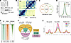 Figure 1
cGAS CUT&RUN analysis reveals genome-wide cGAS distributions that are independent of its DNA-binding and nucleosome-binding abilities. (A) Immunoblot (IB) analysis of indicated cellular fractions from MDA-MB-231 cells. (B) Chromatin interaction heatmaps of Micro-C data in the chr20 region. (C) A Venn diagram showing identified cGAS CUT&RUN peaks in indicated MDA-MB-231 cells. (D) cGAS CUT&RUN signals are enriched in a narrower window from both WT- and KKEA-cGAS–expressing MDA-MB-231 cells. (E) A heatmap for all indicated CUT&RUN peaks spanning –2.5 to 2.5 kb regions. H3K4me3, histone H3 trimethylated at lysine 4. (F) A Venn diagram showing identified cGAS CUT&RUN peaks in indicated MDA-MB-231 cells. sh, shRNA. (G) Average signal plots of z-normalized cGAS CUT&RUN signal from indicated MDA-MB-231 cells ± 1 kb around peak centers for each cGAS-binding category.
Given that nuclear cGAS has functions beyond innate immunity, including inhibition of PARP1-mediated DNA repair (21), regulation of replication fork progression (7), and centromere binding (8), we asked whether chromatin-associated cGAS regulates transcription. CUT&RUN analysis of EV-, WT- and KKEA-cGAS-expressing MDA-MB-231 cGAS−/− cells revealed robust genome-wide cGAS binding. WT- and KKEA-cGAS showed highly overlapping peaks (Figure 1, C–E), indicating similar genomic localization independent of DNA binding. These peaks partially overlapped with H3K4me3, a promoter-associated mark (Figure 1E), which was further confirmed at individual loci (Supplemental Figure 1L). Thus, chromatin-associated cGAS localizes genome-wide independently of its DNA-binding activity.
Because cGAS binds the acidic patch of nucleosomes (15–20), we included the nucleosome-binding–deficient R255E-cGAS mutant in CUT&RUN analysis. To control for altered cGAS expression or localization following reconstitution, we also mapped endogenous cGAS in parental shScr cells. Approximately 50% of endogenous cGAS peaks were recapitulated by reconstituted WT-cGAS (Figure 1F and Supplemental Figure 1, M–O), validating these sites while highlighting potential reconstitution-associated false positives. We therefore classified peaks absent in R255E-cGAS as “nucleosomal” peaks. Remaining peaks were divided into “neo-gained” peaks, unique to R255E-cGAS, and “true-cGAS” peaks, shared by endogenous, WT-, and R255E-cGAS, representing non-nucleosomal cGAS binding sites.
In MDA-MB-231 cells, we identified 212,634 high-confidence cGAS CUT&RUN peaks at non-nucleosomal regions and 6,374 at nucleosomal regions (Figure 1F). Among these, 18,960 true-cGAS peaks represented high-confidence, non-nucleosomal cGAS-binding sites and were the focus of this study. Notably, nucleosome-binding–deficient R255E-cGAS exhibited more and stronger non-nucleosomal peaks than nucleosomal peaks (Figure 1G and Figure 2A), suggesting that nucleosome tethering not only maintains cGAS inactivity (9) but also may restrict its localization to non-nucleosomal chromatin regions.
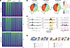 Figure 2
Nuclear cGAS localizes to genomic regions beyond nucleosomes. (A) Heatmaps of z-normalized signal for all indicated CUT&RUN peaks spanning –1 to 1 kb regions. (B) Pie charts showing the distributions of cGAS CUT&RUN peaks in each indicated category. (C) Representative genome browser views for indicated cGAS CUT&RUN peaks at indicated loci. Tracks are replicate averages of the z-normalized signal. Bars and highlights mark labeled cGAS peak categories. (D) A schematic model illustrating that nuclear cGAS exists in 3 distinct states: (i) tethered to nucleosomes via interaction with H2A/H2B acidic patches, (ii) associated with transcription factors to regulate transcription, and (iii) engaging both nucleosomal acidic patches and transcription factors simultaneously.
Further analysis revealed distinct genomic distributions of high-confidence true-cGAS peaks. Promoters were preferentially enriched in true-cGAS peaks (16.9%) compared with nucleosomal (9.3%) and neo-gained (8.7%) peaks (Figure 2B), suggesting a potential role for cGAS in transcriptional regulation. Genome browser analysis confirmed cGAS occupancy across these categories, including overlapping endogenous, WT-, and R255E-cGAS peaks at the LGALS3 promoter (Figure 2C). These findings suggest 3 potential chromatin-associated states of cGAS: nucleosome-tethered, nucleosome- and transcription factor–associated, and transcription factor/chromatin remodeler–associated (Figure 2D). These distinct states are explored below.
Integrative CUT&RUN, RNA-seq, and nuclear cGAS TurboID analyses reveal that cGAS interacts with AP-1 transcription factors to regulate genes involved in cholesterol homeostasis. To determine whether non-nucleosomal chromatin-associated cGAS regulates transcription, we performed RNA-seq in EV-, WT-cGAS–, and KKEA-cGAS–expressing MDA-MB-231 cGAS−/− cells (Figure 3A). Integrative analysis of cGAS CUT&RUN and RNA-seq identified cholesterol homeostasis as the top cGAS-regulated pathway, followed by fatty acid metabolism (Figure 3B and Supplemental Figure 2A). Among cholesterol-related genes, WT- and KKEA-cGAS increased LGALS3 and PPARG transcription relative to EV (Figure 3C and Supplemental Figure 2B). However, high-confidence true-cGAS peaks were detected at the LGALS3, but not PPARG, locus (Figure 3D and Supplemental Figure 2C). WT-, KKEA-, and R255E-cGAS all occupied the LGALS3 promoter (Figure 3E and Figure 2C), and R255E-cGAS promoted LGALS3 transcription similarly to WT-cGAS (Figure 3F). Consistently, cGAS deletion reduced LGALS3 expression in HeLa, A498, and BPH1 cells (Supplemental Figure 2, D–F). These findings identify LGALS3 as a potential direct transcriptional target of cGAS.
 Figure 3
cGAS is recruited to chromatin by AP-1 transcription factors. (A) A volcano plot from RNA-seq analysis displaying overlapping hits between WT- and KKEA-cGAS–expressing MDA-MB-231-cGAS–/– cells compared with EV-expressing cells. (B) A volcano plot showing the integrative analysis results from both cGAS CUT&RUN and RNA-seq analysis from EV-, WT- and KKEA-cGAS–expressing MDA-MB-231-cGAS–/– cells. (C and F) RT-PCR analysis of indicated gene targets from indicated MDA-MB-231 cells. Error bars were calculated as mean ± SD. n = 3. *P < 0.05 (1-way ANOVA test). The red dashed line indicates the normalized expression level of the control group, set to 1. (D) A volcano plot from cGAS CUT&RUN analysis showing difference in counts between WT- versus R255E-cGAS rescue within all cGAS peaks. The closer to the center of the x axis, the more likely the target is a true cGAS transcriptional target. The LGALS3 promoter peak is labeled in red. Peaks are colored by cGAS-binding category. Y axis is limited to 0–50 to improve visualization (102 points removed). (E) Representative genome browser views for indicated cGAS CUT&RUN peaks at the LGALS3 locus.
To explore the recruitment of cGAS to non-nucleosomal genomic regions, we conducted transcription factor motif enrichment analysis. AME (22) and de novo STREME analyses revealed strong enrichment of AP-1 motifs (RTGASTCAY) within true-cGAS CUT&RUN peaks (Figure 4A and Supplemental Figure 2G). Notably, AP-1 motifs were enriched in true-cGAS but depleted in nucleosomal peaks, suggesting that nucleosome tethering may influence cGAS association with AP-1 factors. Notably, we observed a shift in transcription factor motifs from nucleosomal cGAS peaks to true-cGAS peaks (Figure 4A), suggesting that cGAS binding to AP-1 transcription factors is influenced by its tethering to H2A/H2B acidic patches in certain genomic regions. To identify nuclear cGAS-interacting proteins, we performed nuclear cGAS TurboID proteomics (Supplemental Figure 2, H–L), identifying 675 proteins enriched ≥2-fold over controls. Known cGAS partners, including PARP1 (7) and BAF (13), validated the approach. Among the top transcription factors were the AP-1 family members JunB and FosL1 (Figure 4B), consistent with the motif analysis.
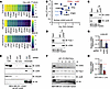 Figure 4
cGAS binds AP-1 transcription factors. (A) Heatmap of –log10(adj.pvalue) for top 10 enriched motifs identified by AME for sequences within cGAS CUT&RUN peaks in each group. (B) A volcano plot showing the top transcription factor hits from nuclear cGAS TurboID analysis from MDA-MB-231 cells. (C and D) IB analysis of indicated immunoprecipitations (IPs) and input from MDA-MB-231 cell chromatin fractions. (E) IB analysis of indicated cellular fractions from indicated MDA-MB-231 cells. (F) IB analysis of chromatin fractions from MDA-MB-231 cells transfected with indicated AP-1 TF-PROTACs. (G and H) RT-PCR analysis of indicated target genes from indicated MDA-MB-231 cells (G) or from MDA-MB-231 cells treated with 1 μM VZP1052 for 24 hours prior to cell collection (H).
We validated cGAS interactions with endogenous JunB and FosL1 in MDA-MB-231 cells (Figure 4, C and D), with similar interactions observed for exogenous cGAS and in THP1 cells (Supplemental Figure 2, M–O). JunB depletion reduced chromatin-associated cGAS (Figure 4E), while FosL1 degradation using the FosL1-PROTAC VZP1052 (23) produced a similar effect in MDA-MB-231 and THP1 cells (Supplemental Figure 2, P–S). Using a transcription factor targeting PROteolysis-TArgeting Chimera (TF-PROTAC) strategy (24, 25), we generated AP-1 TF-PROTACs by conjugating the canonical AP-1–binding sequence TGACTCA to von Hippel-Lindau (VHL) ligands. Several compounds (#7–11) reduced JunB/FosL1 levels and cGAS chromatin association (Figure 4F and Supplemental Figure 2, T and U). Depletion of JunB or FosL1 also reduced LGALS3 transcription (Figure 4, G and H, and Supplemental Figure 2, V–Y). Together, these findings support AP-1–dependent recruitment of nuclear cGAS to non-nucleosomal chromatin, particularly through JunB and FosL1.
cGAS contains TADs necessary for transcription factor binding. To determine how nuclear cGAS promotes transcription through JunB and FosL1, we asked whether cGAS contains TADs, which mediate transcriptional activation (26). The 9aaTAD algorithm (27) predicted 5 putative TADs, with TAD2 identified as a high-confidence TAD containing 2 canonical Φ-Φ-x-x-Φ motifs (Supplemental Figure 3, A and B). Using an AP-1–mCherry reporter assay (28), we found that JunB depletion or FosL1 degradation by VZP1052 reduced AP-1 activity in control but not cGAS−/− cells, where cGAS re-expression restored this activity (Supplemental Figure 3, C–E). Deletion of individual TADs reduced AP-1 reporter activity and cGAS binding to JunB or FosL1 and impaired LGALS3 transcription (Supplemental Figure 3, F–I). Together, these findings indicate that multiple cGAS TADs contribute to its interaction with AP-1 factors and transcriptional activity.
To minimize effects of protein conformational changes caused by TAD deletions, we focused on evolutionarily conserved Phe residues within these domains (Figure 5, A and B). Compared with F226A/F230A, the F491A/F503A and 4FA (F226A/F230A/F491A/F503A) mutants markedly reduced AP-1 reporter activity (Figure 5A) and impaired binding to JunB and FosL1 (Figure 5, C and D, and Supplemental Figure 3, J and K). However, unlike 4FA, the F491A/F503A mutant retained DNA-induced cGAS activation (Figure 5, E and F). Although F491A/F503A showed reduced chromatin localization, it retained nucleosome binding (Figure 5, G and H, and Supplemental Figure 3, L–N) but failed to efficiently promote LGALS3 transcription (Figure 5, I–K). In contrast, DNA-binding–deficient KKEA and nucleosome-binding–deficient R255E cGAS retained JunB/FosL1 binding (Supplemental Figure 3O). Thus, we used F491A/F503A-cGAS as a key model to dissect the transcriptional function of cGAS independently of its canonical immune activity (Supplemental Figure 3P).
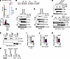 Figure 5
A TAD in cGAS is responsible for AP-1 recognition but dispensable for canonical cGAS activation in innate immunity. (A) Representative FACS analysis of AP-1 cherry reporter activity in indicated MDA-MB-231 cells and quantified. Error bars were calculated as mean ± SD. n = 3. (B) Protein sequence alignment of human and mouse cGAS with critical Phe residues in possible TADs in red. (C and D) IB analysis of Flag-IPs and whole-cell lysate (WCL) from HEK293T cells transfected with indicated DNA constructs. (E) IB analysis of WCL from HEK293T cells transfected with indicated DNA constructs. (F) IB analysis of indicated MDA-MB-231 cells transfected with 5 μg/mL ISD90 for indicated time periods. (G) IB analysis of indicated cellular fractions from indicated MDA-MB-231 cells. (H) IB analysis of indicated Flag-NCP-IPs with indicated bacterially purified GST-cGAS recombinant proteins. (I–K) RT-PCR analysis of mRNA expression of LGALS3 from indicated cells. Error bars represent mean ± SD. n = 3. *P < 0.05 (1-way ANOVA test).
We next performed structural modeling and molecular dynamics (MD) simulations of the chromatin-bound cGAS/AP-1 complex to define their interaction interface. The complex remained stable throughout equilibration, with 4 conserved polar interaction networks and extensive hydrophobic contacts preserved. cGAS engaged AP-1 adjacent to its KKH DNA-binding domain, positioning the functionally critical F503 at the center of the interface (Supplemental Figure 4A). Specifically, cGAS-R499, K427, Y510/N514, and N513 formed persistent interactions with FosL1-E159, E152, E141, and JunB-K299, respectively (Supplemental Figure 4B). F503 also occupied a hydrophobic pocket within FosL1, where its aromatic ring packed against the aliphatic portion of FosL1-E152. These interactions provide a structural basis for the critical role of F503 in stabilizing cGAS/AP-1 binding, consistent with our biochemical findings.
cGAS governs AP-1–dependent transcriptional programs. Beyond LGALS3, we identified cGAS-bound genomic regions enriched for AP-1 motifs and occupied by endogenous, WT-, and R255E-cGAS (Supplemental Figure 5, A–H). RT-qPCR validated several targets and confirmed their dependence on cGAS and its TAD (Supplemental Figure 5, I and J). To define the broader cGAS/AP-1 transcriptional program, we performed RNA-seq in cGAS−/− MDA-MB-231 cells reconstituted with WT-, F491A/F503A-cGAS, or WT-cGAS with JunB depletion (WT-cGAS-shJunB) (Supplemental Figure 5K). We identified 725 genes downregulated by both F491A/F503A-cGAS and JunB depletion relative to WT-cGAS, with LGALS3 among the top 2 affected genes (Supplemental Figure 5, L–N). Gene Ontology analysis linked these cGAS/AP-1 targets to cell-cell junction organization, proliferation, and other tumor-related processes (Supplemental Figure 5O).
cGAS governs transcription by complexing with transcription modulators. Nuclear cGAS TurboID identified, in addition to transcription factors (Figure 4B), multiple chromatin regulators, suggesting their involvement in cGAS/AP-1–dependent transcription. Chromatin fractionation and cGAS immunoprecipitation confirmed interactions with AP-1 factors as well as chromatin regulators SMARCA5 and SMARCB1 and epigenetic modifiers KMT2A and HDAC1 (Supplemental Figure 6A). Depletion of SMARCA5 or SMARCB1 reduced cGAS-dependent LGALS3 transcription (Supplemental Figure 6, B–D), supporting their functional role in this pathway. Although the cGAS N-terminus mediates DNA binding (29, 30) and oligomerization (31), the isolated N-terminus failed to promote LGALS3 transcription, whereas an N-terminal deletion mutant retained this activity (Supplemental Figure 6, E and F). Both constructs localized to the nucleus, but only the N-terminal deletion mutant efficiently associated with chromatin (Supplemental Figure 6, G and H).
Previous studies showed that nuclear cGAS tethering to nucleosomes is regulated by N-terminal phosphorylation by mitotic kinases (10) and mTORC2 (11). We therefore asked whether these pathways also regulate cGAS-dependent transcription. Nocodazole synchronization of HeLa cells followed by washout revealed increased cGAS and AP-1 recruitment to chromatin during late G1, coinciding with enhanced LGALS3 transcription (Supplemental Figure 6, I–K). Conversely, mTORC2 inhibition with Torin 2 increased cytoplasmic cGAS and reduced LGALS3 expression (Supplemental Figure 6, L and M), consistent with decreased nuclear cGAS activity.
DNA-damaging agents such as doxorubicin can promote cGAS nuclear translocation through B-lymphoid tyrosine kinase–dependent phosphorylation (21). Consistent with this, etoposide increased nuclear cGAS accumulation and LGALS3 transcription in MDA-MB-231 cells (Supplemental Figure 6, N and O), likely through enhanced chromatin association. Because AP-1 activity is regulated by JNK-mediated phosphorylation (32), we tested whether JNK activity affects cGAS recruitment. JNK inhibition with SP600125 reduced cGAS–AP-1 association on chromatin (Supplemental Figure 6P), suggesting that JNK-dependent AP-1 activation promotes cGAS recruitment and transcriptional activity.
Metabolomics reveals that the cGAS/LGALS3/ACAA2 axis regulates fatty acid oxidation, acetyl-CoA production, and cholesterol synthesis. Our integrated cGAS CUT&RUN and RNA-seq analyses identified cholesterol homeostasis as the top cGAS-regulated pathway (Figure 3, A and B). Filippin III staining (33) showed that WT-cGAS re-expression reduced cellular cholesterol in MDA-MB-231 cGAS−/− cells, whereas LGALS3 depletion restored cholesterol accumulation (Figure 6A). Consistently, the transcriptionally inactive F491A/F503A-cGAS mutant failed to suppress cholesterol levels in MDA-MB-231, mouse embryonic fibroblast (MEF), BPH1, and THP1 cells (Figure 6, B and C, and Supplemental Figure 7, A–C), consistent with its impaired AP-1 binding and LGALS3 transcription. In contrast, STING deficiency did not increase cholesterol in HeLa cells (Supplemental Figure 7, D and E), indicating that cGAS regulates cholesterol independently of canonical STING signaling. Consistent with these findings, cGas−/− C57BL/6 mouse livers showed increased cholesterol and reduced Lgals3 expression (Figure 6, D and E), supporting a role for the cGAS/LGALS3 axis in suppressing hepatic cholesterol accumulation.
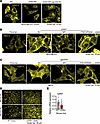 Figure 6
cGAS-driven transcription of LGALS3 inhibits cholesterol production. (A–D) Representative images for Filippin III staining for free cholesterol in indicated MDA-MB-231 cells (A and B), MEF cells (C), or liver slides from indicated C57BL/6 mice (D). (E) RT-PCR analysis of mRNA expression of Lgals3 from indicated mouse livers. Error bars represent mean ± SD. n = 3. *P < 0.05 (1-way ANOVA test). The red dashed line indicates the normalized expression level of the control group, set to 1.
BioGRID analysis of cGAS-interacting proteins identified several metabolic enzymes involved in fatty acid and cholesterol metabolism (Figure 7A). Among these, ACAA2, which catalyzes the final step of mitochondrial fatty acid β-oxidation (34), partially colocalized with LGALS3 in MDA-MB-231 cells (Figure 7B) and directly interacted with LGALS3 in vitro (Figure 7, C and D). Importantly, LGALS3 inhibited ACAA2 enzymatic activity (Figure 7E and Supplemental Figure 7F). Consistent with this mechanism, WT-cGAS re-expression reduced acetyl-CoA levels in MDA-MB-231 cGAS−/− cells, an effect largely reversed by LGALS3 depletion (Figure 7F). Increasing ACAA2 activity elevated cholesterol, whereas ACAA2 inhibition with trimetazidine reduced cholesterol specifically in cGAS-deficient cells (Supplemental Figure 7, G–I). Together, these findings support a cGAS/LGALS3/ACAA2 axis that restrains cholesterol synthesis by limiting fatty acid β-oxidation and acetyl-CoA production.
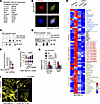 Figure 7
cGAS-driven transcription of LGALS3 inhibits ACAA2 activity, suppressing fatty acid oxidation. (A) A list of LGALS3-binding proteins from BioGRID. (B) Representative confocal immunofluorescence (IF) images for ACAA2 and LGALS3 in MDA-MB-231 cells. (C and D) IB analysis of pulldowns and inputs using indicated bacterially purified recombinant proteins. (E and F) Representative acetyl-CoA ELISAs measuring acetyl-CoA levels in either in vitro ACAA2 reactions with recombinant LGALS3 proteins (E) or indicated MDA-MB-231 cells (F). (G) Normalized levels of [13C2]-citrate (citrate M+2) and [13C16]-palmityl-carnitine in indicated MDA-MB-231 cells relative to parental cells. Data are shown as individual values with mean, n = 2 independent biological replicates. No statistical analysis was performed because n < 3. (H) A heatmap quantifying indicated metabolites in indicated MDA-MB-231 cells with 13C-palmitate tracing for 2 hours. HP, hexose phosphate; FBP, fructose bisphosphate; DHAP/GAP, dihydroxyacetone phosphate/glyceraldehyde-3-phosphate; 2PG/3PG, 2-phosphoglycerate/3-phosphoglycerate; PEP, phosphoenolpyruvate. (I) Representative Filippin III staining images for free cholesterol in indicated MDA-MB-231 cells. Where indicated, 5 μM lovastatin was used to treat cells for 6 hours prior to staining.
Because ACAA2 generates acetyl-CoA, a precursor for cholesterol synthesis (34), we next examined whether cGAS regulates fatty acid oxidation. Metabolomics and 13C-palmitate tracing in parental, cGAS−/−, and cGAS−/− cells reconstituted with WT-cGAS or LGALS3 revealed that cGAS loss increased fatty acid oxidation, reflected by elevated 13C-labeled citrate and palmitoyl-carnitine (Figure 7G). cGAS deletion also increased glycolysis, energy metabolites, acyl-carnitines, and amino acids, changes largely reversed by WT-cGAS or LGALS3 re-expression (Figure 7H). Similar metabolic alterations occurred in HeLa but not STING−/− cells (Supplemental Figure 7, J and K), supporting a STING-independent role for cGAS in metabolic regulation. Additionally, acetyl-CoA serves as the starting substrate for cholesterol synthesis, with the rate-limiting step being the conversion of HMG-CoA to mevalonate by HMG-CoA reductase (HMGCR) (35). Inhibition of HMGCR with lovastatin reduced cholesterol in both WT and cGAS−/− cells (Figure 7I), implicating de novo cholesterol synthesis in the cholesterol accumulation caused by cGAS loss. Together, these findings define a cGAS/LGALS3/ACAA2 axis that restrains fatty acid β-oxidation, acetyl-CoA production, and downstream cholesterol synthesis.
Cholesterol restrains phosphatase and tensin homolog on the plasma membrane to elevate phosphatidylinositol [3,4,5] triphosphate levels and subsequent mTORC2 activation. Cholesterol has been reported to influence lipid raft composition (36), trafficking (37), PI3K activity (38), or other physiological function (39). Consistent with a role in mTOR regulation, WT-cGAS re-expression in cGAS−/− cells reduced plasma membrane phosphatidylinositol [3,4,5] triphosphate [PI(3,4,5)P3] levels, an effect largely reversed by LGALS3 depletion (Figure 8A and Supplemental Figure 8A). Conversely, LGALS3 re-expression reduced PI(3,4,5)P3 in cGAS−/− cells (Supplemental Figure 8B), linking cGAS/LGALS3 signaling to cholesterol-dependent PI345P3 regulation. cGAS loss also reduced membrane localization of phosphatase and tensin homolog (PTEN) (Figure 8B), a key PI(3,4,5)P3 phosphatase (40). Previously, we reported PI(3,4,5)P3 is critical for mTORC2 plasma membrane localization and activation (41). Elevated PI(3,4,5)P3 in cGAS−/− cells was accompanied by increased membrane localization of mTORC2 components Rictor and Sin1 (Figure 8C and Supplemental Figure 8C). Together, these findings suggest that cGAS suppresses mTORC2 activation through the cGAS/LGALS3/cholesterol/PI(3,4,5)P3 axis.
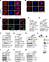 Figure 8
cGAS/LGALS3-regulated cholesterol changes modulate PI(3,4,5)P3 levels and mTORC2/Akt activation. (A) Representative confocal images of PI(3,4,5)P3 staining of indicated MDA-MB-231 cells. (B and C) Representative confocal images of PTEN staining (B) or Rictor staining (C) of indicated MDA-MB-231 cells. (D–H) IB analysis of WCL from indicated MDA-MB-231 cells. (I) IB analysis of WCL from indicated THP1 cells. (J) IB analysis of WCL from indicated MDA-MB-231 cells. Where indicated, 100 μM cholesterol was added to cell culture for indicated time periods prior to cell collection. (K) IB analysis of WCL from indicated MDA-MB-231 cells. Where indicated, cells were pretreated with 5 μM lovastatin for 2 hours followed by addition of 100 μM cholesterol for 1.5 hours prior to cell collection. (L) A schematic illustration of the proposed model: cGAS promotes LGALS3 transcription, which binds to and suppresses ACAA2 activity, leading to reduced cellular acetyl-CoA levels, decreased cholesterol production, and diminished mTORC2/Akt activation.
We next examined mTORC2 activity and found that cGAS deletion increased Akt-pS473, an mTORC2 substrate, in MDA-MB-231 cells, which was reversed by WT-cGAS reconstitution (Figure 8D and Supplemental Figure 8D). Re-expression of LGALS3 similarly suppressed Akt-pS473 induced by cGAS deletion (Figure 8E), whereas LGALS3 depletion or ACAA2 expression largely reversed the suppression mediated by WT-cGAS (Figure 8, F and G). Consistent with cGAS regulating mTORC2 independently of its innate immune function, KKEA- and R255E-cGAS, but not F491A/F503A-cGAS, suppressed Akt-pS473 (Figure 8, H and I, and Supplemental Figure 8, E and F). Cholesterol addition enhanced Akt-pS473, whereas lovastatin suppressed this effect (Figure 8, J and K, and Supplemental Figure 8, E–I). Collectively, these findings suggest that the cGAS/LGALS3/ACAA2 axis negatively regulates cholesterol levels to suppress mTORC2 activity (Figure 8L).
Inhibition of Akt, but not STING, reverses cholesterol accumulation in cGAS-deficient mouse livers. To further establish the role of mTORC2/Akt signaling in cGAS-mediated cholesterol homeostasis, we treated cGas–/– C57BL/6 mice with either Akt inhibitor capivasertib or STING inhibitor H-151. Capivasertib, but not H-151, reduced hepatic cholesterol levels in cGas–/– mice (Figure 9A), without affecting body weight (Supplemental Figure 9A), supporting a role for mTORC2/Akt signaling in regulating cholesterol independently of STING.
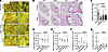 Figure 9
Inhibition of Akt, but not STING, reverses cholesterol accumulation in cGAS-deficient mouse livers. (A) Representative images for Filippin III staining for free cholesterol in indicated mouse livers. Where indicated, indicated C57BL/6 mice were treated with Akt inhibitor capivasertib or STING inhibitor H-151 prior to liver collection. Refer to Methods for details. (B) Lung histopathology in 4-month-old WT and cGAS-KO mice. Representative H&E-stained lung sections from both sexes show preserved airway and alveolar architecture. Left: low magnification, intermediate section of the left lung lobe, scale bar = 0.5 mm. Right: high magnification of insets, scale bar = 50 μm. (C) RT-PCR analysis of Lgals3 mRNA expression in lungs from indicated mice with indicated treatments. Error bars represent mean ± SD; n = 3. *P < 0.05 by 1-way ANOVA. (D–G) Bronchoalveolar lavage (BAL) cell composition in cGas-KO and WT littermates under indicated treatment conditions. (D) Total BAL cell counts. (E–G) Differential cell counts showing macrophages, neutrophils, and lymphocytes.
Because hepatic cholesterol accumulation and dysregulation are associated with pulmonary inflammation, fibrosis, and impaired surfactant homeostasis (42), and a 2024 preprint (43) reported that aged male cGas–/– mice develop spontaneous pro-inflammatory phenotypes in both the lung and liver, we asked whether cGAS deficiency causes pulmonary abnormalities in young adult mice. Histological analysis of 4-month-old cGas–/– mice and Wt littermates of both sexes revealed no overt differences in airway or alveolar architecture (Figure 9B). As in the liver, Lgals3 expression was markedly reduced in cGas–/– lungs and was not restored by capivasertib or H-151 (Figure 9C), supporting mTORC2/Akt as a downstream pathway of the cGAS/LGALS3 axis. Bronchoalveolar lavage (BAL) analysis showed that macrophages predominated in both genotypes, with no significant differences in macrophage, neutrophil, or lymphocyte numbers (Figure 9, D–G). Lung macrophages from cGas–/– mice also lacked morphological features of activation or polarization (Supplemental Figure 9B). Body weight and lung wet/dry ratios were comparable between genotypes (Supplemental Figure 9, C–F). Thus, cGas–/– mice do not exhibit overt pulmonary inflammation or pathology under basal conditions at 4 months of age, despite reduced Lgals3 expression.
However, under the stress of CO2 euthanasia, cGas–/– lungs appeared visibly enlarged, whereas the liver, heart, kidney, spleen, and overall body weight were unchanged (Supplemental Figure 10, A–K). Compared with Wt lungs, cGas–/– lungs exhibited reduced Lgals3 expression, increased cholesterol levels, and elevated expression of inflammatory/fibrosis-associated genes, including B2m, Cxcl9, and Ifitm3 (Supplemental Figure 10, L–P). These findings suggest that the cGAS/LGALS3/cholesterol axis may influence stress-induced pulmonary responses. However, CO2 euthanasia can itself induce hypoxia, hypercapnia, acidosis, and rapid changes in lung morphology, inflammation, gene expression, metabolism, and immune phenotypes (44–47). Thus, these findings should be interpreted cautiously, and further studies using euthanasia methods that minimize such artifacts are needed to determine whether cGAS deficiency promotes pulmonary pathology under stress or other challenge conditions.
cGAS/LGALS3/mTORC2 signaling controls cell size and tumor growth. The mTORC2/Akt pathway promotes cell proliferation and survival (48–50). We therefore examined whether the cGAS/LGALS3/ACAA2/mTORC2 axis regulates cell behavior. cGAS deletion increased mTORC2/Akt activation and cell size, which was phenocopied by LGALS3 depletion (Figure 10A and Figure 8F). This effect was independent of canonical cGAS/STING signaling, as STING depletion did not alter cell size (Figure 10, B and C, and Supplemental Figure 11, A–E). Re-expression of WT-, R255E-, or KKEA-cGAS suppressed anchorage-independent growth of cGAS-deficient MDA-MB-231 cells, accompanied by reduced cell size (Figure 10, D and E). In contrast, the AP-1–binding–deficient F491A/F503A-cGAS mutant failed to suppress soft agar growth (Figure 10, F and G). Consistent with LGALS3 being a major cGAS transcriptional target, LGALS3 re-expression similarly suppressed anchorage-independent growth (Figure 10, H and I).
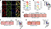 Figure 10
The transcriptional activity of cGAS influences cell size and 3D growth. (A) Representative IF images of phalloidin and vinculin in the indicated MDA-MB-231 cells. (B and C) Representative FACS analysis showing cell size distributions in the indicated MDA-MB-231 cells. (D–I) Representative images of cell colonies grown in soft agar using the indicated MDA-MB-231 cells, with quantification shown in E, G, and I. Error bars represent mean ± SD. n = 3. *P < 0.05 (1-way ANOVA test).
To further define the role of the cGAS/LGALS3/ACAA2/acetyl-CoA/cholesterol/mTORC2 axis in tumor growth, we performed xenograft studies in nude mice. Whereas STING deletion had minimal effects, cGAS deletion markedly accelerated tumor growth in both HeLa and MDA-MB-231 xenografts (Figure 11, A–C, and Supplemental Figure 11, F–J), accompanied by increased mTORC2/Akt activation (Supplemental Figure 11, K–M). LGALS3 re-expression largely reversed the enhanced tumor growth and lipid accumulation caused by cGAS deletion (Figure 11, D–H, and Supplemental Figure 11, N and O), supporting LGALS3 as a key downstream effector of cGAS-mediated tumor suppression and lipid homeostasis. Consistently, unlike WT-cGAS, the transcriptionally defective F491A/F503A-cGAS mutant failed to suppress tumor growth in cGAS-deficient MDA-MB-231 xenografts (Figure 11, I–L, and Supplemental Figure 11, P and Q). Finally, reduced LGALS3 expression was associated with poorer survival in ovarian, lung, and gastric cancers (Supplemental Figure 11, R–T), further supporting a tumor-suppressive role for the cGAS/LGALS3 axis.
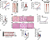 Figure 11
The transcriptional activity of cGAS regulates xenografted tumor growth. (A and D) Tumor volume measurements over time for xenografted cell lines. Error bars represent mean ± SD. n = 8. *P < 0.05 (1-way ANOVA test). (B and E) Images of isolated tumors from A and D with corresponding tumor weights shown in C and F. Error bars represent mean ± SD. n = 8. *P < 0.05 (1-way ANOVA test). (G and H) Representative H&E staining images of fixed tumor sections from the indicated xenografted tumors. (I) Tumor volume measurements over time for xenografted cell lines. Error bars represent mean ± SD. n = 9. *P < 0.05 (1-way ANOVA test). (J) Images of isolated tumors from I with corresponding tumor weights shown in K. Error bars represent mean ± SD. n = 9. *P < 0.05 (1-way ANOVA test). (L) RT-PCR analysis of LGALS3 mRNA levels in indicated collected tumors from J. Error bars represent mean ± SD. n = 9. *P < 0.05 (1-way ANOVA test).
cGAS/LGALS3/mTORC2 signaling regulates tumor therapeutic response. mTORC2/Akt hyperactivation not only promotes tumor growth but also enhances apoptosis resistance (50), resists therapy (51), and drives angiogenic and metastatic processes (52). We therefore asked whether the cGAS/LGALS3/ACAA2/mTORC2 axis regulates therapeutic response. We started from chemotherapy because it is the first-line treatment for various cancer types (53, 54). In MDA-MB-231 and HeLa cells, cGAS deletion conferred resistance to doxorubicin and etoposide, which was reversed by WT-cGAS re-expression (Figure 12A and Supplemental Figure 12, A–E). LGALS3 re-expression similarly restored drug sensitivity, whereas the transcriptionally defective F491A/F503A-cGAS mutant did not (Figure 12, B–E, and Supplemental Figure 12, F and G). Consistent with elevated cholesterol and mTORC2/Akt activity in cGAS-deficient cells, cGAS deletion also conferred resistance to cholesterol-lowering agents, including lovastatin, atorvastatin, and triheptanoin, as well as the mTOR inhibitor Torin 2 and Akt inhibitor GSK2110183 (Figure 12, F–J). WT-cGAS, but not F491A/F503A-cGAS, largely restored drug sensitivity, which depended on LGALS3 expression (Figure 12, K–M). Long-term 3D soft agar assays further confirmed the requirement for cGAS transcriptional activity in responses to doxorubicin and lovastatin (Figure 13, A–D). Together, these findings identify the LGALS3/cholesterol/mTORC2 axis as a key downstream effector of cGAS that regulates responses to chemotherapy, cholesterol-lowering agents, and mTOR/Akt inhibition (Supplemental Figure 12H).
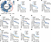 Figure 12
The transcriptional activity of cGAS influences cellular responses to therapies. (A) Heatmap displaying cell responses to the indicated treatments. (B–M) MTT assays using the indicated MDA-MB-231 cells treated for 3 days with indicated agents at the indicated doses. Error bars represent mean ± SD. n = 3. *P < 0.05 (1-way ANOVA test).
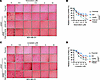 Figure 13
Transcriptional activity of cGAS influences cellular responses to therapy in 3D soft agar assays. (A and C) Representative 3D soft agar assay images of indicated MDA-MB-231 cells treated with indicated doses of doxorubicin (A) or lovastatin (C) for 3 weeks and quantified in B and D. Compounds were refreshed every 3 days. Error bars represent mean ± SD. n = 3. *P < 0.05 (1-way ANOVA test).


Copyright © 2026 American Society for Clinical Investigation
ISSN: 0021-9738 (print), 1558-8238 (online)