
Advertisement
Research ArticleCardiologyCell biologyClinical Research
Open Access |  10.1172/JCI201684
Gut microbial trimethylamine N-oxide generation promotes risk of atrial fibrillation via muscarinic receptor–mediated autonomic dysfunction
Selvam Arjunan,1 Isaiah Pemberton,1 Xinmin S. Li,1 Naseer Sangwan,1 Lydia Akino,2,3 Emmanuel Opoku,1 Dmitriy Verbovetskiy,1 Ina Nemet,1 Hyun Su Kim,1 Haruko Masumiya,1 Zeneng Wang,1 Joseph A. Lupica,1 Melissa Y. Tian,1 Karis Mao,1 Deepthi P. Mallela,1 Maradumane L. Mohan,1 Sarah M. Schumacher,1 Julie H. Rennison,1 Sathyamangla V. Naga Prasad,1 Kenneth R. Laurita,2,3 Vamsi Chodisetty,1 Mina K. Chung,1 David R. Van Wagoner,1 John Barnard,1 Jonathan D. Smith,1 Oussama Wazni,1,4 Stanley L. Hazen,1,4 and Robert A. Koeth1,4
1Department of Heart, Blood & Kidney Research, Cleveland Clinic Research, Cleveland Clinic, Cleveland, Ohio, USA.
2Department of Biomedical Engineering, Case School of Engineering School of Medicine, Case Western Reserve University, Cleveland, Ohio, USA.
3Heart and Vascular Research Center, MetroHealth Campus, Case Western Reserve University, Cleveland, Ohio, USA.
4Department of Cardiovascular Medicine, Heart, Vascular & Thoracic Institute, Cleveland Clinic, Cleveland, Ohio, USA.
Address correspondence to: Robert A. Koeth, Department of Cardiovascular Medicine and Department of Heart, Blood & Kidney Research, Cleveland Clinic Research, Cleveland Clinic, 9500 Euclid Avenue, J2-2, Cleveland, Ohio 44195, USA. Phone: 216.445.7610; Email: koethr@ccf.org.
Find articles by Arjunan, S. in: PubMed | Google Scholar
1Department of Heart, Blood & Kidney Research, Cleveland Clinic Research, Cleveland Clinic, Cleveland, Ohio, USA.
2Department of Biomedical Engineering, Case School of Engineering School of Medicine, Case Western Reserve University, Cleveland, Ohio, USA.
3Heart and Vascular Research Center, MetroHealth Campus, Case Western Reserve University, Cleveland, Ohio, USA.
4Department of Cardiovascular Medicine, Heart, Vascular & Thoracic Institute, Cleveland Clinic, Cleveland, Ohio, USA.
Address correspondence to: Robert A. Koeth, Department of Cardiovascular Medicine and Department of Heart, Blood & Kidney Research, Cleveland Clinic Research, Cleveland Clinic, 9500 Euclid Avenue, J2-2, Cleveland, Ohio 44195, USA. Phone: 216.445.7610; Email: koethr@ccf.org.
Find articles by Pemberton, I. in: PubMed | Google Scholar
1Department of Heart, Blood & Kidney Research, Cleveland Clinic Research, Cleveland Clinic, Cleveland, Ohio, USA.
2Department of Biomedical Engineering, Case School of Engineering School of Medicine, Case Western Reserve University, Cleveland, Ohio, USA.
3Heart and Vascular Research Center, MetroHealth Campus, Case Western Reserve University, Cleveland, Ohio, USA.
4Department of Cardiovascular Medicine, Heart, Vascular & Thoracic Institute, Cleveland Clinic, Cleveland, Ohio, USA.
Address correspondence to: Robert A. Koeth, Department of Cardiovascular Medicine and Department of Heart, Blood & Kidney Research, Cleveland Clinic Research, Cleveland Clinic, 9500 Euclid Avenue, J2-2, Cleveland, Ohio 44195, USA. Phone: 216.445.7610; Email: koethr@ccf.org.
Find articles by Li, X. in: PubMed | Google Scholar
1Department of Heart, Blood & Kidney Research, Cleveland Clinic Research, Cleveland Clinic, Cleveland, Ohio, USA.
2Department of Biomedical Engineering, Case School of Engineering School of Medicine, Case Western Reserve University, Cleveland, Ohio, USA.
3Heart and Vascular Research Center, MetroHealth Campus, Case Western Reserve University, Cleveland, Ohio, USA.
4Department of Cardiovascular Medicine, Heart, Vascular & Thoracic Institute, Cleveland Clinic, Cleveland, Ohio, USA.
Address correspondence to: Robert A. Koeth, Department of Cardiovascular Medicine and Department of Heart, Blood & Kidney Research, Cleveland Clinic Research, Cleveland Clinic, 9500 Euclid Avenue, J2-2, Cleveland, Ohio 44195, USA. Phone: 216.445.7610; Email: koethr@ccf.org.
Find articles by Sangwan, N. in: PubMed | Google Scholar
1Department of Heart, Blood & Kidney Research, Cleveland Clinic Research, Cleveland Clinic, Cleveland, Ohio, USA.
2Department of Biomedical Engineering, Case School of Engineering School of Medicine, Case Western Reserve University, Cleveland, Ohio, USA.
3Heart and Vascular Research Center, MetroHealth Campus, Case Western Reserve University, Cleveland, Ohio, USA.
4Department of Cardiovascular Medicine, Heart, Vascular & Thoracic Institute, Cleveland Clinic, Cleveland, Ohio, USA.
Address correspondence to: Robert A. Koeth, Department of Cardiovascular Medicine and Department of Heart, Blood & Kidney Research, Cleveland Clinic Research, Cleveland Clinic, 9500 Euclid Avenue, J2-2, Cleveland, Ohio 44195, USA. Phone: 216.445.7610; Email: koethr@ccf.org.
Find articles by Akino, L. in: PubMed | Google Scholar
1Department of Heart, Blood & Kidney Research, Cleveland Clinic Research, Cleveland Clinic, Cleveland, Ohio, USA.
2Department of Biomedical Engineering, Case School of Engineering School of Medicine, Case Western Reserve University, Cleveland, Ohio, USA.
3Heart and Vascular Research Center, MetroHealth Campus, Case Western Reserve University, Cleveland, Ohio, USA.
4Department of Cardiovascular Medicine, Heart, Vascular & Thoracic Institute, Cleveland Clinic, Cleveland, Ohio, USA.
Address correspondence to: Robert A. Koeth, Department of Cardiovascular Medicine and Department of Heart, Blood & Kidney Research, Cleveland Clinic Research, Cleveland Clinic, 9500 Euclid Avenue, J2-2, Cleveland, Ohio 44195, USA. Phone: 216.445.7610; Email: koethr@ccf.org.
Find articles by Opoku, E. in: PubMed | Google Scholar
1Department of Heart, Blood & Kidney Research, Cleveland Clinic Research, Cleveland Clinic, Cleveland, Ohio, USA.
2Department of Biomedical Engineering, Case School of Engineering School of Medicine, Case Western Reserve University, Cleveland, Ohio, USA.
3Heart and Vascular Research Center, MetroHealth Campus, Case Western Reserve University, Cleveland, Ohio, USA.
4Department of Cardiovascular Medicine, Heart, Vascular & Thoracic Institute, Cleveland Clinic, Cleveland, Ohio, USA.
Address correspondence to: Robert A. Koeth, Department of Cardiovascular Medicine and Department of Heart, Blood & Kidney Research, Cleveland Clinic Research, Cleveland Clinic, 9500 Euclid Avenue, J2-2, Cleveland, Ohio 44195, USA. Phone: 216.445.7610; Email: koethr@ccf.org.
Find articles by Verbovetskiy, D. in: PubMed | Google Scholar
1Department of Heart, Blood & Kidney Research, Cleveland Clinic Research, Cleveland Clinic, Cleveland, Ohio, USA.
2Department of Biomedical Engineering, Case School of Engineering School of Medicine, Case Western Reserve University, Cleveland, Ohio, USA.
3Heart and Vascular Research Center, MetroHealth Campus, Case Western Reserve University, Cleveland, Ohio, USA.
4Department of Cardiovascular Medicine, Heart, Vascular & Thoracic Institute, Cleveland Clinic, Cleveland, Ohio, USA.
Address correspondence to: Robert A. Koeth, Department of Cardiovascular Medicine and Department of Heart, Blood & Kidney Research, Cleveland Clinic Research, Cleveland Clinic, 9500 Euclid Avenue, J2-2, Cleveland, Ohio 44195, USA. Phone: 216.445.7610; Email: koethr@ccf.org.
Find articles by Nemet, I. in: PubMed | Google Scholar
1Department of Heart, Blood & Kidney Research, Cleveland Clinic Research, Cleveland Clinic, Cleveland, Ohio, USA.
2Department of Biomedical Engineering, Case School of Engineering School of Medicine, Case Western Reserve University, Cleveland, Ohio, USA.
3Heart and Vascular Research Center, MetroHealth Campus, Case Western Reserve University, Cleveland, Ohio, USA.
4Department of Cardiovascular Medicine, Heart, Vascular & Thoracic Institute, Cleveland Clinic, Cleveland, Ohio, USA.
Address correspondence to: Robert A. Koeth, Department of Cardiovascular Medicine and Department of Heart, Blood & Kidney Research, Cleveland Clinic Research, Cleveland Clinic, 9500 Euclid Avenue, J2-2, Cleveland, Ohio 44195, USA. Phone: 216.445.7610; Email: koethr@ccf.org.
Find articles by Kim, H. in: PubMed | Google Scholar
1Department of Heart, Blood & Kidney Research, Cleveland Clinic Research, Cleveland Clinic, Cleveland, Ohio, USA.
2Department of Biomedical Engineering, Case School of Engineering School of Medicine, Case Western Reserve University, Cleveland, Ohio, USA.
3Heart and Vascular Research Center, MetroHealth Campus, Case Western Reserve University, Cleveland, Ohio, USA.
4Department of Cardiovascular Medicine, Heart, Vascular & Thoracic Institute, Cleveland Clinic, Cleveland, Ohio, USA.
Address correspondence to: Robert A. Koeth, Department of Cardiovascular Medicine and Department of Heart, Blood & Kidney Research, Cleveland Clinic Research, Cleveland Clinic, 9500 Euclid Avenue, J2-2, Cleveland, Ohio 44195, USA. Phone: 216.445.7610; Email: koethr@ccf.org.
Find articles by Masumiya, H. in: PubMed | Google Scholar
1Department of Heart, Blood & Kidney Research, Cleveland Clinic Research, Cleveland Clinic, Cleveland, Ohio, USA.
2Department of Biomedical Engineering, Case School of Engineering School of Medicine, Case Western Reserve University, Cleveland, Ohio, USA.
3Heart and Vascular Research Center, MetroHealth Campus, Case Western Reserve University, Cleveland, Ohio, USA.
4Department of Cardiovascular Medicine, Heart, Vascular & Thoracic Institute, Cleveland Clinic, Cleveland, Ohio, USA.
Address correspondence to: Robert A. Koeth, Department of Cardiovascular Medicine and Department of Heart, Blood & Kidney Research, Cleveland Clinic Research, Cleveland Clinic, 9500 Euclid Avenue, J2-2, Cleveland, Ohio 44195, USA. Phone: 216.445.7610; Email: koethr@ccf.org.
Find articles by
Wang, Z.
in:
PubMed
|
Google Scholar
|

1Department of Heart, Blood & Kidney Research, Cleveland Clinic Research, Cleveland Clinic, Cleveland, Ohio, USA.
2Department of Biomedical Engineering, Case School of Engineering School of Medicine, Case Western Reserve University, Cleveland, Ohio, USA.
3Heart and Vascular Research Center, MetroHealth Campus, Case Western Reserve University, Cleveland, Ohio, USA.
4Department of Cardiovascular Medicine, Heart, Vascular & Thoracic Institute, Cleveland Clinic, Cleveland, Ohio, USA.
Address correspondence to: Robert A. Koeth, Department of Cardiovascular Medicine and Department of Heart, Blood & Kidney Research, Cleveland Clinic Research, Cleveland Clinic, 9500 Euclid Avenue, J2-2, Cleveland, Ohio 44195, USA. Phone: 216.445.7610; Email: koethr@ccf.org.
Find articles by Lupica, J. in: PubMed | Google Scholar
1Department of Heart, Blood & Kidney Research, Cleveland Clinic Research, Cleveland Clinic, Cleveland, Ohio, USA.
2Department of Biomedical Engineering, Case School of Engineering School of Medicine, Case Western Reserve University, Cleveland, Ohio, USA.
3Heart and Vascular Research Center, MetroHealth Campus, Case Western Reserve University, Cleveland, Ohio, USA.
4Department of Cardiovascular Medicine, Heart, Vascular & Thoracic Institute, Cleveland Clinic, Cleveland, Ohio, USA.
Address correspondence to: Robert A. Koeth, Department of Cardiovascular Medicine and Department of Heart, Blood & Kidney Research, Cleveland Clinic Research, Cleveland Clinic, 9500 Euclid Avenue, J2-2, Cleveland, Ohio 44195, USA. Phone: 216.445.7610; Email: koethr@ccf.org.
Find articles by Tian, M. in: PubMed | Google Scholar
1Department of Heart, Blood & Kidney Research, Cleveland Clinic Research, Cleveland Clinic, Cleveland, Ohio, USA.
2Department of Biomedical Engineering, Case School of Engineering School of Medicine, Case Western Reserve University, Cleveland, Ohio, USA.
3Heart and Vascular Research Center, MetroHealth Campus, Case Western Reserve University, Cleveland, Ohio, USA.
4Department of Cardiovascular Medicine, Heart, Vascular & Thoracic Institute, Cleveland Clinic, Cleveland, Ohio, USA.
Address correspondence to: Robert A. Koeth, Department of Cardiovascular Medicine and Department of Heart, Blood & Kidney Research, Cleveland Clinic Research, Cleveland Clinic, 9500 Euclid Avenue, J2-2, Cleveland, Ohio 44195, USA. Phone: 216.445.7610; Email: koethr@ccf.org.
Find articles by Mao, K. in: PubMed | Google Scholar
1Department of Heart, Blood & Kidney Research, Cleveland Clinic Research, Cleveland Clinic, Cleveland, Ohio, USA.
2Department of Biomedical Engineering, Case School of Engineering School of Medicine, Case Western Reserve University, Cleveland, Ohio, USA.
3Heart and Vascular Research Center, MetroHealth Campus, Case Western Reserve University, Cleveland, Ohio, USA.
4Department of Cardiovascular Medicine, Heart, Vascular & Thoracic Institute, Cleveland Clinic, Cleveland, Ohio, USA.
Address correspondence to: Robert A. Koeth, Department of Cardiovascular Medicine and Department of Heart, Blood & Kidney Research, Cleveland Clinic Research, Cleveland Clinic, 9500 Euclid Avenue, J2-2, Cleveland, Ohio 44195, USA. Phone: 216.445.7610; Email: koethr@ccf.org.
Find articles by Mallela, D. in: PubMed | Google Scholar
1Department of Heart, Blood & Kidney Research, Cleveland Clinic Research, Cleveland Clinic, Cleveland, Ohio, USA.
2Department of Biomedical Engineering, Case School of Engineering School of Medicine, Case Western Reserve University, Cleveland, Ohio, USA.
3Heart and Vascular Research Center, MetroHealth Campus, Case Western Reserve University, Cleveland, Ohio, USA.
4Department of Cardiovascular Medicine, Heart, Vascular & Thoracic Institute, Cleveland Clinic, Cleveland, Ohio, USA.
Address correspondence to: Robert A. Koeth, Department of Cardiovascular Medicine and Department of Heart, Blood & Kidney Research, Cleveland Clinic Research, Cleveland Clinic, 9500 Euclid Avenue, J2-2, Cleveland, Ohio 44195, USA. Phone: 216.445.7610; Email: koethr@ccf.org.
Find articles by Mohan, M. in: PubMed | Google Scholar
1Department of Heart, Blood & Kidney Research, Cleveland Clinic Research, Cleveland Clinic, Cleveland, Ohio, USA.
2Department of Biomedical Engineering, Case School of Engineering School of Medicine, Case Western Reserve University, Cleveland, Ohio, USA.
3Heart and Vascular Research Center, MetroHealth Campus, Case Western Reserve University, Cleveland, Ohio, USA.
4Department of Cardiovascular Medicine, Heart, Vascular & Thoracic Institute, Cleveland Clinic, Cleveland, Ohio, USA.
Address correspondence to: Robert A. Koeth, Department of Cardiovascular Medicine and Department of Heart, Blood & Kidney Research, Cleveland Clinic Research, Cleveland Clinic, 9500 Euclid Avenue, J2-2, Cleveland, Ohio 44195, USA. Phone: 216.445.7610; Email: koethr@ccf.org.
Find articles by Schumacher, S. in: PubMed | Google Scholar
1Department of Heart, Blood & Kidney Research, Cleveland Clinic Research, Cleveland Clinic, Cleveland, Ohio, USA.
2Department of Biomedical Engineering, Case School of Engineering School of Medicine, Case Western Reserve University, Cleveland, Ohio, USA.
3Heart and Vascular Research Center, MetroHealth Campus, Case Western Reserve University, Cleveland, Ohio, USA.
4Department of Cardiovascular Medicine, Heart, Vascular & Thoracic Institute, Cleveland Clinic, Cleveland, Ohio, USA.
Address correspondence to: Robert A. Koeth, Department of Cardiovascular Medicine and Department of Heart, Blood & Kidney Research, Cleveland Clinic Research, Cleveland Clinic, 9500 Euclid Avenue, J2-2, Cleveland, Ohio 44195, USA. Phone: 216.445.7610; Email: koethr@ccf.org.
Find articles by Rennison, J. in: PubMed | Google Scholar
1Department of Heart, Blood & Kidney Research, Cleveland Clinic Research, Cleveland Clinic, Cleveland, Ohio, USA.
2Department of Biomedical Engineering, Case School of Engineering School of Medicine, Case Western Reserve University, Cleveland, Ohio, USA.
3Heart and Vascular Research Center, MetroHealth Campus, Case Western Reserve University, Cleveland, Ohio, USA.
4Department of Cardiovascular Medicine, Heart, Vascular & Thoracic Institute, Cleveland Clinic, Cleveland, Ohio, USA.
Address correspondence to: Robert A. Koeth, Department of Cardiovascular Medicine and Department of Heart, Blood & Kidney Research, Cleveland Clinic Research, Cleveland Clinic, 9500 Euclid Avenue, J2-2, Cleveland, Ohio 44195, USA. Phone: 216.445.7610; Email: koethr@ccf.org.
Find articles by Prasad, S. in: PubMed | Google Scholar
1Department of Heart, Blood & Kidney Research, Cleveland Clinic Research, Cleveland Clinic, Cleveland, Ohio, USA.
2Department of Biomedical Engineering, Case School of Engineering School of Medicine, Case Western Reserve University, Cleveland, Ohio, USA.
3Heart and Vascular Research Center, MetroHealth Campus, Case Western Reserve University, Cleveland, Ohio, USA.
4Department of Cardiovascular Medicine, Heart, Vascular & Thoracic Institute, Cleveland Clinic, Cleveland, Ohio, USA.
Address correspondence to: Robert A. Koeth, Department of Cardiovascular Medicine and Department of Heart, Blood & Kidney Research, Cleveland Clinic Research, Cleveland Clinic, 9500 Euclid Avenue, J2-2, Cleveland, Ohio 44195, USA. Phone: 216.445.7610; Email: koethr@ccf.org.
Find articles by Laurita, K. in: PubMed | Google Scholar
1Department of Heart, Blood & Kidney Research, Cleveland Clinic Research, Cleveland Clinic, Cleveland, Ohio, USA.
2Department of Biomedical Engineering, Case School of Engineering School of Medicine, Case Western Reserve University, Cleveland, Ohio, USA.
3Heart and Vascular Research Center, MetroHealth Campus, Case Western Reserve University, Cleveland, Ohio, USA.
4Department of Cardiovascular Medicine, Heart, Vascular & Thoracic Institute, Cleveland Clinic, Cleveland, Ohio, USA.
Address correspondence to: Robert A. Koeth, Department of Cardiovascular Medicine and Department of Heart, Blood & Kidney Research, Cleveland Clinic Research, Cleveland Clinic, 9500 Euclid Avenue, J2-2, Cleveland, Ohio 44195, USA. Phone: 216.445.7610; Email: koethr@ccf.org.
Find articles by Chodisetty, V. in: PubMed | Google Scholar
1Department of Heart, Blood & Kidney Research, Cleveland Clinic Research, Cleveland Clinic, Cleveland, Ohio, USA.
2Department of Biomedical Engineering, Case School of Engineering School of Medicine, Case Western Reserve University, Cleveland, Ohio, USA.
3Heart and Vascular Research Center, MetroHealth Campus, Case Western Reserve University, Cleveland, Ohio, USA.
4Department of Cardiovascular Medicine, Heart, Vascular & Thoracic Institute, Cleveland Clinic, Cleveland, Ohio, USA.
Address correspondence to: Robert A. Koeth, Department of Cardiovascular Medicine and Department of Heart, Blood & Kidney Research, Cleveland Clinic Research, Cleveland Clinic, 9500 Euclid Avenue, J2-2, Cleveland, Ohio 44195, USA. Phone: 216.445.7610; Email: koethr@ccf.org.
Find articles by Chung, M. in: PubMed | Google Scholar
1Department of Heart, Blood & Kidney Research, Cleveland Clinic Research, Cleveland Clinic, Cleveland, Ohio, USA.
2Department of Biomedical Engineering, Case School of Engineering School of Medicine, Case Western Reserve University, Cleveland, Ohio, USA.
3Heart and Vascular Research Center, MetroHealth Campus, Case Western Reserve University, Cleveland, Ohio, USA.
4Department of Cardiovascular Medicine, Heart, Vascular & Thoracic Institute, Cleveland Clinic, Cleveland, Ohio, USA.
Address correspondence to: Robert A. Koeth, Department of Cardiovascular Medicine and Department of Heart, Blood & Kidney Research, Cleveland Clinic Research, Cleveland Clinic, 9500 Euclid Avenue, J2-2, Cleveland, Ohio 44195, USA. Phone: 216.445.7610; Email: koethr@ccf.org.
Find articles by
Van Wagoner, D.
in:
PubMed
|
Google Scholar
|
1Department of Heart, Blood & Kidney Research, Cleveland Clinic Research, Cleveland Clinic, Cleveland, Ohio, USA.
2Department of Biomedical Engineering, Case School of Engineering School of Medicine, Case Western Reserve University, Cleveland, Ohio, USA.
3Heart and Vascular Research Center, MetroHealth Campus, Case Western Reserve University, Cleveland, Ohio, USA.
4Department of Cardiovascular Medicine, Heart, Vascular & Thoracic Institute, Cleveland Clinic, Cleveland, Ohio, USA.
Address correspondence to: Robert A. Koeth, Department of Cardiovascular Medicine and Department of Heart, Blood & Kidney Research, Cleveland Clinic Research, Cleveland Clinic, 9500 Euclid Avenue, J2-2, Cleveland, Ohio 44195, USA. Phone: 216.445.7610; Email: koethr@ccf.org.
Find articles by
Barnard, J.
in:
PubMed
|
Google Scholar
|
1Department of Heart, Blood & Kidney Research, Cleveland Clinic Research, Cleveland Clinic, Cleveland, Ohio, USA.
2Department of Biomedical Engineering, Case School of Engineering School of Medicine, Case Western Reserve University, Cleveland, Ohio, USA.
3Heart and Vascular Research Center, MetroHealth Campus, Case Western Reserve University, Cleveland, Ohio, USA.
4Department of Cardiovascular Medicine, Heart, Vascular & Thoracic Institute, Cleveland Clinic, Cleveland, Ohio, USA.
Address correspondence to: Robert A. Koeth, Department of Cardiovascular Medicine and Department of Heart, Blood & Kidney Research, Cleveland Clinic Research, Cleveland Clinic, 9500 Euclid Avenue, J2-2, Cleveland, Ohio 44195, USA. Phone: 216.445.7610; Email: koethr@ccf.org.
Find articles by
Smith, J.
in:
PubMed
|
Google Scholar
|
1Department of Heart, Blood & Kidney Research, Cleveland Clinic Research, Cleveland Clinic, Cleveland, Ohio, USA.
2Department of Biomedical Engineering, Case School of Engineering School of Medicine, Case Western Reserve University, Cleveland, Ohio, USA.
3Heart and Vascular Research Center, MetroHealth Campus, Case Western Reserve University, Cleveland, Ohio, USA.
4Department of Cardiovascular Medicine, Heart, Vascular & Thoracic Institute, Cleveland Clinic, Cleveland, Ohio, USA.
Address correspondence to: Robert A. Koeth, Department of Cardiovascular Medicine and Department of Heart, Blood & Kidney Research, Cleveland Clinic Research, Cleveland Clinic, 9500 Euclid Avenue, J2-2, Cleveland, Ohio 44195, USA. Phone: 216.445.7610; Email: koethr@ccf.org.
Find articles by Wazni, O. in: PubMed | Google Scholar
1Department of Heart, Blood & Kidney Research, Cleveland Clinic Research, Cleveland Clinic, Cleveland, Ohio, USA.
2Department of Biomedical Engineering, Case School of Engineering School of Medicine, Case Western Reserve University, Cleveland, Ohio, USA.
3Heart and Vascular Research Center, MetroHealth Campus, Case Western Reserve University, Cleveland, Ohio, USA.
4Department of Cardiovascular Medicine, Heart, Vascular & Thoracic Institute, Cleveland Clinic, Cleveland, Ohio, USA.
Address correspondence to: Robert A. Koeth, Department of Cardiovascular Medicine and Department of Heart, Blood & Kidney Research, Cleveland Clinic Research, Cleveland Clinic, 9500 Euclid Avenue, J2-2, Cleveland, Ohio 44195, USA. Phone: 216.445.7610; Email: koethr@ccf.org.
Find articles by
Hazen, S.
in:
PubMed
|
Google Scholar
|
1Department of Heart, Blood & Kidney Research, Cleveland Clinic Research, Cleveland Clinic, Cleveland, Ohio, USA.
2Department of Biomedical Engineering, Case School of Engineering School of Medicine, Case Western Reserve University, Cleveland, Ohio, USA.
3Heart and Vascular Research Center, MetroHealth Campus, Case Western Reserve University, Cleveland, Ohio, USA.
4Department of Cardiovascular Medicine, Heart, Vascular & Thoracic Institute, Cleveland Clinic, Cleveland, Ohio, USA.
Address correspondence to: Robert A. Koeth, Department of Cardiovascular Medicine and Department of Heart, Blood & Kidney Research, Cleveland Clinic Research, Cleveland Clinic, 9500 Euclid Avenue, J2-2, Cleveland, Ohio 44195, USA. Phone: 216.445.7610; Email: koethr@ccf.org.
Find articles by Koeth, R. in: PubMed | Google Scholar
Published July 24, 2026 - More info
J Clin Invest. 2026;136(17):e201684. https://doi.org/10.1172/JCI201684.
© 2026 Arjunan et al. This work is licensed under the Creative Commons Attribution 4.0 International License. To view a copy of this license, visit http://creativecommons.org/licenses/by/4.0/.
Received: November 11, 2025; Accepted: July 9, 2026


-
Results
Plasma TMAO concentrations independently associate with prevalent AF. Subjects were stratified into tertiles based on increasing plasma levels of TMAO, choline, and betaine. A significant association was observed between tertiles and history of AF for all 3 metabolites (Figure 1A). Tertiles of TMAO, choline, and betaine were also significantly associated with AF risk in an unadjusted model (Figure 1B, open circles), after adjustment for subject characteristics, comorbidities, and laboratory values (Figure 1B, gray circles, model 1) or after adjustment for subject characteristics, comorbidities, laboratory values, and kidney function (Figure 1B, black circles, model 1 + estimated glomerular filtration rate [eGFR]). Cubic spline plots of the unadjusted ORs demonstrated that low levels of plasma TMAs (TMAO, choline, and betaine) are associated with a lower likelihood of having AF and that TMAs at higher plasma concentrations are associated with a higher likelihood of having AF (Figure 1C). Forest plots of the ORs of TMAO tertiles and prevalent AF are shown for individual patient characteristics and laboratory values, and similar trends were observed across all key subgroups, with no statistically significant differences noted (Figure 2).
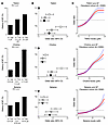 Figure 1
Plasma TMAO, choline, and betaine independently associate with prevalent AF. (A) The relationship between tertiles (T) of increasing plasma concentrations of TMAO, choline, and betaine and the proportion of prevalent AF in GeneBank subjects. The P value represents a Pearson’s χ2 test. (B) Forest plots of the OR of prevalent AF and tertiles of increasing concentrations of plasma TMAO, choline, and betaine with no adjustment (white circles), adjustment for patient characteristics and comorbidities (model 1: age, sex, diabetes mellitus, CVD, systolic blood pressure, current smoker, BMI, and high-sensitivity C-reactive protein; gray circles), and after full model adjustment (model 1 and eGFR; black circles). The circles represent the OR and the horizontal bars extend from the lower limit to the upper limit of the 95% CI. (C) Cubic spline plots of plasma concentrations of TMAO, choline, and betaine and the OR of prevalent AF. The solid red line represents the estimated OR from unadjusted logistic regression analysis at increasing values of the metabolite, and the dashed blue lines represent the 95% CI. **P < 0.01.
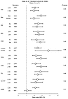 Figure 2
Association of baseline TMAO tertiles with odds of AF. Forest plots show OR for AF with increasing TMAO levels in the indicated subgroups. Open circles represent OR for tertile 3 (T3) versus tertile 1 (T1); horizontal lines indicate 95% CIs. P interaction (P) values were obtained from likelihood ratio tests comparing logistic models with and without the interaction between TMAO tertiles and each subgroup factor. Units for continuous variables: age (years), BMI (kg/m2), HDL cholesterol (HDLc; mg/dL), LDL cholesterol (LDLc; mg/dL), triglycerides (TG; mg/dL), high-sensitivity C-reactive protein (hs-CRP; mg/L), and eGFR (mL/min/1.73 m2). DM, diabetes mellitus.
Dietary TMAO supplementation accelerates AF onset and persistence in CREM-IbΔC-X transgenic mice. The finding that TMAO is independently associated with AF in humans suggested that it may contribute to AF pathogenesis and that inhibition of the gut microbial TMAO pathway could represent a potential preventive therapeutic approach. The CREM-IbΔC-X mouse is a model that develops spontaneous AF; it recapitulates human AF by first developing paroxysmal AF and then ultimately developing persistent AF (31, 32). Male and female CREM-IbΔC-X mice supplemented with dietary TMAO (0.3%) developed paroxysmal AF (first time onset of AF) and persistent AF (10 serial ECGs demonstrating AF) sooner compared with those fed a chow diet using serial needle electrode ECG measurements (Figure 3A and Supplemental Tables 1 and 2; supplemental material available online with this article; https://doi.org/10.1172/JCI201684DS1). CREM-IbΔC-X mice supplemented with dietary TMAO had significantly higher plasma levels of TMAO (Figure 3B). Similar trends were observed for the onset of paroxysmal and persistent AF and plasma TMA (TMA-containing compounds TMAO, choline, and betaine) levels in both male and female CREM-IbΔC-X mice, but female CREM-IbΔC-X mice failed to reach statistical significance for the onset of paroxysmal AF (first time AF) (Supplemental Figures 1 and 2 and Supplemental Tables 3 and 4). Notably, male and female CREM-IbΔC-X mice fed chow and TMAO-supplemented diets did not differ significantly in body weight (Supplemental Figure 3). These data suggest TMAO and, by extension, the gut microbiome has a direct role in the pathogenesis of AF.
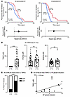 Figure 3
Kaplan-Meier plots and HRs of the incidence of AF in CREM-IbΔC-X mice on TMAO diet and plasma TMA concentrations. (A) The incidence of paroxysmal and persistent AF in all CREM-IbΔC-X mice on a chow or TMAO-supplemented diet, as represented by Kaplan-Meier plots and HRs. The circles represent the HR, and the horizontal bars extend from the lower limit to the upper limit of the 95% CI of the estimate of the HR. HRs between respective dietary groups were calculated using a Cox proportional hazard model. (B) Plasma concentrations of TMAs in all CREM-IbΔC-X mice used in the TMAO mouse ECG study. A Mann-Whitney test was used to assess significant differences between groups. Data represent mean ± SD. (C) Trials of AF percent induction in C57BL/6J mice treated with TMAO and chow diets. (D) TMAO levels and AF percent induction correlation in all C57BL/6J mice. Pearson’s correlation coefficient measuring the relationship between TMAO levels and AF percentage of AF. P values were determined using a linear regression model. *P < 0.05, **P < 0.01, ****P < 0.0001.
Dietary TMAO supplementation mediated an increase in AF susceptibility in C57BL/6J mice. To demonstrate that the effects of TMAO were not strain dependent, we validated the key findings of our study using an alternative mouse model of AF. We performed a transesophageal AF induction study in male and female C57BL/6J mice on and off a TMAO diet. AF inducibility was increased in C57BL/6J mice fed a TMAO-supplemented diet compared with chow-fed controls (Figure 3C). Notably, TMAO supplementation resulted in an 11-fold increase in AF inducibility relative to controls (Figure 3C). When stratified by sex, both male and female C57BL/6J mice demonstrated similarly significant increases in AF inducibility (Supplemental Figure 4).
Plasma TMAO concentrations in C57BL/6J mice used in transesophageal pacing studies were also significantly increased in all C57BL/6J mice on a TMAO-supplemented diet compared with chow-fed controls (Supplemental Figure 5). Additionally, plasma levels of TMAO were significantly correlated with AF percent induction, and similar trends were observed when stratified by sex, but the correlation remained significant only in C57BL/6J male mice (Figure 3D and Supplemental Figure 6A). No consistent significant relationship was noted between AF percent induction and other plasma TMAs (Supplemental Figure 6, B and C).
Inhibition of gut microbial TMAO production with the TMA lyase inhibitor iodomethylcholine significantly delays the onset of paroxysmal and persistent AF in CREM-IbΔC-X transgenic mice. We next sought to determine whether targeting the gut microbial TMAO pathway could provide a potential therapeutic strategy for AF. CREM-IbΔC-X mice fed a choline-supplemented diet developed paroxysmal AF and persistent AF earlier (Figure 4, A and B, and Supplemental Tables 5 and 6) than chow-fed controls. Administration of iodomethylcholine (IMC), an inhibitor of microbial choline to TMA pathway (7), in animals maintained on a high-choline diet (choline + IMC) resulted in marked suppression of choline diet–induced AF (Figure 4, A and B, and Supplemental Tables 5 and 6). Comparable trends in the onset of paroxysmal and persistent AF were observed in each sex, but CREM-IbΔC-X female mice on choline-supplemented diet did not show a significant increase in paroxysmal AF (first time AF) compared with chow-fed controls (Supplemental Tables 7 and 8). Notably, in chow-fed mice (choline content 0.8%–0.9%) (1), IMC supplementation significantly delayed the development of paroxysmal AF compared with controls and showed a trend toward delayed onset of persistent AF (Figure 4, A and B, and Supplemental Tables 5 and 6).
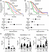 Figure 4
Kaplan-Meier plots and HRs of the incidence of paroxysmal AF and persistent AF in CREM-IbΔC-X mice on respective diets. (A and B) The incidence of paroxysmal (A) and persistent AF (B) in all CREM-IbΔC-X mice, as represented by a Kaplan-Meier plots and HRs, on a chow diet, a choline-supplemented diet, a chow + IMC-supplemented diet, and a choline + IMC-supplemented diet. The circles represent the HR, and the horizontal bars extend from the lower limit to the upper limit of the 95% CI of the estimate of the HR. HRs between respective dietary groups were calculated using a Cox proportional hazard model. (C) Average plasma concentrations of TMAO, choline, and betaine in all CREM-IbΔC-X mice used in the ECG study. Data represent mean ± SD. A Kruskal-Wallis test with Dunn’s post hoc analysis was used to determine the significant differences among the medians of 4 independent diet groups, and multiple paired comparison adjustments were performed. *P < 0.05, **P < 0.01, ***P < 0.001, ****P < 0.0001.
CREM-IbΔC-X mice fed a choline-supplemented diet had significantly higher plasma concentrations of TMAO (Figure 4C) and TMA (Supplemental Figure 9) compared with the chow-fed (control) mice. Dietary supplementation of choline in combination with IMC led to marked reduction in plasma concentration TMAO and TMA (Figure 4C and Supplemental Figure 9). TMAO and TMA levels were not significantly reduced in mice fed a chow (control) diet in combination with IMC (Figure 4C and Supplemental Figure 9).
Consistent with the well-established observation that FMO3 (the liver enzyme responsible for the oxidation of TMA to TMAO) is expressed at higher levels in female than male mice (9), CREM-IbΔC-X female mice had higher circulation TMAO than male CREM-IbΔC-X mice (Supplemental Figure 10). This observation was also true of CREM-IbΔC-X female and male mice on a chow diet (CREM-IbΔC-X female chow TMAO, 3.1 ± 1.5 μM; CREM-IbΔC-X male chow TMAO, 0.5 ± 0.24 μM). Choline plasma concentrations were higher in the choline diet compared with the chow diet in both male and female CREM-IbΔC-X mice (Figure 4C). Betaine plasma concentrations tended to be highest in supplemented mice supplemented with choline + IMC compared with the other diets (Figure 4C and Supplemental Figure 10).
Plasma TMAO concentrations significantly predicted the earlier onset of paroxysmal AF in all male CREM-IbΔC-X mice and approached significance in female CREM-IbΔC-X mice (Figure 5A). Higher plasma betaine concentrations associated with later onset of paroxysmal AF in male CREM-IbΔC-X mice, whereas plasma choline levels had no association with the onset of paroxysmal AF (Figure 5A). Higher TMAO levels predicted the earlier onset of persistent AF in both male and female CREM-IbΔC-X mice, whereas plasma betaine and choline plasma concentrations did not (Figure 5A).
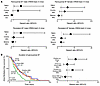 Figure 5
HRs and Kaplan-Meier plot of the incidence of paroxysmal and persistent AF. (A) The incidence of paroxysmal and persistent AF and average plasma TMAO, choline, and betaine concentrations in male and female CREM-IbΔC-X mice. (B) Kaplan-Meier plot and HRs of the duration of paroxysmal AF in male and female CREM-IbΔC-X mice. In A and B, the circles represent the HR, and the horizontal bars extend from the lower limit to the upper limit of the 95% CI of the estimate of the HR. HRs between respective dietary groups were calculated using a Cox proportional hazard model. Each HR represents the risk over the entire analyte concentration range. *P < 0.05, **P < 0.01.
In general, CREM-IbΔC-X female mice developed AF earlier and progressed to persistent AF more rapidly than male CREM-IbΔC-X mice (Supplemental Figures 7 and 8). In contrast, serial needle electrode ECGs performed in WT mice fed either chow or a choline-supplemented diet revealed no onset of AF over the study period (Supplemental Figure 11). Notably, CREM-IbΔC-X mice in the ECG studies showed no significant differences in body weight at the time of euthanasia, and no appreciable liver pathology was observed (Supplemental Figures 12 and 13). To determine whether the earlier development of persistent AF in CREM-IbΔC-X mice was simply due to earlier AF onset, we examined the duration of paroxysmal AF (the time between the onset of AF and persistent AF). This duration was significantly shorter in choline-supplemented CREM-IbΔC-X mice compared with chow-fed or IMC-treated mice (Figure 5B and Supplemental Table 9). Collectively, these data suggested that TMAO contributes to both the onset and progression of AF and that inhibition of the gut microbial TMAO pathway may represent a potential strategy to prevent AF development and progression.
Pharmacological targeting of gut microbial CutC/D resides in the gut and is not readily absorbed. To evaluate the potential of IMC as pharmacological therapy, we first assessed its absorption in mice receiving long-term IMC supplementation. Notably, the majority of IMC remained in the cecal contents of CREM-IbΔC-X mice, with minimal levels detected in plasma (Figure 6A). In addition, IMC treatment had no significant impact on hepatic FMO activity, as monitored by direct examination of the metabolic transformation activity of TMA into TMAO in liver homogenates (Figure 6B). In contrast, mice chronically treated with IMC showed potent inhibition in cecal microbial enzymatic transformation capacity of d9-choline into d9-TMA under both aerobic and anaerobic conditions (Figure 6, C and D). We observed that plasma TMAO concentrations in CREM-IbΔC-X mice fed a choline-supplemented diet and IMC were persistently suppressed for the duration of the ECG study (~5 months) (Supplemental Figure 14). In contrast, CREM-IbΔC-X mice fed a choline diet alone exhibited persistently high plasma TMAO levels over the same period (Supplemental Figure 14). The persistent suppression or production of TMAO over time suggested that there were stable microbiota alterations present. These data also suggest that IMC effectively and chronically inhibits the TMA/TMAO gut microbial pathway, is not the result of off-target effects on liver FMO activity, and could be considered a long-term preventive therapeutic pharmacological agent.
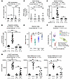 Figure 6
IMC concentrations in plasma and cecum samples and assessment of CutC/D activity. (A) Quantification of IMC in cecum and plasma samples of CREM-IbΔC-X mice on respective diets. (B) Quantification of the conversion of d9-TMA to d9-TMAO in liver samples of mice on respective diets. (C and D) Quantification of the conversion of d9-choline to d9-TMA in cecum of CREM-IbΔC-X mice under aerobic (C) and anaerobic (D) conditions on respective diets. Replicates represent 4 independent samples from each mouse. (E and F) α-Diversity plot (E; Shannon’s index) and principal component analysis (F) of β-diversity plot of CREM-IbΔC-X mice on respective diets. Permutational ANOVA (Permanova) was used to compare the β-diversity distributions of dietary groups. (G) CutC/D mRNA counts in cecum samples from all, male, and female CREM-IbΔC-X mice utilized in the mouse ECG study. In A–E and G, data represent mean ± SD. A Kruskal-Wallis test with Dunn’s post hoc analysis was used to determine the significant difference among the medians of 4 independent diet groups, and multiple paired comparison adjustments were performed. CCA, canonical correspondence analysis. *P < 0.05, **P < 0.01, ***P < 0.001, ****P < 0.0001.
Choline-supplemented diet altered gut microbiota composition in CREM-IbΔC-X transgenic mice, and treatment with IMC reversed these alterations. We next characterized the gut microbiota using metagenomic sequence analysis of cecum in a subset of male and female CREM-IbΔC-X mice used in the mouse ECG study. CREM-IbΔC-X mice on the choline-supplemented diet in the ECG study showed a significant decrease in gut microbiome diversity, which was reversed by IMC treatment (Figure 6E). A canonical correspondence analysis plot demonstrated that mice on a choline diet showed differences in diversity with microbial taxa compared with respective control groups (Figure 6F). Metagenomic associations are provided in Supplemental Tables 10 and 11. Compositional differences of the gut microbiota showed that the relative abundance of the phylum Firmicutes was higher in CREM-IbΔC-X mice supplemented with a choline diet compared with chow and choline + IMC controls (Supplemental Figure 15A, species highlighted in yellow). At the species level, the relative abundance of GGB29005_SGB41723 was greater in the choline-supplemented diet compared with the chow and choline + IMC dietary control groups (Supplemental Figure 15, A and B). The species GGB18827_SGB41440 of the phylum Firmicutes was less abundant in the choline group compared with the respective dietary control groups (Supplemental Figure 15, A and B).
CutC/D (catalytic subunit of choline TMA lyase) mRNA counts were significantly higher in the choline diet compared with the chow and choline + IMC groups (Figure 6G). The same trend was observed in CREM-IbΔC-X mice after stratification by sex (Figure 6G). We next investigated the association of genera that positively and inversely associated with the choline diet with the TMA, TMAO, and CutC/D levels in CREM-IbΔC-X mice. A strong and significant positive correlation between the relative abundance of the gut microbial species GGB29005_SGB41723 and GGB18827_SGB41440 was observed for TMA, TMAO, and CutC/D levels in CREM-IbΔC-X mice (Figure 7A). Although no significant inverse association was found between gut microbial species Lactobacillus_SGB5158 and GGB28980_SGB41692 with TMA and TMAO concentration, CutC/D levels showed a significant inverse relationship (Figure 7A). Other gut microbial species noted to have high or low relative abundances with CREM-IbΔC-X mice supplemented with a choline diet did not show a significant association with TMA, TMAO, or CutC/D (Supplemental Figure 16).
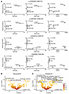 Figure 7
CutC levels and correlation with gut microbiome and differential abundance of gut microbial species with the time of onset of paroxysmal and persistent AF. (A) Gut microbiota taxa abundance and its association with mouse plasma TMA and TMAO and CutC/D mRNA counts in linked cecal samples of male and female CREM-IbΔC-X mice on respective diets. The circles represent the mean, and the bars represent SEM with regression and P values determined using the linear regression model. *P < 0.05, **P < 0.01. (B and C) Volcano plot showing the association of differential abundance of gut microbial species with the time of onset of paroxysmal (B) and persistent (C) AF. In the plot, the x axis shows the coefficient (effect size and direction), and the y axis shows the –log10(q value). Each circle represents a specific microbial species, colored on a yellow-to-red gradient indicating q values, with yellow being more significant. The dashed dark red line at y ≈ 1.30 marks the statistical significance threshold (q value = 0.05). Circles above this line and labeled are statistically significant (q < 0.05). Circles in the upper corners of the plot, far from 0 on the x axis and high on the y axis, represent features with both large effect sizes and high statistical significance. Benjamini-Hochberg false discovery rate correction was used to adjust q values for multiple testing.
Gut microbial taxa associate with the onset of paroxysmal and persistent AF. In the final set of gut microbial studies, we assessed the changes in cecal microbial communities and the association with the time to onset of paroxysmal AF and persistent AF in CREM-IbΔC-X mice. Volcano plots (Figure 7, B and C) showed that several significant gut microbial species were associated with the time of onset of paroxysmal AF and persistent AF. In contrast, the duration of AF did not show any significant association with gut microbial species (Supplemental Figure 17). Parvibacter caecicola was the only species significantly associated with the time to paroxysmal AF. GGB30408_SGB43438 was significantly associated with the time to persistent AF. None of the gut microbial bacterial species had overlap with species that significantly associated with a choline diet.
A TMAO-promoting diet was associated with atrial remodeling in CREM-IbΔC-X and C57BL/6J mice. We next investigated whether dietary choline supplementation, with and without IMC, in CREM-IbΔC-X mice was associated with structural and functional changes in the atria and ventricles. Left atria (LA) size, measured in both diastole and systole, was significantly increased in male and female CREM-IbΔC-X mice fed a choline-supplemented diet compared with chow-fed controls (P < 0.01) at both 5 weeks (Supplemental Figure 18) and 8 weeks of age (Figure 8, A–C). Notably, the LA size in both systole and diastole was significantly attenuated in the choline + IMC dietary group at 8 weeks of age (Figure 8, A–C), suggesting that pharmacological treatment with IMC can prevent TMAO-associated structural changes. In contrast, no significant differences were observed in left ventricular (LV) mass, LV ejection fraction, cardiac output, fractional shortening, or heart rate among dietary groups, as measured by echocardiography at 5 and 8 weeks of age (Supplemental Figures 19–22). Since inflammation may play a role in atrial remodeling and AF, we also evaluated circulating inflammatory markers. Serum amyloid A and IL-6 were not significantly different between choline-supplemented mice and chow-fed controls (Supplemental Figure 23).
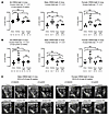 Figure 8
Choline diet increases the LA size during diastolic and systolic function. (A) LA diastolic size in male and female CREM-IbΔC-X mice at 8 weeks of age on respective diets. (B) LA systolic size in male and female CREM-IbΔC-X mice at 8 weeks of age on respective diets. (C) Echocardiography of LA size in female and male CREM-IbΔC-X mice at 8 weeks of age on respective diets. In A and B, data represent mean ± SD. A Kruskal-Wallis test with Dunn’s post hoc analysis was used to determine the significant difference among the medians of 4 independent diet groups, and multiple paired comparison adjustments were performed. *P < 0.05, **P < 0.01, ****P < 0.00001.
We also performed echocardiograms on C57BL/6J male and female mice used in the transesophageal AF induction studies. Similar to CREM-IbΔC-X mice, C57BL/6J mice on a TMAO-supplemented diet showed an increase in LA size that was significantly greater compared with chow-fed C57BL/6J mice in both atrial diastole (P < 0.01) and systole (P < 0.01) (Supplemental Figure 24, A and B). Similar trends were observed when stratifying mice by sex (Supplemental Figure 24A). In contrast, no significant differences were observed in LV mass, LV ejection fraction, cardiac output, fractional shortening, and heart rate, as measured by echocardiography among TMAO and chow groups in C57BL/6J mice at 6 weeks of age (Supplemental Figure 25).
The NLRP3 inflammasome is not a significant contributor to TMAO-promoted AF. We performed in vitro and in vivo studies examining the role of the NLRP3 (NOD-, LRR-, and pyrin domain-containing protein 3) inflammasome in the early onset of AF in choline-supplemented CREM-IbΔC-X mice. We first exposed multiple cell types to physiological relevant levels of TMAO and examined the expression of NLRP3 and IL-1β. The cell types included human cardiac fibroblasts (Supplemental Figure 26, A and B), atrial engineered heart tissues (aEHTs) (Supplemental Figure 27, A and B), primary mouse atrial and ventricular cardiomyocytes, and primary mouse atrial and ventricular fibroblast treated with vehicle or TMAO (Supplemental Figures 28 and 29). We observed no significant increase in the relative mRNA expression of NLRP3 or IL-1β in cells exposed to TMAO. However, a significant decrease was detected in IL-1β expression in primary atrial fibroblasts treated with TMAO at high concentrations (1 mM) compared with control (Supplemental Figures 29).
To investigate the role of the NLRP3 inflammasome in the early onset of AF observed in choline-supplemented CREM-IbΔC-X mice, we performed mouse ECG experiments using a potent and selective inhibitor of NLRP3 inflammasome, MCC950. CREM-IbΔC-X mice supplemented with both choline and MCC950 in water had no significant change in developing paroxysmal AF compared with CREM-IbΔC-X mice supplemented with a choline diet (Supplemental Figure 30A and Supplemental Table 12). CREM-IbΔC-X mice supplemented with both choline in combination with MCC950 developed persistent AF significantly earlier compared with CREM-IbΔC-X mice supplemented with a choline diet alone (Supplemental Figure 30B and Supplemental Table 13). We confirmed that MCC950 systemically inhibited the NLRP3 inflammasome in CREM-IbΔC-X mice by performing LPS i.p. injections and measuring plasma concentrations of IL-1β (a specific cytokine product of the NLRP3 inflammasome) and TNF-α, a nondependent NLRP3 cytokine. Compared with vehicle (PBS), there was no significant decrease in plasma concentrations of TNF-α, but there was a significant decrease in the concentration of IL-1β, consistent with MCC950 NLRP3 inhibition (Supplemental Figure 30, C and D). We also observed that the same CREM-IbΔC-X mice treated with choline in combination with MCC950 had higher TMAO levels compared with the control mice fed a chow diet (Supplemental Figure 30E). Together, these data suggest that the NLRP3 inflammasome is not a significant pathogenic mechanism in TMAO-promoted AF in CREM-IbΔC-X mice.
A choline diet is associated with atrial electrophysiological dysfunction. To understand the electrophysiological implications of a choline-supplemented diet, we performed optical action potential mapping studies on female CREM-IbΔC-X mice (Figure 9A). Female CREM-IbΔC-X mice were selected due to relatively higher plasma TMAO levels, as observed in this study and previous studies (33). Representative examples of atrial action potentials, activation isochrone maps, and local conduction velocity (CV) vectors are shown in Figure 9B. Measurement of average local CV vectors demonstrated a nonsignificant decrease in CREM-IbΔC-X female mice fed a choline-supplemented diet compared with mice fed a chow diet (Figure 9C). In contrast, action potential duration at 80% repolarization (APD80) and maximum APD80 were significantly reduced in the choline group, while minimum APD80 showed a trend toward significance (Figure 9, D–F). Shortening of APD is a well-established mechanism underlying increased susceptibility to AF (34–39). Consistent with this, the calculated cardiac wavelength (CV × APD80) was also significantly shorter in mice receiving choline supplementation (Figure 9G). Notably, reduced wavelength is associated with an increased vulnerability to AF (34–39).
 Figure 9
Analysis of CV and APD in CREM-IbΔC-X mouse hearts using ex vivo optical mapping on chow versus choline-supplemented diets. (A) Schematic of the optical mapping experimental setup used for whole-heart atrial action potential measurements (see text for details). (B) Representative examples of action potentials (left) measured from the LA and posterior wall for chow (black) and choline (red), and atrial activation maps (posterior view) during 130 ms pacing (asterisk) with local CV vectors superimposed for chow (middle) and choline (right) RA, right atria. (C–G) Grouped data of CV (C), APD80 (D), APD80 maximum (E), APD 80 minimum (F), and wavelength (G) at a pacing cycle length of 130 ms. All P values shown were determined using 2-tailed Student’s t test to compare the means of 2 independent groups, after confirmation of normality using Shapiro-Wilk and Kolmogorov-Smirnov tests. *P < 0.05, **P < 0.01.
In contrast, we found no significant change in beat rate, APD at 50% repolarization (APD50), or calcium amplitude in aEHTs exposed to TMAO (Supplemental Figure 31). We also performed an ex vivo study of male and female CREM-IbΔC-X Langendorff perfused mouse hearts with direct TMAO exposure. Similarly, we found no significant changes in APD50, APD80, or CV in optical mapping studies performed on ex vivo male and female CREM-IbΔC-X Langendorff perfused mouse hearts with direct TMAO exposure (Supplemental Figures 32–34).
TMAO promotes autonomic dysfunction by inhibition of muscarinic receptor 2. TMAO has structural similarities to acetylcholine. Optical mapping studies demonstrated that mice fed a choline-supplemented diet exhibited significantly shortened APD (Figure 9D). In addition, a prior report has shown that direct injection of TMAO into the ganglion plexi of the cardiac autonomic nervous system shortens the effective refractory period, a surrogate for APD duration, and increased AF inducibility in canine myocardium using a burst pacing protocol (30). Together, these data suggested that TMAO may promote AF through autonomic dysfunction, potentially via interaction with muscarinic receptors. To explore this mechanism, we generated HEK293 cells stably expressing M2 muscarinic receptor (M2R) (Figure 10 and Supplemental Figure 35A; clone 1 was used for subsequent experiments), the most abundant muscarinic receptor subtype in the myocardium. In this system, TMAO acted as a significant inhibitor of M2R signaling in the presence of the M2R agonist carbachol (Figure 10A). Furthermore, heart rate measurements in unanesthetized male and female CREM-IbΔC-X mice demonstrated that choline-supplemented mice had significantly higher heart rates, which were accompanied by elevated plasma TMAO levels (Figure 10B and Supplemental Figure 35). Collectively, these data support a role for TMAO in autonomic dysfunction and the promotion of AF.
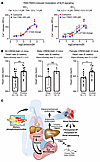 Figure 10
TMAO modulates autonomic signaling. (A) TMA/TMAO induced inhibition of M2R (clone 1) signaling in HEK293 cells. Unpaired 2-tailed Welch’s t test was used to obtain the fold change P value by comparing the mean EC50 values of carbachol (Car) and TMA/TMAO (n = 6 independent experiments). (B) Heart rate in CREM-IbΔC-X mice treated with choline and chow diet. Data represent mean ± SD. A Mann-Whitney test was used to assess significant differences between groups. *P < 0.05, **P < 0.01. (C) Schematic demonstrating that IMC inhibits TMA/ TMAO formation, AF induction, and progression. Choline diet alters gut microbial TMA and its metabolite formation. TMAO formation plays a key role in autonomic dysfunction via inhibition of M2R and the resulting increased sympathetic tone and susceptibility to AF. IMC that inhibits formation of TMA/TMAO can reduce AF induction and progression.


Copyright © 2026 American Society for Clinical Investigation
ISSN: 0021-9738 (print), 1558-8238 (online)