
Advertisement
Research ArticleAutoimmunityDermatologyImmunology
Open Access |  10.1172/JCI200316
Hyperglycemia aggravates vitiligo through succinate/SUCNR1-mediated T cell activation
Pan Kang,1 Yuqian Chang,1 Tingting Wang,1 Xiuli Yi,1 Yinghan Wang,1 Pengran Du,1 Jiaxi Chen,1 Baizhang Li,1 Shuli Li,1 Zhongjun Shao,2 Jianru Chen,1,3 and Chunying Li1
1Department of Dermatology, Xijing Hospital, and
2Department of Epidemiology, School of Public Health, Fourth Military Medical University, Xi’an, Shaanxi, China.
3National Key Laboratory of Immunity and Inflammation, Institute of Immunology, Naval Medical University, Shanghai, China.
Address correspondence to: Chunying Li or Jianru Chen, Department of Dermatology, Xijing Hospital, Fourth Military Medical University, No. 127 of West Changle Road, Xi’an, Shaanxi, 710032, China. Phone: 86.29.84775406; Email: lichying@fmmu.edu.cn (CL). Phone: 86.29.84775406; Email: chenjr0516@fmmu.edu.cn (JC).
Authorship note: PK, YC, and TW are co–first authors.
Find articles by Kang, P. in: PubMed | Google Scholar
1Department of Dermatology, Xijing Hospital, and
2Department of Epidemiology, School of Public Health, Fourth Military Medical University, Xi’an, Shaanxi, China.
3National Key Laboratory of Immunity and Inflammation, Institute of Immunology, Naval Medical University, Shanghai, China.
Address correspondence to: Chunying Li or Jianru Chen, Department of Dermatology, Xijing Hospital, Fourth Military Medical University, No. 127 of West Changle Road, Xi’an, Shaanxi, 710032, China. Phone: 86.29.84775406; Email: lichying@fmmu.edu.cn (CL). Phone: 86.29.84775406; Email: chenjr0516@fmmu.edu.cn (JC).
Authorship note: PK, YC, and TW are co–first authors.
Find articles by Chang, Y. in: PubMed | Google Scholar
1Department of Dermatology, Xijing Hospital, and
2Department of Epidemiology, School of Public Health, Fourth Military Medical University, Xi’an, Shaanxi, China.
3National Key Laboratory of Immunity and Inflammation, Institute of Immunology, Naval Medical University, Shanghai, China.
Address correspondence to: Chunying Li or Jianru Chen, Department of Dermatology, Xijing Hospital, Fourth Military Medical University, No. 127 of West Changle Road, Xi’an, Shaanxi, 710032, China. Phone: 86.29.84775406; Email: lichying@fmmu.edu.cn (CL). Phone: 86.29.84775406; Email: chenjr0516@fmmu.edu.cn (JC).
Authorship note: PK, YC, and TW are co–first authors.
Find articles by Wang, T. in: PubMed | Google Scholar
1Department of Dermatology, Xijing Hospital, and
2Department of Epidemiology, School of Public Health, Fourth Military Medical University, Xi’an, Shaanxi, China.
3National Key Laboratory of Immunity and Inflammation, Institute of Immunology, Naval Medical University, Shanghai, China.
Address correspondence to: Chunying Li or Jianru Chen, Department of Dermatology, Xijing Hospital, Fourth Military Medical University, No. 127 of West Changle Road, Xi’an, Shaanxi, 710032, China. Phone: 86.29.84775406; Email: lichying@fmmu.edu.cn (CL). Phone: 86.29.84775406; Email: chenjr0516@fmmu.edu.cn (JC).
Authorship note: PK, YC, and TW are co–first authors.
Find articles by Yi, X. in: PubMed | Google Scholar
1Department of Dermatology, Xijing Hospital, and
2Department of Epidemiology, School of Public Health, Fourth Military Medical University, Xi’an, Shaanxi, China.
3National Key Laboratory of Immunity and Inflammation, Institute of Immunology, Naval Medical University, Shanghai, China.
Address correspondence to: Chunying Li or Jianru Chen, Department of Dermatology, Xijing Hospital, Fourth Military Medical University, No. 127 of West Changle Road, Xi’an, Shaanxi, 710032, China. Phone: 86.29.84775406; Email: lichying@fmmu.edu.cn (CL). Phone: 86.29.84775406; Email: chenjr0516@fmmu.edu.cn (JC).
Authorship note: PK, YC, and TW are co–first authors.
Find articles by Wang, Y. in: PubMed | Google Scholar
1Department of Dermatology, Xijing Hospital, and
2Department of Epidemiology, School of Public Health, Fourth Military Medical University, Xi’an, Shaanxi, China.
3National Key Laboratory of Immunity and Inflammation, Institute of Immunology, Naval Medical University, Shanghai, China.
Address correspondence to: Chunying Li or Jianru Chen, Department of Dermatology, Xijing Hospital, Fourth Military Medical University, No. 127 of West Changle Road, Xi’an, Shaanxi, 710032, China. Phone: 86.29.84775406; Email: lichying@fmmu.edu.cn (CL). Phone: 86.29.84775406; Email: chenjr0516@fmmu.edu.cn (JC).
Authorship note: PK, YC, and TW are co–first authors.
Find articles by Du, P. in: PubMed | Google Scholar
1Department of Dermatology, Xijing Hospital, and
2Department of Epidemiology, School of Public Health, Fourth Military Medical University, Xi’an, Shaanxi, China.
3National Key Laboratory of Immunity and Inflammation, Institute of Immunology, Naval Medical University, Shanghai, China.
Address correspondence to: Chunying Li or Jianru Chen, Department of Dermatology, Xijing Hospital, Fourth Military Medical University, No. 127 of West Changle Road, Xi’an, Shaanxi, 710032, China. Phone: 86.29.84775406; Email: lichying@fmmu.edu.cn (CL). Phone: 86.29.84775406; Email: chenjr0516@fmmu.edu.cn (JC).
Authorship note: PK, YC, and TW are co–first authors.
Find articles by Chen, J. in: PubMed | Google Scholar
1Department of Dermatology, Xijing Hospital, and
2Department of Epidemiology, School of Public Health, Fourth Military Medical University, Xi’an, Shaanxi, China.
3National Key Laboratory of Immunity and Inflammation, Institute of Immunology, Naval Medical University, Shanghai, China.
Address correspondence to: Chunying Li or Jianru Chen, Department of Dermatology, Xijing Hospital, Fourth Military Medical University, No. 127 of West Changle Road, Xi’an, Shaanxi, 710032, China. Phone: 86.29.84775406; Email: lichying@fmmu.edu.cn (CL). Phone: 86.29.84775406; Email: chenjr0516@fmmu.edu.cn (JC).
Authorship note: PK, YC, and TW are co–first authors.
Find articles by Li, B. in: PubMed | Google Scholar
1Department of Dermatology, Xijing Hospital, and
2Department of Epidemiology, School of Public Health, Fourth Military Medical University, Xi’an, Shaanxi, China.
3National Key Laboratory of Immunity and Inflammation, Institute of Immunology, Naval Medical University, Shanghai, China.
Address correspondence to: Chunying Li or Jianru Chen, Department of Dermatology, Xijing Hospital, Fourth Military Medical University, No. 127 of West Changle Road, Xi’an, Shaanxi, 710032, China. Phone: 86.29.84775406; Email: lichying@fmmu.edu.cn (CL). Phone: 86.29.84775406; Email: chenjr0516@fmmu.edu.cn (JC).
Authorship note: PK, YC, and TW are co–first authors.
Find articles by Li, S. in: PubMed | Google Scholar
1Department of Dermatology, Xijing Hospital, and
2Department of Epidemiology, School of Public Health, Fourth Military Medical University, Xi’an, Shaanxi, China.
3National Key Laboratory of Immunity and Inflammation, Institute of Immunology, Naval Medical University, Shanghai, China.
Address correspondence to: Chunying Li or Jianru Chen, Department of Dermatology, Xijing Hospital, Fourth Military Medical University, No. 127 of West Changle Road, Xi’an, Shaanxi, 710032, China. Phone: 86.29.84775406; Email: lichying@fmmu.edu.cn (CL). Phone: 86.29.84775406; Email: chenjr0516@fmmu.edu.cn (JC).
Authorship note: PK, YC, and TW are co–first authors.
Find articles by Shao, Z. in: PubMed | Google Scholar
1Department of Dermatology, Xijing Hospital, and
2Department of Epidemiology, School of Public Health, Fourth Military Medical University, Xi’an, Shaanxi, China.
3National Key Laboratory of Immunity and Inflammation, Institute of Immunology, Naval Medical University, Shanghai, China.
Address correspondence to: Chunying Li or Jianru Chen, Department of Dermatology, Xijing Hospital, Fourth Military Medical University, No. 127 of West Changle Road, Xi’an, Shaanxi, 710032, China. Phone: 86.29.84775406; Email: lichying@fmmu.edu.cn (CL). Phone: 86.29.84775406; Email: chenjr0516@fmmu.edu.cn (JC).
Authorship note: PK, YC, and TW are co–first authors.
Find articles by Chen, J. in: PubMed | Google Scholar
1Department of Dermatology, Xijing Hospital, and
2Department of Epidemiology, School of Public Health, Fourth Military Medical University, Xi’an, Shaanxi, China.
3National Key Laboratory of Immunity and Inflammation, Institute of Immunology, Naval Medical University, Shanghai, China.
Address correspondence to: Chunying Li or Jianru Chen, Department of Dermatology, Xijing Hospital, Fourth Military Medical University, No. 127 of West Changle Road, Xi’an, Shaanxi, 710032, China. Phone: 86.29.84775406; Email: lichying@fmmu.edu.cn (CL). Phone: 86.29.84775406; Email: chenjr0516@fmmu.edu.cn (JC).
Authorship note: PK, YC, and TW are co–first authors.
Find articles by Li, C. in: PubMed | Google Scholar
Published June 15, 2026 - More info
J Clin Invest. 2026;136(12):e200316. https://doi.org/10.1172/JCI200316.
© 2026 Kang et al. This work is licensed under the Creative Commons Attribution 4.0 International License. To view a copy of this license, visit http://creativecommons.org/licenses/by/4.0/.
Received: September 16, 2025; Accepted: April 10, 2026

Related article:
Abstract
Vitiligo is a depigmenting disease marked by progressive T cell–driven destruction of melanocytes in the skin, hair, and mucosa. While vitiligo is known to be a T cell–mediated autoimmune disease, its triggers have remained poorly understood and treatment options limited. In this issue of the JCI, Kang et al. demonstrated how hyperglycemia exacerbates vitiligo progression through the succinate/SUCNR1 axis. These findings identify succinate as a potential biomarker for disease activity and highlight an independent pathway for targeting in therapeutic intervention. More broadly, the findings linking succinate and glucose metabolism to vitiligo suggest that lifestyle factors could be modified to slow development of vitiligo and other autoimmune diseases linked to succinate.
Authors
Kaitlyn G. O’Donnell, I. Caroline Le Poole

-
Results
Hyperglycemia promotes the progression of vitiligo by enhancing CD8+ T cell immune response. We initially conducted a hospital-based case-control study to verify the association between hyperglycemia and vitiligo. Participants’ demographic and clinical characteristics are in Supplemental Tables 1–4; supplemental material available online with this article; https://doi.org/10.1172/JCI200316DS1 Our results showed that patients with vitiligo had a higher incidence of hyperglycemia than healthy controls (11.84% vs. 7.01%, P = 0.012) (Figure 1A and Supplemental Table 5). After adjusting for potential confounders including age, body mass index (BMI), sex, smoking, and drinking, bivariate logistic regression analysis showed that hyperglycemia was notably positively correlated with vitiligo (adjusted odds ratio [OR] = 1.781, 95% confidence interval [CI] = 1.131–2.806, P = 0.013). Moreover, patients with progressive vitiligo had a substantially higher incidence of hyperglycemia than those with stable vitiligo (15.02% vs. 5.56%, P = 0.013) (Figure 1B and Supplemental Table 6). Following adjustment for confounding factors, bivariate logistic regression analysis showed that hyperglycemia was remarkably positively correlated with vitiligo activity (adjusted OR = 3.006, 95% CI = 1.216–7.430, P = 0.017). Additionally, there was a significant difference in the incidence of hyperglycemia among different groups of vitiligo severity (3.36% vs. 15.92% vs. 20.00, P < 0.001) (Figure 1C and Supplemental Table 7). After adjustment for confounding factors, ordinal multivariable logistic regression analysis revealed that hyperglycemia was markedly associated with vitiligo severity (adjusted OR = 2.707, 95% CI = 1.306–5.609, P = 0.007). However, no significant difference was found in the incidence of hyperglycemia between segmental and nonsegmental patients with vitiligo (5.56% vs. 13.00%, P = 0.198) (Supplemental Figure 1A and Supplemental Table 8). The undetermined vitiligo group (n = 8) was excluded from this analysis as it contained no cases with hyperglycemia. These findings highlight hyperglycemia as a potential risk factor for the development and progression of vitiligo.
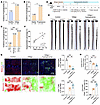 Figure 1
Hyperglycemia aggravates vitiligo. (A–C) Incidence of hyperglycemia in vitiligo patients and healthy controls (A), vitiligo patients with different activity (B), and vitiligo patients with different severity (C). (D) Schematic presentation of the vitiligo and hyperglycemia mouse model establishment. Thus, 3 groups of mice (Control, Vitiligo, and Vitiligo + Hyperglycemia) were studied here. (E) Representative tail images of mice in each group at week 10. (F) The correlation between tail depigmentation percentage and FBG levels of vitiligo-induced mice at week 10 (n = 12). (G) Representative whole-mount immunofluorescence images and quantification of melanocytes (shown in red) and CD8+ T cells (green) in tail epidermis of mice in each group (n = 6). (H) Quantification of CD45–CD117+ melanocytes and CD3+CD8+ T cells in tail epidermis of mice in each group (n = 6). Data are presented as frequency (A–C) or mean ± SEM (G and H) and analyzed by χ2 test (A–C) or 1-way ANOVA (G and H). *P < 0.05; **P < 0.01; ***P < 0.001. FBG, fasting blood glucose.
To further clarify the role of hyperglycemia in the development of vitiligo in vivo, we employed a melanoma-Treg-induced vitiligo mouse model that recapitulated the pathological characteristics of CD8+ T cell–mediated melanocyte destruction in human vitiligo as previously described (32–34). Following the treatment with vitiligo induction, a high-fat diet combined with 100 mg/kg streptozotocin (STZ) intraperitoneal injection was applied to induce hyperglycemia (Figure 1D). Compared with control and vitiligo mice, strongly elevated glucose levels were observed in hyperglycemia-treated vitiligo mice after hyperglycemia induction (Supplemental Figure 1B). After 10 weeks of hyperglycemia, we analyzed and quantified the depigmented area on the tails in each group. As shown, the total depigmented area on the tails of hyperglycemia-treated vitiligo mice was substantially larger than that in vitiligo mice (Figure 1E and Supplemental Figure 1, C and D). Importantly, depigmented areas showed a positive correlation with glucose levels at week 10 (Figure 1F). Progressive vitiligo lesions are characterized by focal CD8+ T cell infiltration and melanocyte destruction in the skin (1, 35, 36). Whole-mount immunofluorescence staining and flow cytometric analysis revealed that hyperglycemia-treated vitiligo mice exhibited more extensive melanocyte loss accompanied by increased CD8+ T cell infiltration compared with vitiligo mice without hyperglycemia (Figure 1, G and H, and Supplemental Figure 1, E and F). Taken together, hyperglycemia might aggravate the progression of vitiligo by increasing the infiltration of CD8+ T cells in skin.
Succinate is the key metabolite associated with hyperglycemia-aggravated vitiligo. To identify the metabolite that mediates hyperglycemia-induced vitiligo progression, we performed targeted metabolomic analyses on serum samples from 30 patients with vitiligo and 30 age- and sex-matched healthy individuals. The individuals were comparable in BMI, smoking, and drinking (Supplemental Table 9). Levels of glucose and 11 TCA cycle intermediates including pyruvate, acetyl-CoA, citrate, itaconate, α-ketoglutarate, succinyl-CoA, succinate, fumarate, malate, oxaloacetate, and lactate were measured. The DModX test plot indicated none of the samples exhibited a severe deviation (Supplemental Figure 2A). As shown by orthogonal partial least squares discriminant analysis, there was a significant distinction between the 2 groups (Q2 = 0.659) (Supplemental Figure 2B). The heatmap showed substantial changes in the metabolic level in the vitiligo group (Figure 2A). Of these, apart from glucose, the levels of acetyl-CoA, itaconate, succinyl-CoA, succinate, and fumarate were increased in the serum of patients with vitiligo, while those of citrate, α-ketoglutarate, malate, and oxaloacetate were decreased (Figure 2B). Among the upregulated metabolites, glucose, itaconate, succinate, and fumarate showed VIP > 1 (indicating higher difference compared with controls), while downregulated counterparts were oxaloacetate and malate (Figure 3A). Correlation analysis showed that glucose was positively correlated with succinate, itaconate, and fumarate; no statistically significant correlation was observed between glucose and oxaloacetate, nor with malate (Figure 3B and Supplemental Figure 3A). Itaconate and fumarate are well-established immunosuppressive metabolites (31, 37–39). Notably, under conditions of immune activation, itaconate and fumarate tend to be just compensatively upregulated to balance the immune response (37). Further correlation analysis showed a positive correlation between succinate and assessed Vitiligo Area Scoring Index (VASI) scores of vitiligo (Figure 3C). Therefore, we further examined the implication of succinate in hyperglycemia-mediated vitiligo progression. Total succinate levels in the serum samples from the case-control study were measured using a succinate assay kit. As a result, serum succinate levels of patients with vitiligo were markedly higher than those of healthy controls (219.00 μmol/L vs. 80.95 μmol/L, P = 0.0001) (Figure 3D). Concordantly, higher succinate levels were found in progressive vitiligo (Figure 3E), serum succinate levels in moderate vitiligo were higher than those in mild vitiligo (Supplemental Figure 3B), and there were no differences between the vitiligo types (Supplemental Figure 3C). Moreover, succinate was strikingly elevated in lesional blister fluid compared with nonlesional blister fluid in patients with vitiligo (Figure 3F). Sequential in vivo experiments showed that circulating succinate levels were considerably higher in hyperglycemic vitiligo mice than in vitiligo mice (Figure 3G). A positive correlation between serum glucose and succinate levels was observed (Figure 3H). Moreover, we found a notable positive correlation between serum succinate levels and depigmentation (Figure 3I). The above results suggest that succinate may serve as a key metabolite mediating hyperglycemia-induced vitiligo progression.
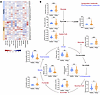 Figure 2
Altered serum levels of glucose-related metabolites in patients with vitiligo. (A) Heatmap of normalized metabolite abundance in serum samples from vitiligo patients and healthy controls (n = 30). (B) Violin plots showing individuals’ indicated metabolism levels in each group. Data are presented as median ± IQR and analyzed by Mann-Whitney U test (B). *P < 0.05; **P < 0.01; ***P < 0.001.
 Figure 3
Succinate is the potential metabolite mediating the exacerbation of vitiligo by hyperglycemia. (A) VIP scores of indicated metabolites. The colored boxes on the right indicate the relative concentrations of the corresponding metabolite. (B) Correlation between serum glucose and succinate concentration of vitiligo patients and healthy controls (n = 60). (C) Correlation between VASI scores and serum succinate concentration of vitiligo patients (n = 30). (D and E) Serum succinate levels of vitiligo patients and healthy controls (D) and vitiligo patients with different activity (E). (F) The level of succinate in blister fluid from lesional and nonlesional skin areas of vitiligo patients. (G) Serum succinate levels of mice in each group (n = 6). (H) Correlation between FBG and serum succinate concentration of mice at week 10 (n = 18). (I) The correlation between serum succinate concentrations and the tail depigmentation percentage of mice at week 10 (n = 18). Data are presented as median ± IQR (D, E, and F) or mean ± SEM (G). Data are analyzed by Mann-Whitney U test (D and E), Wilcoxon’s signed-rank test (F), or 1-way ANOVA (G). *P < 0.05, ***P < 0.001. VIP, variable importance in projection; VASI, vitiligo area scoring index; FBG, fasting blood glucose.
SUCNR1 mediates succinate-induced overactivation of CD8+ T cells in hyperglycemia. We further explored how succinate promotes the CD8+ T cell–mediated immune response. First, we analyzed published single-cell RNA-seq data (40) and found that SUCNR1 expression was markedly higher in circulating CD8+ T cells from patients with vitiligo compared with those from healthy controls (Supplemental Figure 4A). qRT-PCR, Western blot, and flow cytometric analyses confirmed that the expression of SUCNR1 in CD8+ T cells isolated from the peripheral blood of patients with vitiligo was higher than that in healthy controls (Figure 4, A–C, and Supplemental Figure 4B). Moreover, significant SUCNR1 expression was evident in the CD8+ T cells infiltrating the lesions of patients with vitiligo, as demonstrated by immunofluorescence analysis (Figure 4D). Importantly, both high glucose and succinate considerably upregulated SUCNR1 expression in CD8+ T cells derived from patients with vitiligo (Figure 4, E–G, and Supplemental Figure 4C). Further flow cytometry analysis showed that CD8+ T cell activation was enhanced by high-glucose or succinate treatment, which could be attenuated by SUCNR1-specific antagonist NF-56-EJ40 (Figure 5A and Supplemental Figure 5A). Similar results were observed in effector function molecules of CD8+ T cells, including granzyme B and perforin (Figure 5B and Supplemental Figure 5B). Collectively, these findings suggest that SUCNR1 is the receptor through which high glucose and succinate mediate enhanced activation of CD8+ T cells.
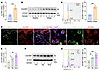 Figure 4
Upregulated SUCNR1 expression in vitiligo CD8+ T cells is modulated by hyperglycemia and succinate. (A and B) The mRNA (A) and protein (B) levels of SUCNR1 in CD8+ T cells from vitiligo patients and healthy controls. (C) Proportion of SUCNR1+ cells in CD8+ T cells of vitiligo patients and healthy controls (n = 5). (D) Representative immunofluorescence images for SUCNR1 (red) in CD8+ T cells in perilesional skin of vitiligo patients. CD8+ T cells were characterized by CD8 (green); nuclei were counterstained with DAPI (blue). (E and F) The mRNA (E) and protein expression (F) levels of SUCNR1 in CD8+ T cells of vitiligo patients treated with high glucose or succinate. (G) Proportion of SUCNR1+ cells in CD8+ T cells of vitiligo patients (n = 5) treated with high glucose or succinate. Data are presented as mean ± SEM (A, C, E, and G) and analyzed by 2-tailed Student’s t test (A and C) or 1-way ANOVA (E and G). *P < 0.05; **P < 0.01; ***P < 0.001. Ctrl, control; Hg, high glucose; Succ, succinate.
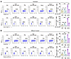 Figure 5
SUCNR1 mediates succinate-induced activation of CD8+ T cells in hyperglycemia. (A and B) Proportion of CD69+, CD137+ (A), granzyme B+, and perforin+ cells (B) in CD8+ T cells of vitiligo patients pretreated with NF-56-EJ40 before high-glucose or succinate treatment. Data are presented as mean ± SEM and analyzed by 1-way ANOVA (A and B). *P < 0.05; **P < 0.01; ***P < 0.001. Ctrl, control; Hg, high glucose; Succ, succinate.
Succinate induces CD8+ T cells’ hyperactivation and accelerated depigmentation through SUCNR1. To further clarify the role of SUCNR1 in mediating the regulation of CD8+ T cell immune responses influenced by hyperglycemia and succinate, we employed Sucnr1fl/fl Cd8-Cre+/– mice (referred to as Cd8-Sucnr1-KO mice), in which Sucnr1 was knocked out in CD8+ T cells (Supplemental Figure 6, A and B). As expected, SUCNR1 expression was strongly reduced in blood and lymph node CD8+ T cells of Cd8-Sucnr1-KO mice when compared with Sucnr1fl/fl mice, while no changes were observed in other tissues, such as the kidney and liver (Supplemental Figure 6C). Following vitiligo modeling, mice underwent hyperglycemia induction or succinate treatment (Figure 6A). The total depigmented area on the tail was drastically larger in both hyperglycemia- and succinate-treated vitiligo mice compared with vitiligo mice. In contrast, Cd8-Sucnr1-KO mice exhibited an evidently smaller tail depigmented area than Sucnr1fl/fl control mice (Figure 6B and Supplemental Figure 7A). In addition, whole-mount immunofluorescence staining and flow cytometric analysis showed that Cd8-Sucnr1-KO mice exhibited markedly attenuated CD8+ T cell infiltration and melanocyte destruction compared with Sucnr1fl/fl mice (Figure 6, C and D, and Supplemental Figure 7, B and C). The results above suggest that hyperglycemia and succinate regulate the progression of vitiligo through SUCNR1.
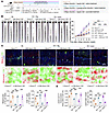 Figure 6
Succinate aggravates vitiligo through SUCNR1. (A) Schematic presentation of vitiligo, hyperglycemia, and succinate-treated mouse model establishment. Thus, 3 groups of mice (Vitiligo, Vitiligo + Hyperglycemia, Vitiligo + Succinate) were studied here. (B) Representative tail skin images of mice in each group at week 10 and the tail pigmentation percentages of mice in each group (n = 4) in 10 consecutive weeks. (C) Representative whole-mount immunofluorescence staining images (upper) and corresponding heatmaps (low) of melanocytes (red) and CD8+ T cells (green) in the tail skin epidermis of mice in each group. Numbers of melanocytes and CD8+ T cells in each group (n = 4) are shown. (D) Quantification of CD45–CD117+ melanocytes and CD3+CD8+ T cells in tail epidermis of mice in each group (n = 4). Data are presented as mean ± SEM and analyzed by 1-way ANOVA (B–D). **P < 0.01; ***P < 0.001. Vit, vitiligo; Hg, hyperglycemia; Succ, succinate.
We further evaluated the effects of hyperglycemia and succinate on CD8+ T cell–mediated immune response in vivo. Consistent with previous results, knockdown of SUCNR1 in CD8+ T cells markedly ameliorated their skin infiltration and peripheral accumulation in vitiligo mice under hyperglycemic conditions or following succinate treatment (Figure 7, A and B, and Supplemental Figure 8A). Similar results were obtained in the primary draining lymph nodes of mouse tail skin (Supplemental Figure 8, B and C). Sequentially, CD8+ T cell activation and effector function were substantially increased in the hyperglycemia and succinate group in peripheral blood and lymph node, and the activation and cytotoxicity were inhibited considerably in CD8+ T cells from the Cd8-Sucnr1-KO mice compared with Sucnr1fl/fl mice (Figure 7, C and D, and Supplemental Figure 9), though the phenomenon was not observed in tail skin (Supplemental Figure 10). Overall, these results demonstrate that hyperglycemia and succinate lead to increased number, activation, and effector function of CD8+ T cells by binding to SUCNR1 on CD8+ T cells.
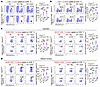 Figure 7
Succinate promotes hyperactivation of CD8+ T cells via SUCNR1. (A) Proportion of CD8+ T cells in the tail skin of mice in each group (n = 4). (B) Proportion of CD8+ T cells in the blood of mice in each group (n = 4). (C and D) Proportion of CD69+CD8+ T cells, CD137+CD8+ T cells (C), granzyme B+CD8+ T cells, and perforin+CD8+ T cells (D) in the blood of mice in each group (n = 4). Data are presented as mean ± SEM and analyzed by 1-way ANOVA (A–D). *P < 0.05; **P < 0.01; ***P < 0.001. Vit, vitiligo; Hg, hyperglycemia; Succ, succinate.
Succinate-induced chemokine secretion via SUCNR1 in keratinocytes promotes CD8+ T cell migration. As shown in Figure 3F, blister fluid from vitiligo lesional skin contained higher levels of succinate than that from nonlesional skin, indicating that elevated succinate in the skin microenvironment contributes to vitiligo development. Moreover, we found that SUCNR1 expression levels were increased in lesional keratinocytes of patients with vitiligo than those in healthy controls based on published single-cell RNA-Seq data (32) (Supplemental Figure 11A). To confirm this finding at the protein level, we performed immunofluorescence analysis, demonstrating increased SUCNR1 expression in keratinocytes of patients with vitiligo (Figure 8A). In addition, succinate treatment markedly elevated the mRNA and protein levels of SUCNR1 in normal human keratinocytes (NHKs) (Figure 8B and Supplemental Figure 11B). Immunofluorescence analysis confirmed the upregulation of SUCNR1 in succinate-treated NHKs (Figure 8C). Studies have illustrated that keratinocyte-derived chemokines CXCL9, CXCL10, and CXCL16 recruit CD8+ T cells to the epidermis to kill melanocytes in vitiligo (41, 42). Our results showed that, compared with control, the mRNA and secretion levels of CXCL9 and CXCL10 were increased in succinate-treated NHKs (Figure 8, D and E), while there were no significant changes in the mRNA and secretion levels of CXCL16 (Supplemental Figure 11, C and D). Increased expression of CXCL9 and CXCL10 was observed in keratinocytes of succinate-treated vitiligo mice by immunofluorescence analysis (Supplemental Figure 11E). Notably, pretreatment with SUCNR1 siRNA or SUCNR1 antagonist NF-56-EJ40 markedly dampened succinate-induced increase of CXCL9 and CXCL10 in both mRNA and protein levels (Figure 8, F and G, and Supplemental Figure 11, F and G). Transwell assay showed that the medium from succinate-treated NHKs facilitated migration of vitiligo patient–derived CD8+ T cells, which could be attenuated by either knockdown or inhibition of SUCNR1 in NHKs but reversed by recombinant human CXCL9 or CXCL10 supplement (Figure 8H and Supplemental Figure 11H). Our findings demonstrate that keratinocyte SUCNR1-mediated secretion of CXCL9 and CXCL10 under succinate promotes CD8+ T cell migration.
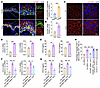 Figure 8
Succinate-induced chemokine secretion via SUCNR1 in keratinocytes promotes CD8+ T cell migration. (A) Representative immunofluorescence images and quantification of SUCNR1 (red) in keratinocytes from vitiligo patients and healthy controls (n = 5). Keratinocytes were characterized by cytokeratin 14 (green); nuclei were counterstained with DAPI (blue). Scale bars: 50 μm (left), 10 μm (middle and right). (B) The mRNA level of SUCNR1 in NHKs treated with succinate. (C) Representative images of SUCNR1 expression in succinate-treated NHKs. (D) The mRNA levels of CXCL9 and CXCL10 in NHKs treated with succinate. (E) The secretion of CXCL9 and CXCL10 in NHKs treated with succinate. (F and G) The mRNA (F) and secretion (G) levels of CXCL9 and CXCL10 in NHKs treated with succinate or pretreated with SUCNR1 siRNA prior to succinate stimulation. (H) Transwell assay showing the percentage of migrated CD8+ T cells in response to culture supernatants from succinate-treated NHKs with knockdown of SUCNR1 or with the addition of rhCXCL9 or rhCXCL10 into the Transwell system. Data are presented as mean ± SEM (A, B, and D–H) and analyzed by Student’s t test (A, B, D, and E) or 1-way ANOVA (F–H). *P < 0.05, **P < 0.01, ***P < 0.001. NHKs, normal human keratinocytes; Ctrl, control; Succ, succinate; rhCXCL9, recombinant human CXCL9; rhCXCL10, recombinant human CXCL10; rMFI, relative mean fluorescence intensity.
Succinate stabilizes HIF-1α through SUCNR1 to upregulate CXCL9 and CXCL10. Next, we investigated the mechanisms by which succinate induces the secretion of CXCL9 and CXCL10 in NHKs. Hypoxia-inducible factor-1α (HIF-1α) is well known as a hallmark of hypoxia and has been shown to mediate the secretion of pro-inflammatory cytokines induced by the succinate/SUCNR1 axis, such as IL-1β (43, 44). As shown by immunofluorescence, the expression of HIF-1α was upregulated in keratinocytes from vitiligo lesions versus in those from healthy skin (Figure 9A). Western blot assay confirmed that succinate markedly increased the expression of HIF-1α, and both SUCNR1 siRNA and NF-56-EJ40 pretreatment reversed succinate-induced HIF-1α upregulation (Figure 9B and Supplemental Figure 12A). Importantly, HIF-1α siRNA pretreatment could ameliorate the elevated CXCL9 and CXCL10 transcription and secretion levels in succinate-stimulated NHKs (Figure 9, C and D). To further explore the mechanism underlying the regulation of CXCL9 and CXCL10, as shown by Western blot assay, the strongly increased expression of HIF-1α was mainly observed in the cell nucleus; both SUCNR1 siRNA and NF-56-EJ40 decreased HIF-1α expression in the cell nucleus (Figure 9E and Supplemental Figure 12B). Immunofluorescence analysis also showed that both SUCNR1 siRNA and NF-56-EJ40 inhibited the succinate-induced nuclear translocation of HIF-1α in NHKs (Figure 9F and Supplemental Figure 12C). Studies have shown that nuclear localized HIF-1α functions primarily as a transcription factor to upregulate hypoxia-responsive genes, including chemokines such as CCL2 and CXCL8 (45, 46). To further investigate whether HIF-1α directly regulates the transcription of CXCL9 and CXCL10, we screened the JASPAR database (http://jaspar.genereg.net), analyzed the –2,000 to +1 region of the CXCL9 and CXCL10 promoter sequences, and identified 6 and 3 putative HIF-1α–responsive elements, respectively (Supplemental Figure 12D). ChIP assay showed that HIF-1α binds to the first predicted binding site in the promoter region of CXCL9 and the second predicted binding site in the promoter region of CXCL10 (Figure 9G). Notably, our results indicate that SUCNR1 siRNA or NF-56-EJ40 diminished the binding of HIF-1α to the CXCL9 and CXCL10 promoter regions induced by succinate (Figure 9H and Supplemental Figure 12E). As shown in Figure 9B and Supplemental Figure 12A, succinate promoted the expression of HIF-1α in NHKs, while it had no significant effect on HIF-1α transcription levels (Figure 10A). HIF-1α is a well-known proline hydroxylation substrate, and the modification of HIF-1α by prolyl hydroxylase domain-containing protein 2 (PHD2) tightly regulates the protein level of HIF-1α in the cells by regulating its ubiquitination and proteasome-dependent protein degradation (47). Thus, we investigated whether succinate stabilizes HIF-1α by inhibiting its prolyl hydroxylation and ubiquitination by SUCNR1. Western blot and co-immunoprecipitation (co-IP) assays showed that knockdown or inhibition of SUCNR1 can reverse the decrease of prolyl hydroxylation and concomitantly ubiquitination of HIF-1α induced by succinate (Figure 10, B–E). Moreover, co-IP assays demonstrated that succinate markedly decreased the expression of PHD2 and the interaction of HIF-1α with PHD2 in NHKs; knockdown or inhibition of SUCNR1 reversed the conditions (Figure 10, F and G). Taken together, these findings suggest that succinate decreased the interaction of HIF-1α with PHD2 via SUCNR1, preventing its prolyl hydroxylation, ubiquitination, and degradation and thus increasing its transcriptional activity, which promotes the transcription of CXCL9 and CXCL10.
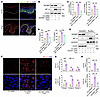 Figure 9
Succinate promotes HIF-1α nuclear translocation and its binding to CXCL9/CXCL10 promoters in keratinocytes via SUCNR1. (A) The expression of HIF-1α (red) in keratinocytes in vitiligo patients and healthy controls. Keratinocytes were characterized by cytokeratin 14 (green); nuclei were counterstained with DAPI (blue). (B) Protein levels of SUCNR1 and HIF-1α in NHKs pretransfected with SUCNR1 siRNA prior to succinate stimulation. (C and D) The mRNA (C) and secretion (D) levels of CXCL9 and CXCL10 in NHKs pretransfected with HIF-1α siRNA prior to succinate stimulation. (E) The expression of SUCNR1 and HIF-1α in the cytoplasm and nucleus of NHKs pretransfected with HIF-1α siRNA prior to succinate stimulation. (F) The expression of HIF-1α in NHKs pretransfected with SUCNR1 siRNA prior to succinate stimulation. (G) Binding of HIF-1α to CXCL9 and CXCL10 promoter regions in NHKs. (H) Binding of HIF-1α to CXCL9 and CXCL10 promoter region in NHKs pretreated with SUCNR1 siRNA prior to succinate treatment. Data are presented as mean ± SEM (C, D, G, and H) and analyzed by 1-way ANOVA (C, D, and H) or 2-tailed Student’s t test (G). **P < 0.01; ***P < 0.001; NHKs, normal human keratinocytes; Ctrl, control; Succ, succinate.
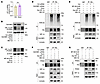 Figure 10
Succinate enhances HIF-1α stability through SUCNR1. (A) The mRNA level of HIF-1α in NHKs treated with succinate. (B and C) Expression of HIF-1α-OH in NHKs pretreated with SUCNR1 siRNA (B) or NF-56-EJ40 (C) prior to succinate treatment. (D and E) The ubiquitination of HIF-1α in NHKs pretreated with SUCNR1 siRNA (D) or NF-56-EJ40 (E) prior to succinate treatment. (F and G) The binding of HIF-1α and PHD2 in NHKs pretreated with SUCNR1 siRNA (F) or NF-56-EJ40 (G) prior to succinate treatment. Data are presented as mean ± SEM and analyzed by 2-tailed Student’s t test (A). NHKs, normal human keratinocytes; Ctrl, control; Succ, succinate.


Copyright © 2026 American Society for Clinical Investigation
ISSN: 0021-9738 (print), 1558-8238 (online)