
Advertisement
Research ArticleCell biologyGeneticsVascular biology
Open Access |  10.1172/JCI197609
Loss of Smad4 drives vascular malformations via c-KIT–dependent high–fluid shear stress mimicry
Johannes Gahn,1 Fan Wu,1 Qing Zhang,1 Yuxi Di,1 Tanmaya Behera,1 Yonggang Ren,2,3 Zohrah Hashemi,1 Kuheli Banerjee,1 Julio Cordero,2,3 Claudia Gherman,4 Kornelia Andorfer,5 Caroline T. Seebauer,6 Fatemeh Mirzapour-Shafiyi,7 Gergana Dobreva,2,3 Martin A. Schwartz,8 and Roxana Ola1
1Experimental Pharmacology Mannheim (EPM) and
2Department of Cardiovascular Genomics and Epigenomics, European Center for Angioscience (ECAS), Medical Faculty Mannheim, Heidelberg University, Mannheim, Germany.
3German Centre for Cardiovascular Research (DZHK), Mannheim, Germany.
4Ion Chiricuta Oncological Institute, Cluj Napoca, Romania.
5Department of Otorhinolaryngology, Regensburg University Medical Center, Regensburg, Germany.
6Department of Otorhinolaryngology, Head and Neck Surgery, Luzerner Kantonsspital, Lucerne, Switzerland.
7Center for Synthetic Biology, Technical University Darmstadt, Darmstadt, Germany.
8Yale Cardiovascular Research Center, Departments of Internal Medicine (Cardiology), Cell Biology, and Biomedical Engineering, Yale School of Medicine, New Haven, Connecticut, USA.
Address correspondence to: Roxana Ola, Experimental Pharmacology Mannheim (EPM), European Center for Angioscience (ECAS), Medical Faculty Mannheim, Heidelberg University, Ludolf-Krehl-Str. 13 – 17, 68167 Mannheim, Germany. Phone: 49.621.383.71518; Email: roxana.ola@medma.uni-heidelberg.de.
Authorship note: JG, FW, and QZ contributed equally to this work and are co–first authors.
Find articles by
Gahn, J.
in:
PubMed
|
Google Scholar
|

1Experimental Pharmacology Mannheim (EPM) and
2Department of Cardiovascular Genomics and Epigenomics, European Center for Angioscience (ECAS), Medical Faculty Mannheim, Heidelberg University, Mannheim, Germany.
3German Centre for Cardiovascular Research (DZHK), Mannheim, Germany.
4Ion Chiricuta Oncological Institute, Cluj Napoca, Romania.
5Department of Otorhinolaryngology, Regensburg University Medical Center, Regensburg, Germany.
6Department of Otorhinolaryngology, Head and Neck Surgery, Luzerner Kantonsspital, Lucerne, Switzerland.
7Center for Synthetic Biology, Technical University Darmstadt, Darmstadt, Germany.
8Yale Cardiovascular Research Center, Departments of Internal Medicine (Cardiology), Cell Biology, and Biomedical Engineering, Yale School of Medicine, New Haven, Connecticut, USA.
Address correspondence to: Roxana Ola, Experimental Pharmacology Mannheim (EPM), European Center for Angioscience (ECAS), Medical Faculty Mannheim, Heidelberg University, Ludolf-Krehl-Str. 13 – 17, 68167 Mannheim, Germany. Phone: 49.621.383.71518; Email: roxana.ola@medma.uni-heidelberg.de.
Authorship note: JG, FW, and QZ contributed equally to this work and are co–first authors.
Find articles by Wu, F. in: PubMed | Google Scholar
1Experimental Pharmacology Mannheim (EPM) and
2Department of Cardiovascular Genomics and Epigenomics, European Center for Angioscience (ECAS), Medical Faculty Mannheim, Heidelberg University, Mannheim, Germany.
3German Centre for Cardiovascular Research (DZHK), Mannheim, Germany.
4Ion Chiricuta Oncological Institute, Cluj Napoca, Romania.
5Department of Otorhinolaryngology, Regensburg University Medical Center, Regensburg, Germany.
6Department of Otorhinolaryngology, Head and Neck Surgery, Luzerner Kantonsspital, Lucerne, Switzerland.
7Center for Synthetic Biology, Technical University Darmstadt, Darmstadt, Germany.
8Yale Cardiovascular Research Center, Departments of Internal Medicine (Cardiology), Cell Biology, and Biomedical Engineering, Yale School of Medicine, New Haven, Connecticut, USA.
Address correspondence to: Roxana Ola, Experimental Pharmacology Mannheim (EPM), European Center for Angioscience (ECAS), Medical Faculty Mannheim, Heidelberg University, Ludolf-Krehl-Str. 13 – 17, 68167 Mannheim, Germany. Phone: 49.621.383.71518; Email: roxana.ola@medma.uni-heidelberg.de.
Authorship note: JG, FW, and QZ contributed equally to this work and are co–first authors.
Find articles by Zhang, Q. in: PubMed | Google Scholar
1Experimental Pharmacology Mannheim (EPM) and
2Department of Cardiovascular Genomics and Epigenomics, European Center for Angioscience (ECAS), Medical Faculty Mannheim, Heidelberg University, Mannheim, Germany.
3German Centre for Cardiovascular Research (DZHK), Mannheim, Germany.
4Ion Chiricuta Oncological Institute, Cluj Napoca, Romania.
5Department of Otorhinolaryngology, Regensburg University Medical Center, Regensburg, Germany.
6Department of Otorhinolaryngology, Head and Neck Surgery, Luzerner Kantonsspital, Lucerne, Switzerland.
7Center for Synthetic Biology, Technical University Darmstadt, Darmstadt, Germany.
8Yale Cardiovascular Research Center, Departments of Internal Medicine (Cardiology), Cell Biology, and Biomedical Engineering, Yale School of Medicine, New Haven, Connecticut, USA.
Address correspondence to: Roxana Ola, Experimental Pharmacology Mannheim (EPM), European Center for Angioscience (ECAS), Medical Faculty Mannheim, Heidelberg University, Ludolf-Krehl-Str. 13 – 17, 68167 Mannheim, Germany. Phone: 49.621.383.71518; Email: roxana.ola@medma.uni-heidelberg.de.
Authorship note: JG, FW, and QZ contributed equally to this work and are co–first authors.
Find articles by Di, Y. in: PubMed | Google Scholar
1Experimental Pharmacology Mannheim (EPM) and
2Department of Cardiovascular Genomics and Epigenomics, European Center for Angioscience (ECAS), Medical Faculty Mannheim, Heidelberg University, Mannheim, Germany.
3German Centre for Cardiovascular Research (DZHK), Mannheim, Germany.
4Ion Chiricuta Oncological Institute, Cluj Napoca, Romania.
5Department of Otorhinolaryngology, Regensburg University Medical Center, Regensburg, Germany.
6Department of Otorhinolaryngology, Head and Neck Surgery, Luzerner Kantonsspital, Lucerne, Switzerland.
7Center for Synthetic Biology, Technical University Darmstadt, Darmstadt, Germany.
8Yale Cardiovascular Research Center, Departments of Internal Medicine (Cardiology), Cell Biology, and Biomedical Engineering, Yale School of Medicine, New Haven, Connecticut, USA.
Address correspondence to: Roxana Ola, Experimental Pharmacology Mannheim (EPM), European Center for Angioscience (ECAS), Medical Faculty Mannheim, Heidelberg University, Ludolf-Krehl-Str. 13 – 17, 68167 Mannheim, Germany. Phone: 49.621.383.71518; Email: roxana.ola@medma.uni-heidelberg.de.
Authorship note: JG, FW, and QZ contributed equally to this work and are co–first authors.
Find articles by Behera, T. in: PubMed | Google Scholar
1Experimental Pharmacology Mannheim (EPM) and
2Department of Cardiovascular Genomics and Epigenomics, European Center for Angioscience (ECAS), Medical Faculty Mannheim, Heidelberg University, Mannheim, Germany.
3German Centre for Cardiovascular Research (DZHK), Mannheim, Germany.
4Ion Chiricuta Oncological Institute, Cluj Napoca, Romania.
5Department of Otorhinolaryngology, Regensburg University Medical Center, Regensburg, Germany.
6Department of Otorhinolaryngology, Head and Neck Surgery, Luzerner Kantonsspital, Lucerne, Switzerland.
7Center for Synthetic Biology, Technical University Darmstadt, Darmstadt, Germany.
8Yale Cardiovascular Research Center, Departments of Internal Medicine (Cardiology), Cell Biology, and Biomedical Engineering, Yale School of Medicine, New Haven, Connecticut, USA.
Address correspondence to: Roxana Ola, Experimental Pharmacology Mannheim (EPM), European Center for Angioscience (ECAS), Medical Faculty Mannheim, Heidelberg University, Ludolf-Krehl-Str. 13 – 17, 68167 Mannheim, Germany. Phone: 49.621.383.71518; Email: roxana.ola@medma.uni-heidelberg.de.
Authorship note: JG, FW, and QZ contributed equally to this work and are co–first authors.
Find articles by Ren, Y. in: PubMed | Google Scholar
1Experimental Pharmacology Mannheim (EPM) and
2Department of Cardiovascular Genomics and Epigenomics, European Center for Angioscience (ECAS), Medical Faculty Mannheim, Heidelberg University, Mannheim, Germany.
3German Centre for Cardiovascular Research (DZHK), Mannheim, Germany.
4Ion Chiricuta Oncological Institute, Cluj Napoca, Romania.
5Department of Otorhinolaryngology, Regensburg University Medical Center, Regensburg, Germany.
6Department of Otorhinolaryngology, Head and Neck Surgery, Luzerner Kantonsspital, Lucerne, Switzerland.
7Center for Synthetic Biology, Technical University Darmstadt, Darmstadt, Germany.
8Yale Cardiovascular Research Center, Departments of Internal Medicine (Cardiology), Cell Biology, and Biomedical Engineering, Yale School of Medicine, New Haven, Connecticut, USA.
Address correspondence to: Roxana Ola, Experimental Pharmacology Mannheim (EPM), European Center for Angioscience (ECAS), Medical Faculty Mannheim, Heidelberg University, Ludolf-Krehl-Str. 13 – 17, 68167 Mannheim, Germany. Phone: 49.621.383.71518; Email: roxana.ola@medma.uni-heidelberg.de.
Authorship note: JG, FW, and QZ contributed equally to this work and are co–first authors.
Find articles by Hashemi, Z. in: PubMed | Google Scholar
1Experimental Pharmacology Mannheim (EPM) and
2Department of Cardiovascular Genomics and Epigenomics, European Center for Angioscience (ECAS), Medical Faculty Mannheim, Heidelberg University, Mannheim, Germany.
3German Centre for Cardiovascular Research (DZHK), Mannheim, Germany.
4Ion Chiricuta Oncological Institute, Cluj Napoca, Romania.
5Department of Otorhinolaryngology, Regensburg University Medical Center, Regensburg, Germany.
6Department of Otorhinolaryngology, Head and Neck Surgery, Luzerner Kantonsspital, Lucerne, Switzerland.
7Center for Synthetic Biology, Technical University Darmstadt, Darmstadt, Germany.
8Yale Cardiovascular Research Center, Departments of Internal Medicine (Cardiology), Cell Biology, and Biomedical Engineering, Yale School of Medicine, New Haven, Connecticut, USA.
Address correspondence to: Roxana Ola, Experimental Pharmacology Mannheim (EPM), European Center for Angioscience (ECAS), Medical Faculty Mannheim, Heidelberg University, Ludolf-Krehl-Str. 13 – 17, 68167 Mannheim, Germany. Phone: 49.621.383.71518; Email: roxana.ola@medma.uni-heidelberg.de.
Authorship note: JG, FW, and QZ contributed equally to this work and are co–first authors.
Find articles by
Banerjee, K.
in:
PubMed
|
Google Scholar
|
1Experimental Pharmacology Mannheim (EPM) and
2Department of Cardiovascular Genomics and Epigenomics, European Center for Angioscience (ECAS), Medical Faculty Mannheim, Heidelberg University, Mannheim, Germany.
3German Centre for Cardiovascular Research (DZHK), Mannheim, Germany.
4Ion Chiricuta Oncological Institute, Cluj Napoca, Romania.
5Department of Otorhinolaryngology, Regensburg University Medical Center, Regensburg, Germany.
6Department of Otorhinolaryngology, Head and Neck Surgery, Luzerner Kantonsspital, Lucerne, Switzerland.
7Center for Synthetic Biology, Technical University Darmstadt, Darmstadt, Germany.
8Yale Cardiovascular Research Center, Departments of Internal Medicine (Cardiology), Cell Biology, and Biomedical Engineering, Yale School of Medicine, New Haven, Connecticut, USA.
Address correspondence to: Roxana Ola, Experimental Pharmacology Mannheim (EPM), European Center for Angioscience (ECAS), Medical Faculty Mannheim, Heidelberg University, Ludolf-Krehl-Str. 13 – 17, 68167 Mannheim, Germany. Phone: 49.621.383.71518; Email: roxana.ola@medma.uni-heidelberg.de.
Authorship note: JG, FW, and QZ contributed equally to this work and are co–first authors.
Find articles by
Cordero, J.
in:
PubMed
|
Google Scholar
|
1Experimental Pharmacology Mannheim (EPM) and
2Department of Cardiovascular Genomics and Epigenomics, European Center for Angioscience (ECAS), Medical Faculty Mannheim, Heidelberg University, Mannheim, Germany.
3German Centre for Cardiovascular Research (DZHK), Mannheim, Germany.
4Ion Chiricuta Oncological Institute, Cluj Napoca, Romania.
5Department of Otorhinolaryngology, Regensburg University Medical Center, Regensburg, Germany.
6Department of Otorhinolaryngology, Head and Neck Surgery, Luzerner Kantonsspital, Lucerne, Switzerland.
7Center for Synthetic Biology, Technical University Darmstadt, Darmstadt, Germany.
8Yale Cardiovascular Research Center, Departments of Internal Medicine (Cardiology), Cell Biology, and Biomedical Engineering, Yale School of Medicine, New Haven, Connecticut, USA.
Address correspondence to: Roxana Ola, Experimental Pharmacology Mannheim (EPM), European Center for Angioscience (ECAS), Medical Faculty Mannheim, Heidelberg University, Ludolf-Krehl-Str. 13 – 17, 68167 Mannheim, Germany. Phone: 49.621.383.71518; Email: roxana.ola@medma.uni-heidelberg.de.
Authorship note: JG, FW, and QZ contributed equally to this work and are co–first authors.
Find articles by Gherman, C. in: PubMed | Google Scholar
1Experimental Pharmacology Mannheim (EPM) and
2Department of Cardiovascular Genomics and Epigenomics, European Center for Angioscience (ECAS), Medical Faculty Mannheim, Heidelberg University, Mannheim, Germany.
3German Centre for Cardiovascular Research (DZHK), Mannheim, Germany.
4Ion Chiricuta Oncological Institute, Cluj Napoca, Romania.
5Department of Otorhinolaryngology, Regensburg University Medical Center, Regensburg, Germany.
6Department of Otorhinolaryngology, Head and Neck Surgery, Luzerner Kantonsspital, Lucerne, Switzerland.
7Center for Synthetic Biology, Technical University Darmstadt, Darmstadt, Germany.
8Yale Cardiovascular Research Center, Departments of Internal Medicine (Cardiology), Cell Biology, and Biomedical Engineering, Yale School of Medicine, New Haven, Connecticut, USA.
Address correspondence to: Roxana Ola, Experimental Pharmacology Mannheim (EPM), European Center for Angioscience (ECAS), Medical Faculty Mannheim, Heidelberg University, Ludolf-Krehl-Str. 13 – 17, 68167 Mannheim, Germany. Phone: 49.621.383.71518; Email: roxana.ola@medma.uni-heidelberg.de.
Authorship note: JG, FW, and QZ contributed equally to this work and are co–first authors.
Find articles by Andorfer, K. in: PubMed | Google Scholar
1Experimental Pharmacology Mannheim (EPM) and
2Department of Cardiovascular Genomics and Epigenomics, European Center for Angioscience (ECAS), Medical Faculty Mannheim, Heidelberg University, Mannheim, Germany.
3German Centre for Cardiovascular Research (DZHK), Mannheim, Germany.
4Ion Chiricuta Oncological Institute, Cluj Napoca, Romania.
5Department of Otorhinolaryngology, Regensburg University Medical Center, Regensburg, Germany.
6Department of Otorhinolaryngology, Head and Neck Surgery, Luzerner Kantonsspital, Lucerne, Switzerland.
7Center for Synthetic Biology, Technical University Darmstadt, Darmstadt, Germany.
8Yale Cardiovascular Research Center, Departments of Internal Medicine (Cardiology), Cell Biology, and Biomedical Engineering, Yale School of Medicine, New Haven, Connecticut, USA.
Address correspondence to: Roxana Ola, Experimental Pharmacology Mannheim (EPM), European Center for Angioscience (ECAS), Medical Faculty Mannheim, Heidelberg University, Ludolf-Krehl-Str. 13 – 17, 68167 Mannheim, Germany. Phone: 49.621.383.71518; Email: roxana.ola@medma.uni-heidelberg.de.
Authorship note: JG, FW, and QZ contributed equally to this work and are co–first authors.
Find articles by Seebauer, C. in: PubMed | Google Scholar
1Experimental Pharmacology Mannheim (EPM) and
2Department of Cardiovascular Genomics and Epigenomics, European Center for Angioscience (ECAS), Medical Faculty Mannheim, Heidelberg University, Mannheim, Germany.
3German Centre for Cardiovascular Research (DZHK), Mannheim, Germany.
4Ion Chiricuta Oncological Institute, Cluj Napoca, Romania.
5Department of Otorhinolaryngology, Regensburg University Medical Center, Regensburg, Germany.
6Department of Otorhinolaryngology, Head and Neck Surgery, Luzerner Kantonsspital, Lucerne, Switzerland.
7Center for Synthetic Biology, Technical University Darmstadt, Darmstadt, Germany.
8Yale Cardiovascular Research Center, Departments of Internal Medicine (Cardiology), Cell Biology, and Biomedical Engineering, Yale School of Medicine, New Haven, Connecticut, USA.
Address correspondence to: Roxana Ola, Experimental Pharmacology Mannheim (EPM), European Center for Angioscience (ECAS), Medical Faculty Mannheim, Heidelberg University, Ludolf-Krehl-Str. 13 – 17, 68167 Mannheim, Germany. Phone: 49.621.383.71518; Email: roxana.ola@medma.uni-heidelberg.de.
Authorship note: JG, FW, and QZ contributed equally to this work and are co–first authors.
Find articles by Mirzapour-Shafiyi, F. in: PubMed | Google Scholar
1Experimental Pharmacology Mannheim (EPM) and
2Department of Cardiovascular Genomics and Epigenomics, European Center for Angioscience (ECAS), Medical Faculty Mannheim, Heidelberg University, Mannheim, Germany.
3German Centre for Cardiovascular Research (DZHK), Mannheim, Germany.
4Ion Chiricuta Oncological Institute, Cluj Napoca, Romania.
5Department of Otorhinolaryngology, Regensburg University Medical Center, Regensburg, Germany.
6Department of Otorhinolaryngology, Head and Neck Surgery, Luzerner Kantonsspital, Lucerne, Switzerland.
7Center for Synthetic Biology, Technical University Darmstadt, Darmstadt, Germany.
8Yale Cardiovascular Research Center, Departments of Internal Medicine (Cardiology), Cell Biology, and Biomedical Engineering, Yale School of Medicine, New Haven, Connecticut, USA.
Address correspondence to: Roxana Ola, Experimental Pharmacology Mannheim (EPM), European Center for Angioscience (ECAS), Medical Faculty Mannheim, Heidelberg University, Ludolf-Krehl-Str. 13 – 17, 68167 Mannheim, Germany. Phone: 49.621.383.71518; Email: roxana.ola@medma.uni-heidelberg.de.
Authorship note: JG, FW, and QZ contributed equally to this work and are co–first authors.
Find articles by
Dobreva, G.
in:
PubMed
|
Google Scholar
|
1Experimental Pharmacology Mannheim (EPM) and
2Department of Cardiovascular Genomics and Epigenomics, European Center for Angioscience (ECAS), Medical Faculty Mannheim, Heidelberg University, Mannheim, Germany.
3German Centre for Cardiovascular Research (DZHK), Mannheim, Germany.
4Ion Chiricuta Oncological Institute, Cluj Napoca, Romania.
5Department of Otorhinolaryngology, Regensburg University Medical Center, Regensburg, Germany.
6Department of Otorhinolaryngology, Head and Neck Surgery, Luzerner Kantonsspital, Lucerne, Switzerland.
7Center for Synthetic Biology, Technical University Darmstadt, Darmstadt, Germany.
8Yale Cardiovascular Research Center, Departments of Internal Medicine (Cardiology), Cell Biology, and Biomedical Engineering, Yale School of Medicine, New Haven, Connecticut, USA.
Address correspondence to: Roxana Ola, Experimental Pharmacology Mannheim (EPM), European Center for Angioscience (ECAS), Medical Faculty Mannheim, Heidelberg University, Ludolf-Krehl-Str. 13 – 17, 68167 Mannheim, Germany. Phone: 49.621.383.71518; Email: roxana.ola@medma.uni-heidelberg.de.
Authorship note: JG, FW, and QZ contributed equally to this work and are co–first authors.
Find articles by
Schwartz, M.
in:
PubMed
|
Google Scholar
|
1Experimental Pharmacology Mannheim (EPM) and
2Department of Cardiovascular Genomics and Epigenomics, European Center for Angioscience (ECAS), Medical Faculty Mannheim, Heidelberg University, Mannheim, Germany.
3German Centre for Cardiovascular Research (DZHK), Mannheim, Germany.
4Ion Chiricuta Oncological Institute, Cluj Napoca, Romania.
5Department of Otorhinolaryngology, Regensburg University Medical Center, Regensburg, Germany.
6Department of Otorhinolaryngology, Head and Neck Surgery, Luzerner Kantonsspital, Lucerne, Switzerland.
7Center for Synthetic Biology, Technical University Darmstadt, Darmstadt, Germany.
8Yale Cardiovascular Research Center, Departments of Internal Medicine (Cardiology), Cell Biology, and Biomedical Engineering, Yale School of Medicine, New Haven, Connecticut, USA.
Address correspondence to: Roxana Ola, Experimental Pharmacology Mannheim (EPM), European Center for Angioscience (ECAS), Medical Faculty Mannheim, Heidelberg University, Ludolf-Krehl-Str. 13 – 17, 68167 Mannheim, Germany. Phone: 49.621.383.71518; Email: roxana.ola@medma.uni-heidelberg.de.
Authorship note: JG, FW, and QZ contributed equally to this work and are co–first authors.
Find articles by Ola, R. in: PubMed | Google Scholar
Published October 1, 2026 - More info
J Clin Invest. 2026;136(19):e197609. https://doi.org/10.1172/JCI197609.
© 2026 Gahn et al. This work is licensed under the Creative Commons Attribution 4.0 International License. To view a copy of this license, visit http://creativecommons.org/licenses/by/4.0/.
Received: July 8, 2025; Accepted: August 7, 2026

Related article:
Abstract
Hereditary hemorrhagic telangiectasia (HHT) is characterized by fragile, enlarged vessels with arteriovenous connections, which can form arteriovenous malformations (AVMs) that are prone to rupture. It is driven by mutations in TGF‑β pathway genes that disrupt endothelial cell responses and blood vessel remodeling. Two studies published in this issue of the JCI identify c‑KIT as a central mediator of HHT vascular pathology. Gahn et al. showed that SMAD4 loss derepressed c‑KIT, whose activity in the junctional mechanosensory receptor complex of endothelial cells reset the fluid shear stress set point to drive maladaptive vascular remodeling. Drapé et al. demonstrated that ALK1 loss in a mouse model of HHT induced a regional pro‑angiogenic c‑KIT+ endothelial state in brain AVMs. Both studies showed that pharmacologic c-KIT inhibition mitigated disease, revealing an unexpected, potentially ligand‑independent role for c‑KIT and highlighting a promising therapeutic target.
Authors
Ruilin Wu, Jason E. Fish

-
Results
SMAD4 restricts both the magnitude and duration of flow-induced signaling. We previously reported that SMAD4 restrains flow induction of KLF4 and activation of Akt to maintain the FSS set point (4). To investigate upstream mechanisms, we assessed flow signaling through the junctional complex in HUVECs subjected to laminar FSS (1-5-12 dynes/cm2) for 2 h (h) with/without SMAD4 using short interfering RNA (siRNA). Specificity of the SMAD4 siRNA and SMAD4 antibody was confirmed by Western blot (WB) (Figure 1, A and B). We examined FSS-induced phosphorylation of SRC family kinases (SFKY416), VE-CadherinY658, and VEGFR2Y1054; the downstream phosphorylation of AktS473; and the phosphorylation-dependent ERK5 gel shift (upper band) as markers of activation (Figure 1A). When ECs were subjected to FSS at increasing magnitudes, in comparison with control siRNA (CTRL siRNA), SMAD4 knockdown (SMAD4 KD) augmented FSS-dependent activation of VE-Cadherin, VEGFR2, ERK5, and Akt. Interestingly, SMAD4 KD not only increased phosphorylation of SFKY418 and VE-CadherinY658 at low physiological FSS but also prevented their inhibition at high FSS (Figure 1A, quantified in Figure 1B). Thus, SMAD4 restricts mechanotransduction pathways upstream of KLF4/Akt to establish the FSS set point.
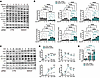 Figure 1
SMAD4 restricts both the magnitude and duration of flow-induced signaling. (A and C) Western blot (WB) for the indicated proteins in HUVECs transfected with CTRL or SMAD4 siRNAs grown statically or subjected to 1-5-12 dynes/cm2 for 2 h (A) or subjected to 12 dynes/cm2 in a time course of 0–240 minutes (C). (B and D) Quantifications of SMAD4, SFK (Y416), VE-Cadherin (Y658), VEGFR2 (Y1054), and Akt (S473) levels normalized to GAPDH or total SRC, VE-Cadherin, VEGFR2, and Akt, respectively, and of ERK5 gel shift (quantified by densitometry) (n = 3/group). p-, phosphorylated. (E) qPCR for KLF4 (fold-change) in CTRL and SMAD4 siRNA HUVECs grown statically (0 minute) versus 30 and 120 minutes of 12 dynes/cm2 (n = 4). Data are represented as mean ± SEM. Two-way ANOVA was used to determine statistical significance in B, D, and E. *P < 0.05, **P < 0.01, ***P < 0.005, ****P < 0.001.
We next assayed effects of SMAD4 loss on these pathways over 5–240 minutes under 12 dynes/cm2 FSS, a typical physiological level (Figure 1, C–E). SFK activation is the earliest flow-responsive event downstream of PECAM-1, followed by phosphorylation of VE-Cadherin, VEGFRs, PI3K, and Akt (14). SMAD4 depletion increased SFKY416, VE-CadherinY658, and AktS473 in this context as well and converted the transient activation to a sustained time course, suggesting loss of a physiological negative feedback mechanism. FSS-mediated ERK5 activation was also markedly amplified, driving excessive KLF4 transcription and persistent Akt activation (Figure 1, C–E). Sustained junctional component activation and heightened SFK/ERK5/KLF4 signaling after SMAD4 KD were also evident at 24 h (Supplemental Figure 1, A–E; supplemental material available online with this article; https://doi.org/10.1172/JCI197609DS1). Taken together, these data suggest that SMAD4 restricts FSS signals through the junctional mechanosensory receptor complex in a time- and magnitude-dependent manner.
SMAD4 restricts junctional mechanotransduction upstream of MEKK3/ERK5/KLF4/Akt activation. We next evaluated the role of sustained SFK activity in downstream ERK5/KLF4/Akt signaling. CTRL and SMAD4 siRNA ECs were examined at 2 h under 12 dynes/cm2 FSS with and without the SFK inhibitor, PP2. SFK inhibition reduced ERK5 and the downstream KLF4/Akt activation in both CTRL and SMAD4 siRNA cells (Figure 2, A–C). As SFKs are essential for activation of the mechanosensory receptor complex, we next addressed the requirement for junctional PECAM-1, VE-Cadherin, and VEGFRs. Depletion of any of these components reduced the high ERK5/KLF4/Akt levels in SMAD4 siRNA HUVECs (Figure 2, D–F, and Supplemental Figure 2, A and B). Together, these results support a requirement for junctional SFK activation within the mechanosensory receptor complex for augmented ERK5/KLF4/Akt after SMAD4 KD.
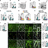 Figure 2
SMAD4 restricts junctional signals upstream of MEKK3/ERK5/KLF4/Akt activation. (A, D, and G) WB for the indicated proteins in HUVECs transfected with CTRL or SMAD4 siRNAs subjected to 12 dynes/cm2 for 2 h and treated with DMSO or PP2 inhibitor (A) or transfected additionally with CD31, CDH5, and KDR/FLT4 siRNAs (D) or with MEKK3 siRNA (G). (B, E, and H) Quantification of ERK5 gel shift (upper band, quantified by densitometry) and of p-Akt normalized to total Akt, respectively (n = 4/group in B and H; n = 3/group in E). (C, F, and I) KLF4 fold-change in the indicated genotypes and treatment conditions; n = 6/group in C and F; n = 4/group in I. (J) Colabeling of Smad4fl/fl and Smad4iΔEC P6 retinas with p-ERK5 (green, upper panel), KLF4 (green, middle panel), pS6 (green, lower panel), and Isolectin B4 (IB4, white). Red, blue, and yellow arrowheads indicate arteries (a), veins (v), and capillaries, respectively. Scale bar: 50 μm. (K) Quantification of pERK5, KLF4, and pS6 in retina vascular plexus was calculated by normalizing fluorescence intensity threshold value per IB4+ vascular plexus capillary area (%) in Smad4fl/fl and Smad4iΔEC neonates (n = 3 retinas per genotype). Data are represented as mean ± SEM. Two-way ANOVA (B, C, E, F, H, and I) and Student’s t test (K) were used to determine statistical significance. *P < 0.05, **P < 0.01, ***P < 0.005, ****P < 0.001.
To determine the functional role of ERK5 in the context of SMAD4 LOF, HUVECs were additionally depleted of MEKK3 (Figure 2, G–I) or treated with the MEK5 inhibitor, BIX02189 (Supplemental Figure 2, D–F). Cells were subjected to 12 dynes/cm2 FSS for 2 h and signaling pathways were assayed. SMAD4 KD induced the MEKK3 gel shift, indicating its activation (Figure 2G and Supplemental Figure 2C). Inhibition of MEK5-MEKK3 abrogated FSS-mediated ERK5 activation and KLF4 induction, and the downstream Akt activation, in both control and SMAD4 KDECs (Figure 2, G–I, and Supplemental Figure 2, D–F).
To assess ERK5 activation in vivo, we labeled postnatal day 6 (P6) retinas from Smad4fl/fl and Smad4iΔEC (EC-specific tamoxifen-inducible [Tx-inducible] Smad4 deletion) neonates for p-ERK5 (green), KLF4 (green), phosphorylated ribosomal S6 (p-S6, green), along with Isolectin B4 (IB4; white) to identify ECs (Figure 2J). In control Smad4fl/fl retinas, ERK5 activation was stronger in arteries than in veins. In Smad4iΔEC retinas, increased ERK5 marked the entire vascular plexus, including the AVMs. KLF4 was highly elevated; and ribosomal S6, which is downstream of Akt, was highly phosphorylated (Figure 2, J and K) (4, 36). Thus, derepression of MEKK3-ERK5 in Smad4 KO/KD ECs augments KLF4 expression and Akt activation in AVM lesions.
Upregulation of c-KIT in high-flow but low-FSS AVMs. We assessed FSS levels in retinal AVMs using computational flow simulation on the PolNet platform (38). As previously reported in murine wild-type developing retinas, the simulated wall shear stress (WSS) is the lowest at the sprouting front, is low to medium in the veins, and the highest in arteries and first branch point capillaries, with the latter being the region prone to AVM formation (27, 35). Interestingly, simulated WSS within the AVM in Smad4iΔEC was lower (between 1 and 5 Pascals) compared with Smad4fl/fl retinal arteries and veins. In vessels beyond the AVMs (toward periphery), WSS was nearly 0 (Figure 3, A and B), accounting for low KLF4 in these regions (39). Although flow rates within AVMs are relatively high, FSS is inversely proportional to vessel radius (40). As a result, ECs within these enlarged, high-flow AVMs experience low FSS while exhibiting activation of pathways typical of high FSS.
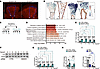 Figure 3
c-KIT upregulation characterizes the high-flow but low-FSS AVMs. (A and B) Whole-mount staining of IB4 (red) (A) and computer-simulated WSS modeling (B) of the Smad4fl/fl and Smad4iΔEC P6 mouse retinas. Magnified vascular plexus (boxed) is shown in B. a, artery; v, vein. Scale bars: 100 μm (left) and 50 μm (right). (C) Experimental design of bulk RNA-seq in CTRL and SMAD4 siRNA HUVECs grown statically or subjected to increasing magnitudes of 1-5-12 dynes/cm2 FSS for 2 h. (D) Significantly upregulated GO terms. (E and F) Expression of KIT by qPCR (E) and c-KIT protein by WB (F) in CTRL and SMAD4 siRNA HUVECs grown statically or subjected to increasing magnitudes of 1-5-12 dynes/cm2 FSS for 2 h. (G) Quantification of c-KIT/GAPDH ratio (n = 3/group). (H and I) KIT fold-change in BMP9-stimulated (10 ng) CTRL and SMAD4 siRNA HUVECs (n = 4/group) (H) and in HUVECs grown statically versus at 12 dynes/cm2 FSS for 2 h (n = 4/group) (I). Data are represented as mean ± SEM. Two-way ANOVA (E and G–I) was used to determine statistical significance. *P < 0.05, **P < 0.01, ***P < 0.005, ****P < 0.001.
To investigate the mechanisms upstream of SFK/ERK5/KLF4/Akt driving sustained mechanotransduction upon SMAD4 loss, we performed bulk RNA sequencing (RNA-seq) in HUVECs with/without SMAD4, grown statically or under low, medium, and physiological FSS (1-5-12 dynes/cm2) for 2 h (Figure 3C). Principal component analysis revealed a clear separation between the CTRL and SMAD4 siRNA groups (Supplemental Figure 3A). Major gene ontology (GO) terms for the differentially expressed genes (DEGs) upregulated upon SMAD4 KD (comparison of all CTRL versus SMAD4 siRNA groups) included MAPK pathway, cell migration, vascular development, and cell adhesion, all of which are pertinent to EC mechanobiology (Figure 3D). Among the downregulated DEGs, the cell cycle–related terms predominated (Supplemental Figure 3, D and E). Interestingly, listing the top 5 upregulated genes within each GO term, KIT gene was present in 5 out of the 8 GO terms (Figure 3D). Reactome pathway analysis further identified upregulated c-KIT signaling, thus suggesting its role in FSS pathway deregulation after SMAD4 LOF (Supplemental Figure 3B). We therefore focused on the c-KIT RTK, the receptor for stem cell factor (SCF), encoded by KITL.
Visualizing RNA-seq samples in Ridgeplot, KIT in CTRL siRNA cells was downregulated by FSS at all magnitudes. In SMAD4 KD cells, KIT was strongly upregulated under static conditions, and the downregulation by flow was reduced, resulting in higher levels of KIT under all conditions (Supplemental Figure 3C). Independent analysis of mRNA by RT-PCR revealed similar strong upregulation by SMAD4 siRNA without flow and minimal downregulation by flow in SMAD4 KD cells, with a decreasing trend for KIT mRNA from lower to higher flow (Figure 3E). Protein analysis showed that SMAD4 KD upregulated c-KIT protein and blocked its inhibition by flow of all magnitudes (Figure 3, F and G). SMAD4 loss thus potently upregulates KIT via both transcriptional and posttranscriptional mechanisms.
We next assessed BMP9 in regulation of KIT by FSS. HUVECs under static versus physiological FSS were stimulated with 1 ng BMP9 for 2 h. Similar to FSS, BMP9 downregulated KIT expression, which required SMAD4 (Figure 3H) and augmented the inhibitory effect of FSS on KIT expression (Figure 3I). To investigate whether c-KIT is similarly regulated by SMAD4 upstream signaling components, HUVECs were depleted for SMAD1/5 and ALK1 and stimulated with 12 dynes/cm2 FSS for 2 h. Similar to SMAD4 depletion, loss of either ALK1 or SMAD1/5 led to an increase in KIT expression in static and in flow conditions but also to augmented activation of ERK5/KLF4/Akt upon physiological FSS (Supplemental Figure 3, F–I). Thus, c-KIT and heightened flow signaling are part of a conserved mechanism across multiple AVM models.
SMAD4 restricts c-KIT expression in vivo. To corroborate our findings in vivo, we first reanalyzed published single-cell RNA-sequencing (scRNA-seq) data from P6 wild-type retinas (41). Kit transcripts were expressed at low levels in tip cells and some venous like ECs and absent in arterial ECs (Figure 4A). Labeling P6 Smad4fl/fl versus Smad4iΔEC retinas for c-KIT showed enhanced expression of c-KIT in tip cells and a few capillaries of Smad4fl/fl (arrowheads in Figure 4B, quantified in Figure 4, C and D), consistent with scRNA-seq results and confirming antibody specificity. In Smad4iΔEC retinas, c-KIT was strongly upregulated in all ECs, including AVMs (arrowheads in Figure 4B, quantified in Figure 4, C and D).
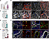 Figure 4
SMAD4 restricts c-KIT expression in vivo. (A) Reanalysis of Kit expression in scRNA-seq of P6 wild-type retinas (Tip, retina tip ECs). P values are above violin plots. (B) Colabeling for c-KIT (white) and IB4 (red) of retinal flatmounts from Tx-induced P6 Smad4fl/fl and Smad4iΔEC. Blue arrowheads point to c-KIT expression in tip cells (left panel) and vascular plexus (right panel). (C and D) Quantification of c-KIT fluorescence labeling intensity in ECs in the vascular front area (C) and in the vascular plexus (D) (n = 3 retinas/group). (E) Immunolabeling of human HHT2 patient nasal mucosa biopsy sections for c-KIT (red), ENDOGLIN (green), α-SMA (white), and DAPI (blue). Yellow arrowheads in non-AVM regions indicate ECs negative for c-KIT. Blue arrowheads in the insets indicate expression of c-KIT in the ECs in human AVM. (F) Quantification of c-KIT fluorescence labeling intensity in ENDOGLIN+ arterial versus venous endothelium (%), in non-AVM and AVM regions (n = 8, 4 pictures/patient biopsy). Scale bars: 50 μm in B and E left panels, 20 μm in insets in E. a, artery; v, vein. Data are represented as mean ± SEM. Bimod test was used in A; Student’s t test (C and D) and 1-way ANOVA (F) were used to determine statistical significance. *P < 0.05, **P < 0.01, ****P < 0.001.
We next examined c-KIT expression in 2 specimens from human HHT type 2 (HHT2) nasal telangiectasia biopsies and compared AVM region versus non-AVM regions within the same biopsy. Sections were stained for c-KIT, ENDOGLIN (to visualize the endothelium), α–smooth muscle actin (α-SMA; for smooth muscle cells), and DAPI (to visualize the nuclei). We observed a segment of the AVM endothelium strongly positive for c-KIT (blue versus yellow arrowheads in Figure 4E, quantified in Figure 4F), suggesting de novo expression, while the arterial and venous endothelium within the non-AVM regions was negative. Notably, mast cells also stained strongly for c-KIT. Thus, Smad4 inactivation triggers c-KIT upregulation in both mouse and human lesion ECs in vivo.
SMAD4 loss promotes c-KIT–dependent amplification of junctional flow signaling. We next considered the functional role of c-KIT in FSS mechanotransduction. Specificity of KIT siRNA and c-KIT antibody was confirmed by immunoblotting (Supplemental Figure 4A). In control cells, KIT KD had very little effect on FSS-mediated mechanotransduction (Supplemental Figure 4, A–C). Yet, in the context of SMAD4 depletion, KIT KD reduced the elevated p-SFK and p–VE-Cadherin and ERK5/KLF4/Akt activation, all components of or downstream of the junctional receptor complex (Figure 5, A–C). Thus, upregulated c-KIT in SMAD4 KDECs leads to increased activation of junction-dependent signals.
 Figure 5
SMAD4 loss promotes c-KIT–dependent amplification of junctional flow signaling. (A) WB for the indicated proteins in HUVECs transfected with CTRL, SMAD4, and SMAD4 KIT siRNAs grown statically (0 minute) and 5-30-120 minutes 12 dynes/cm2 FSS. (B) Quantifications of activated SFK (Y418), VE-Cadherin (Y658), and Akt (S473) levels normalized to total SRC, VE-Cadherin, and Akt, respectively, and of ERK5 gel shift (upper band, quantified by densitometry) (n = 4/group). (C) KLF4 fold-change in the indicated time points and conditions (n = 4/group). (D and E) CTRL versus SMAD4 siRNA HUVECs grown statically and at 12 dynes/cm2 for 2 h were subjected to anti–c-KIT (D) and anti–VE-Cadherin (E) IP, and WB was performed for the indicated proteins. Data are represented as mean ± SEM. Two-way ANOVA (B and C) was used to determine statistical significance. *P < 0.05, **P < 0.01, ****P < 0.001.
To determine whether c-KIT acts within the mechanosensory receptor complex, we immunoprecipitated c-KIT and VE-Cadherin and probed immunoprecipitants for junctional components using cells with/without SMAD4 under static and 12 dynes/cm2 FSS conditions (Figure 5D and Supplemental Figure 4, D and E). c-KIT associated with VE-Cadherin and VEGFR2 under static and flow, in both control and SMAD4 KD cells (Figure 5D). VE-Cadherin immunoprecipitation (IP) confirmed interaction with c-KIT and VEGFR2 (Figure 5E). Upon 2-hr FSS, control ECs showed reduced c-KIT expression and association with VE-Cadherin, despite comparable VE-Cadherin IP efficiency, accompanied by diminished VE-Cadherin Tyr phosphorylation. In contrast, SMAD4-deficient cells retained elevated c-KIT expression and sustained junctional association with increased VE-Cadherin activation, consistent with prolonged c-KIT–dependent junctional signaling (Figure 5, D and E). c-KIT thus shows physical and functional interactions with the junctional complex after its upregulation by SMAD4 LOF.
KIT inhibition in SMAD4 KD cells restores the FSS set point for EC behaviors. We next evaluated the role of c-KIT in the enhanced FSS-mediated EC behaviors in SMAD4 KD cells. We first assessed the EC elongation (ratio of length to width of individual ECs) and the EC alignment parallel to the direction of flow (≤30° angle directionality) in HUVECs depleted of KIT or SMAD4, or with double depletion of KIT and SMAD4, grown in static versus 1 and 12 dynes/cm2 FSS for 48 h (Figure 6, A–D). Under both magnitudes, HUVECs depleted of KIT alone showed no difference compared to CTRL cells. By contrast, KIT KD in SMAD4 KD cells restored the augmented flow-induced EC elongation and alignment (Figure 6, A–D). We next labeled cells for the Golgi apparatus (GM130, red), VE-Cadherin (white), and DAPI (blue), and we measured the nucleus-Golgi axis angle (at an angle ≤ 45° in relation to EC orientation) (Supplemental Figure 4, F and G; quantified in Figure 6, E and F). As previously shown (4), SMAD4 KD HUVECs oriented against the flow direction under 1 dyne, whereas KIT-KD cells showed no orientation (Supplemental Figure 4F and Figure 6E). Under 12 dynes/cm2, KD of SMAD4 reduced orientation, whereas KIT KD similar to CTRL cells oriented against the flow direction (Supplemental Figure 4F; quantified in Figure 6F). Interestingly, KIT KD restored the aberrant axial polarity in SMAD4-depleted HUVECs under both low and physiological FSS ((Supplemental Figure 4, F, and G; quantified in Figure 6, E and F).
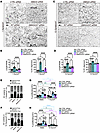 Figure 6
KIT inactivation in SMAD4 KD cells restores the FSS set point for EC behaviors. (A and C) VE-Cadherin staining of CTRL, KIT, SMAD4, and SMAD4 KIT siRNA–treated HUVECs subjected to 1 dyne/cm2 (A) and 12 dynes/cm2 (C) for 48 h. Scale bars: 50 μm. Flow direction: right to left. (B and D) Quantification of length-to-width ratio (%) and of EC alignment parallel to flow direction (%) in the mentioned genotypes upon 1 dyne/cm2 (B) or 12 dynes/cm2 (D) (n = 12 average of 4 images [70–140 cells/image] per 3 independent experiments/group). (E and F) Quantification of EC polarization (%) against, toward the flow’s direction, or nonoriented (neutral) of HUVECs transfected with CTRL, KIT, SMAD4, and SMAD4 KIT siRNA subjected to 1 dyne/cm2 (E) and 12 dynes/cm2 (F) for 48 h (n = 15–18 images [50–100 cells/image] per 3 independent experiments/group). (G and H) S phase ratio (EdU+) per total number of DAPI+ nuclei (%) in CTRL, KIT, SMAD4, and SMAD4 KIT siRNA–treated HUVECs grown statically or subjected to 1 dyne/cm2 (G) and 12 dynes/cm2 (H) for 24 h (n = 15 images [200–250 cells/image] per 3 independent experiments/group). Data are represented as mean ± SEM. Two-way ANOVA (B–H) was used to determine statistical significance. *P < 0.05, **P < 0.01, ***P < 0.005, ****P < 0.001.
To further assess the effect of KIT KD in EC proliferation, we performed 5-ethynyl-2′-deoxyuridine (EdU) labeling to identify ECs in S phase. In both KIT and CTRL siRNA cells, 12 dynes/cm2 promoted cell cycle arrest, while 1 dyne had no effect (Figure 6, G and H). In contrast, 1 dyne/cm2 reduced DNA synthesis in SMAD4-depleted HUVECs, which was reversed by KIT siRNA (Figure 6G). Together, these results show that resetting the FSS set point to lower values by SMAD4 KD requires c-KIT.
Soluble ligand–independent c-KIT activation in SMAD4 KD sustains flow signaling. We next investigated whether c-KIT function in mechanotransduction requires its ligand, SCF. In our RNA-seq dataset, KITL was selectively upregulated under physiological FSS only in SMAD4 KD cells (Figure 7A), which was confirmed by qPCR (Figure 7B). To elucidate the role of SCF in mediating c-KIT events, we blocked SCF using a specific neutralizing antibody (SCF blAb). Interestingly, SCF blockade did not attenuate the elevated SFK/ERK5/KLF4/Akt activation in SMAD4 KD cells (Figure 7, C–E). Conversely, stimulation with the soluble SCF transiently increased p-SFK and p-Akt levels at 5 minutes in both CTRL and SMAD4 siRNA cells, but it did not further enhance ERK5/KLF4 activation (Figure 7, F–H). Collectively, these data suggest that c-KIT driving augmented and persistent mechanotransduction in SMAD4 KD cells occurs independently of its soluble ligand.
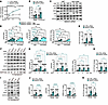 Figure 7
Ligand-independent c-KIT activation in SMAD4 KD sustains flow signaling. (A) Relative KITL mRNA expression in RNA-seq groups (n = 3/group). (B) KITL qPCR in CTRL and SMAD4 siRNA HUVECs grown statically versus 2 h of 12 dynes/cm2 (n = 4/group). (C) WB for the indicated proteins in SMAD4 siRNA HUVECs grown statically or subjected to 12 dynes/cm2 for 5-30-120 minutes and treated with IgG or SCF blocking antibodies (SCF blAb). (D) Quantifications of SFK (Y418) and Akt (S473) levels normalized to total SRC and Akt, respectively, and of ERK5 gel shift (upper band, quantified by densitometry) (n = 3/group). (E, H, and K) KLF4 qPCR in CTRL and SMAD4 siRNA HUVECs treated with SCF blAb subjected to 12 dynes/cm2 FSS for 2 h (n = 4/group) (E) or treated with SCF for 2 h and subjected to 12 dynes/cm2 FSS (n = 4/group) (H) or subjected to 2 h of 12 dynes/cm2 treated with DMSO versus avapritinib (Ava) (n = 4/group) (K). (F) WB for the indicated proteins in CTRL and SMAD4 siRNA HUVECs subjected to 12 dynes/cm2 for 2 h in the presence of SCF (50 ng/mL) for the indicated time (0 minute — no SCF; 5 and 120 minutes of SCF). (G) Quantifications of SFK (Y416) and Akt (S473) levels normalized to total SRC and Akt, respectively, and of ERK5 gel shift (n = 3/group). (I) WB for the indicated proteins in SMAD4 siRNA HUVECs subjected to 12 dynes/cm2 for 2 h treated with DMSO or Ava. (J) Quantifications of SFK (Y416) and Akt (S473) levels normalized to total SRC and Akt, respectively, and of ERK5 gel shift (n = 3/group). Data are represented as mean ± SEM. Statistical significance for the interaction between siRNA groups and shear stress was determined by DESeq2 Likelihood Ratio Test (A). Two-way ANOVA (B, D, E, G, H, J, and K) was used to determine statistical significance. *P < 0.05, **P < 0.01, ***P < 0.005, ****P < 0.001.
We next tested the effects of avapritinib, an FDA-approved selective inhibitor of c-KIT catalytic activity, which also targets PDGFRA (42, 43). Avapritinib efficiently suppressed SCF-induced c-KIT phosphorylation at Y719 (PI3K activation site) (44) and downstream SFK, Akt, and ERK1/2 activation in CTRL and SMAD4 KD cells, confirming pathway suppression (Supplemental Figure 5, A and B).
Interestingly, in HUVECs with/without SMAD4 subjected to 2 h of 12 dynes/cm2 FSS, pharmacological inhibition of c-KIT activity selectively rescued FSS-induced junctional SFK and the ERK5/KLF4/Akt activation (Figure 7, I–K). Analysis of our RNA-seq data showed that PDGFRA was unchanged following SMAD4 depletion, despite robust induction of KIT, suggesting that altered PDGFRA signaling is unlikely to account for the observed effects of avapritinib.
Inhibition of c-KIT kinase activity reverses AVM formation. We next assessed the effect of avapritinib in rescuing AVM formation in Smad4iΔEC mice. Tx was administered at P0 and P1 in Smad4fl/fl and Smad4iΔEC littermates, followed by intraperitoneal (i.p.) administration of avapritinib at P3–P5, with retinas analyzed at P6 (Figure 8A). Avapritinib rescued the number of AVMs and the increased vascular density at the angiogenic front in Smad4iΔEC retinas, whereas no effect was observed in Smad4fl/fl littermates (Figure 8, B–E).
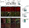 Figure 8
Inhibition of c-KIT kinase activity reverses AVM formation. (A) Schematic of Tx administration (black arrowheads), DMSO or Avapritinib (Ava) treatment (blue arrowheads), and harvesting (red arrowhead) in Smad4fl/fl and Smad4iΔEC P6 neonates. (B) IB4 staining of Smad4iΔEC P6 retinas, DMSO versus Ava treated. (C) Quantification of AVMs’ number per retina in Smad4iΔEC DMSO- and Ava-treated neonates (n = 10 retinas/genotype). (D) Labeling for IB4 (red, upper panel) and IB4 (red), ERG (green), and KI67 (white) in lower panel of Smad4fl/fl and Smad4iΔEC retinas from DMSO- and Ava-treated neonates. (E) Quantification of retinal front vascular density in DMSO- and Ava-treated neonates (n = 6 retinas/genotype). (F) Quantification of KI67+ERG+ per total ERG+ ECs in vascular plexus capillaries of Smad4fl/fl and Smad4iΔEC retinas (n = 6–11 images from 3 retinas/group). (G) IB4 (green), ERG (white), and GOLPH4 (red) colabeling of P6 Smad4iΔEC retina explants isolated from DMSO- and Ava-treated neonates. (H) Quantification of EC polarization: against, with flow, and neutral (nonpolarized) in capillaries from P6 Smad4iΔEC retinas harvested from DMSO- and Ava-treated neonates (n = 2 images per retina from 3 retinas/group). Scale bars: 100 μm in B, 50 μm in D and G, and 20 μm in the insets in D and G. Yellow arrowheads in D indicate AVMs. Arrows in G indicate direction of EC migration. a, artery; v, vein. Data are represented as mean ± SEM. Student’s t test (C and H) and 2-way ANOVA (E and F) were used to determine statistical significance. **P < 0.01, ****P < 0.001.
To analyze the role of c-KIT activation in cell cycle progression, we labeled retinas of Smad4fl/fl and Smad4iΔEC DMSO- and avapritinib-treated neonates for IB4 (red), ERG (green), and KI67 (white). Avapritinib treatment reduced the high EC proliferation in the Smad4-deficient vascular plexus with no effect on Smad4fl/fl (Figure 8, D and F). We further evaluated axial polarity by labeling retinas for ERG1 (white), GOLPH4 (red), and IB4 (green) (Figure 8G). Compared with DMSO-treated Smad4iΔEC, avapritinib treatment blunted the increased axial polarity in Smad4iΔEC (Figure 8, G and H). Thus, elevated c-KIT kinase activity promotes cell cycle progression and aberrant EC migration in Smad4iΔEC leading to AVM formation.
Last, we tested avapritinib in established AVMs by administering the inhibitor at P4 (Supplemental Figure 5, C–F). In this setting, avapritinib did not change the number of AVMs but significantly decreased their diameter (Figure 8, E and F). To validate rescue of AVMs by c-KIT inhibition, Smad4iΔEC neonates in an mTmG background (that allows visualization of gene recombination upon Tx administration) were treated with masitinib, another potent and selective RTK inhibitor that was shown to target c-KIT (45). Masitinib also rescued the number of AVMs in Smad4iΔEC mice (Supplemental Figure 5, G and I). Thus, 2 distinct inhibitors of c-KIT kinase activity are potential treatments for AVMs in patients with HHT.

-
Methods
Sex as a biological variable. Sex was not considered as a biological variable in the present study. Our study examined male and female neonates, and similar findings are reported for both sexes.
RNA-seq and analysis. Total RNA from HUVECs transfected with CTRL and SMAD4 siRNAs and grown in static versus 1-5-12 dynes/cm2 conditions for 2 h in 2% FBS was isolated using the Quick-RNA Miniprep Kit (Zymo Research). The RNA 6000 Nano Kit (Agilent) was used to assess the RNA integrity on a Bioanalyzer 2100 (Agilent). Library preparation and sequencing were performed on the BGISEQ-500 platform. Quality of RNA-seq reads was assessed with the MultiQC tool (v1.13) and trimmed of adapters using Trimmomatic (v0.39). Reads were mapped by STAR (v2.7.10a) with the following settings: -alignIntronMin 20 and -alignIntronMax 500,000 to the hg38 reference genome. Tag directories were created with makeTagDirectory, and reads were counted by the analyzeRepeats.pl function (rna hg38 -strad both -count exons -noadj), both from HOMER (v4.7.2). Differential expression was quantified and normalized using DESeq2. Rpkm.default from EdgeR was used to determine average reads per millions mapped (RPKM). Heatmaps were created by using heatmapper.ca from the RPKM values and represent the row-based Z-scores.
Animal experiments. To generate Smad4iΔEC neonates, we intercrossed 8-week-old Smad4fl/fl mice (Smad4tm2.1Cxd; The Jackson Laboratory) and Tx-inducible Cdh5-CreERT2 mice. To generate mTmG Smad4iΔEC mice, we subsequently intercrossed Smad4iΔEC with an mTmG reporter mouse line (The Jackson Laboratory). Gene deletion was achieved by i.p. injections of 100 μg Tx (Sigma-Aldrich) into Smad4iΔECand mTmG Smad4iΔEC at P0–P2. Tx-injected Cre-negative littermates (Smad4fl/fl) were used as controls. c-KIT kinase inhibitor avapritinib (MedChemExpress, 50 mg/kg/d) was administered i.p. either at P3–P5 or at P4–P5, and masitinib (MedChemExpress, 50 mg/kg/d) was administered i.p. at P3–P5. Mice were kept in a controlled environment, adhering to standard specific pathogen–free conditions.
Quantitative real-time PCR. RNA was extracted using RNeasy Mini Kit (QIAGEN). The RNA was reverse-transcribed with High-Capacity cDNA Reverse Transcription Kit (Thermo Fisher Scientific), and qPCR was performed using PowerUP SYBR Green Master Mix (Thermo Fisher Scientific) with a QuantStudio 3 (Thermo Fisher Scientific) according to the manufacturer’s protocol. For a detailed list of primer sequences, see Supplemental Table 1.
Immunostaining. Eyes of P6 pups or plated cells on coverslips were fixed in 4% paraformaldehyde for 17 minutes at room temperature. Staining with specific antibodies (Supplemental Table 2) was performed as previously described (4). High-resolution images were captured using a Zeiss confocal microscope equipped with an Airyscan Detector and analyzed with Zeiss ZEN software. Quantification of retinal vasculature and of labeled cells was performed using Fiji.
Cells and cell culture reagents. HUVECs were obtained from PromoCell (no. 12203) and cultured in Endothelial Cell Growth Medium MV2 (PromoCell, C-22022). Depletion of SMAD4, MEKK3, KIT, ALK1, SMAD1/5, KIT, CD31, CDH5, KDR, and FLT4 was achieved by transfecting 15 pmol of siRNA using Lipofectamine RNAiMax (Invitrogen) in 2% OptiMEM (Gibco). The sequences of specific siRNAs can be found in Supplemental Table 3. Transfection efficiency was assessed by WB and qPCR. Experiments were performed 48–72 h after transfection, and results were compared with CTRL siRNA. Inhibition of MEK5 was achieved by using 10 μM BIX02189, added 2 h before the onset of FSS; 5 μM PP2 was added 30 minutes before FSS; and 1 μM avapritinib was added 10 h before the FSS onset. Corresponding volumes of DMSO were added to CTRL cells. SCF pathway stimulation was achieved with 50 ng/mL SCF for 5 and 120 minutes in addition to 12 dynes/cm2 flow stimulation. Blockade of SCF using a human-specific blocking antibody (2 μg/mL) was done 1 hr before onset of flow. Before experiments, cells were starved for 4–8 h in 2% FCS. Chemicals are listed in Supplemental Table 4. For long-term flow experiments (48 h), cells were maintained in full medium.
Exposure of ECs to shear stress. Following siRNA transfection of HUVECs, fully confluent cells plated in 6-well plates were supplemented with 2 mL of cell culture media. The plates were subsequently placed on an orbital shaker (Rotamax120, Heidolph Instruments), which was set to specific rotations per minute (rpm) to generate shear stress levels of 1, 5, and 12 dynes/cm2. The rpm to achieve the desired shear stress was calculated using the following formula (Equation 1):

where a denotes the orbital radius of rotation in cm, η indicates the viscosity of the medium, ρ represents the density of the culture medium, and f is the frequency of rotation (in rotations per second) (54).
WB. HUVECs were washed with PBS and subsequently lysed using Laemmli buffer (Bio-Rad). The resulting samples were separated through 8% or 10% SDS-PAGE gels and transferred onto 0.2 μm nitrocellulose membranes (GE Healthcare, now Cytiva). Western blots were developed with Clarity Western ECL Substrate (Bio-Rad) utilizing a Luminescent Image Analyzer, Fusion FX (Vilber). Band intensities were quantified using Fiji software. The list for primary and secondary antibodies can be found in Supplemental Tables 5 and 6.
IP. For the IP procedure, cell lysates were prepared utilizing RIPA buffer (Carl Roth 23T1.3) and subsequently subjected to centrifugation at 17,000g for 20 minutes. The supernatants were incubated overnight at 4°C with specific antibodies: c-KIT (Cell Signaling Technology 3074S, 1:50, D13A2), VE-Cadherin (Cell Signaling Technology 2500S, 1:50, D87F2), or Rabbit IgG (Cell Signaling Technology 2729S, 1:200, polyclonal). Following this incubation, protein A/G–coated Dynabeads (Thermo Fisher Scientific, 88803) were incorporated, and the mixture was allowed to incubate for an additional 2 h at 4°C. The beads were washed thoroughly 4 times with PBS containing 0.05% Tween 20. The captured proteins were eluted at 25°C with gentle agitation using a soft elution buffer composed of 50 mM Tris-HCl (pH 8.0), 2% SDS, and 0.1% Tween 20. The resulting eluate was combined in a 1:4 ratio with 4× Laemmli Buffer (Bio-Rad 1610747) and incubated at 65°C for 10 minutes, followed by SDS-PAGE and subsequent immunoblotting analysis.
Biopsies. Nasal mucosa biopsies containing AVMs and surrounding non-AVM tissue were subsequently embedded in paraffin and stained as previously described (36). Antibodies used in this study are included in Supplemental Table 2. Slides were imaged with the LSM800 Confocal microscope with Airyscan Detector and the Zeiss ZEN software. Quantification of retina and human samples was done using Fiji.
Statistics. Data are expressed as the mean ± SEM. The statistical analysis was performed using the 2-tailed Student’s t test as well as 1-way and 2-way ANOVA, followed by Tukey’s method for multiple comparisons, employing GraphPad Prism 9.0 (GraphPad Software). A P value of less than 0.05 was considered the lowest statistically significant value.
Study approval. The animal procedures received approval from the animal welfare commission of the Regierungspräsidium Karlsruhe, Karlsruhe, Germany. Nasal biopsies containing AVMs and normal surrounding tissue was obtained from patients who underwent surgery for vascular malformations associated with HHT between 2018 and 2023 within the Department of Otorhinolaryngology, University Medical Center Regensburg, Germany, in accordance with the principles of the Declaration of Helsinki and was approved by the local ethics committee, Regensburg, Germany (No. 17-854-101). Written informed consent was obtained from all participants.
Data availability. RNA-seq datasets have been deposited at National Center for Biotechnology Information Gene Expression Omnibus with accession number GSE330070. The numerical data underlying all graphs and summary statistics are provided in the Supporting Data Values file.


Copyright © 2026 American Society for Clinical Investigation
ISSN: 0021-9738 (print), 1558-8238 (online)