
Advertisement
Research ArticleInflammationNeurosciencePulmonology
Open Access |  10.1172/JCI195110
Cerebellar and frontal lobe alterations in pulmonary hypertension implicate an inflammatory lung vascular/brain axis
Satoshi Okawa,1,2,3 Yinan Shi,1 Somanshu Banerjee,4 Yi Yin Tai,1 Jamal Nabhanizadeh,5 Stefan Guenther,6 Anisha N. Shah,1 Siyi Jiang,1,7 Timothy Kim,8 Yassmin Al Aaraj,1 Caroline Pidro,9 Metin Aytekin,1 Omolara Fatukasi,10 Nishita Kalepalli,1 Joseph Mettenburg,11 Timothy D. Girard,9 James T. Becker,10 Kathiravan Kaliyappan,12,13 Shanshan Song,12,13 Sandra Breuils-Bonnet,14 Steeve Provencher,14 Sébastien Bonnet,5,14 Theodore Huppert,15 Lloyd D. Harvey,1 Seyed Mehdi Nouraie,16 Rajkumar Savai,5,6,14 Dandan Sun,12,13 Soban Umar,4 Soni S. Pullamsetti,5,6,14 and Stephen Y. Chan1,8
1Center for Pulmonary Vascular Biology and Medicine, Pittsburgh Heart, Lung, and Blood Vascular Medicine Institute, Division of Cardiology, Department of Medicine;
2Department of Computational and Systems Biology; and
3McGowan Institute for Regenerative Medicine, University of Pittsburgh School of Medicine, Pittsburgh, Pennsylvania, USA.
4Department of Anesthesiology and Perioperative Medicine, Division of Molecular Medicine, University of California, Los Angeles, Los Angeles, California, USA.
5Institute for Lung Health, Department of Internal Medicine, German Center for Lung Research (DZL), Cardio-pulmonary Institute (CPI), Justus Liebig University, Giessen, Germany.
6Max Planck Institute for Heart and Lung Research, DZL, CPI, Bad Nauheim, Germany.
7Cellular and Molecular Pathology Graduate Program, University of Pittsburgh, Pittsburgh, Pennsylvania, USA.
8Heart and Vascular Institute, University of Pittsburgh Medical Center (UPMC), Pittsburgh, Pennsylvania, USA.
9Department of Critical Care Medicine,
10Department of Psychiatry,
11Department of Radiology, and
12Department of Neurology, University of Pittsburgh School of Medicine, Pittsburgh, Pennsylvania, USA.
13Pittsburgh Veteran’s Affairs Research Center, Pittsburgh, Pennsylvania, USA.
14PAH Research Group, Institut Universitaire de Cardiologie et Pneumologie de Québec Research Center, Department of Medicine, Université Laval, Quebec City, Québec, Canada.
15Department of Electrical and Computer Engineering, Swanson School of Engineering, University of Pittsburgh, Pittsburgh, Pennsylvania, USA.
16Division of Pulmonary, Allergy, and Critical Care Medicine, Department of Medicine, University of Pittsburgh School of Medicine, Pittsburgh, Pennsylvania, USA.
Address for correspondence. Stephen Y. Chan, University of Pittsburgh School of Medicine and UPMC, 200 Lothrop Street, BST E1240, Pittsburgh, Pennsylvania 15261, USA. Phone: 412.383.6990; Email: chansy@pitt.edu. Or: Satoshi Okawa, University of Pittsburgh School of Medicine and UPMC, 200 Lothrop Street, BST E1047, Pittsburgh, Pennsylvania 15261, USA. Phone: 412.383.1388; Email: okawas@pitt.edu.
Authorship note: SO and YS contributed equally to this work.
Find articles by Okawa, S. in: PubMed | Google Scholar
1Center for Pulmonary Vascular Biology and Medicine, Pittsburgh Heart, Lung, and Blood Vascular Medicine Institute, Division of Cardiology, Department of Medicine;
2Department of Computational and Systems Biology; and
3McGowan Institute for Regenerative Medicine, University of Pittsburgh School of Medicine, Pittsburgh, Pennsylvania, USA.
4Department of Anesthesiology and Perioperative Medicine, Division of Molecular Medicine, University of California, Los Angeles, Los Angeles, California, USA.
5Institute for Lung Health, Department of Internal Medicine, German Center for Lung Research (DZL), Cardio-pulmonary Institute (CPI), Justus Liebig University, Giessen, Germany.
6Max Planck Institute for Heart and Lung Research, DZL, CPI, Bad Nauheim, Germany.
7Cellular and Molecular Pathology Graduate Program, University of Pittsburgh, Pittsburgh, Pennsylvania, USA.
8Heart and Vascular Institute, University of Pittsburgh Medical Center (UPMC), Pittsburgh, Pennsylvania, USA.
9Department of Critical Care Medicine,
10Department of Psychiatry,
11Department of Radiology, and
12Department of Neurology, University of Pittsburgh School of Medicine, Pittsburgh, Pennsylvania, USA.
13Pittsburgh Veteran’s Affairs Research Center, Pittsburgh, Pennsylvania, USA.
14PAH Research Group, Institut Universitaire de Cardiologie et Pneumologie de Québec Research Center, Department of Medicine, Université Laval, Quebec City, Québec, Canada.
15Department of Electrical and Computer Engineering, Swanson School of Engineering, University of Pittsburgh, Pittsburgh, Pennsylvania, USA.
16Division of Pulmonary, Allergy, and Critical Care Medicine, Department of Medicine, University of Pittsburgh School of Medicine, Pittsburgh, Pennsylvania, USA.
Address for correspondence. Stephen Y. Chan, University of Pittsburgh School of Medicine and UPMC, 200 Lothrop Street, BST E1240, Pittsburgh, Pennsylvania 15261, USA. Phone: 412.383.6990; Email: chansy@pitt.edu. Or: Satoshi Okawa, University of Pittsburgh School of Medicine and UPMC, 200 Lothrop Street, BST E1047, Pittsburgh, Pennsylvania 15261, USA. Phone: 412.383.1388; Email: okawas@pitt.edu.
Authorship note: SO and YS contributed equally to this work.
Find articles by Shi, Y. in: PubMed | Google Scholar
1Center for Pulmonary Vascular Biology and Medicine, Pittsburgh Heart, Lung, and Blood Vascular Medicine Institute, Division of Cardiology, Department of Medicine;
2Department of Computational and Systems Biology; and
3McGowan Institute for Regenerative Medicine, University of Pittsburgh School of Medicine, Pittsburgh, Pennsylvania, USA.
4Department of Anesthesiology and Perioperative Medicine, Division of Molecular Medicine, University of California, Los Angeles, Los Angeles, California, USA.
5Institute for Lung Health, Department of Internal Medicine, German Center for Lung Research (DZL), Cardio-pulmonary Institute (CPI), Justus Liebig University, Giessen, Germany.
6Max Planck Institute for Heart and Lung Research, DZL, CPI, Bad Nauheim, Germany.
7Cellular and Molecular Pathology Graduate Program, University of Pittsburgh, Pittsburgh, Pennsylvania, USA.
8Heart and Vascular Institute, University of Pittsburgh Medical Center (UPMC), Pittsburgh, Pennsylvania, USA.
9Department of Critical Care Medicine,
10Department of Psychiatry,
11Department of Radiology, and
12Department of Neurology, University of Pittsburgh School of Medicine, Pittsburgh, Pennsylvania, USA.
13Pittsburgh Veteran’s Affairs Research Center, Pittsburgh, Pennsylvania, USA.
14PAH Research Group, Institut Universitaire de Cardiologie et Pneumologie de Québec Research Center, Department of Medicine, Université Laval, Quebec City, Québec, Canada.
15Department of Electrical and Computer Engineering, Swanson School of Engineering, University of Pittsburgh, Pittsburgh, Pennsylvania, USA.
16Division of Pulmonary, Allergy, and Critical Care Medicine, Department of Medicine, University of Pittsburgh School of Medicine, Pittsburgh, Pennsylvania, USA.
Address for correspondence. Stephen Y. Chan, University of Pittsburgh School of Medicine and UPMC, 200 Lothrop Street, BST E1240, Pittsburgh, Pennsylvania 15261, USA. Phone: 412.383.6990; Email: chansy@pitt.edu. Or: Satoshi Okawa, University of Pittsburgh School of Medicine and UPMC, 200 Lothrop Street, BST E1047, Pittsburgh, Pennsylvania 15261, USA. Phone: 412.383.1388; Email: okawas@pitt.edu.
Authorship note: SO and YS contributed equally to this work.
Find articles by Banerjee, S. in: PubMed | Google Scholar
1Center for Pulmonary Vascular Biology and Medicine, Pittsburgh Heart, Lung, and Blood Vascular Medicine Institute, Division of Cardiology, Department of Medicine;
2Department of Computational and Systems Biology; and
3McGowan Institute for Regenerative Medicine, University of Pittsburgh School of Medicine, Pittsburgh, Pennsylvania, USA.
4Department of Anesthesiology and Perioperative Medicine, Division of Molecular Medicine, University of California, Los Angeles, Los Angeles, California, USA.
5Institute for Lung Health, Department of Internal Medicine, German Center for Lung Research (DZL), Cardio-pulmonary Institute (CPI), Justus Liebig University, Giessen, Germany.
6Max Planck Institute for Heart and Lung Research, DZL, CPI, Bad Nauheim, Germany.
7Cellular and Molecular Pathology Graduate Program, University of Pittsburgh, Pittsburgh, Pennsylvania, USA.
8Heart and Vascular Institute, University of Pittsburgh Medical Center (UPMC), Pittsburgh, Pennsylvania, USA.
9Department of Critical Care Medicine,
10Department of Psychiatry,
11Department of Radiology, and
12Department of Neurology, University of Pittsburgh School of Medicine, Pittsburgh, Pennsylvania, USA.
13Pittsburgh Veteran’s Affairs Research Center, Pittsburgh, Pennsylvania, USA.
14PAH Research Group, Institut Universitaire de Cardiologie et Pneumologie de Québec Research Center, Department of Medicine, Université Laval, Quebec City, Québec, Canada.
15Department of Electrical and Computer Engineering, Swanson School of Engineering, University of Pittsburgh, Pittsburgh, Pennsylvania, USA.
16Division of Pulmonary, Allergy, and Critical Care Medicine, Department of Medicine, University of Pittsburgh School of Medicine, Pittsburgh, Pennsylvania, USA.
Address for correspondence. Stephen Y. Chan, University of Pittsburgh School of Medicine and UPMC, 200 Lothrop Street, BST E1240, Pittsburgh, Pennsylvania 15261, USA. Phone: 412.383.6990; Email: chansy@pitt.edu. Or: Satoshi Okawa, University of Pittsburgh School of Medicine and UPMC, 200 Lothrop Street, BST E1047, Pittsburgh, Pennsylvania 15261, USA. Phone: 412.383.1388; Email: okawas@pitt.edu.
Authorship note: SO and YS contributed equally to this work.
Find articles by
Tai, Y.
in:
PubMed
|
Google Scholar
|

1Center for Pulmonary Vascular Biology and Medicine, Pittsburgh Heart, Lung, and Blood Vascular Medicine Institute, Division of Cardiology, Department of Medicine;
2Department of Computational and Systems Biology; and
3McGowan Institute for Regenerative Medicine, University of Pittsburgh School of Medicine, Pittsburgh, Pennsylvania, USA.
4Department of Anesthesiology and Perioperative Medicine, Division of Molecular Medicine, University of California, Los Angeles, Los Angeles, California, USA.
5Institute for Lung Health, Department of Internal Medicine, German Center for Lung Research (DZL), Cardio-pulmonary Institute (CPI), Justus Liebig University, Giessen, Germany.
6Max Planck Institute for Heart and Lung Research, DZL, CPI, Bad Nauheim, Germany.
7Cellular and Molecular Pathology Graduate Program, University of Pittsburgh, Pittsburgh, Pennsylvania, USA.
8Heart and Vascular Institute, University of Pittsburgh Medical Center (UPMC), Pittsburgh, Pennsylvania, USA.
9Department of Critical Care Medicine,
10Department of Psychiatry,
11Department of Radiology, and
12Department of Neurology, University of Pittsburgh School of Medicine, Pittsburgh, Pennsylvania, USA.
13Pittsburgh Veteran’s Affairs Research Center, Pittsburgh, Pennsylvania, USA.
14PAH Research Group, Institut Universitaire de Cardiologie et Pneumologie de Québec Research Center, Department of Medicine, Université Laval, Quebec City, Québec, Canada.
15Department of Electrical and Computer Engineering, Swanson School of Engineering, University of Pittsburgh, Pittsburgh, Pennsylvania, USA.
16Division of Pulmonary, Allergy, and Critical Care Medicine, Department of Medicine, University of Pittsburgh School of Medicine, Pittsburgh, Pennsylvania, USA.
Address for correspondence. Stephen Y. Chan, University of Pittsburgh School of Medicine and UPMC, 200 Lothrop Street, BST E1240, Pittsburgh, Pennsylvania 15261, USA. Phone: 412.383.6990; Email: chansy@pitt.edu. Or: Satoshi Okawa, University of Pittsburgh School of Medicine and UPMC, 200 Lothrop Street, BST E1047, Pittsburgh, Pennsylvania 15261, USA. Phone: 412.383.1388; Email: okawas@pitt.edu.
Authorship note: SO and YS contributed equally to this work.
Find articles by Nabhanizadeh, J. in: PubMed | Google Scholar
1Center for Pulmonary Vascular Biology and Medicine, Pittsburgh Heart, Lung, and Blood Vascular Medicine Institute, Division of Cardiology, Department of Medicine;
2Department of Computational and Systems Biology; and
3McGowan Institute for Regenerative Medicine, University of Pittsburgh School of Medicine, Pittsburgh, Pennsylvania, USA.
4Department of Anesthesiology and Perioperative Medicine, Division of Molecular Medicine, University of California, Los Angeles, Los Angeles, California, USA.
5Institute for Lung Health, Department of Internal Medicine, German Center for Lung Research (DZL), Cardio-pulmonary Institute (CPI), Justus Liebig University, Giessen, Germany.
6Max Planck Institute for Heart and Lung Research, DZL, CPI, Bad Nauheim, Germany.
7Cellular and Molecular Pathology Graduate Program, University of Pittsburgh, Pittsburgh, Pennsylvania, USA.
8Heart and Vascular Institute, University of Pittsburgh Medical Center (UPMC), Pittsburgh, Pennsylvania, USA.
9Department of Critical Care Medicine,
10Department of Psychiatry,
11Department of Radiology, and
12Department of Neurology, University of Pittsburgh School of Medicine, Pittsburgh, Pennsylvania, USA.
13Pittsburgh Veteran’s Affairs Research Center, Pittsburgh, Pennsylvania, USA.
14PAH Research Group, Institut Universitaire de Cardiologie et Pneumologie de Québec Research Center, Department of Medicine, Université Laval, Quebec City, Québec, Canada.
15Department of Electrical and Computer Engineering, Swanson School of Engineering, University of Pittsburgh, Pittsburgh, Pennsylvania, USA.
16Division of Pulmonary, Allergy, and Critical Care Medicine, Department of Medicine, University of Pittsburgh School of Medicine, Pittsburgh, Pennsylvania, USA.
Address for correspondence. Stephen Y. Chan, University of Pittsburgh School of Medicine and UPMC, 200 Lothrop Street, BST E1240, Pittsburgh, Pennsylvania 15261, USA. Phone: 412.383.6990; Email: chansy@pitt.edu. Or: Satoshi Okawa, University of Pittsburgh School of Medicine and UPMC, 200 Lothrop Street, BST E1047, Pittsburgh, Pennsylvania 15261, USA. Phone: 412.383.1388; Email: okawas@pitt.edu.
Authorship note: SO and YS contributed equally to this work.
Find articles by Guenther, S. in: PubMed | Google Scholar
1Center for Pulmonary Vascular Biology and Medicine, Pittsburgh Heart, Lung, and Blood Vascular Medicine Institute, Division of Cardiology, Department of Medicine;
2Department of Computational and Systems Biology; and
3McGowan Institute for Regenerative Medicine, University of Pittsburgh School of Medicine, Pittsburgh, Pennsylvania, USA.
4Department of Anesthesiology and Perioperative Medicine, Division of Molecular Medicine, University of California, Los Angeles, Los Angeles, California, USA.
5Institute for Lung Health, Department of Internal Medicine, German Center for Lung Research (DZL), Cardio-pulmonary Institute (CPI), Justus Liebig University, Giessen, Germany.
6Max Planck Institute for Heart and Lung Research, DZL, CPI, Bad Nauheim, Germany.
7Cellular and Molecular Pathology Graduate Program, University of Pittsburgh, Pittsburgh, Pennsylvania, USA.
8Heart and Vascular Institute, University of Pittsburgh Medical Center (UPMC), Pittsburgh, Pennsylvania, USA.
9Department of Critical Care Medicine,
10Department of Psychiatry,
11Department of Radiology, and
12Department of Neurology, University of Pittsburgh School of Medicine, Pittsburgh, Pennsylvania, USA.
13Pittsburgh Veteran’s Affairs Research Center, Pittsburgh, Pennsylvania, USA.
14PAH Research Group, Institut Universitaire de Cardiologie et Pneumologie de Québec Research Center, Department of Medicine, Université Laval, Quebec City, Québec, Canada.
15Department of Electrical and Computer Engineering, Swanson School of Engineering, University of Pittsburgh, Pittsburgh, Pennsylvania, USA.
16Division of Pulmonary, Allergy, and Critical Care Medicine, Department of Medicine, University of Pittsburgh School of Medicine, Pittsburgh, Pennsylvania, USA.
Address for correspondence. Stephen Y. Chan, University of Pittsburgh School of Medicine and UPMC, 200 Lothrop Street, BST E1240, Pittsburgh, Pennsylvania 15261, USA. Phone: 412.383.6990; Email: chansy@pitt.edu. Or: Satoshi Okawa, University of Pittsburgh School of Medicine and UPMC, 200 Lothrop Street, BST E1047, Pittsburgh, Pennsylvania 15261, USA. Phone: 412.383.1388; Email: okawas@pitt.edu.
Authorship note: SO and YS contributed equally to this work.
Find articles by Shah, A. in: PubMed | Google Scholar
1Center for Pulmonary Vascular Biology and Medicine, Pittsburgh Heart, Lung, and Blood Vascular Medicine Institute, Division of Cardiology, Department of Medicine;
2Department of Computational and Systems Biology; and
3McGowan Institute for Regenerative Medicine, University of Pittsburgh School of Medicine, Pittsburgh, Pennsylvania, USA.
4Department of Anesthesiology and Perioperative Medicine, Division of Molecular Medicine, University of California, Los Angeles, Los Angeles, California, USA.
5Institute for Lung Health, Department of Internal Medicine, German Center for Lung Research (DZL), Cardio-pulmonary Institute (CPI), Justus Liebig University, Giessen, Germany.
6Max Planck Institute for Heart and Lung Research, DZL, CPI, Bad Nauheim, Germany.
7Cellular and Molecular Pathology Graduate Program, University of Pittsburgh, Pittsburgh, Pennsylvania, USA.
8Heart and Vascular Institute, University of Pittsburgh Medical Center (UPMC), Pittsburgh, Pennsylvania, USA.
9Department of Critical Care Medicine,
10Department of Psychiatry,
11Department of Radiology, and
12Department of Neurology, University of Pittsburgh School of Medicine, Pittsburgh, Pennsylvania, USA.
13Pittsburgh Veteran’s Affairs Research Center, Pittsburgh, Pennsylvania, USA.
14PAH Research Group, Institut Universitaire de Cardiologie et Pneumologie de Québec Research Center, Department of Medicine, Université Laval, Quebec City, Québec, Canada.
15Department of Electrical and Computer Engineering, Swanson School of Engineering, University of Pittsburgh, Pittsburgh, Pennsylvania, USA.
16Division of Pulmonary, Allergy, and Critical Care Medicine, Department of Medicine, University of Pittsburgh School of Medicine, Pittsburgh, Pennsylvania, USA.
Address for correspondence. Stephen Y. Chan, University of Pittsburgh School of Medicine and UPMC, 200 Lothrop Street, BST E1240, Pittsburgh, Pennsylvania 15261, USA. Phone: 412.383.6990; Email: chansy@pitt.edu. Or: Satoshi Okawa, University of Pittsburgh School of Medicine and UPMC, 200 Lothrop Street, BST E1047, Pittsburgh, Pennsylvania 15261, USA. Phone: 412.383.1388; Email: okawas@pitt.edu.
Authorship note: SO and YS contributed equally to this work.
Find articles by Jiang, S. in: PubMed | Google Scholar
1Center for Pulmonary Vascular Biology and Medicine, Pittsburgh Heart, Lung, and Blood Vascular Medicine Institute, Division of Cardiology, Department of Medicine;
2Department of Computational and Systems Biology; and
3McGowan Institute for Regenerative Medicine, University of Pittsburgh School of Medicine, Pittsburgh, Pennsylvania, USA.
4Department of Anesthesiology and Perioperative Medicine, Division of Molecular Medicine, University of California, Los Angeles, Los Angeles, California, USA.
5Institute for Lung Health, Department of Internal Medicine, German Center for Lung Research (DZL), Cardio-pulmonary Institute (CPI), Justus Liebig University, Giessen, Germany.
6Max Planck Institute for Heart and Lung Research, DZL, CPI, Bad Nauheim, Germany.
7Cellular and Molecular Pathology Graduate Program, University of Pittsburgh, Pittsburgh, Pennsylvania, USA.
8Heart and Vascular Institute, University of Pittsburgh Medical Center (UPMC), Pittsburgh, Pennsylvania, USA.
9Department of Critical Care Medicine,
10Department of Psychiatry,
11Department of Radiology, and
12Department of Neurology, University of Pittsburgh School of Medicine, Pittsburgh, Pennsylvania, USA.
13Pittsburgh Veteran’s Affairs Research Center, Pittsburgh, Pennsylvania, USA.
14PAH Research Group, Institut Universitaire de Cardiologie et Pneumologie de Québec Research Center, Department of Medicine, Université Laval, Quebec City, Québec, Canada.
15Department of Electrical and Computer Engineering, Swanson School of Engineering, University of Pittsburgh, Pittsburgh, Pennsylvania, USA.
16Division of Pulmonary, Allergy, and Critical Care Medicine, Department of Medicine, University of Pittsburgh School of Medicine, Pittsburgh, Pennsylvania, USA.
Address for correspondence. Stephen Y. Chan, University of Pittsburgh School of Medicine and UPMC, 200 Lothrop Street, BST E1240, Pittsburgh, Pennsylvania 15261, USA. Phone: 412.383.6990; Email: chansy@pitt.edu. Or: Satoshi Okawa, University of Pittsburgh School of Medicine and UPMC, 200 Lothrop Street, BST E1047, Pittsburgh, Pennsylvania 15261, USA. Phone: 412.383.1388; Email: okawas@pitt.edu.
Authorship note: SO and YS contributed equally to this work.
Find articles by Kim, T. in: PubMed | Google Scholar
1Center for Pulmonary Vascular Biology and Medicine, Pittsburgh Heart, Lung, and Blood Vascular Medicine Institute, Division of Cardiology, Department of Medicine;
2Department of Computational and Systems Biology; and
3McGowan Institute for Regenerative Medicine, University of Pittsburgh School of Medicine, Pittsburgh, Pennsylvania, USA.
4Department of Anesthesiology and Perioperative Medicine, Division of Molecular Medicine, University of California, Los Angeles, Los Angeles, California, USA.
5Institute for Lung Health, Department of Internal Medicine, German Center for Lung Research (DZL), Cardio-pulmonary Institute (CPI), Justus Liebig University, Giessen, Germany.
6Max Planck Institute for Heart and Lung Research, DZL, CPI, Bad Nauheim, Germany.
7Cellular and Molecular Pathology Graduate Program, University of Pittsburgh, Pittsburgh, Pennsylvania, USA.
8Heart and Vascular Institute, University of Pittsburgh Medical Center (UPMC), Pittsburgh, Pennsylvania, USA.
9Department of Critical Care Medicine,
10Department of Psychiatry,
11Department of Radiology, and
12Department of Neurology, University of Pittsburgh School of Medicine, Pittsburgh, Pennsylvania, USA.
13Pittsburgh Veteran’s Affairs Research Center, Pittsburgh, Pennsylvania, USA.
14PAH Research Group, Institut Universitaire de Cardiologie et Pneumologie de Québec Research Center, Department of Medicine, Université Laval, Quebec City, Québec, Canada.
15Department of Electrical and Computer Engineering, Swanson School of Engineering, University of Pittsburgh, Pittsburgh, Pennsylvania, USA.
16Division of Pulmonary, Allergy, and Critical Care Medicine, Department of Medicine, University of Pittsburgh School of Medicine, Pittsburgh, Pennsylvania, USA.
Address for correspondence. Stephen Y. Chan, University of Pittsburgh School of Medicine and UPMC, 200 Lothrop Street, BST E1240, Pittsburgh, Pennsylvania 15261, USA. Phone: 412.383.6990; Email: chansy@pitt.edu. Or: Satoshi Okawa, University of Pittsburgh School of Medicine and UPMC, 200 Lothrop Street, BST E1047, Pittsburgh, Pennsylvania 15261, USA. Phone: 412.383.1388; Email: okawas@pitt.edu.
Authorship note: SO and YS contributed equally to this work.
Find articles by Al Aaraj, Y. in: PubMed | Google Scholar
1Center for Pulmonary Vascular Biology and Medicine, Pittsburgh Heart, Lung, and Blood Vascular Medicine Institute, Division of Cardiology, Department of Medicine;
2Department of Computational and Systems Biology; and
3McGowan Institute for Regenerative Medicine, University of Pittsburgh School of Medicine, Pittsburgh, Pennsylvania, USA.
4Department of Anesthesiology and Perioperative Medicine, Division of Molecular Medicine, University of California, Los Angeles, Los Angeles, California, USA.
5Institute for Lung Health, Department of Internal Medicine, German Center for Lung Research (DZL), Cardio-pulmonary Institute (CPI), Justus Liebig University, Giessen, Germany.
6Max Planck Institute for Heart and Lung Research, DZL, CPI, Bad Nauheim, Germany.
7Cellular and Molecular Pathology Graduate Program, University of Pittsburgh, Pittsburgh, Pennsylvania, USA.
8Heart and Vascular Institute, University of Pittsburgh Medical Center (UPMC), Pittsburgh, Pennsylvania, USA.
9Department of Critical Care Medicine,
10Department of Psychiatry,
11Department of Radiology, and
12Department of Neurology, University of Pittsburgh School of Medicine, Pittsburgh, Pennsylvania, USA.
13Pittsburgh Veteran’s Affairs Research Center, Pittsburgh, Pennsylvania, USA.
14PAH Research Group, Institut Universitaire de Cardiologie et Pneumologie de Québec Research Center, Department of Medicine, Université Laval, Quebec City, Québec, Canada.
15Department of Electrical and Computer Engineering, Swanson School of Engineering, University of Pittsburgh, Pittsburgh, Pennsylvania, USA.
16Division of Pulmonary, Allergy, and Critical Care Medicine, Department of Medicine, University of Pittsburgh School of Medicine, Pittsburgh, Pennsylvania, USA.
Address for correspondence. Stephen Y. Chan, University of Pittsburgh School of Medicine and UPMC, 200 Lothrop Street, BST E1240, Pittsburgh, Pennsylvania 15261, USA. Phone: 412.383.6990; Email: chansy@pitt.edu. Or: Satoshi Okawa, University of Pittsburgh School of Medicine and UPMC, 200 Lothrop Street, BST E1047, Pittsburgh, Pennsylvania 15261, USA. Phone: 412.383.1388; Email: okawas@pitt.edu.
Authorship note: SO and YS contributed equally to this work.
Find articles by Pidro, C. in: PubMed | Google Scholar
1Center for Pulmonary Vascular Biology and Medicine, Pittsburgh Heart, Lung, and Blood Vascular Medicine Institute, Division of Cardiology, Department of Medicine;
2Department of Computational and Systems Biology; and
3McGowan Institute for Regenerative Medicine, University of Pittsburgh School of Medicine, Pittsburgh, Pennsylvania, USA.
4Department of Anesthesiology and Perioperative Medicine, Division of Molecular Medicine, University of California, Los Angeles, Los Angeles, California, USA.
5Institute for Lung Health, Department of Internal Medicine, German Center for Lung Research (DZL), Cardio-pulmonary Institute (CPI), Justus Liebig University, Giessen, Germany.
6Max Planck Institute for Heart and Lung Research, DZL, CPI, Bad Nauheim, Germany.
7Cellular and Molecular Pathology Graduate Program, University of Pittsburgh, Pittsburgh, Pennsylvania, USA.
8Heart and Vascular Institute, University of Pittsburgh Medical Center (UPMC), Pittsburgh, Pennsylvania, USA.
9Department of Critical Care Medicine,
10Department of Psychiatry,
11Department of Radiology, and
12Department of Neurology, University of Pittsburgh School of Medicine, Pittsburgh, Pennsylvania, USA.
13Pittsburgh Veteran’s Affairs Research Center, Pittsburgh, Pennsylvania, USA.
14PAH Research Group, Institut Universitaire de Cardiologie et Pneumologie de Québec Research Center, Department of Medicine, Université Laval, Quebec City, Québec, Canada.
15Department of Electrical and Computer Engineering, Swanson School of Engineering, University of Pittsburgh, Pittsburgh, Pennsylvania, USA.
16Division of Pulmonary, Allergy, and Critical Care Medicine, Department of Medicine, University of Pittsburgh School of Medicine, Pittsburgh, Pennsylvania, USA.
Address for correspondence. Stephen Y. Chan, University of Pittsburgh School of Medicine and UPMC, 200 Lothrop Street, BST E1240, Pittsburgh, Pennsylvania 15261, USA. Phone: 412.383.6990; Email: chansy@pitt.edu. Or: Satoshi Okawa, University of Pittsburgh School of Medicine and UPMC, 200 Lothrop Street, BST E1047, Pittsburgh, Pennsylvania 15261, USA. Phone: 412.383.1388; Email: okawas@pitt.edu.
Authorship note: SO and YS contributed equally to this work.
Find articles by Aytekin, M. in: PubMed | Google Scholar
1Center for Pulmonary Vascular Biology and Medicine, Pittsburgh Heart, Lung, and Blood Vascular Medicine Institute, Division of Cardiology, Department of Medicine;
2Department of Computational and Systems Biology; and
3McGowan Institute for Regenerative Medicine, University of Pittsburgh School of Medicine, Pittsburgh, Pennsylvania, USA.
4Department of Anesthesiology and Perioperative Medicine, Division of Molecular Medicine, University of California, Los Angeles, Los Angeles, California, USA.
5Institute for Lung Health, Department of Internal Medicine, German Center for Lung Research (DZL), Cardio-pulmonary Institute (CPI), Justus Liebig University, Giessen, Germany.
6Max Planck Institute for Heart and Lung Research, DZL, CPI, Bad Nauheim, Germany.
7Cellular and Molecular Pathology Graduate Program, University of Pittsburgh, Pittsburgh, Pennsylvania, USA.
8Heart and Vascular Institute, University of Pittsburgh Medical Center (UPMC), Pittsburgh, Pennsylvania, USA.
9Department of Critical Care Medicine,
10Department of Psychiatry,
11Department of Radiology, and
12Department of Neurology, University of Pittsburgh School of Medicine, Pittsburgh, Pennsylvania, USA.
13Pittsburgh Veteran’s Affairs Research Center, Pittsburgh, Pennsylvania, USA.
14PAH Research Group, Institut Universitaire de Cardiologie et Pneumologie de Québec Research Center, Department of Medicine, Université Laval, Quebec City, Québec, Canada.
15Department of Electrical and Computer Engineering, Swanson School of Engineering, University of Pittsburgh, Pittsburgh, Pennsylvania, USA.
16Division of Pulmonary, Allergy, and Critical Care Medicine, Department of Medicine, University of Pittsburgh School of Medicine, Pittsburgh, Pennsylvania, USA.
Address for correspondence. Stephen Y. Chan, University of Pittsburgh School of Medicine and UPMC, 200 Lothrop Street, BST E1240, Pittsburgh, Pennsylvania 15261, USA. Phone: 412.383.6990; Email: chansy@pitt.edu. Or: Satoshi Okawa, University of Pittsburgh School of Medicine and UPMC, 200 Lothrop Street, BST E1047, Pittsburgh, Pennsylvania 15261, USA. Phone: 412.383.1388; Email: okawas@pitt.edu.
Authorship note: SO and YS contributed equally to this work.
Find articles by Fatukasi, O. in: PubMed | Google Scholar
1Center for Pulmonary Vascular Biology and Medicine, Pittsburgh Heart, Lung, and Blood Vascular Medicine Institute, Division of Cardiology, Department of Medicine;
2Department of Computational and Systems Biology; and
3McGowan Institute for Regenerative Medicine, University of Pittsburgh School of Medicine, Pittsburgh, Pennsylvania, USA.
4Department of Anesthesiology and Perioperative Medicine, Division of Molecular Medicine, University of California, Los Angeles, Los Angeles, California, USA.
5Institute for Lung Health, Department of Internal Medicine, German Center for Lung Research (DZL), Cardio-pulmonary Institute (CPI), Justus Liebig University, Giessen, Germany.
6Max Planck Institute for Heart and Lung Research, DZL, CPI, Bad Nauheim, Germany.
7Cellular and Molecular Pathology Graduate Program, University of Pittsburgh, Pittsburgh, Pennsylvania, USA.
8Heart and Vascular Institute, University of Pittsburgh Medical Center (UPMC), Pittsburgh, Pennsylvania, USA.
9Department of Critical Care Medicine,
10Department of Psychiatry,
11Department of Radiology, and
12Department of Neurology, University of Pittsburgh School of Medicine, Pittsburgh, Pennsylvania, USA.
13Pittsburgh Veteran’s Affairs Research Center, Pittsburgh, Pennsylvania, USA.
14PAH Research Group, Institut Universitaire de Cardiologie et Pneumologie de Québec Research Center, Department of Medicine, Université Laval, Quebec City, Québec, Canada.
15Department of Electrical and Computer Engineering, Swanson School of Engineering, University of Pittsburgh, Pittsburgh, Pennsylvania, USA.
16Division of Pulmonary, Allergy, and Critical Care Medicine, Department of Medicine, University of Pittsburgh School of Medicine, Pittsburgh, Pennsylvania, USA.
Address for correspondence. Stephen Y. Chan, University of Pittsburgh School of Medicine and UPMC, 200 Lothrop Street, BST E1240, Pittsburgh, Pennsylvania 15261, USA. Phone: 412.383.6990; Email: chansy@pitt.edu. Or: Satoshi Okawa, University of Pittsburgh School of Medicine and UPMC, 200 Lothrop Street, BST E1047, Pittsburgh, Pennsylvania 15261, USA. Phone: 412.383.1388; Email: okawas@pitt.edu.
Authorship note: SO and YS contributed equally to this work.
Find articles by Kalepalli, N. in: PubMed | Google Scholar
1Center for Pulmonary Vascular Biology and Medicine, Pittsburgh Heart, Lung, and Blood Vascular Medicine Institute, Division of Cardiology, Department of Medicine;
2Department of Computational and Systems Biology; and
3McGowan Institute for Regenerative Medicine, University of Pittsburgh School of Medicine, Pittsburgh, Pennsylvania, USA.
4Department of Anesthesiology and Perioperative Medicine, Division of Molecular Medicine, University of California, Los Angeles, Los Angeles, California, USA.
5Institute for Lung Health, Department of Internal Medicine, German Center for Lung Research (DZL), Cardio-pulmonary Institute (CPI), Justus Liebig University, Giessen, Germany.
6Max Planck Institute for Heart and Lung Research, DZL, CPI, Bad Nauheim, Germany.
7Cellular and Molecular Pathology Graduate Program, University of Pittsburgh, Pittsburgh, Pennsylvania, USA.
8Heart and Vascular Institute, University of Pittsburgh Medical Center (UPMC), Pittsburgh, Pennsylvania, USA.
9Department of Critical Care Medicine,
10Department of Psychiatry,
11Department of Radiology, and
12Department of Neurology, University of Pittsburgh School of Medicine, Pittsburgh, Pennsylvania, USA.
13Pittsburgh Veteran’s Affairs Research Center, Pittsburgh, Pennsylvania, USA.
14PAH Research Group, Institut Universitaire de Cardiologie et Pneumologie de Québec Research Center, Department of Medicine, Université Laval, Quebec City, Québec, Canada.
15Department of Electrical and Computer Engineering, Swanson School of Engineering, University of Pittsburgh, Pittsburgh, Pennsylvania, USA.
16Division of Pulmonary, Allergy, and Critical Care Medicine, Department of Medicine, University of Pittsburgh School of Medicine, Pittsburgh, Pennsylvania, USA.
Address for correspondence. Stephen Y. Chan, University of Pittsburgh School of Medicine and UPMC, 200 Lothrop Street, BST E1240, Pittsburgh, Pennsylvania 15261, USA. Phone: 412.383.6990; Email: chansy@pitt.edu. Or: Satoshi Okawa, University of Pittsburgh School of Medicine and UPMC, 200 Lothrop Street, BST E1047, Pittsburgh, Pennsylvania 15261, USA. Phone: 412.383.1388; Email: okawas@pitt.edu.
Authorship note: SO and YS contributed equally to this work.
Find articles by Mettenburg, J. in: PubMed | Google Scholar
1Center for Pulmonary Vascular Biology and Medicine, Pittsburgh Heart, Lung, and Blood Vascular Medicine Institute, Division of Cardiology, Department of Medicine;
2Department of Computational and Systems Biology; and
3McGowan Institute for Regenerative Medicine, University of Pittsburgh School of Medicine, Pittsburgh, Pennsylvania, USA.
4Department of Anesthesiology and Perioperative Medicine, Division of Molecular Medicine, University of California, Los Angeles, Los Angeles, California, USA.
5Institute for Lung Health, Department of Internal Medicine, German Center for Lung Research (DZL), Cardio-pulmonary Institute (CPI), Justus Liebig University, Giessen, Germany.
6Max Planck Institute for Heart and Lung Research, DZL, CPI, Bad Nauheim, Germany.
7Cellular and Molecular Pathology Graduate Program, University of Pittsburgh, Pittsburgh, Pennsylvania, USA.
8Heart and Vascular Institute, University of Pittsburgh Medical Center (UPMC), Pittsburgh, Pennsylvania, USA.
9Department of Critical Care Medicine,
10Department of Psychiatry,
11Department of Radiology, and
12Department of Neurology, University of Pittsburgh School of Medicine, Pittsburgh, Pennsylvania, USA.
13Pittsburgh Veteran’s Affairs Research Center, Pittsburgh, Pennsylvania, USA.
14PAH Research Group, Institut Universitaire de Cardiologie et Pneumologie de Québec Research Center, Department of Medicine, Université Laval, Quebec City, Québec, Canada.
15Department of Electrical and Computer Engineering, Swanson School of Engineering, University of Pittsburgh, Pittsburgh, Pennsylvania, USA.
16Division of Pulmonary, Allergy, and Critical Care Medicine, Department of Medicine, University of Pittsburgh School of Medicine, Pittsburgh, Pennsylvania, USA.
Address for correspondence. Stephen Y. Chan, University of Pittsburgh School of Medicine and UPMC, 200 Lothrop Street, BST E1240, Pittsburgh, Pennsylvania 15261, USA. Phone: 412.383.6990; Email: chansy@pitt.edu. Or: Satoshi Okawa, University of Pittsburgh School of Medicine and UPMC, 200 Lothrop Street, BST E1047, Pittsburgh, Pennsylvania 15261, USA. Phone: 412.383.1388; Email: okawas@pitt.edu.
Authorship note: SO and YS contributed equally to this work.
Find articles by Girard, T. in: PubMed | Google Scholar
1Center for Pulmonary Vascular Biology and Medicine, Pittsburgh Heart, Lung, and Blood Vascular Medicine Institute, Division of Cardiology, Department of Medicine;
2Department of Computational and Systems Biology; and
3McGowan Institute for Regenerative Medicine, University of Pittsburgh School of Medicine, Pittsburgh, Pennsylvania, USA.
4Department of Anesthesiology and Perioperative Medicine, Division of Molecular Medicine, University of California, Los Angeles, Los Angeles, California, USA.
5Institute for Lung Health, Department of Internal Medicine, German Center for Lung Research (DZL), Cardio-pulmonary Institute (CPI), Justus Liebig University, Giessen, Germany.
6Max Planck Institute for Heart and Lung Research, DZL, CPI, Bad Nauheim, Germany.
7Cellular and Molecular Pathology Graduate Program, University of Pittsburgh, Pittsburgh, Pennsylvania, USA.
8Heart and Vascular Institute, University of Pittsburgh Medical Center (UPMC), Pittsburgh, Pennsylvania, USA.
9Department of Critical Care Medicine,
10Department of Psychiatry,
11Department of Radiology, and
12Department of Neurology, University of Pittsburgh School of Medicine, Pittsburgh, Pennsylvania, USA.
13Pittsburgh Veteran’s Affairs Research Center, Pittsburgh, Pennsylvania, USA.
14PAH Research Group, Institut Universitaire de Cardiologie et Pneumologie de Québec Research Center, Department of Medicine, Université Laval, Quebec City, Québec, Canada.
15Department of Electrical and Computer Engineering, Swanson School of Engineering, University of Pittsburgh, Pittsburgh, Pennsylvania, USA.
16Division of Pulmonary, Allergy, and Critical Care Medicine, Department of Medicine, University of Pittsburgh School of Medicine, Pittsburgh, Pennsylvania, USA.
Address for correspondence. Stephen Y. Chan, University of Pittsburgh School of Medicine and UPMC, 200 Lothrop Street, BST E1240, Pittsburgh, Pennsylvania 15261, USA. Phone: 412.383.6990; Email: chansy@pitt.edu. Or: Satoshi Okawa, University of Pittsburgh School of Medicine and UPMC, 200 Lothrop Street, BST E1047, Pittsburgh, Pennsylvania 15261, USA. Phone: 412.383.1388; Email: okawas@pitt.edu.
Authorship note: SO and YS contributed equally to this work.
Find articles by Becker, J. in: PubMed | Google Scholar
1Center for Pulmonary Vascular Biology and Medicine, Pittsburgh Heart, Lung, and Blood Vascular Medicine Institute, Division of Cardiology, Department of Medicine;
2Department of Computational and Systems Biology; and
3McGowan Institute for Regenerative Medicine, University of Pittsburgh School of Medicine, Pittsburgh, Pennsylvania, USA.
4Department of Anesthesiology and Perioperative Medicine, Division of Molecular Medicine, University of California, Los Angeles, Los Angeles, California, USA.
5Institute for Lung Health, Department of Internal Medicine, German Center for Lung Research (DZL), Cardio-pulmonary Institute (CPI), Justus Liebig University, Giessen, Germany.
6Max Planck Institute for Heart and Lung Research, DZL, CPI, Bad Nauheim, Germany.
7Cellular and Molecular Pathology Graduate Program, University of Pittsburgh, Pittsburgh, Pennsylvania, USA.
8Heart and Vascular Institute, University of Pittsburgh Medical Center (UPMC), Pittsburgh, Pennsylvania, USA.
9Department of Critical Care Medicine,
10Department of Psychiatry,
11Department of Radiology, and
12Department of Neurology, University of Pittsburgh School of Medicine, Pittsburgh, Pennsylvania, USA.
13Pittsburgh Veteran’s Affairs Research Center, Pittsburgh, Pennsylvania, USA.
14PAH Research Group, Institut Universitaire de Cardiologie et Pneumologie de Québec Research Center, Department of Medicine, Université Laval, Quebec City, Québec, Canada.
15Department of Electrical and Computer Engineering, Swanson School of Engineering, University of Pittsburgh, Pittsburgh, Pennsylvania, USA.
16Division of Pulmonary, Allergy, and Critical Care Medicine, Department of Medicine, University of Pittsburgh School of Medicine, Pittsburgh, Pennsylvania, USA.
Address for correspondence. Stephen Y. Chan, University of Pittsburgh School of Medicine and UPMC, 200 Lothrop Street, BST E1240, Pittsburgh, Pennsylvania 15261, USA. Phone: 412.383.6990; Email: chansy@pitt.edu. Or: Satoshi Okawa, University of Pittsburgh School of Medicine and UPMC, 200 Lothrop Street, BST E1047, Pittsburgh, Pennsylvania 15261, USA. Phone: 412.383.1388; Email: okawas@pitt.edu.
Authorship note: SO and YS contributed equally to this work.
Find articles by Kaliyappan, K. in: PubMed | Google Scholar
1Center for Pulmonary Vascular Biology and Medicine, Pittsburgh Heart, Lung, and Blood Vascular Medicine Institute, Division of Cardiology, Department of Medicine;
2Department of Computational and Systems Biology; and
3McGowan Institute for Regenerative Medicine, University of Pittsburgh School of Medicine, Pittsburgh, Pennsylvania, USA.
4Department of Anesthesiology and Perioperative Medicine, Division of Molecular Medicine, University of California, Los Angeles, Los Angeles, California, USA.
5Institute for Lung Health, Department of Internal Medicine, German Center for Lung Research (DZL), Cardio-pulmonary Institute (CPI), Justus Liebig University, Giessen, Germany.
6Max Planck Institute for Heart and Lung Research, DZL, CPI, Bad Nauheim, Germany.
7Cellular and Molecular Pathology Graduate Program, University of Pittsburgh, Pittsburgh, Pennsylvania, USA.
8Heart and Vascular Institute, University of Pittsburgh Medical Center (UPMC), Pittsburgh, Pennsylvania, USA.
9Department of Critical Care Medicine,
10Department of Psychiatry,
11Department of Radiology, and
12Department of Neurology, University of Pittsburgh School of Medicine, Pittsburgh, Pennsylvania, USA.
13Pittsburgh Veteran’s Affairs Research Center, Pittsburgh, Pennsylvania, USA.
14PAH Research Group, Institut Universitaire de Cardiologie et Pneumologie de Québec Research Center, Department of Medicine, Université Laval, Quebec City, Québec, Canada.
15Department of Electrical and Computer Engineering, Swanson School of Engineering, University of Pittsburgh, Pittsburgh, Pennsylvania, USA.
16Division of Pulmonary, Allergy, and Critical Care Medicine, Department of Medicine, University of Pittsburgh School of Medicine, Pittsburgh, Pennsylvania, USA.
Address for correspondence. Stephen Y. Chan, University of Pittsburgh School of Medicine and UPMC, 200 Lothrop Street, BST E1240, Pittsburgh, Pennsylvania 15261, USA. Phone: 412.383.6990; Email: chansy@pitt.edu. Or: Satoshi Okawa, University of Pittsburgh School of Medicine and UPMC, 200 Lothrop Street, BST E1047, Pittsburgh, Pennsylvania 15261, USA. Phone: 412.383.1388; Email: okawas@pitt.edu.
Authorship note: SO and YS contributed equally to this work.
Find articles by Song, S. in: PubMed | Google Scholar
1Center for Pulmonary Vascular Biology and Medicine, Pittsburgh Heart, Lung, and Blood Vascular Medicine Institute, Division of Cardiology, Department of Medicine;
2Department of Computational and Systems Biology; and
3McGowan Institute for Regenerative Medicine, University of Pittsburgh School of Medicine, Pittsburgh, Pennsylvania, USA.
4Department of Anesthesiology and Perioperative Medicine, Division of Molecular Medicine, University of California, Los Angeles, Los Angeles, California, USA.
5Institute for Lung Health, Department of Internal Medicine, German Center for Lung Research (DZL), Cardio-pulmonary Institute (CPI), Justus Liebig University, Giessen, Germany.
6Max Planck Institute for Heart and Lung Research, DZL, CPI, Bad Nauheim, Germany.
7Cellular and Molecular Pathology Graduate Program, University of Pittsburgh, Pittsburgh, Pennsylvania, USA.
8Heart and Vascular Institute, University of Pittsburgh Medical Center (UPMC), Pittsburgh, Pennsylvania, USA.
9Department of Critical Care Medicine,
10Department of Psychiatry,
11Department of Radiology, and
12Department of Neurology, University of Pittsburgh School of Medicine, Pittsburgh, Pennsylvania, USA.
13Pittsburgh Veteran’s Affairs Research Center, Pittsburgh, Pennsylvania, USA.
14PAH Research Group, Institut Universitaire de Cardiologie et Pneumologie de Québec Research Center, Department of Medicine, Université Laval, Quebec City, Québec, Canada.
15Department of Electrical and Computer Engineering, Swanson School of Engineering, University of Pittsburgh, Pittsburgh, Pennsylvania, USA.
16Division of Pulmonary, Allergy, and Critical Care Medicine, Department of Medicine, University of Pittsburgh School of Medicine, Pittsburgh, Pennsylvania, USA.
Address for correspondence. Stephen Y. Chan, University of Pittsburgh School of Medicine and UPMC, 200 Lothrop Street, BST E1240, Pittsburgh, Pennsylvania 15261, USA. Phone: 412.383.6990; Email: chansy@pitt.edu. Or: Satoshi Okawa, University of Pittsburgh School of Medicine and UPMC, 200 Lothrop Street, BST E1047, Pittsburgh, Pennsylvania 15261, USA. Phone: 412.383.1388; Email: okawas@pitt.edu.
Authorship note: SO and YS contributed equally to this work.
Find articles by Breuils-Bonnet, S. in: PubMed | Google Scholar
1Center for Pulmonary Vascular Biology and Medicine, Pittsburgh Heart, Lung, and Blood Vascular Medicine Institute, Division of Cardiology, Department of Medicine;
2Department of Computational and Systems Biology; and
3McGowan Institute for Regenerative Medicine, University of Pittsburgh School of Medicine, Pittsburgh, Pennsylvania, USA.
4Department of Anesthesiology and Perioperative Medicine, Division of Molecular Medicine, University of California, Los Angeles, Los Angeles, California, USA.
5Institute for Lung Health, Department of Internal Medicine, German Center for Lung Research (DZL), Cardio-pulmonary Institute (CPI), Justus Liebig University, Giessen, Germany.
6Max Planck Institute for Heart and Lung Research, DZL, CPI, Bad Nauheim, Germany.
7Cellular and Molecular Pathology Graduate Program, University of Pittsburgh, Pittsburgh, Pennsylvania, USA.
8Heart and Vascular Institute, University of Pittsburgh Medical Center (UPMC), Pittsburgh, Pennsylvania, USA.
9Department of Critical Care Medicine,
10Department of Psychiatry,
11Department of Radiology, and
12Department of Neurology, University of Pittsburgh School of Medicine, Pittsburgh, Pennsylvania, USA.
13Pittsburgh Veteran’s Affairs Research Center, Pittsburgh, Pennsylvania, USA.
14PAH Research Group, Institut Universitaire de Cardiologie et Pneumologie de Québec Research Center, Department of Medicine, Université Laval, Quebec City, Québec, Canada.
15Department of Electrical and Computer Engineering, Swanson School of Engineering, University of Pittsburgh, Pittsburgh, Pennsylvania, USA.
16Division of Pulmonary, Allergy, and Critical Care Medicine, Department of Medicine, University of Pittsburgh School of Medicine, Pittsburgh, Pennsylvania, USA.
Address for correspondence. Stephen Y. Chan, University of Pittsburgh School of Medicine and UPMC, 200 Lothrop Street, BST E1240, Pittsburgh, Pennsylvania 15261, USA. Phone: 412.383.6990; Email: chansy@pitt.edu. Or: Satoshi Okawa, University of Pittsburgh School of Medicine and UPMC, 200 Lothrop Street, BST E1047, Pittsburgh, Pennsylvania 15261, USA. Phone: 412.383.1388; Email: okawas@pitt.edu.
Authorship note: SO and YS contributed equally to this work.
Find articles by
Provencher, S.
in:
PubMed
|
Google Scholar
|
1Center for Pulmonary Vascular Biology and Medicine, Pittsburgh Heart, Lung, and Blood Vascular Medicine Institute, Division of Cardiology, Department of Medicine;
2Department of Computational and Systems Biology; and
3McGowan Institute for Regenerative Medicine, University of Pittsburgh School of Medicine, Pittsburgh, Pennsylvania, USA.
4Department of Anesthesiology and Perioperative Medicine, Division of Molecular Medicine, University of California, Los Angeles, Los Angeles, California, USA.
5Institute for Lung Health, Department of Internal Medicine, German Center for Lung Research (DZL), Cardio-pulmonary Institute (CPI), Justus Liebig University, Giessen, Germany.
6Max Planck Institute for Heart and Lung Research, DZL, CPI, Bad Nauheim, Germany.
7Cellular and Molecular Pathology Graduate Program, University of Pittsburgh, Pittsburgh, Pennsylvania, USA.
8Heart and Vascular Institute, University of Pittsburgh Medical Center (UPMC), Pittsburgh, Pennsylvania, USA.
9Department of Critical Care Medicine,
10Department of Psychiatry,
11Department of Radiology, and
12Department of Neurology, University of Pittsburgh School of Medicine, Pittsburgh, Pennsylvania, USA.
13Pittsburgh Veteran’s Affairs Research Center, Pittsburgh, Pennsylvania, USA.
14PAH Research Group, Institut Universitaire de Cardiologie et Pneumologie de Québec Research Center, Department of Medicine, Université Laval, Quebec City, Québec, Canada.
15Department of Electrical and Computer Engineering, Swanson School of Engineering, University of Pittsburgh, Pittsburgh, Pennsylvania, USA.
16Division of Pulmonary, Allergy, and Critical Care Medicine, Department of Medicine, University of Pittsburgh School of Medicine, Pittsburgh, Pennsylvania, USA.
Address for correspondence. Stephen Y. Chan, University of Pittsburgh School of Medicine and UPMC, 200 Lothrop Street, BST E1240, Pittsburgh, Pennsylvania 15261, USA. Phone: 412.383.6990; Email: chansy@pitt.edu. Or: Satoshi Okawa, University of Pittsburgh School of Medicine and UPMC, 200 Lothrop Street, BST E1047, Pittsburgh, Pennsylvania 15261, USA. Phone: 412.383.1388; Email: okawas@pitt.edu.
Authorship note: SO and YS contributed equally to this work.
Find articles by Bonnet, S. in: PubMed | Google Scholar
1Center for Pulmonary Vascular Biology and Medicine, Pittsburgh Heart, Lung, and Blood Vascular Medicine Institute, Division of Cardiology, Department of Medicine;
2Department of Computational and Systems Biology; and
3McGowan Institute for Regenerative Medicine, University of Pittsburgh School of Medicine, Pittsburgh, Pennsylvania, USA.
4Department of Anesthesiology and Perioperative Medicine, Division of Molecular Medicine, University of California, Los Angeles, Los Angeles, California, USA.
5Institute for Lung Health, Department of Internal Medicine, German Center for Lung Research (DZL), Cardio-pulmonary Institute (CPI), Justus Liebig University, Giessen, Germany.
6Max Planck Institute for Heart and Lung Research, DZL, CPI, Bad Nauheim, Germany.
7Cellular and Molecular Pathology Graduate Program, University of Pittsburgh, Pittsburgh, Pennsylvania, USA.
8Heart and Vascular Institute, University of Pittsburgh Medical Center (UPMC), Pittsburgh, Pennsylvania, USA.
9Department of Critical Care Medicine,
10Department of Psychiatry,
11Department of Radiology, and
12Department of Neurology, University of Pittsburgh School of Medicine, Pittsburgh, Pennsylvania, USA.
13Pittsburgh Veteran’s Affairs Research Center, Pittsburgh, Pennsylvania, USA.
14PAH Research Group, Institut Universitaire de Cardiologie et Pneumologie de Québec Research Center, Department of Medicine, Université Laval, Quebec City, Québec, Canada.
15Department of Electrical and Computer Engineering, Swanson School of Engineering, University of Pittsburgh, Pittsburgh, Pennsylvania, USA.
16Division of Pulmonary, Allergy, and Critical Care Medicine, Department of Medicine, University of Pittsburgh School of Medicine, Pittsburgh, Pennsylvania, USA.
Address for correspondence. Stephen Y. Chan, University of Pittsburgh School of Medicine and UPMC, 200 Lothrop Street, BST E1240, Pittsburgh, Pennsylvania 15261, USA. Phone: 412.383.6990; Email: chansy@pitt.edu. Or: Satoshi Okawa, University of Pittsburgh School of Medicine and UPMC, 200 Lothrop Street, BST E1047, Pittsburgh, Pennsylvania 15261, USA. Phone: 412.383.1388; Email: okawas@pitt.edu.
Authorship note: SO and YS contributed equally to this work.
Find articles by Huppert, T. in: PubMed | Google Scholar
1Center for Pulmonary Vascular Biology and Medicine, Pittsburgh Heart, Lung, and Blood Vascular Medicine Institute, Division of Cardiology, Department of Medicine;
2Department of Computational and Systems Biology; and
3McGowan Institute for Regenerative Medicine, University of Pittsburgh School of Medicine, Pittsburgh, Pennsylvania, USA.
4Department of Anesthesiology and Perioperative Medicine, Division of Molecular Medicine, University of California, Los Angeles, Los Angeles, California, USA.
5Institute for Lung Health, Department of Internal Medicine, German Center for Lung Research (DZL), Cardio-pulmonary Institute (CPI), Justus Liebig University, Giessen, Germany.
6Max Planck Institute for Heart and Lung Research, DZL, CPI, Bad Nauheim, Germany.
7Cellular and Molecular Pathology Graduate Program, University of Pittsburgh, Pittsburgh, Pennsylvania, USA.
8Heart and Vascular Institute, University of Pittsburgh Medical Center (UPMC), Pittsburgh, Pennsylvania, USA.
9Department of Critical Care Medicine,
10Department of Psychiatry,
11Department of Radiology, and
12Department of Neurology, University of Pittsburgh School of Medicine, Pittsburgh, Pennsylvania, USA.
13Pittsburgh Veteran’s Affairs Research Center, Pittsburgh, Pennsylvania, USA.
14PAH Research Group, Institut Universitaire de Cardiologie et Pneumologie de Québec Research Center, Department of Medicine, Université Laval, Quebec City, Québec, Canada.
15Department of Electrical and Computer Engineering, Swanson School of Engineering, University of Pittsburgh, Pittsburgh, Pennsylvania, USA.
16Division of Pulmonary, Allergy, and Critical Care Medicine, Department of Medicine, University of Pittsburgh School of Medicine, Pittsburgh, Pennsylvania, USA.
Address for correspondence. Stephen Y. Chan, University of Pittsburgh School of Medicine and UPMC, 200 Lothrop Street, BST E1240, Pittsburgh, Pennsylvania 15261, USA. Phone: 412.383.6990; Email: chansy@pitt.edu. Or: Satoshi Okawa, University of Pittsburgh School of Medicine and UPMC, 200 Lothrop Street, BST E1047, Pittsburgh, Pennsylvania 15261, USA. Phone: 412.383.1388; Email: okawas@pitt.edu.
Authorship note: SO and YS contributed equally to this work.
Find articles by
Harvey, L.
in:
PubMed
|
Google Scholar
|
1Center for Pulmonary Vascular Biology and Medicine, Pittsburgh Heart, Lung, and Blood Vascular Medicine Institute, Division of Cardiology, Department of Medicine;
2Department of Computational and Systems Biology; and
3McGowan Institute for Regenerative Medicine, University of Pittsburgh School of Medicine, Pittsburgh, Pennsylvania, USA.
4Department of Anesthesiology and Perioperative Medicine, Division of Molecular Medicine, University of California, Los Angeles, Los Angeles, California, USA.
5Institute for Lung Health, Department of Internal Medicine, German Center for Lung Research (DZL), Cardio-pulmonary Institute (CPI), Justus Liebig University, Giessen, Germany.
6Max Planck Institute for Heart and Lung Research, DZL, CPI, Bad Nauheim, Germany.
7Cellular and Molecular Pathology Graduate Program, University of Pittsburgh, Pittsburgh, Pennsylvania, USA.
8Heart and Vascular Institute, University of Pittsburgh Medical Center (UPMC), Pittsburgh, Pennsylvania, USA.
9Department of Critical Care Medicine,
10Department of Psychiatry,
11Department of Radiology, and
12Department of Neurology, University of Pittsburgh School of Medicine, Pittsburgh, Pennsylvania, USA.
13Pittsburgh Veteran’s Affairs Research Center, Pittsburgh, Pennsylvania, USA.
14PAH Research Group, Institut Universitaire de Cardiologie et Pneumologie de Québec Research Center, Department of Medicine, Université Laval, Quebec City, Québec, Canada.
15Department of Electrical and Computer Engineering, Swanson School of Engineering, University of Pittsburgh, Pittsburgh, Pennsylvania, USA.
16Division of Pulmonary, Allergy, and Critical Care Medicine, Department of Medicine, University of Pittsburgh School of Medicine, Pittsburgh, Pennsylvania, USA.
Address for correspondence. Stephen Y. Chan, University of Pittsburgh School of Medicine and UPMC, 200 Lothrop Street, BST E1240, Pittsburgh, Pennsylvania 15261, USA. Phone: 412.383.6990; Email: chansy@pitt.edu. Or: Satoshi Okawa, University of Pittsburgh School of Medicine and UPMC, 200 Lothrop Street, BST E1047, Pittsburgh, Pennsylvania 15261, USA. Phone: 412.383.1388; Email: okawas@pitt.edu.
Authorship note: SO and YS contributed equally to this work.
Find articles by Nouraie, S. in: PubMed | Google Scholar
1Center for Pulmonary Vascular Biology and Medicine, Pittsburgh Heart, Lung, and Blood Vascular Medicine Institute, Division of Cardiology, Department of Medicine;
2Department of Computational and Systems Biology; and
3McGowan Institute for Regenerative Medicine, University of Pittsburgh School of Medicine, Pittsburgh, Pennsylvania, USA.
4Department of Anesthesiology and Perioperative Medicine, Division of Molecular Medicine, University of California, Los Angeles, Los Angeles, California, USA.
5Institute for Lung Health, Department of Internal Medicine, German Center for Lung Research (DZL), Cardio-pulmonary Institute (CPI), Justus Liebig University, Giessen, Germany.
6Max Planck Institute for Heart and Lung Research, DZL, CPI, Bad Nauheim, Germany.
7Cellular and Molecular Pathology Graduate Program, University of Pittsburgh, Pittsburgh, Pennsylvania, USA.
8Heart and Vascular Institute, University of Pittsburgh Medical Center (UPMC), Pittsburgh, Pennsylvania, USA.
9Department of Critical Care Medicine,
10Department of Psychiatry,
11Department of Radiology, and
12Department of Neurology, University of Pittsburgh School of Medicine, Pittsburgh, Pennsylvania, USA.
13Pittsburgh Veteran’s Affairs Research Center, Pittsburgh, Pennsylvania, USA.
14PAH Research Group, Institut Universitaire de Cardiologie et Pneumologie de Québec Research Center, Department of Medicine, Université Laval, Quebec City, Québec, Canada.
15Department of Electrical and Computer Engineering, Swanson School of Engineering, University of Pittsburgh, Pittsburgh, Pennsylvania, USA.
16Division of Pulmonary, Allergy, and Critical Care Medicine, Department of Medicine, University of Pittsburgh School of Medicine, Pittsburgh, Pennsylvania, USA.
Address for correspondence. Stephen Y. Chan, University of Pittsburgh School of Medicine and UPMC, 200 Lothrop Street, BST E1240, Pittsburgh, Pennsylvania 15261, USA. Phone: 412.383.6990; Email: chansy@pitt.edu. Or: Satoshi Okawa, University of Pittsburgh School of Medicine and UPMC, 200 Lothrop Street, BST E1047, Pittsburgh, Pennsylvania 15261, USA. Phone: 412.383.1388; Email: okawas@pitt.edu.
Authorship note: SO and YS contributed equally to this work.
Find articles by
Savai, R.
in:
PubMed
|
Google Scholar
|
1Center for Pulmonary Vascular Biology and Medicine, Pittsburgh Heart, Lung, and Blood Vascular Medicine Institute, Division of Cardiology, Department of Medicine;
2Department of Computational and Systems Biology; and
3McGowan Institute for Regenerative Medicine, University of Pittsburgh School of Medicine, Pittsburgh, Pennsylvania, USA.
4Department of Anesthesiology and Perioperative Medicine, Division of Molecular Medicine, University of California, Los Angeles, Los Angeles, California, USA.
5Institute for Lung Health, Department of Internal Medicine, German Center for Lung Research (DZL), Cardio-pulmonary Institute (CPI), Justus Liebig University, Giessen, Germany.
6Max Planck Institute for Heart and Lung Research, DZL, CPI, Bad Nauheim, Germany.
7Cellular and Molecular Pathology Graduate Program, University of Pittsburgh, Pittsburgh, Pennsylvania, USA.
8Heart and Vascular Institute, University of Pittsburgh Medical Center (UPMC), Pittsburgh, Pennsylvania, USA.
9Department of Critical Care Medicine,
10Department of Psychiatry,
11Department of Radiology, and
12Department of Neurology, University of Pittsburgh School of Medicine, Pittsburgh, Pennsylvania, USA.
13Pittsburgh Veteran’s Affairs Research Center, Pittsburgh, Pennsylvania, USA.
14PAH Research Group, Institut Universitaire de Cardiologie et Pneumologie de Québec Research Center, Department of Medicine, Université Laval, Quebec City, Québec, Canada.
15Department of Electrical and Computer Engineering, Swanson School of Engineering, University of Pittsburgh, Pittsburgh, Pennsylvania, USA.
16Division of Pulmonary, Allergy, and Critical Care Medicine, Department of Medicine, University of Pittsburgh School of Medicine, Pittsburgh, Pennsylvania, USA.
Address for correspondence. Stephen Y. Chan, University of Pittsburgh School of Medicine and UPMC, 200 Lothrop Street, BST E1240, Pittsburgh, Pennsylvania 15261, USA. Phone: 412.383.6990; Email: chansy@pitt.edu. Or: Satoshi Okawa, University of Pittsburgh School of Medicine and UPMC, 200 Lothrop Street, BST E1047, Pittsburgh, Pennsylvania 15261, USA. Phone: 412.383.1388; Email: okawas@pitt.edu.
Authorship note: SO and YS contributed equally to this work.
Find articles by Sun, D. in: PubMed | Google Scholar
1Center for Pulmonary Vascular Biology and Medicine, Pittsburgh Heart, Lung, and Blood Vascular Medicine Institute, Division of Cardiology, Department of Medicine;
2Department of Computational and Systems Biology; and
3McGowan Institute for Regenerative Medicine, University of Pittsburgh School of Medicine, Pittsburgh, Pennsylvania, USA.
4Department of Anesthesiology and Perioperative Medicine, Division of Molecular Medicine, University of California, Los Angeles, Los Angeles, California, USA.
5Institute for Lung Health, Department of Internal Medicine, German Center for Lung Research (DZL), Cardio-pulmonary Institute (CPI), Justus Liebig University, Giessen, Germany.
6Max Planck Institute for Heart and Lung Research, DZL, CPI, Bad Nauheim, Germany.
7Cellular and Molecular Pathology Graduate Program, University of Pittsburgh, Pittsburgh, Pennsylvania, USA.
8Heart and Vascular Institute, University of Pittsburgh Medical Center (UPMC), Pittsburgh, Pennsylvania, USA.
9Department of Critical Care Medicine,
10Department of Psychiatry,
11Department of Radiology, and
12Department of Neurology, University of Pittsburgh School of Medicine, Pittsburgh, Pennsylvania, USA.
13Pittsburgh Veteran’s Affairs Research Center, Pittsburgh, Pennsylvania, USA.
14PAH Research Group, Institut Universitaire de Cardiologie et Pneumologie de Québec Research Center, Department of Medicine, Université Laval, Quebec City, Québec, Canada.
15Department of Electrical and Computer Engineering, Swanson School of Engineering, University of Pittsburgh, Pittsburgh, Pennsylvania, USA.
16Division of Pulmonary, Allergy, and Critical Care Medicine, Department of Medicine, University of Pittsburgh School of Medicine, Pittsburgh, Pennsylvania, USA.
Address for correspondence. Stephen Y. Chan, University of Pittsburgh School of Medicine and UPMC, 200 Lothrop Street, BST E1240, Pittsburgh, Pennsylvania 15261, USA. Phone: 412.383.6990; Email: chansy@pitt.edu. Or: Satoshi Okawa, University of Pittsburgh School of Medicine and UPMC, 200 Lothrop Street, BST E1047, Pittsburgh, Pennsylvania 15261, USA. Phone: 412.383.1388; Email: okawas@pitt.edu.
Authorship note: SO and YS contributed equally to this work.
Find articles by Umar, S. in: PubMed | Google Scholar
1Center for Pulmonary Vascular Biology and Medicine, Pittsburgh Heart, Lung, and Blood Vascular Medicine Institute, Division of Cardiology, Department of Medicine;
2Department of Computational and Systems Biology; and
3McGowan Institute for Regenerative Medicine, University of Pittsburgh School of Medicine, Pittsburgh, Pennsylvania, USA.
4Department of Anesthesiology and Perioperative Medicine, Division of Molecular Medicine, University of California, Los Angeles, Los Angeles, California, USA.
5Institute for Lung Health, Department of Internal Medicine, German Center for Lung Research (DZL), Cardio-pulmonary Institute (CPI), Justus Liebig University, Giessen, Germany.
6Max Planck Institute for Heart and Lung Research, DZL, CPI, Bad Nauheim, Germany.
7Cellular and Molecular Pathology Graduate Program, University of Pittsburgh, Pittsburgh, Pennsylvania, USA.
8Heart and Vascular Institute, University of Pittsburgh Medical Center (UPMC), Pittsburgh, Pennsylvania, USA.
9Department of Critical Care Medicine,
10Department of Psychiatry,
11Department of Radiology, and
12Department of Neurology, University of Pittsburgh School of Medicine, Pittsburgh, Pennsylvania, USA.
13Pittsburgh Veteran’s Affairs Research Center, Pittsburgh, Pennsylvania, USA.
14PAH Research Group, Institut Universitaire de Cardiologie et Pneumologie de Québec Research Center, Department of Medicine, Université Laval, Quebec City, Québec, Canada.
15Department of Electrical and Computer Engineering, Swanson School of Engineering, University of Pittsburgh, Pittsburgh, Pennsylvania, USA.
16Division of Pulmonary, Allergy, and Critical Care Medicine, Department of Medicine, University of Pittsburgh School of Medicine, Pittsburgh, Pennsylvania, USA.
Address for correspondence. Stephen Y. Chan, University of Pittsburgh School of Medicine and UPMC, 200 Lothrop Street, BST E1240, Pittsburgh, Pennsylvania 15261, USA. Phone: 412.383.6990; Email: chansy@pitt.edu. Or: Satoshi Okawa, University of Pittsburgh School of Medicine and UPMC, 200 Lothrop Street, BST E1047, Pittsburgh, Pennsylvania 15261, USA. Phone: 412.383.1388; Email: okawas@pitt.edu.
Authorship note: SO and YS contributed equally to this work.
Find articles by Pullamsetti, S. in: PubMed | Google Scholar
1Center for Pulmonary Vascular Biology and Medicine, Pittsburgh Heart, Lung, and Blood Vascular Medicine Institute, Division of Cardiology, Department of Medicine;
2Department of Computational and Systems Biology; and
3McGowan Institute for Regenerative Medicine, University of Pittsburgh School of Medicine, Pittsburgh, Pennsylvania, USA.
4Department of Anesthesiology and Perioperative Medicine, Division of Molecular Medicine, University of California, Los Angeles, Los Angeles, California, USA.
5Institute for Lung Health, Department of Internal Medicine, German Center for Lung Research (DZL), Cardio-pulmonary Institute (CPI), Justus Liebig University, Giessen, Germany.
6Max Planck Institute for Heart and Lung Research, DZL, CPI, Bad Nauheim, Germany.
7Cellular and Molecular Pathology Graduate Program, University of Pittsburgh, Pittsburgh, Pennsylvania, USA.
8Heart and Vascular Institute, University of Pittsburgh Medical Center (UPMC), Pittsburgh, Pennsylvania, USA.
9Department of Critical Care Medicine,
10Department of Psychiatry,
11Department of Radiology, and
12Department of Neurology, University of Pittsburgh School of Medicine, Pittsburgh, Pennsylvania, USA.
13Pittsburgh Veteran’s Affairs Research Center, Pittsburgh, Pennsylvania, USA.
14PAH Research Group, Institut Universitaire de Cardiologie et Pneumologie de Québec Research Center, Department of Medicine, Université Laval, Quebec City, Québec, Canada.
15Department of Electrical and Computer Engineering, Swanson School of Engineering, University of Pittsburgh, Pittsburgh, Pennsylvania, USA.
16Division of Pulmonary, Allergy, and Critical Care Medicine, Department of Medicine, University of Pittsburgh School of Medicine, Pittsburgh, Pennsylvania, USA.
Address for correspondence. Stephen Y. Chan, University of Pittsburgh School of Medicine and UPMC, 200 Lothrop Street, BST E1240, Pittsburgh, Pennsylvania 15261, USA. Phone: 412.383.6990; Email: chansy@pitt.edu. Or: Satoshi Okawa, University of Pittsburgh School of Medicine and UPMC, 200 Lothrop Street, BST E1047, Pittsburgh, Pennsylvania 15261, USA. Phone: 412.383.1388; Email: okawas@pitt.edu.
Authorship note: SO and YS contributed equally to this work.
Find articles by
Chan, S.
in:
PubMed
|
Google Scholar
|
Published November 2, 2026 - More info
J Clin Invest. 2026;136(21):e195110. https://doi.org/10.1172/JCI195110.
© 2026 Okawa et al. This work is licensed under the Creative Commons Attribution 4.0 International License. To view a copy of this license, visit http://creativecommons.org/licenses/by/4.0/.
Received: May 9, 2025; Accepted: September 3, 2026

Related article:
Abstract
Growing evidence shows that the risk of cognitive impairment is increased across a range of metabolic and cardiovascular diseases, but underlying mechanisms remain poorly understood. One possibility involves activation of microglia, a glial cell type of the central nervous system with morphological and transcriptional features comparable to resident macrophages in other tissues that plays specialized roles in supporting neuronal and brain function. Combining clinical investigation in pulmonary arterial hypertension (PAH) cohorts with mouse model and microglial cell culture studies, a study by Okawa, Shi, and colleagues in this issue of the JCI adds compelling, albeit indirect, evidence linking impaired lysosomal lipid handling in both endothelial cells and microglia to a specific neurological disorder associated with PAH. The findings open opportunities for understanding how cardiopulmonary disease predisposes to neurodegeneration.
Authors
Michael W. Schwartz, Karin E. Bornfeldt
Related video:
An inflammatory lung vascular/brain axis is implicated in pulmonary hypertension
In this episode, Stephen Chan, Satoshi Okawa, and Yinan Shi describe how connections between vascular and neurocognitive diseases are emerging but remain poorly defined. Cognitive impairment is increasingly recognized in pulmonary hypertension, a disease traditionally viewed as limited to the heart and lungs. Their findings identify NCOA7-linked inflammatory dysfunction in the cerebellar-frontal axis in PH, redefining this disease with neurologic consequences and thus offering neural targets for diagnostic, prognostic, and therapeutic development.

-
Results
A multicohort human PH study of brain structure and function performed across discovery and validation cohorts. The United Kingdom Biobank (UKBB) is a large-scale biomedical repository of deidentified data from 502,367 participants, generally representing mild-to-moderate prevalent disease states rather than severe illness (40, 41). It includes approximately 50,000 individuals with brain MRI from the initial imaging visit. Within this population, a PH cohort (n = 248) was defined using combinations of ICD-9/ICD-10 and OPCS codes established in prior studies (Figure 1A and Supplemental Table 1; supplemental material available online with this article; https://doi.org/10.1172/JCI195110DS1; Supplemental Methods). To improve case ascertainment beyond ICD coding alone, we applied a prior administrative data approach (42) that incorporated PH diagnosis codes with associated diagnoses and/or PAH-specific vasodilator use. This strategy increased confidence in identifying PH or PAH cases across multiple diagnostic and treatment domains, including connective tissue disease, left heart disease (LHD), and pulmonary vasodilator prescriptions. The resulting cohort represented a PH population spanning WSPH groups, in which inflammatory phenotypes have been broadly described.
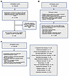 Figure 1
Flowchart to establish UK Biobank cohorts. (A) UK Biobank (UKBB) PH cohort. (B) UKBB control cohort. (C) UKBB comparator cohorts. List of diseases excluded from the control cohort is available in Supplemental Figure 1, and University of Pennsylvania Medical Center (UPMC) patient enrollment scheme is shown in Supplemental Figure 2. Sets of ICD-9/10 codes used to define each cohort are shown in Supplemental Table 1. T1-weighted (T1w) brain MRI.
The UKBB PH cohort had a median age of 68 years (IQR 61, 73); 44% were female, 41% had a college degree, and 50% had ever smoked. Comorbidities included essential hypertension (26.4%), ischemic or hemorrhagic stroke (1.6%), and diabetes with or without complications (7.5%), with a median BMI of 27.0 (IQR 25.18, 30.62) (Supplemental Table 2). The median time from PH diagnosis to MRI was 7.2 years (IQR 3, 13). Although precise WSPH group assignment was not possible in UKBB, prior ICD diagnoses of LHD, chronic obstructive pulmonary disease (COPD), or interstitial lung disease (ILD) were used to identify likely group 2–3 PH–related disease backgrounds. The blood sampling–MRI interval was similar between PH patients and controls [9.71 years (IQR 8.2, 10.9) vs. 9.34 years (IQR 7.75, 10.53)]. Notably, only 1 participant had a documented erectile dysfunction diagnosis (ICD-10: N48.4), suggesting that any vasodilator exposure in this group most likely reflects PAH-directed therapy rather than treatment for erectile dysfunction.
The UKBB control cohort (n = 11,656) was derived by excluding individuals with physician-diagnosed chronic disease, PH, chronic cardiopulmonary diseases listed in Figure 1C, or ICD codes indicating chronic infection or inflammation (Figure 1B and Supplemental Figure 1). The remaining cohort represented generally healthy controls with no known chronic disease and low prevalence of substance use disorder diagnoses (2%). Controls had a median age of 63 years (IQR 57, 69), 54% were female, 47% had a college degree, 51% had ever smoked, and median BMI was 25.5 (IQR 23.1, 27.6) (Supplemental Table 2A). Because more than 95% of both PH and control cohorts were White British (UKBB Data-Field 21000), individuals of other ethnic backgrounds were excluded to reduce population stratification and potential confounding.
The single-center, prospectively recruited UPMC validation cohort included 10 patients with PAH and 10 age-, sex-, and education-matched non-PAH healthy controls for T1/T2-weighted MRI and resting-state functional MRI comparisons (Supplemental Figure 2 and Supplemental Table 3). This focused independent validation cohort prioritized clinical homogeneity of idiopathic PAH over sample size, with stringent eligibility criteria selecting mild-to-moderate disease severity (NYHA Functional Class II–III), preserved hemodynamics, and MRI compatibility. Participants were European Caucasian and predominantly female (80%), with similar median age and education between PAH and controls (40 vs. 39 years; 15 vs. 16 years). No participants carried a diagnosis or were treated for erectile dysfunction. The median time from PAH diagnosis to MRI was 3.7 years (IQR 2.4, 7.2), compared with 7.2 years in UKBB (Supplemental Table 2B). In contrast with the >9-year blood sampling–MRI interval in UKBB, blood draw and MRI were nearly contemporaneous in UPMC: 8 of 10 PAH patients and 7 of 10 controls had same-day sampling and imaging, with only minor deviations in the remaining participants. Thus, UKBB and UPMC provided complementary information on PH-associated brain changes and their blood biomarker associations across different sampling intervals.
Specific structural and functional connectivity alterations observed in PH/PAH across the left cerebellum/superior frontal gyrus axis. To identify brain regions affected structurally and functionally by PH, we analyzed T1-weighted MRI (T1w-MRI) and resting-state functional MRI (rfMRI) data using covariate-adjusted ordinary lease squares (OLS) regression, with Benjamini-Hochberg correction for multiple comparisons (Supplemental Methods and Supplemental Table 2). Among 424 T1w-MRI–derived phenotypes, 5 showed significant reductions in the UKBB PH cohort compared with healthy controls: left superior frontal gyrus thickness (L-SFG; –2.08%, Padj = 0.035), left precentral gyrus thickness (L-PG; –2.55%, Padj = 0.035), left paracentral lobule thickness (L-PL; –2.83%, Padj = 0.035), right caudal middle frontal gyrus thickness (R-CMF; –1.83%, Padj = 0.039), and left cerebellar white matter volume (L-CRBL-WM; –4.39%, Padj = 0.049) (Figure 2A and Supplemental Table 4).
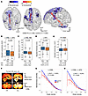 Figure 2
Brain structural and functional connectivity changes in UKBB PH. (A) UK Biobank (UKBB) T1-weighted (T1w) MRI and resting-state functional MRI showing PH-associated changes in brain volume, cortical thickness, and functional connectivity. Percentage differences reflect raw group means; significance was assessed by covariate-adjusted ordinary least squares (OLS) regression with Benjamini-Hochberg correction (Padj ≤ 0.05; Supplemental Table 4). Views: ventral, dorsal, and left lateral. CRBL-WM (cerebellar white matter), SFG (superior frontal gyrus), PG (precentral gyrus), PL (paracentral lobule), CMF (caudal middle frontal gyrus). (B–D) Covariate-adjusted L-CRBL–L-SFG connectivity, L-CRBL-WM volume, and L-SFG thickness in UKBB PH and controls, shown as OLS residuals with the group effect reintroduced. Significance was assessed by OLS regression with Benjamini-Hochberg correction: median (IQR), mean ± SEM underneath; n = 248 cases, n = 11,656 controls. (E) Covariate-adjusted perivascular white matter hyperintensity (PV WMH) from T2-weighted (T2w) FLAIR MRI: median (IQR), mean ± SEM underneath; n = 228 cases, n = 11,242 controls. (F) Representative T2w FLAIR images showing CRBL-WM WMH. (G and H) Global betweenness centrality across graph density thresholds for L-CRBL and L-CRBL–L-SFG; group differences were tested using AUC by OLS regression with permutation-derived P values (mean ± bootstrapped 95% CI; n = 193 cases, n = 10,242 controls).
To assess functional correlates of these structural deficits, we quantified resting-state functional connectivity among the affected regions by rfMRI. Because blood oxygen level–dependent signal generally reflects neuronal postsynaptic activity (43–45), the entire left cerebellum, rather than only cerebellar white matter, was used for connectivity analysis. PH was associated with significantly reduced L-CRBL–L-SFG connectivity (AUC = –22.8%, Padj = 0.03) (Figure 2, A and B, and Supplemental Table 4), along with reduced covariate-adjusted L-CRBL-WM volume and L-SFG thickness (Figure 2, C and D, and Supplemental Table 4). White matter hyperintensity was also increased in PH (5.6%, Padj = 0.046), including in the cerebellum, consistent with cerebellar white matter injury (Figure 2, E and F). Global connectivity analysis also showed reduced betweenness centrality in L-CRBL and the L-CRBL–L-SFG connection, indicating diminished network hub function and information flow in PH. Specifically, PH was associated with reduced L-CRBL regional betweenness centrality (AUC = –13.8%, P = 0.003) and lower L-CRBL–L-SFG edge betweenness centrality (AUC = –10.9%, P = 0.025) (Figure 2, G and H).
Reduced right ventricular (RV) ejection fraction (<45%) was observed in only 16 of 232 PH participants with cardiac MRI data (6.9%), consistent with the mild-to-moderate disease severity represented in UKBB (40, 41). L-CRBL–L-SFG abnormalities were not significantly associated with RV systolic volume, RV dilation, or overall cardiac MRI-derived RV function (Supplemental Table 5). Similarly, no association was observed with SNPs carrying known genome-wide associations with PAH risk (46), including rs10103692, rs2856830, rs13266183, or available linked tag SNPs (R2 > 0.8) in UKBB (Supplemental Table 6). Pathogenic BMPR2 mutations were too infrequent for meaningful association analysis.
Validation analysis in the UPMC PAH cohort focused on brain regions significantly altered in the UKBB PH cohort. UPMC patients with PAH showed replicated reductions in L-SFG thickness (–3.9%, P = 0.005), L-CRBL-WM volume (–0.15%, P = 0.048), and L-CRBL–L-SFG functional connectivity (AUC = –39.4%, P = 0.017) (Figure 3, A–D, and Supplemental Table 7). Because all UPMC patients with PAH were receiving PAH-directed vasodilator therapy, treatment effects could not be isolated within this cohort, and future longitudinal studies of treatment-naive patients with PAH before and after therapy initiation will be needed. Nonetheless, replication of these abnormalities in a clinically phenotyped PAH cohort supports the interpretation that they are not explained by vasodilator exposure alone. Together, these findings demonstrate consistent structural and functional deficits within the L-CRBL–L-SFG axis across discovery and validation cohorts, spanning median diagnosis-to-MRI intervals of 3.7–9.7 years.
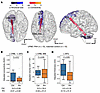 Figure 3
Brain structural and functional connectivity changes in UPMC PAH. (A) University of Pennsylvania Medical Center (UPMC) T1-weighted MRI (T1w-MRI) and resting-state functional MRI showing PAH-associated changes in brain volume, cortical thickness, and functional connectivity. Percentage differences reflect raw group means; significance was assessed by covariate-adjusted ordinary least squares (OLS) regression with permutation-derived P values (P ≤ 0.05; Supplemental Table 7). Views: ventral, dorsal, and left lateral. CRBL-WM, cerebellar white matter; SFG, superior frontal gyrus. (B–D) Covariate-adjusted L-CRBL–L-SFG connectivity, L-CRBL-WM volume, and L-SFG thickness: median (IQR), mean ± SEM underneath; n = 10 cases, n = 10 matched controls.
We next tested whether cognitive function was reduced in PH. In UKBB, all patients with PH from the initial assessment visit were analyzed, including those without MRI. PH was associated with impaired fluid intelligence, reflecting abstract reasoning, problem-solving, and working memory (Data-Field 20016; β = –0.222, Padj = 0.037), and slower matching performance, reflecting visual attention and processing speed (Data-Field 20023; β = 5.705, Padj = 0.046) (Supplemental Table 8). In the UPMC cohort, NIH Toolbox testing showed that patients with PAH performed worse on the Flanker inhibition test, reflecting interference control and executive function (paired 2-tailed t = –12.621, P = 0.009) (47, 48), and the Pattern Comparison test, reflecting processing speed and working memory (paired t = –12.962, P = 0.004) (Supplemental Table 8) (48). Thus, both cohorts showed PH-associated deficits in reasoning, executive function, and processing speed, consistent with prior reports of cognitive impairment in PAH and with the roles of frontal and cerebellar circuits in memory and executive function (21, 25, 49).
Deficits in the L-CRBL–L-SFG axis are specific for PH. We next assessed whether L-CRBL–L-SFG deficits could be explained by hemodynamic compromise or hypoxia. In UKBB, available cardiac and blood pressure measures showed only minimal group differences despite some statistically significant findings (Supplemental Table 2A). LV cardiac output, overall cardiac output, and ventricular rate were only modestly elevated in PH, whereas central systolic blood pressure did not differ significantly. These values remained within ranges expected for relatively mild-to-moderate disease and were unlikely to explain the observed brain abnormalities. Similar findings were observed in the UPMC validation cohort, where pulse oximetry values were largely normal (92%–99%) and elevated pulmonary arterial systolic pressures confirmed PAH without evidence of sustained hypoxemia or overt hemodynamic compromise (Supplemental Table 2B and Supplemental Table 3). Together, these physiologic profiles suggest that PH-associated L-CRBL–L-SFG abnormalities are not readily explained by hypoxia, systemic hypotension, or impaired cardiac output alone.
Cardiac hemodynamic disturbances, including reduced cardiac output or systemic hypotension from therapeutic vasodilators, can cause cerebral hypoperfusion and affect brain structure and function. Because PH therapy has also been linked to cognitive changes (20), we examined whether oral vasodilator treatment (UOVT) might contribute to the observed brain alterations. UKBB medication records do not distinguish vasodilator indication or continuous versus intermittent use; therefore, we evaluated a non-PH comparator cohort defined by UOVT exposure alone. Although this cohort was entirely male, it was not defined solely by erectile dysfunction and included diagnoses compatible with other vasodilator indications, including Raynaud’s-related, autoimmune, or vascular conditions — suggesting that vasodilators could have been prescribed for either intermittent or chronic use. The non-PH UOVT cohort showed no significant changes in brain volume or functional connectivity (Figure 4A and Supplemental Table 9), arguing against generalized vasodilator exposure alone as a sufficient explanation for the L-CRBL–L-SFG abnormalities in PH or PAH.
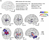 Figure 4
Brain structural and functional connectivity changes in UKBB comparator cohorts. (A) By T1-weighted (T1w) MRI and resting state functional MRI (rfMRI) in the UK Biobank (UKBB) population, when comparing patients with a range of chronic illnesses to a healthy control cohort (n = 11,656), no significant changes in volume and cortical thickness or functional connectivity were observed across any brain regions. In contrast, as compared with healthy controls, patients with specific conditions displayed significant changes: (B) HIV (n = 35). (C) COPD (n = 359). Left: ventral view, middle: dorsal view, right: left lateral view. VDC, ventral diencephalon; THA, thalamus; PAL, pallidum; HIP, hippocampus; CRBL, cerebellum; WM, white matter; BS, brain stem; AMYG, amygdala. Percentage differences reflect raw group means. Significance cutoff, Padj ≤ 0.05; NS, Padj > 0.05, by ordinary least squares (OLS) regression adjusted by Benjamini-Hochberg method (see also Supplemental Table 10).
We next identified non-PH UKBB comparator cohorts with comorbidities associated with major PH groups to test whether the PH brain phenotype was reproduced by related conditions (Figure 1C and Supplemental Table 9). These included group 1–associated conditions, including congenital heart disease (CHD), autoimmune diseases, and infection by HIV (HIV); group 2–associated LHD; group 3–associated COPD, ILD, and sleep disordered breathing; and group 5–associated metabolic disease and renal failure. Deep venous thrombosis and pulmonary embolism (group 4) and hematologic disorders (group 5) were considered but excluded because PH-free sample sizes were too small (n < 10), primarily because many such individuals also carried a PH diagnosis.
Seven of 9 comparator cohorts showed no significant brain alterations (Figure 4A and Supplemental Table 10). Significant structural changes were observed only in HIV and COPD. HIV was associated with reduced volumes of bilateral ventral diencephalon, right thalamus, pallidum, and hippocampus (Figure 4B and Supplemental Table 10). COPD showed broader reductions involving bilateral ventral diencephalon, thalami, hippocampi, cerebellar white matter, left cerebellum, left amygdala, and brain stem, with several reduced functional connectivity interactions among these regions (Figure 4C and Supplemental Table 10). However, neither COPD nor other major cardiopulmonary comparator cohorts, including LHD and ILD, reproduced the combined L-CRBL–L-SFG structural and functional deficiency pattern observed in PH. To further assess potential confounding by LHD or chronic hypoxic lung disease within the PH cohort, we adjusted for prior LHD, COPD, or ILD diagnosed ≥6 months before PH diagnosis (28/248, 11.3%). This adjustment did not materially change the PH associations, and L-CRBL–L-SFG abnormalities remained significant with the same direction of effect. A broader definition including LHD/COPD/ILD diagnoses any time on or before PH diagnosis (42/248, 16.9%) yielded consistent results: the L-CRBL-WM volume association became borderline (Padj = 0.053) with essentially unchanged effect size (β = −408.73 vs. −408.52), while the remaining L-CRBL–L-SFG structural and functional associations were preserved. These findings support that the PH cerebellar–frontal phenotype was not reproduced in the comparator cohorts tested and was not explained by preceding LHD, COPD, or ILD alone.
Because patients with PH may carry potentially confounding cardiovascular, endocrine, and neuropsychiatric diagnoses, including hypertension, diabetes, anxiety, and depression (50), we performed the same structural and functional connectivity analyses in non-PH UKBB cohorts with these conditions (Figure 1C and Supplemental Table 9). Significant brain volume changes were observed in hypertensive disorders (HD), diabetes mellitus (DM), and depression disorders (DD). HD showed increased right choroid plexus volume and reduced right accumbens volume (Figure 5A and Supplemental Table 10). DM showed broader structural alterations, including increased bilateral choroid plexus volume and reduced volumes or thicknesses across ventral diencephalon, parietal, frontal, thalamic, and related cortical regions, with multiple reduced functional connectivity interactions among these regions (Figure 5B and Supplemental Table 10). DD was associated with reduced bilateral hippocampal volumes (Figure 5C and Supplemental Table 10).
 Figure 5
Brain structural and functional connectivity changes in UKBB potential confounding disease cohorts. (A) Hypertension (n = 46). (B) Diabetes mellitus (n = 1,162). (C) Depression (n = 912). Left: ventral view, middle: dorsal view, right: left lateral view. VDC, ventral diencephalon; THA, thalamus; PAL, pallidum; HIP, hippocampus; CHP, choroid plexus; ACC, accumbens; SPL, superior parietal lobe; IPL, inferior parietal lobule; SPM, supramarginal gyrus; SFG, superior frontal gyrus; RMFG, rostral middle frontal gyrus; PCNU, precuneus; PG, precentral gyrus; PSA, pars triangularis; PL, paracentral lobule; CMF, caudal middle frontal gyrus. Percentage differences reflect raw group means. Significance cutoff, Padj ≤ 0.05; NS, Padj > 0.05, by ordinary least squares (OLS) regression adjusted by Benjamini-Hochberg method (see also Supplemental Table 10).
Thus, the deficit in the L-CRBL–L-SFG axis was not reproduced in the comparator cohorts tested and was not associated with other comorbidities linked to PH or with vasodilator use independent of PH.
L-CRBL–L-SFG axis deficiencies are associated with plasma biomarkers of inflammation. We next examined plasma biomarker associations with L-CRBL–L-SFG deficiencies to identify molecular pathways potentially underlying these brain alterations. Analyses were performed within the PH cohort to capture PH-specific biomarker–brain relationships rather than case-control differences. Among 30 UKBB blood biochemical markers, 11 were significantly associated with L-CRBL-WM volume or L-SFG thickness (Figure 6A and Supplemental Table 11). IGF-1, testosterone, and hemoglobin A1c (HbA1c) were associated with both regions; urea, cystatin C, urate, SHBG, creatinine, and albumin were associated only with L-CRBL-WM volume; and vitamin D and C-reactive protein (CRP) were associated only with L-SFG thickness. Importantly, 10 of these 11 markers have known links to chronic vascular or neurologic inflammation, including HbA1c and CRP, which have been associated with mild cognitive impairment (MCI) in other contexts (51–53). To test whether these associations reflected metabolic dysregulation alone, we repeated the analysis in non-PH DM participants. In contrast with PH, neither HbA1c nor testosterone was significantly associated with L-CRBL-WM volume or L-SFG thickness, suggesting that these biomarker–brain relationships are not explained by diabetes-related metabolic variation alone.
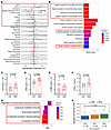 Figure 6
Systemic and cerebellar inflammatory signatures in PAH revealed by blood biomarkers. (A) Forest plot showing 30 blood biochemical markers and their statistical associations with L-CRBL-WM volume and L-SFG thickness among UK Biobank (UKBB) PH participants. X axis indicates standardized regression β-coefficient + CIs. Significant associations are indicated by filled circles. Significance cutoff, Padj ≤ 0.05; NS, Padj > 0.05, by ordinary least squares (OLS) regression using a generalized linear model (GLM) with bootstrapped P values and Benjamini-Hochberg adjustment (original n = 27–239; bootstrap resamples = 1,000; see also Supplemental Table 11). Top red square indicates markers significantly associated with at least 1 of the PH anatomic brain abnormalities. (B) Bar chart showing Gene Ontology Biological Processes (GOBP) categories enriched among the union of significantly negatively associated plasma proteins by Olink plasma proteomic analysis. Significance cutoff, Padj ≤ 0.05; NS, Padj > 0.05, by enrichR and FDR adjustment (see also Supplemental Figure 3 and Supplemental Tables 12 and 13). Red squares indicate immune response/inflammation categories. (C–F) Orthogonal ELISA validation of inflammatory plasma proteins in University of Pennsylvania Medical Center (UPMC) PAH individuals (n = 10) and age- and sex-matched healthy controls (n = 10). Data are mean ± SD. Significance cutoff, P ≤ 0.05; NS, P > 0.05. P values were determined by the Mann-Whitney U test (C) and 2-tailed Welch’s unpaired t tests (D–F). (G) Rank-based GOBP pathway fast gene set enrichment analysis of the rs11154337_G–Olink association in UK Biobank (UKBB) PH participants. Significance cutoff, Padj ≤ 0.05; NS, Padj > 0.05 by FDR adjustment. (H) Covariate-adjusted L-CRBL–L-SFG functional connectivity strength for UKBB PH cohort stratified by rs11154337 SNP allele genotype. Significance cutoff, P ≤ 0.05; NS, P > 0.05, by ordinary least squares (OLS) regress ion: median (IQR); n = 40 C/C, n = 94 G/C, n = 42 G/G alleles.
We next analyzed UKBB plasma proteomic data from the Olink platform. Among 2,923 circulating proteins, multivariate analysis identified 30 proteins associated with L-CRBL-WM volume, 11 with L-SFG thickness, and 13 with L-CRBL–L-SFG connectivity (Supplemental Figure 3 and Supplemental Table 12). Gene Ontology Biological Processes (GOBP) enrichment of inversely associated proteins, representing higher expression with greater L-CRBL–L-SFG deficiency, revealed immune-response pathways across all 3 analyses (Figure 6B and Supplemental Table 13). Notably, top enriched categories included microglial activation and neuroinflammatory responses, with GRN, PTPRC, and CX3CL1 highlighting abnormal microglial states as part of the L-CRBL–L-SFG inflammatory signature. When inversely and positively associated proteins were pooled, additional pathways related to chemokine response, cytokine signaling, cell proliferation, immune-cell chemotaxis, and Wnt signaling were identified (Supplemental Table 13). To assess whether hypoxic mechanisms contributed to these associations, we analyzed 66 curated hypoxia-related Olink proteins (Supplemental Table 14); none showed significant association, contrasting with the enrichment of immune and inflammatory proteins.
Although precise WSPH classification was not feasible in UKBB, we examined whether plasma biomarker associations were detectable within PH strata, including the more prevalent LHD-associated PH subgroup. PH participants were stratified by LHD diagnosis before or at PH diagnosis and analyzed using identical regression frameworks. Routine biochemical markers, including urea, cystatin C, creatinine, urate, HbA1c, CRP, lipid-related markers, and vitamin D, showed significant associations with PH in the same direction in both LHD-associated and non-LHD PH strata (Supplemental Table 15). Similarly, 459 Olink proteins were significantly associated with PH in both strata, supporting shared inflammatory biomarker patterns across LHD-associated and non-LHD PH contexts (Supplemental Table 15).
To independently validate key inflammatory proteins, we selected significant Olink proteins with commercial ELISA kits (CFD, GRN, C1QBP, and CX3CL1) and measured plasma levels by ELISA in the UPMC cohort (Figure 6, C–F). CFD (P = 0.0232), GRN (P = 0.0082), and CX3CL1 (P = 0.0247) were significantly elevated in PAH compared with controls, whereas C1QBP was not significantly different (P = 0.4904). Together, UKBB and UPMC data support a robust circulating inflammatory signature across PH contexts, including PAH and LHD-associated PH, despite differences in blood sampling timing relative to brain MRI. These associations suggest that persistent inflammatory signaling may contribute to the structural and functional brain deficiencies observed in PH.
Intronic NCOA7 SNP rs11154337 is associated with the L-CRBL–L-SFG connectivity strength. We recently identified the NCOA7–lysosomal–oxysterol axis as a regulator of pulmonary vascular inflammation and mortality in PAH, with the rs11154337 G-allele conferring protection by increasing NCOA7 expression and reducing oxysterol-induced inflammation (39). Because lysosomal dysfunction and oxysterol production are also linked to neuroinflammation and neurodegeneration (37, 38), we hypothesized that NCOA7 deficiency may represent a shared inflammatory mechanism across pulmonary vascular and cerebellar compartments. In UKBB patients with PH, proteins negatively associated with rs11154337_G showed pathway-level enrichment for systemic inflammatory programs, including complement activation, classical complement activation, acute-phase response, and lymphocyte-mediated immunity, whereas regulation of mitochondrial membrane potential was enriched in the positive direction (Figure 6G). Cardiopulmonary comparator groups, including LHD, COPD, and ILD, did not show a fully comparable PH-like inflammatory pattern (Supplemental Table 16), and only in DM were other inflammatory pathways significantly associated with rs11154337_G. Taken together, these data support an association between rs11154337 and systemic immune/inflammatory biology that is primarily represented in PH.
We next tested whether rs11154337 was associated with preservation of the L-CRBL–L-SFG axis. In UKBB patients with PH stratified by genotype [C/C (n = 40), G/C (n = 94), G/G (n = 42)], the G-allele was not significantly associated with L-CRBL-WM volume or L-SFG size but was associated with improved L-CRBL–L-SFG connectivity under an additive model (β = 0.020, P = 0.035), with connectivity increasing stepwise by G-allele dosage (Figure 6H). This association was not observed in comparator populations, except for L-CRBL-WM volume in a small CHD cohort with limited power (n = 22; Supplemental Table 17). Together, these Olink–genotype and imaging–genotype findings link NCOA7-related inflammatory biology to preservation of cerebellar–frontal connectivity in PH.
PAH cerebellar tissue shows increased microglial abundance and multicellular induction of NCOA7. To define inflammatory changes and NCOA7 involvement in PAH cerebellum, we performed immunofluorescence staining of postmortem cerebellar tissue from patients with PAH and healthy controls (Supplemental Table 18). Ionized calcium-binding adapter molecule 1 (IBA1) staining showed significantly increased IBA1+ microglia in both molecular and granular layers of PAH cerebellum (Figure 7, A–E). Co-immunofluorescence further showed increased NCOA7 signal within IBA1+ microglia in both layers (Figure 7, F–I), as well as in CALBINDIN+ Purkinje neurons (Figure 7, J and K) and NEUN+ granule neurons (Figure 7, L and M), but not in PARVALBUMIN+ molecular layer neurons (Supplemental Figure 4, A and B). NCOA7 was also expressed in CD31+ endothelial cells but was not detected at high levels in GFAP+ astrocytes or MBP+ ODCs (Supplemental Figure 4, A–E). Consistent with increased sterol hydroxylation and downstream vascular inflammation controlled by NCOA7 in PAH (39), CH25H was also significantly upregulated in PAH cerebellum (Figure 7, N and O). Together, these findings indicate that PAH cerebellum is characterized by increased microglial abundance and cell type–selective NCOA7 protein induction across microglial and neuronal compartments. Such upregulation is consistent with our observations in human diseased pulmonary vasculature in PAH (39), where increased NCOA7 ensures appropriate lysosomal function and prevents overexuberant inflammation. This dependence on inducible levels of NCOA7 thus identified these cells as those most likely to be vulnerable in contexts of NCOA7 deficiency.
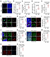 Figure 7
Immunofluorescence analysis of microglia and NCOA7 expression in postmortem human PAH cerebellum. (A–E) Ionized calcium-binding adapter molecule 1 (IBA1) immunofluorescence and quantification of IBA1+ microglia in postmortem human cerebellar tissue from control and PAH individuals. (F–I) Co-immunofluorescence of IBA1 and NCOA7, with quantification of NCOA7 within IBA1+ microglia in the molecular and granular layers. White arrows indicate NCOA7 expression in IBA1+ microglia. (J and K) Co-immunofluorescence and quantification of NCOA7 within CALBINDIN+ Purkinje neurons. White arrows indicate NCOA7 expression in CALBINDIN+ Purkinje neurons. (L and M) Co-immunofluorescence and quantification of NCOA7 within NEUN+ granule neurons. (N and O) CH25H immunofluorescence and quantification in the molecular layer. Data are mean ± SD. For A–M, n = 3 controls and n = 5 PAH cases; for N and O, n = 3 controls and n = 3 PAH cases. Significance cutoff, P ≤ 0.05; NS, P > 0.05, by 2-tailed Welch’s unpaired t test. Scale bars: 50 μm (A, J, and L), 25 μm (F and H). ML, molecular layer; PL, Purkinje layer; GL, granular layer.
An inflammatory PH mouse model recapitulates cerebellar volume loss, microglial expansion, and multicellular NCOA7 induction. To determine whether the cerebellar abnormalities observed in human PAH were reproduced in an experimental PH model, we used an Il6-Tg mouse model that recapitulates inflammatory PH without confounding from direct ischemic or toxic brain injury, such as monocrotaline exposure (54). Co-immunofluorescence showed increased NCOA7 signal within IBA1+ microglia in both layers (Figure 8, A–D), as well as in CALBINDIN+ Purkinje neurons (Figure 8, E and F), NEUN+ granule neurons (Figure 8, G and H), and PARVALBUMIN+ molecular layer neurons (Figure 8, I and J). Together, these findings indicate that the Il6-Tg PH model recapitulates cell type–selective NCOA7 induction across microglial and neuronal compartments observed in human PAH cerebellum.
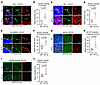 Figure 8
Immunofluorescence analysis of NCOA7 expression in inflammatory PH mouse models. (A–D) Co-immunofluorescence of ionized calcium-binding adapter molecule 1 (IBA1) and NCOA7 with quantification of NCOA7 within IBA1+ microglia in the molecular and granular layers. (E and F) Co-immunofluorescence and quantification of NCOA7 within CALBINDIN+ Purkinje neurons. (G and H) Co-immunofluorescence and quantification of NCOA7 within NEUN+ granule neurons. (I and J) Co-immunofluorescence and quantification of NCOA7 within PARVALBUMIN+ molecular layer neurons. Data are mean ± SD (n = 5 WT and n = 5 Il6-Tg), and P values were determined by 2-tailed Welch’s unpaired t test. Arrows indicate representative cells positive for both markers. Scale bars: 50 μm (E, G, and I) and 25 μm (A and C). ML, molecular layer; PL, Purkinje layer; GL, granular layer.
Next, to investigate mechanisms controlling cerebellar pathology in PH, we used an Ncoa7-knockout Il6 mouse model (Il6-Tg Ncoa7–/–). Because full-length Ncoa7 deletion is constitutive, we assessed whether its loss alone caused developmental brain abnormalities. Prior work reported normal brain development in full-length Ncoa7–/– mice (55), and our MRI analyses similarly showed no reduction in total brain volume (1.8%, P = 0.765) or cerebellar volume (2.1%, P = 0.636) compared with WT mice (Supplemental Figure 5, A and B). We then examined Il6-Tg Ncoa7–/– mice to determine whether Ncoa7 deficiency exacerbated cerebellar pathology in PH (39). Ex vivo T2w-MRI with NeAt atlas-based parcellation (56, 57), adjusted for age and total brain volume, showed reduced cerebellar volume in Il6-Tg mice compared with WT controls (–4.9%, Padj = 0.048; Figure 9A), with a further reduction in Il6-Tg Ncoa7–/– mice compared with Il6-Tg mice (–7.1%, Padj = 0.029; Figure 9A), indicating an interaction between inflammatory PH and Ncoa7 deficiency rather than a primary effect of Ncoa7 loss. Il6-Tg Ncoa7–/– mice also showed greater IBA1 expression in the molecular and granular layers (Figure 9, B–D) compared with Il6-Tg mice, together with increased CH25H expression as a marker of inflammatory activation (Figure 9, E and F). These findings indicate that Ncoa7 modulates cerebellar neuroinflammation and volume loss in PH. To exclude the possibility that Ncoa7 deletion alone causes baseline cardiopulmonary abnormalities that secondarily affect brain structure, we assessed WT and Ncoa7–/– mice by echocardiography and right-heart catheterization. No significant differences were observed in cardiac structure, function, or pulmonary pressures (Supplemental Table 19), indicating that Ncoa7 deletion alone does not induce baseline cardiopulmonary pathology.
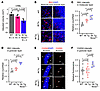 Figure 9
Evaluation of brain structure and histology in an inflammatory PH mouse model with Ncoa7 deficiency. (A) Covariate-adjusted cerebellar volume by ex vivo T2-weighted (T2w) MRI in WT, Il6-Tg, and Il6-Tg Ncoa7–/– mice using ordinary least squares (OLS) regression with permutation-derived P value with Holm’s correction. (B) Representative cerebellar ionized calcium-binding adapter molecule 1 (IBA1) immunofluorescence in WT, Il6-Tg, and Il6-Tg Ncoa7–/– mice. (C and D) Quantification of IBA1 expression in the molecular and granular layers, respectively. (E) Representative cerebellar CH25H immunofluorescence and (F) quantification of CH25H expression in the granular layer. Data are mean ± SEM (A) or mean ± SD (C, D, and F). For A, n = 5 WT, n = 4 Il6-Tg, and n = 4 Il6-Tg Ncoa7–/–; for B–D, n = 5 WT, n = 5 Il6-Tg, and n = 4 Il6-Tg Ncoa7–/–; and for E and F, n = 9 WT, n = 10 Il6-Tg, and n = 4 Il6-Tg Ncoa7–/–. P values for C, D, and F were determined by 1-way ANOVA with post hoc Bonferroni’s testing. Significance cutoff, Padj ≤ 0.05; NS, Padj > 0.05. Scale bars: 50 μm (B and F). ML, molecular layer; PL, Purkinje layer; GL, granular layer; WM, white matter.
NCOA7 regulates microglial lysosomal function and inflammatory activation. To define how NCOA7 deficiency may contribute to neuroinflammation, we built on prior evidence that NCOA7 regulates lysosomal acidification and CH25H-dependent oxysterol–bile acid inflammatory signaling (39). Given our finding of cerebellar microglial NCOA7 upregulation in human and mouse PAH models (Figure 7, F–I, and Figure 8, A–D), we focused on microglia as a candidate effector linking NCOA7 deficiency to neuroinflammation. In the HMC3 cultured human microglial cell line, shRNA-mediated NCOA7 knockdown was confirmed by immunoblotting (Figure 10, A and B) and caused lysosomal dysfunction, including reduced lysosomal acidification, diminished cathepsin D activity, and increased intracellular lipid accumulation (Figure 10, C–K). NCOA7-deficient microglia also showed impaired phagocytosis of pHrodo-labeled synaptosomes (Figure 10, L–N) and increased expression of inflammatory cytokines IL1A, IL1B, IL6, and TNFA and chemokines CCL2 and CXCL10 (Figure 10, O–T). These lysosomal and inflammatory phenotypes were independently reproduced using siRNA-mediated NCOA7 silencing (Supplemental Figure 6). Together, these findings identify NCOA7 as a regulator of microglial lysosomal integrity, phagocytic function, and basal inflammatory activation, providing a mechanistic link between NCOA7 deficiency and PH-associated cerebellar neuroinflammation.
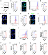 Figure 10
Functional consequences of NCOA7 loss in cultured human microglial cells. (A) Immunoblot of cultured HMC3 cells under shRNA control (shCtrl) and shRNA-mediated NCOA7 knockdown (shNCOA7) conditions, with (B) corresponding densitometric quantification (mean ± SD; n = 4 shCtrl, n = 4 shNCOA7). (C–N) Functional assays in HMC3 cells under shCtrl (n = 3) and shNCOA7 (n = 3) conditions. Representative images and quantification are shown for lysosomal acidification (LysoSensor; C–E), lysosomal protease activity/lysosome signal (SiR-lysosome; F–H), intracellular lipid accumulation (BODIPY; I–K), and phagocytic uptake of pHrodo-labeled brain synaptosomes (L–N). For each assay, panels show representative fluorescence images with DAPI counterstain (C, F, I, and L), flow cytometry histograms (D, G, J, and M), and corresponding quantitative analyses (mean ± SD) (E, H, K, and N). Significance cutoff, P ≤ 0.05; NS, P > 0.05, by 2-tailed Welch’s unpaired t test. (O–T) Relative mRNA expression levels of IL1α, IL1B, IL6, TNFα, CCL2, and CXCL10 in shCtrl- and shNCOA7-treated HMC3 cells (mean ± SD; n = 3 shCtrl, n = 3 shNCOA7). Significance cutoff, P ≤ 0.05; NS, P > 0.05 by 2-tailed Welch’s unpaired t test.
Single-nucleus and spatial transcriptomics reveal region-specific cerebellar vulnerability to PH and Ncoa7 loss. To investigate the cellular and molecular basis of PH-associated cerebellar alterations and the contribution of Ncoa7 deficiency, we performed single-nucleus RNA-seq (snRNA-seq) of cerebellar tissue from WT, Il6-Tg, and Il6-Tg Ncoa7–/– mice. After quality control and reference-based annotation (58), 248,660 nuclei were retained, resolving major cerebellar neuronal, glial, immune, vascular, ventricular, and stromal populations (Figure 11A). Granule neurons and MLIs were further subdivided into Granule_main/Granule_other and MLI1/MLI2, respectively, with cell type assignments supported by canonical marker gene enrichment from the reference cerebellar atlas (Supplemental Figure 7A). No significant changes in cell type composition were detected across the conditions (Supplemental Table 20). Ncoa7 nuclear transcripts were broadly but lowly detected across diverse cell types (Figure 11B and Supplemental Figure 7, B–I). However, unlike quantitative co-immunofluorescence (Figure 8), nuclear Ncoa7 transcript abundance was not significantly increased in PH, likely reflecting the inability of single-nucleus sequencing to distinguish changes in inflammation-responsive isoform usage and cytoplasmic RNA processing previously linked to increased NCOA7 protein expression (39, 59). Thus, snRNA-seq provided a cell type map of Ncoa7 transcript detection and a cerebellar reference for downstream transcriptional reprogramming and spatial transcriptomics deconvolution.
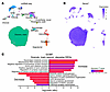 Figure 11
Single-nucleus transcriptomic analyses of PH- and Ncoa7-dependent cerebellar remodeling. (A) Uniform manifold approximation and projection (UMAP) visualization of cerebellar single-nucleus RNA-seq (snRNA-seq) nuclei from WT (n = 3), Il6-Tg (n = 4), and Il6-Tg Ncoa7–/– (n = 4) mice annotated by reference-based label transfer. MLI, molecular layer interneuron; ODC, oligodendrocyte; OPC, oligodendrocyte progenitor cell; PLI, Purkinje layer interneuron; UBC, unipolar brush cell. (B) Feature plot showing Ncoa7 transcript detection across annotated snRNA-seq cell types. (C) Gene Ontology Biological Process (GOBP) enrichment of stepwise differentially expressed genes (DEGs) in Granule_main neurons across Il6-Tg vs. WT and Il6-Tg Ncoa7–/– vs. Il6-Tg comparisons. Significance cutoff, Padj ≤ 0.05; NS, Padj > 0.05 by FDR adjustment.
We then performed cell type–specific differential expression analyses for Il6-Tg versus WT and Il6-Tg Ncoa7–/– versus Il6-Tg, with complete differentially expressed gene (DEG) and gene set enrichment analysis (GSEA) results provided in Supplemental Tables 21 and 22. We focused on DEGs changing in the same direction across both comparisons (stepwise DEGs) in Ncoa7-expressing cell types supported by both snRNA-seq and immunofluorescence: granule neurons, MLIs, Purkinje cells, and microglia (Supplemental Tables 23 and 24). In Granule_main neurons, stepwise increased DEGs were enriched in pathways of mitochondrial RNA metabolism/processing, synapse organization, axon extension, lipid/glycerolipid metabolism, and intracellular protein transport, while decreased DEGs involved dendritic spine morphogenesis, neuron projection/axon development, and synaptic regulation (Figure 11C). Kyoto Encyclopedia of Genes and Genomes analysis additionally identified circadian rhythm among stepwise decreased DEGs (Supplemental Figure 7J). Together, these findings indicate progressive disruption of granule neuron neurite/synaptic regulatory programs in PH, exacerbated with Ncoa7 deficiency, accompanied by metabolic, mitochondrial RNA–processing, and synaptic responses, consistent with the known role of NCOA7 in sterol and lipid regulations (39). MLI1 showed more restricted enrichment for glutamate receptor signaling and chemical synaptic transmission, whereas Purkinje cell and microglial analyses were limited by lower cell recovery and did not yield significant stepwise DEG-GSEA enrichment (Supplemental Table 24).
Next, spatial transcriptomic analyses were conducted to map PH- and Ncoa7 deficiency–associated programs within defined cerebellar microenvironments. This approach enabled transcriptional changes to be localized to anatomical domains enriched for less abundant populations, including Purkinje neurons and microglia (60). Unbiased clustering identified 6 anatomically consistent cerebellar regions (Figure 12A), and integration with the snRNA-seq reference supported these annotations by defining dominant cell type composition and selectively enriched signatures across clusters (Supplemental Figure 7, K and L). Cluster 0 (C0) corresponded to the granule cell layer, cluster 1 (C1) to a molecular layer/interneuron-enriched region, cluster 2 (C2) to the Purkinje cell layer, cluster 3 (C3) to ODC-enriched white matter, cluster 4 (C4) to a choroid plexus-associated ventricular region, and cluster 5 (C5) to a fourth ventricle–adjacent periventricular white matter/medullary velum border. No significant changes in predicted cell fraction were detected (Supplemental Table 25).
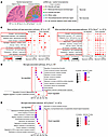 Figure 12
Spatial transcriptomic analyses of PH- and Ncoa7-dependent cerebellar remodeling. (A) Spatial transcriptomic captured image and unbiased clustering of mouse cerebellar sections. Subsequent cell type annotation of each cluster was carried out by integrating the snRNA-seq reference (see Supplemental Methods) (n = 5 WT, n = 5 Il6-Tg, and n = 3 Il6-Tg Ncoa7–/–). (B and C) Most enriched cell type–targeted spatial DEG-GSEA within each spatial cluster for Il6-Tg vs. WT and Il6-Tg Ncoa7–/– vs. Il6-Tg, respectively, after adjustment for other local cell type signals; pathways were grouped into biological supercategories. Red squares indicate pathways also enriched in snRNA-seq Granule_main neurons. (D and E) Microglia-targeted spatial DEG-GSEA for Il6-Tg vs. WT and Il6-Tg Ncoa7–/– vs. Il6-Tg, respectively, showing selected up- or downregulated pathways retained after removal of pathways identified in the corresponding most enriched cell type–targeted analysis. For all analyses, significance was defined as FDR-adjusted Padj ≤ 0.05; NS, Padj > 0.05. Cluster labels are abbreviated C0–C5.
We next performed most enriched cell type–targeted DEG-GSEA within each spatial cluster while adjusting for other local cell type signals (Supplemental Tables 26 and 27), enabling identification of programs associated with the principal anatomical/cellular signature of each cerebellar region. In neuron-enriched cortical layers (i.e., C0–2), Il6-Tg versus WT showed upregulated apoptosis/cell death, oxidative stress, lipid/metabolic stress, synaptic/neuronal projection, and proteostasis-related programs, together with downregulated mitochondrial respiration/ATP metabolism, ion transport/excitability, calcium signaling, and neuronal/synaptic regulatory pathways (Figure 12B). Such enrichment was also observed in pathways highlighted by snRNA-seq analysis alone in Granule_main neurons (Figure 11C), suggesting the consistency between the single nuclear and spatial datasets. In Il6-Tg Ncoa7–/– versus Il6-Tg, these changes became broader and with increased lipid/stress, inflammatory, and apoptotic pathway engagement across C0–C2 (Figure 12C). Together, these findings support the MRI and histologic data by showing that PH is associated with anatomically distributed cerebellar transcriptional remodeling, while Ncoa7 deficiency further shifts neuron-enriched compartments toward metabolic and neuronal dysfunction with amplified inflammatory and cell death signaling.
Disease-associated remodeling extended beyond neuron-enriched cortical layers. In particular, under Il6-Tg conditions, C3 showed downregulation of axonal and myelin homeostatic programs, including axonogenesis, axon development, and regulation of supramolecular fiber organization (Figure 12B and Supplemental Table 27). Altered regulation of neuronal apoptotic processes further suggested cell death–associated vulnerability within this compartment. In Il6-Tg Ncoa7–/– mice, this white matter–associated disruption was more pronounced, with reduced aerobic respiration, oxidative phosphorylation, and proteasomal protein catabolism, along with increased apoptotic programs (Figure 12C and Supplemental Table 27). These findings support the notion that chronic neuroinflammation promotes white matter injury and brain volume loss by disrupting glial survival, myelin maintenance, axonal integrity, and lysosomal-autophagic protein/lipid handling (61–65). Together, these data provide a molecular context for MRI-detected cerebellar white matter volume loss in PH, potentially via disrupted axonal and myelin support.
Finally, we examined microglia-associated signals within these spatial domains beyond those captured by the principal anatomical or cellular signatures. Microglia-targeted DEG-GSEA was performed within each spatial cluster after removing pathways enriched in the corresponding most enriched cell type–targeted analysis (Supplemental Tables 28 and 29). In Il6-Tg versus WT, retained microglia-associated programs involved cytokine production, protein-complex organization, protein transport, ubiquitin/proteasome-mediated catabolism, ERAD/unfolded protein response, calcium homeostasis, and synaptic signaling-related processes (Figure 12D). Il6-Tg Ncoa7–/– versus Il6-Tg further involved IL-6 signaling, lysosomal targeting, autophagy, ER-to-Golgi/vesicle transport, oxidative and chemical stress responses, phospholipid/membrane-raft biology, and apoptosis (Figure 12E). These pathways converged on functional axes identified by NCOA7 knockdown in vitro, including lysosomal/autophagic regulation, lipid and membrane metabolism, proteostasis/ER stress, oxidative stress, inflammatory signaling, and synaptic-material handling (Figure 10). Based on this cluster-specific distribution, Ncoa7 deficiency promoted a regionally dependent microglial stress response program, rather than a globally uniform cerebellar response. Taken together, across PH and exacerbated by Ncoa7 deficiency, these findings defined a landscape of cell type–specific neuronal and microglial transcriptional reprogramming involving lysosomal and inflammatory cerebellar pathobiology.


Copyright © 2026 American Society for Clinical Investigation
ISSN: 0021-9738 (print), 1558-8238 (online)