
Advertisement
Research ArticleAIDS/HIVImmunologyOncology
Open Access |  10.1172/JCI194449
TRAIL splice variant TRAILshort disrupts T cell receptor signaling and promotes immune tolerance in vivo
Shahrzad Jalali,1 Sekar Natesampillai,1 Zilin Nie,1 Ying Zhang,1 Ismail Can,2 Aswath P. Chandrasekar,1,3 Brianna M. Hameister,1 Cristina Correia,4 Tuantuan V. Zhao,5 Dong-Gi Mun,3 Enrique Garcia-Rivera,6 Robert Matson,7 Ashton Krogman,1 Mark A. Maynes,1 Robin Batchelor,1 Dileep D. Monie,4,8 Hu Li,4 Atta Behfar,9 Saad S. Kenderian,2,10 Akhilesh Pandey,3,11,12 Stephen M. Ansell,2 Timucin Taner,13,14 Cornelia Weyand,3 Daniel D. Billadeau,13 and Andrew D. Badley1,10
1Division of Infectious Diseases,
2Division of Hematology,
3Department of Laboratory Medicine and Pathology, and
4Molecular Pharmacology and Experimental Therapeutics, Mayo Clinic, Rochester, Minnesota, USA.
5Department of Medicine, Mayo Clinic College of Medicine and Science, Rochester, Minnesota, USA.
6Formation Bio, New York, New York, USA.
7nference Inc., Cambridge, Massachusetts, USA.
8Department of Neurological Surgery, Albert Einstein College of Medicine-Montefiore Medical Center, Bronx, New York, USA.
9Department of Cardiovascular Medicine,
10Department of Molecular Medicine, and
11Center for Individualized Medicine, Mayo Clinic, Rochester, Minnesota, USA.
12Manipal Academy of Higher Education, Manipal, Karnataka, India.
13Department of Immunology, and
14Department of Surgery, Mayo Clinic, Rochester, Minnesota, USA.
Address correspondence to: Andrew D. Badley, Division of Infectious Diseases and Department of Molecular Medicine, Room 5-20B Guggenheim Building, Mayo Clinic, 200 First Street SW, Rochester, Minnesota 55902 USA. Email: badley.andrew@mayo.edu.
Authorship note: SJ, SN, and ZN have been designated as co–first authors.
Find articles by Jalali, S. in: PubMed | Google Scholar
1Division of Infectious Diseases,
2Division of Hematology,
3Department of Laboratory Medicine and Pathology, and
4Molecular Pharmacology and Experimental Therapeutics, Mayo Clinic, Rochester, Minnesota, USA.
5Department of Medicine, Mayo Clinic College of Medicine and Science, Rochester, Minnesota, USA.
6Formation Bio, New York, New York, USA.
7nference Inc., Cambridge, Massachusetts, USA.
8Department of Neurological Surgery, Albert Einstein College of Medicine-Montefiore Medical Center, Bronx, New York, USA.
9Department of Cardiovascular Medicine,
10Department of Molecular Medicine, and
11Center for Individualized Medicine, Mayo Clinic, Rochester, Minnesota, USA.
12Manipal Academy of Higher Education, Manipal, Karnataka, India.
13Department of Immunology, and
14Department of Surgery, Mayo Clinic, Rochester, Minnesota, USA.
Address correspondence to: Andrew D. Badley, Division of Infectious Diseases and Department of Molecular Medicine, Room 5-20B Guggenheim Building, Mayo Clinic, 200 First Street SW, Rochester, Minnesota 55902 USA. Email: badley.andrew@mayo.edu.
Authorship note: SJ, SN, and ZN have been designated as co–first authors.
Find articles by Natesampillai, S. in: PubMed | Google Scholar
1Division of Infectious Diseases,
2Division of Hematology,
3Department of Laboratory Medicine and Pathology, and
4Molecular Pharmacology and Experimental Therapeutics, Mayo Clinic, Rochester, Minnesota, USA.
5Department of Medicine, Mayo Clinic College of Medicine and Science, Rochester, Minnesota, USA.
6Formation Bio, New York, New York, USA.
7nference Inc., Cambridge, Massachusetts, USA.
8Department of Neurological Surgery, Albert Einstein College of Medicine-Montefiore Medical Center, Bronx, New York, USA.
9Department of Cardiovascular Medicine,
10Department of Molecular Medicine, and
11Center for Individualized Medicine, Mayo Clinic, Rochester, Minnesota, USA.
12Manipal Academy of Higher Education, Manipal, Karnataka, India.
13Department of Immunology, and
14Department of Surgery, Mayo Clinic, Rochester, Minnesota, USA.
Address correspondence to: Andrew D. Badley, Division of Infectious Diseases and Department of Molecular Medicine, Room 5-20B Guggenheim Building, Mayo Clinic, 200 First Street SW, Rochester, Minnesota 55902 USA. Email: badley.andrew@mayo.edu.
Authorship note: SJ, SN, and ZN have been designated as co–first authors.
Find articles by Nie, Z. in: PubMed | Google Scholar
1Division of Infectious Diseases,
2Division of Hematology,
3Department of Laboratory Medicine and Pathology, and
4Molecular Pharmacology and Experimental Therapeutics, Mayo Clinic, Rochester, Minnesota, USA.
5Department of Medicine, Mayo Clinic College of Medicine and Science, Rochester, Minnesota, USA.
6Formation Bio, New York, New York, USA.
7nference Inc., Cambridge, Massachusetts, USA.
8Department of Neurological Surgery, Albert Einstein College of Medicine-Montefiore Medical Center, Bronx, New York, USA.
9Department of Cardiovascular Medicine,
10Department of Molecular Medicine, and
11Center for Individualized Medicine, Mayo Clinic, Rochester, Minnesota, USA.
12Manipal Academy of Higher Education, Manipal, Karnataka, India.
13Department of Immunology, and
14Department of Surgery, Mayo Clinic, Rochester, Minnesota, USA.
Address correspondence to: Andrew D. Badley, Division of Infectious Diseases and Department of Molecular Medicine, Room 5-20B Guggenheim Building, Mayo Clinic, 200 First Street SW, Rochester, Minnesota 55902 USA. Email: badley.andrew@mayo.edu.
Authorship note: SJ, SN, and ZN have been designated as co–first authors.
Find articles by Zhang, Y. in: PubMed | Google Scholar
1Division of Infectious Diseases,
2Division of Hematology,
3Department of Laboratory Medicine and Pathology, and
4Molecular Pharmacology and Experimental Therapeutics, Mayo Clinic, Rochester, Minnesota, USA.
5Department of Medicine, Mayo Clinic College of Medicine and Science, Rochester, Minnesota, USA.
6Formation Bio, New York, New York, USA.
7nference Inc., Cambridge, Massachusetts, USA.
8Department of Neurological Surgery, Albert Einstein College of Medicine-Montefiore Medical Center, Bronx, New York, USA.
9Department of Cardiovascular Medicine,
10Department of Molecular Medicine, and
11Center for Individualized Medicine, Mayo Clinic, Rochester, Minnesota, USA.
12Manipal Academy of Higher Education, Manipal, Karnataka, India.
13Department of Immunology, and
14Department of Surgery, Mayo Clinic, Rochester, Minnesota, USA.
Address correspondence to: Andrew D. Badley, Division of Infectious Diseases and Department of Molecular Medicine, Room 5-20B Guggenheim Building, Mayo Clinic, 200 First Street SW, Rochester, Minnesota 55902 USA. Email: badley.andrew@mayo.edu.
Authorship note: SJ, SN, and ZN have been designated as co–first authors.
Find articles by Can, I. in: PubMed | Google Scholar
1Division of Infectious Diseases,
2Division of Hematology,
3Department of Laboratory Medicine and Pathology, and
4Molecular Pharmacology and Experimental Therapeutics, Mayo Clinic, Rochester, Minnesota, USA.
5Department of Medicine, Mayo Clinic College of Medicine and Science, Rochester, Minnesota, USA.
6Formation Bio, New York, New York, USA.
7nference Inc., Cambridge, Massachusetts, USA.
8Department of Neurological Surgery, Albert Einstein College of Medicine-Montefiore Medical Center, Bronx, New York, USA.
9Department of Cardiovascular Medicine,
10Department of Molecular Medicine, and
11Center for Individualized Medicine, Mayo Clinic, Rochester, Minnesota, USA.
12Manipal Academy of Higher Education, Manipal, Karnataka, India.
13Department of Immunology, and
14Department of Surgery, Mayo Clinic, Rochester, Minnesota, USA.
Address correspondence to: Andrew D. Badley, Division of Infectious Diseases and Department of Molecular Medicine, Room 5-20B Guggenheim Building, Mayo Clinic, 200 First Street SW, Rochester, Minnesota 55902 USA. Email: badley.andrew@mayo.edu.
Authorship note: SJ, SN, and ZN have been designated as co–first authors.
Find articles by
Chandrasekar, A.
in:
PubMed
|
Google Scholar
|

1Division of Infectious Diseases,
2Division of Hematology,
3Department of Laboratory Medicine and Pathology, and
4Molecular Pharmacology and Experimental Therapeutics, Mayo Clinic, Rochester, Minnesota, USA.
5Department of Medicine, Mayo Clinic College of Medicine and Science, Rochester, Minnesota, USA.
6Formation Bio, New York, New York, USA.
7nference Inc., Cambridge, Massachusetts, USA.
8Department of Neurological Surgery, Albert Einstein College of Medicine-Montefiore Medical Center, Bronx, New York, USA.
9Department of Cardiovascular Medicine,
10Department of Molecular Medicine, and
11Center for Individualized Medicine, Mayo Clinic, Rochester, Minnesota, USA.
12Manipal Academy of Higher Education, Manipal, Karnataka, India.
13Department of Immunology, and
14Department of Surgery, Mayo Clinic, Rochester, Minnesota, USA.
Address correspondence to: Andrew D. Badley, Division of Infectious Diseases and Department of Molecular Medicine, Room 5-20B Guggenheim Building, Mayo Clinic, 200 First Street SW, Rochester, Minnesota 55902 USA. Email: badley.andrew@mayo.edu.
Authorship note: SJ, SN, and ZN have been designated as co–first authors.
Find articles by Hameister, B. in: PubMed | Google Scholar
1Division of Infectious Diseases,
2Division of Hematology,
3Department of Laboratory Medicine and Pathology, and
4Molecular Pharmacology and Experimental Therapeutics, Mayo Clinic, Rochester, Minnesota, USA.
5Department of Medicine, Mayo Clinic College of Medicine and Science, Rochester, Minnesota, USA.
6Formation Bio, New York, New York, USA.
7nference Inc., Cambridge, Massachusetts, USA.
8Department of Neurological Surgery, Albert Einstein College of Medicine-Montefiore Medical Center, Bronx, New York, USA.
9Department of Cardiovascular Medicine,
10Department of Molecular Medicine, and
11Center for Individualized Medicine, Mayo Clinic, Rochester, Minnesota, USA.
12Manipal Academy of Higher Education, Manipal, Karnataka, India.
13Department of Immunology, and
14Department of Surgery, Mayo Clinic, Rochester, Minnesota, USA.
Address correspondence to: Andrew D. Badley, Division of Infectious Diseases and Department of Molecular Medicine, Room 5-20B Guggenheim Building, Mayo Clinic, 200 First Street SW, Rochester, Minnesota 55902 USA. Email: badley.andrew@mayo.edu.
Authorship note: SJ, SN, and ZN have been designated as co–first authors.
Find articles by Correia, C. in: PubMed | Google Scholar
1Division of Infectious Diseases,
2Division of Hematology,
3Department of Laboratory Medicine and Pathology, and
4Molecular Pharmacology and Experimental Therapeutics, Mayo Clinic, Rochester, Minnesota, USA.
5Department of Medicine, Mayo Clinic College of Medicine and Science, Rochester, Minnesota, USA.
6Formation Bio, New York, New York, USA.
7nference Inc., Cambridge, Massachusetts, USA.
8Department of Neurological Surgery, Albert Einstein College of Medicine-Montefiore Medical Center, Bronx, New York, USA.
9Department of Cardiovascular Medicine,
10Department of Molecular Medicine, and
11Center for Individualized Medicine, Mayo Clinic, Rochester, Minnesota, USA.
12Manipal Academy of Higher Education, Manipal, Karnataka, India.
13Department of Immunology, and
14Department of Surgery, Mayo Clinic, Rochester, Minnesota, USA.
Address correspondence to: Andrew D. Badley, Division of Infectious Diseases and Department of Molecular Medicine, Room 5-20B Guggenheim Building, Mayo Clinic, 200 First Street SW, Rochester, Minnesota 55902 USA. Email: badley.andrew@mayo.edu.
Authorship note: SJ, SN, and ZN have been designated as co–first authors.
Find articles by Zhao, T. in: PubMed | Google Scholar
1Division of Infectious Diseases,
2Division of Hematology,
3Department of Laboratory Medicine and Pathology, and
4Molecular Pharmacology and Experimental Therapeutics, Mayo Clinic, Rochester, Minnesota, USA.
5Department of Medicine, Mayo Clinic College of Medicine and Science, Rochester, Minnesota, USA.
6Formation Bio, New York, New York, USA.
7nference Inc., Cambridge, Massachusetts, USA.
8Department of Neurological Surgery, Albert Einstein College of Medicine-Montefiore Medical Center, Bronx, New York, USA.
9Department of Cardiovascular Medicine,
10Department of Molecular Medicine, and
11Center for Individualized Medicine, Mayo Clinic, Rochester, Minnesota, USA.
12Manipal Academy of Higher Education, Manipal, Karnataka, India.
13Department of Immunology, and
14Department of Surgery, Mayo Clinic, Rochester, Minnesota, USA.
Address correspondence to: Andrew D. Badley, Division of Infectious Diseases and Department of Molecular Medicine, Room 5-20B Guggenheim Building, Mayo Clinic, 200 First Street SW, Rochester, Minnesota 55902 USA. Email: badley.andrew@mayo.edu.
Authorship note: SJ, SN, and ZN have been designated as co–first authors.
Find articles by Mun, D. in: PubMed | Google Scholar
1Division of Infectious Diseases,
2Division of Hematology,
3Department of Laboratory Medicine and Pathology, and
4Molecular Pharmacology and Experimental Therapeutics, Mayo Clinic, Rochester, Minnesota, USA.
5Department of Medicine, Mayo Clinic College of Medicine and Science, Rochester, Minnesota, USA.
6Formation Bio, New York, New York, USA.
7nference Inc., Cambridge, Massachusetts, USA.
8Department of Neurological Surgery, Albert Einstein College of Medicine-Montefiore Medical Center, Bronx, New York, USA.
9Department of Cardiovascular Medicine,
10Department of Molecular Medicine, and
11Center for Individualized Medicine, Mayo Clinic, Rochester, Minnesota, USA.
12Manipal Academy of Higher Education, Manipal, Karnataka, India.
13Department of Immunology, and
14Department of Surgery, Mayo Clinic, Rochester, Minnesota, USA.
Address correspondence to: Andrew D. Badley, Division of Infectious Diseases and Department of Molecular Medicine, Room 5-20B Guggenheim Building, Mayo Clinic, 200 First Street SW, Rochester, Minnesota 55902 USA. Email: badley.andrew@mayo.edu.
Authorship note: SJ, SN, and ZN have been designated as co–first authors.
Find articles by Garcia-Rivera, E. in: PubMed | Google Scholar
1Division of Infectious Diseases,
2Division of Hematology,
3Department of Laboratory Medicine and Pathology, and
4Molecular Pharmacology and Experimental Therapeutics, Mayo Clinic, Rochester, Minnesota, USA.
5Department of Medicine, Mayo Clinic College of Medicine and Science, Rochester, Minnesota, USA.
6Formation Bio, New York, New York, USA.
7nference Inc., Cambridge, Massachusetts, USA.
8Department of Neurological Surgery, Albert Einstein College of Medicine-Montefiore Medical Center, Bronx, New York, USA.
9Department of Cardiovascular Medicine,
10Department of Molecular Medicine, and
11Center for Individualized Medicine, Mayo Clinic, Rochester, Minnesota, USA.
12Manipal Academy of Higher Education, Manipal, Karnataka, India.
13Department of Immunology, and
14Department of Surgery, Mayo Clinic, Rochester, Minnesota, USA.
Address correspondence to: Andrew D. Badley, Division of Infectious Diseases and Department of Molecular Medicine, Room 5-20B Guggenheim Building, Mayo Clinic, 200 First Street SW, Rochester, Minnesota 55902 USA. Email: badley.andrew@mayo.edu.
Authorship note: SJ, SN, and ZN have been designated as co–first authors.
Find articles by Matson, R. in: PubMed | Google Scholar
1Division of Infectious Diseases,
2Division of Hematology,
3Department of Laboratory Medicine and Pathology, and
4Molecular Pharmacology and Experimental Therapeutics, Mayo Clinic, Rochester, Minnesota, USA.
5Department of Medicine, Mayo Clinic College of Medicine and Science, Rochester, Minnesota, USA.
6Formation Bio, New York, New York, USA.
7nference Inc., Cambridge, Massachusetts, USA.
8Department of Neurological Surgery, Albert Einstein College of Medicine-Montefiore Medical Center, Bronx, New York, USA.
9Department of Cardiovascular Medicine,
10Department of Molecular Medicine, and
11Center for Individualized Medicine, Mayo Clinic, Rochester, Minnesota, USA.
12Manipal Academy of Higher Education, Manipal, Karnataka, India.
13Department of Immunology, and
14Department of Surgery, Mayo Clinic, Rochester, Minnesota, USA.
Address correspondence to: Andrew D. Badley, Division of Infectious Diseases and Department of Molecular Medicine, Room 5-20B Guggenheim Building, Mayo Clinic, 200 First Street SW, Rochester, Minnesota 55902 USA. Email: badley.andrew@mayo.edu.
Authorship note: SJ, SN, and ZN have been designated as co–first authors.
Find articles by Krogman, A. in: PubMed | Google Scholar
1Division of Infectious Diseases,
2Division of Hematology,
3Department of Laboratory Medicine and Pathology, and
4Molecular Pharmacology and Experimental Therapeutics, Mayo Clinic, Rochester, Minnesota, USA.
5Department of Medicine, Mayo Clinic College of Medicine and Science, Rochester, Minnesota, USA.
6Formation Bio, New York, New York, USA.
7nference Inc., Cambridge, Massachusetts, USA.
8Department of Neurological Surgery, Albert Einstein College of Medicine-Montefiore Medical Center, Bronx, New York, USA.
9Department of Cardiovascular Medicine,
10Department of Molecular Medicine, and
11Center for Individualized Medicine, Mayo Clinic, Rochester, Minnesota, USA.
12Manipal Academy of Higher Education, Manipal, Karnataka, India.
13Department of Immunology, and
14Department of Surgery, Mayo Clinic, Rochester, Minnesota, USA.
Address correspondence to: Andrew D. Badley, Division of Infectious Diseases and Department of Molecular Medicine, Room 5-20B Guggenheim Building, Mayo Clinic, 200 First Street SW, Rochester, Minnesota 55902 USA. Email: badley.andrew@mayo.edu.
Authorship note: SJ, SN, and ZN have been designated as co–first authors.
Find articles by Maynes, M. in: PubMed | Google Scholar
1Division of Infectious Diseases,
2Division of Hematology,
3Department of Laboratory Medicine and Pathology, and
4Molecular Pharmacology and Experimental Therapeutics, Mayo Clinic, Rochester, Minnesota, USA.
5Department of Medicine, Mayo Clinic College of Medicine and Science, Rochester, Minnesota, USA.
6Formation Bio, New York, New York, USA.
7nference Inc., Cambridge, Massachusetts, USA.
8Department of Neurological Surgery, Albert Einstein College of Medicine-Montefiore Medical Center, Bronx, New York, USA.
9Department of Cardiovascular Medicine,
10Department of Molecular Medicine, and
11Center for Individualized Medicine, Mayo Clinic, Rochester, Minnesota, USA.
12Manipal Academy of Higher Education, Manipal, Karnataka, India.
13Department of Immunology, and
14Department of Surgery, Mayo Clinic, Rochester, Minnesota, USA.
Address correspondence to: Andrew D. Badley, Division of Infectious Diseases and Department of Molecular Medicine, Room 5-20B Guggenheim Building, Mayo Clinic, 200 First Street SW, Rochester, Minnesota 55902 USA. Email: badley.andrew@mayo.edu.
Authorship note: SJ, SN, and ZN have been designated as co–first authors.
Find articles by Batchelor, R. in: PubMed | Google Scholar
1Division of Infectious Diseases,
2Division of Hematology,
3Department of Laboratory Medicine and Pathology, and
4Molecular Pharmacology and Experimental Therapeutics, Mayo Clinic, Rochester, Minnesota, USA.
5Department of Medicine, Mayo Clinic College of Medicine and Science, Rochester, Minnesota, USA.
6Formation Bio, New York, New York, USA.
7nference Inc., Cambridge, Massachusetts, USA.
8Department of Neurological Surgery, Albert Einstein College of Medicine-Montefiore Medical Center, Bronx, New York, USA.
9Department of Cardiovascular Medicine,
10Department of Molecular Medicine, and
11Center for Individualized Medicine, Mayo Clinic, Rochester, Minnesota, USA.
12Manipal Academy of Higher Education, Manipal, Karnataka, India.
13Department of Immunology, and
14Department of Surgery, Mayo Clinic, Rochester, Minnesota, USA.
Address correspondence to: Andrew D. Badley, Division of Infectious Diseases and Department of Molecular Medicine, Room 5-20B Guggenheim Building, Mayo Clinic, 200 First Street SW, Rochester, Minnesota 55902 USA. Email: badley.andrew@mayo.edu.
Authorship note: SJ, SN, and ZN have been designated as co–first authors.
Find articles by
Monie, D.
in:
PubMed
|
Google Scholar
|
1Division of Infectious Diseases,
2Division of Hematology,
3Department of Laboratory Medicine and Pathology, and
4Molecular Pharmacology and Experimental Therapeutics, Mayo Clinic, Rochester, Minnesota, USA.
5Department of Medicine, Mayo Clinic College of Medicine and Science, Rochester, Minnesota, USA.
6Formation Bio, New York, New York, USA.
7nference Inc., Cambridge, Massachusetts, USA.
8Department of Neurological Surgery, Albert Einstein College of Medicine-Montefiore Medical Center, Bronx, New York, USA.
9Department of Cardiovascular Medicine,
10Department of Molecular Medicine, and
11Center for Individualized Medicine, Mayo Clinic, Rochester, Minnesota, USA.
12Manipal Academy of Higher Education, Manipal, Karnataka, India.
13Department of Immunology, and
14Department of Surgery, Mayo Clinic, Rochester, Minnesota, USA.
Address correspondence to: Andrew D. Badley, Division of Infectious Diseases and Department of Molecular Medicine, Room 5-20B Guggenheim Building, Mayo Clinic, 200 First Street SW, Rochester, Minnesota 55902 USA. Email: badley.andrew@mayo.edu.
Authorship note: SJ, SN, and ZN have been designated as co–first authors.
Find articles by
Li, H.
in:
PubMed
|
Google Scholar
|
1Division of Infectious Diseases,
2Division of Hematology,
3Department of Laboratory Medicine and Pathology, and
4Molecular Pharmacology and Experimental Therapeutics, Mayo Clinic, Rochester, Minnesota, USA.
5Department of Medicine, Mayo Clinic College of Medicine and Science, Rochester, Minnesota, USA.
6Formation Bio, New York, New York, USA.
7nference Inc., Cambridge, Massachusetts, USA.
8Department of Neurological Surgery, Albert Einstein College of Medicine-Montefiore Medical Center, Bronx, New York, USA.
9Department of Cardiovascular Medicine,
10Department of Molecular Medicine, and
11Center for Individualized Medicine, Mayo Clinic, Rochester, Minnesota, USA.
12Manipal Academy of Higher Education, Manipal, Karnataka, India.
13Department of Immunology, and
14Department of Surgery, Mayo Clinic, Rochester, Minnesota, USA.
Address correspondence to: Andrew D. Badley, Division of Infectious Diseases and Department of Molecular Medicine, Room 5-20B Guggenheim Building, Mayo Clinic, 200 First Street SW, Rochester, Minnesota 55902 USA. Email: badley.andrew@mayo.edu.
Authorship note: SJ, SN, and ZN have been designated as co–first authors.
Find articles by Behfar, A. in: PubMed | Google Scholar
1Division of Infectious Diseases,
2Division of Hematology,
3Department of Laboratory Medicine and Pathology, and
4Molecular Pharmacology and Experimental Therapeutics, Mayo Clinic, Rochester, Minnesota, USA.
5Department of Medicine, Mayo Clinic College of Medicine and Science, Rochester, Minnesota, USA.
6Formation Bio, New York, New York, USA.
7nference Inc., Cambridge, Massachusetts, USA.
8Department of Neurological Surgery, Albert Einstein College of Medicine-Montefiore Medical Center, Bronx, New York, USA.
9Department of Cardiovascular Medicine,
10Department of Molecular Medicine, and
11Center for Individualized Medicine, Mayo Clinic, Rochester, Minnesota, USA.
12Manipal Academy of Higher Education, Manipal, Karnataka, India.
13Department of Immunology, and
14Department of Surgery, Mayo Clinic, Rochester, Minnesota, USA.
Address correspondence to: Andrew D. Badley, Division of Infectious Diseases and Department of Molecular Medicine, Room 5-20B Guggenheim Building, Mayo Clinic, 200 First Street SW, Rochester, Minnesota 55902 USA. Email: badley.andrew@mayo.edu.
Authorship note: SJ, SN, and ZN have been designated as co–first authors.
Find articles by
Kenderian, S.
in:
PubMed
|
Google Scholar
|
1Division of Infectious Diseases,
2Division of Hematology,
3Department of Laboratory Medicine and Pathology, and
4Molecular Pharmacology and Experimental Therapeutics, Mayo Clinic, Rochester, Minnesota, USA.
5Department of Medicine, Mayo Clinic College of Medicine and Science, Rochester, Minnesota, USA.
6Formation Bio, New York, New York, USA.
7nference Inc., Cambridge, Massachusetts, USA.
8Department of Neurological Surgery, Albert Einstein College of Medicine-Montefiore Medical Center, Bronx, New York, USA.
9Department of Cardiovascular Medicine,
10Department of Molecular Medicine, and
11Center for Individualized Medicine, Mayo Clinic, Rochester, Minnesota, USA.
12Manipal Academy of Higher Education, Manipal, Karnataka, India.
13Department of Immunology, and
14Department of Surgery, Mayo Clinic, Rochester, Minnesota, USA.
Address correspondence to: Andrew D. Badley, Division of Infectious Diseases and Department of Molecular Medicine, Room 5-20B Guggenheim Building, Mayo Clinic, 200 First Street SW, Rochester, Minnesota 55902 USA. Email: badley.andrew@mayo.edu.
Authorship note: SJ, SN, and ZN have been designated as co–first authors.
Find articles by
Pandey, A.
in:
PubMed
|
Google Scholar
|
1Division of Infectious Diseases,
2Division of Hematology,
3Department of Laboratory Medicine and Pathology, and
4Molecular Pharmacology and Experimental Therapeutics, Mayo Clinic, Rochester, Minnesota, USA.
5Department of Medicine, Mayo Clinic College of Medicine and Science, Rochester, Minnesota, USA.
6Formation Bio, New York, New York, USA.
7nference Inc., Cambridge, Massachusetts, USA.
8Department of Neurological Surgery, Albert Einstein College of Medicine-Montefiore Medical Center, Bronx, New York, USA.
9Department of Cardiovascular Medicine,
10Department of Molecular Medicine, and
11Center for Individualized Medicine, Mayo Clinic, Rochester, Minnesota, USA.
12Manipal Academy of Higher Education, Manipal, Karnataka, India.
13Department of Immunology, and
14Department of Surgery, Mayo Clinic, Rochester, Minnesota, USA.
Address correspondence to: Andrew D. Badley, Division of Infectious Diseases and Department of Molecular Medicine, Room 5-20B Guggenheim Building, Mayo Clinic, 200 First Street SW, Rochester, Minnesota 55902 USA. Email: badley.andrew@mayo.edu.
Authorship note: SJ, SN, and ZN have been designated as co–first authors.
Find articles by
Ansell, S.
in:
PubMed
|
Google Scholar
|
1Division of Infectious Diseases,
2Division of Hematology,
3Department of Laboratory Medicine and Pathology, and
4Molecular Pharmacology and Experimental Therapeutics, Mayo Clinic, Rochester, Minnesota, USA.
5Department of Medicine, Mayo Clinic College of Medicine and Science, Rochester, Minnesota, USA.
6Formation Bio, New York, New York, USA.
7nference Inc., Cambridge, Massachusetts, USA.
8Department of Neurological Surgery, Albert Einstein College of Medicine-Montefiore Medical Center, Bronx, New York, USA.
9Department of Cardiovascular Medicine,
10Department of Molecular Medicine, and
11Center for Individualized Medicine, Mayo Clinic, Rochester, Minnesota, USA.
12Manipal Academy of Higher Education, Manipal, Karnataka, India.
13Department of Immunology, and
14Department of Surgery, Mayo Clinic, Rochester, Minnesota, USA.
Address correspondence to: Andrew D. Badley, Division of Infectious Diseases and Department of Molecular Medicine, Room 5-20B Guggenheim Building, Mayo Clinic, 200 First Street SW, Rochester, Minnesota 55902 USA. Email: badley.andrew@mayo.edu.
Authorship note: SJ, SN, and ZN have been designated as co–first authors.
Find articles by Taner, T. in: PubMed | Google Scholar
1Division of Infectious Diseases,
2Division of Hematology,
3Department of Laboratory Medicine and Pathology, and
4Molecular Pharmacology and Experimental Therapeutics, Mayo Clinic, Rochester, Minnesota, USA.
5Department of Medicine, Mayo Clinic College of Medicine and Science, Rochester, Minnesota, USA.
6Formation Bio, New York, New York, USA.
7nference Inc., Cambridge, Massachusetts, USA.
8Department of Neurological Surgery, Albert Einstein College of Medicine-Montefiore Medical Center, Bronx, New York, USA.
9Department of Cardiovascular Medicine,
10Department of Molecular Medicine, and
11Center for Individualized Medicine, Mayo Clinic, Rochester, Minnesota, USA.
12Manipal Academy of Higher Education, Manipal, Karnataka, India.
13Department of Immunology, and
14Department of Surgery, Mayo Clinic, Rochester, Minnesota, USA.
Address correspondence to: Andrew D. Badley, Division of Infectious Diseases and Department of Molecular Medicine, Room 5-20B Guggenheim Building, Mayo Clinic, 200 First Street SW, Rochester, Minnesota 55902 USA. Email: badley.andrew@mayo.edu.
Authorship note: SJ, SN, and ZN have been designated as co–first authors.
Find articles by Weyand, C. in: PubMed | Google Scholar
1Division of Infectious Diseases,
2Division of Hematology,
3Department of Laboratory Medicine and Pathology, and
4Molecular Pharmacology and Experimental Therapeutics, Mayo Clinic, Rochester, Minnesota, USA.
5Department of Medicine, Mayo Clinic College of Medicine and Science, Rochester, Minnesota, USA.
6Formation Bio, New York, New York, USA.
7nference Inc., Cambridge, Massachusetts, USA.
8Department of Neurological Surgery, Albert Einstein College of Medicine-Montefiore Medical Center, Bronx, New York, USA.
9Department of Cardiovascular Medicine,
10Department of Molecular Medicine, and
11Center for Individualized Medicine, Mayo Clinic, Rochester, Minnesota, USA.
12Manipal Academy of Higher Education, Manipal, Karnataka, India.
13Department of Immunology, and
14Department of Surgery, Mayo Clinic, Rochester, Minnesota, USA.
Address correspondence to: Andrew D. Badley, Division of Infectious Diseases and Department of Molecular Medicine, Room 5-20B Guggenheim Building, Mayo Clinic, 200 First Street SW, Rochester, Minnesota 55902 USA. Email: badley.andrew@mayo.edu.
Authorship note: SJ, SN, and ZN have been designated as co–first authors.
Find articles by
Billadeau, D.
in:
PubMed
|
Google Scholar
|
1Division of Infectious Diseases,
2Division of Hematology,
3Department of Laboratory Medicine and Pathology, and
4Molecular Pharmacology and Experimental Therapeutics, Mayo Clinic, Rochester, Minnesota, USA.
5Department of Medicine, Mayo Clinic College of Medicine and Science, Rochester, Minnesota, USA.
6Formation Bio, New York, New York, USA.
7nference Inc., Cambridge, Massachusetts, USA.
8Department of Neurological Surgery, Albert Einstein College of Medicine-Montefiore Medical Center, Bronx, New York, USA.
9Department of Cardiovascular Medicine,
10Department of Molecular Medicine, and
11Center for Individualized Medicine, Mayo Clinic, Rochester, Minnesota, USA.
12Manipal Academy of Higher Education, Manipal, Karnataka, India.
13Department of Immunology, and
14Department of Surgery, Mayo Clinic, Rochester, Minnesota, USA.
Address correspondence to: Andrew D. Badley, Division of Infectious Diseases and Department of Molecular Medicine, Room 5-20B Guggenheim Building, Mayo Clinic, 200 First Street SW, Rochester, Minnesota 55902 USA. Email: badley.andrew@mayo.edu.
Authorship note: SJ, SN, and ZN have been designated as co–first authors.
Find articles by
Badley, A.
in:
PubMed
|
Google Scholar
|
Published August 3, 2026 - More info
J Clin Invest. 2026;136(15):e194449. https://doi.org/10.1172/JCI194449.
© 2026 Jalali et al. This work is licensed under the Creative Commons Attribution 4.0 International License. To view a copy of this license, visit http://creativecommons.org/licenses/by/4.0/.
Received: April 18, 2025; Accepted: June 12, 2026


-
Results
Increased TRAILshort expression is linked to infectious viral diseases and cancer. We previously identified TRAILshort as a dominant-negative TRAIL receptor ligand that confers resistance to full-length TRAIL-induced apoptosis, which is detected in HIV-infected cell cultures and plasma from HIV+ individuals (22). Genetic inhibition or antibody blockade of TRAILshort increases death of HIV-infected cells in vitro, reducing HIV p24+ cells and viral loads (28). Subsequently, we showed that in malignant B cells, TRAILshort expression is similarly detected, and anti-TRAILshort antibodies enhance T cell–mediated killing of autologous B cell tumors (27).
To define the broader disease context of TRAILshort expression, we performed unbiased enrichment analysis of RNA-seq data from the Gene Expression Omnibus (GEO), revealing elevated TRAILshort RNA in viral infections (COVID-19, Zika), various human neoplasms, chronic infections (tuberculosis), premalignant conditions (myelofibrosis), and autoimmune diseases (Crohn’s disease, systemic lupus erythematosus) (Figure 1A and Supplemental Table 1; supplemental material available online with this article; https://doi.org/10.1172/JCI194449DS1). Although HIV showed modest enrichment in this global screen, prior work from our group has confirmed TRAILshort expression in HIV-infected cells and plasma of HIV+ individuals (22, 28), validating HIV as one of a broad spectrum of TRAILshort-high, disease states. Given that TRAILshort is elevated across such diverse disease states, all of which are characterized by varying degrees of immune dysfunction, we hypothesized that TRAILshort may directly modulate immune function.
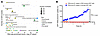 Figure 1
TRAILshort expression and DR5-dependent uptake of TRAILshort-containing EVs. (A) Analysis of human RNA-seq datasets obtained from the GEO database showing disease states associated with high TRAILshort expression. RNA-seq enrichment scores were determined by hypergeometric test with Benjamini-Hochberg FDR correction across high (top 5%) versus low (bottom 5%) TRAILshort-expressing samples. (B) DR5-dependent internalization of TRAILshort EVs. GFP-TRAILshort EVs were first labeled with an anti-TRAILshort antibody conjugated to a pH-sensitive dye and then incubated with wild-type or DR5-knockout 293T cells to monitor EV internalization. Representative line graph showing the kinetics of EV uptake by wild-type (blue) and DR5-knockout (red) 293T cells. **P < 0.001, by 2-way ANOVA. Data are presented as mean ± SEM.
Cellular effects of TRAILshort-containing extracellular vesicles are mediated by the DR5 receptor. We have previously shown that TRAILshort preferentially binds DR5 and that TRAILshort-bearing extracellular vesicles (EVs) convert TRAIL-sensitive cells to a resistant phenotype (26). To determine whether EV-associated TRAILshort can engage DR5 and initiate intracellular signaling, we generated GFP-tagged TRAILshort EVs from TRAIL-KO 293T cells (Supplemental Figure 1, A and B) and monitored their uptake by WT or DR5-KO 293T cells over 12 hours (Figure 1B and Supplemental Figure 1D). TRAILshort EVs were internalized by WT but not DR5-KO cells (P < 0.0001), indicating that EV binding and uptake was DR5 dependent and suggesting that TRAILshort-DR5 engagement may initiate downstream signaling events.
TRAILshort-induced changes in phosphoproteomics pathways. Given the power of large-scale phosphoproteomics as an unbiased, hypothesis-generating tool for exploring signal transduction pathways (29), we used quantitative phosphoproteomics analysis to comprehensively evaluate the effect of TRAILshort on primary T cells. Primary CD4+, CD8+, and CD3+ T cells were treated with TRAILshort peptide or EVs, with or without CD3/CD28 stimulation, and phosphopeptides were quantified across thousands of sites (Figure 2A, Supplemental Figure 2, A and B, and Supplemental Table 2). Kyoto Encyclopedia of Genes and Genomes (KEGG) pathway enrichment analysis revealed significant and reproducible alterations in TCR signaling pathways across independent experiments and T cell populations (Figure 2A and Supplemental Figure 2, A and B), suggesting that TRAILshort broadly affects TCR signaling.
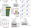 Figure 2
TRAILshort induces changes in the TCR phosphoproteome and inhibits proximal events in the TCR signaling pathway. (A) Schematic of the experimental design: CD3+ T cells were treated with TRAILshort EVs, stimulated with CD3/CD28 beads, and analyzed by phosphoproteomics. Bar graph shows differentially expressed phosphoproteins in TRAILshort EV–treated T cells versus untreated T cells. KEGG pathway enrichment analysis of phosphoproteins with a log2FC of greater than 1.5 or less than –0.9, performed using a hypergeometric test with Benjamini-Hochberg correction (adjusted P < 0.05), identified TCR signaling pathways as preferentially affected. (B) Western blot analysis of p–ZAP-70 following TRAILshort treatment. (C) Quantification showing reduced p–ZAP-70 levels in TRAILshort peptide–treated cells versus scrambled peptide controls (n = 4; *P < 0.05, by 2-tailed, paired t test with Holm correction for multiple comparisons, except control vs. +CD3/CD28, which was analyzed using a 1-sample t test against the normalized control value of 100). (D) Representative flow cytometry plots of CD3+ T cells treated with increasing concentrations (10–100 ng/mL) of TRAILshort or scrambled peptide, followed by CD3/CD28 stimulation and intracellular p–ZAP-70 staining. (E) Paired dot plots showing reduced p–ZAP-70 levels in TRAILshort-treated cells across concentrations (n = 7; **P < 0.01, by paired 2-tailed t test with Holm-adjusted P values). (F) CD3+ cells treated with TRAILshortΔTm or TRAILshort EVs with or without CD3/CD28 beads were subjected to CD3ζ immunoprecipitation and p–ZAP-70 immunoblotting. (G) Western blot analysis of total and p–ZAP-70 in CD3+ cells treated with full-length TRAIL EVs, TRAILshort EVs, or both following CD3/CD28 stimulation. Data are presented as the mean ± SEM.
TRAILshort attenuates ZAP-70 phosphorylation and TCR signaling following CD3/CD28 ligation. T cells recognize peptide-MHC complexes via the TCR: CD8+ T cells engage MHC class I and CD4+ T cells engage MHC class II, triggering downstream signaling that drives T cell activation, proliferation, and cytokine production including IFN-γ. A central mediator is ZAP-70 (a TCRζ chain–associated phosphoprotein), whose deficiency abrogates downstream TCR signaling and causes severe combined immunodeficiency in humans (30). Upon TCR ligation, Lck phosphorylates ZAP-70 at Y492/Y493, enabling its interaction with CD3ζ and propagation of downstream signaling (31). This leads to phosphorylation of linker for activation of T cells (LAT) (32), which recruits and activates phospholipase C gamma 1 (PLCγ1) and other signaling effectors (33).
To test whether TRAILshort modulates proximal TCR signaling, primary CD3+ T cells were pretreated with TRAILshort peptide or BSA and stimulated with CD3/CD28 beads. CD3/CD28 ligation increased phosphorylated ZAP-70 (p–ZAP-70), p-LAT, and p-PLCγ as expected, whereas TRAILshort peptide alone had no effect. However, TRAILshort pretreatment diminished the phosphorylation of all 3 signaling molecules upon CD3/CD28 stimulation (Supplemental Figure 2C). We then evaluated TRAILshort effects under lower, more physiologically relevant concentrations, consistent with prior studies of immunomodulatory TNF superfamily ligands in disease settings. For example, serum B-cell activating factor levels range from 1–20 ng/mL in patients with autoimmune disorders (34), and soluble TRAIL is present at low ng/mL concentrations in individuals with HIV infection or cancer (35). To assess the effect of reduced TRAILshort concentrations on ZAP-70 phosphorylation, using an appropriate control, T cells were treated with TRAILshort or a scrambled peptide of an equivalent amino acid length prior to CD3/CD28 ligation, and then p–ZAP-70 levels were measured. Exposure to TRAILshort decreased p–ZAP-70 levels compared with levels in scrambled peptide–treated T cells (Figure 2, B and C; P < 0.05). Importantly, TRAILshort also attenuated ZAP-70 phosphorylation across a range of lower doses (10–100 ng/mL) relative to matched concentrations of the scrambled control peptide (Figure 2, D and E; P < 0.05).
Since TRAILshort is naturally secreted in EV form, we treated CD4+ and CD8+ T cells with TRAILshort EVs or TRAILshortΔTm EVs, a transmembrane-deficient variant in which TRAILshort is absent from the EV surface and is therefore unable to engage DR5; this construct was used as negative control (Supplemental Figure 1B). TRAILshort EVs suppressed CD3/CD28-induced ZAP-70 phosphorylation in both CD4+ and CD8+ T cell populations (including effector memory subsets), as assessed by Western blotting and flow cytometry (Supplemental Figure 2, D and E), whereas TRAILshortΔTm EVs did not.
To assess whether TRAILshort disrupts the p–ZAP-70–CD3ζ interaction critical for TCR signal propagation, we performed co-immunoprecipitation following CD3/CD28 stimulation with or without TRAILshort EV treatment. We found that TRAILshort EVs reduced the p–ZAP-70 association with CD3ζ compared with TRAILshortΔTm EVs or untreated controls (Figure 2F). Since physiological EVs probably carry both full-length TRAIL and TRAILshort, we also tested their combined effect on p–ZAP-70; TRAILshort retained its inhibitory activity even in the presence of full-length TRAIL (Figure 2G). Together, these findings demonstrate that TRAILshort suppressed the phosphorylation of proximal TCR signaling molecules, indicating an inhibitory effect on antigen-specific T cell function.
TRAILshort reduces antigen-specific ELISpot responses, activation, and proliferation in primary T cells. To examine the effect of TRAILshort on antigen-specific T cell function, we performed ELISpot assays measuring IFN-γ secretion in response to recall antigens. TRAILshort peptide pretreatment reduced IFN-γ–secreting cell frequencies in healthy donor T cells stimulated with tetanus toxoid (Figure 3A; P < 0.001) and in T cells from virally suppressed HIV-infected donors stimulated with HIV recall peptides (Supplemental Figure 3A; P < 0.01). These suppressive effects were replicated with TRAILshort EVs but not TRAILshortΔTm EVs (Figure 3A; P < 0.05; Supplemental Figure 3, A and B; P < 0.01). At lower concentrations (1–100 ng/mL), TRAILshort peptide reduced IFN-γ compared with scrambled control at 100 ng/mL (Figure 3, B and C; P < 0.01). TRAILshort EV treatment also reduced IL-2 secretion in CD3+ T cells cocultured with tetanus toxoid–loaded antigen-presenting cells (APCs) compared with empty EVs (Supplemental Figure 3C; P < 0.05).
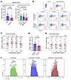 Figure 3
TRAILshort exerts inhibitory effects on the antigen-specific T cell response, cytokine secretion, T cell activation, and primary T cell proliferation. (A) IFN-γ ELISpot assays of CD3+ T cells from uninfected donors (n = 25) stimulated with tetanus toxoid in the presence of TRAILshort peptide or EVs. A Wilcoxon matched-pairs rank test showed that TRAILshort EVs significantly reduced spot formation compared with BSA or TRAILshortΔTm EV controls (*P < 0.05 and ***P < 0.001). (B) Flow cytometric analysis of intracellular IFN-γ in T cells treated with increasing doses (10–100 ng/mL) of scrambled or TRAILshort peptide following CD3/CD28 stimulation. (C) Quantification showing reduced IFN-γ levels in TRAILshort peptide–treated T cells versus scrambled peptide controls (n = 9; *P < 0.05 and **P < 0.01, by paired 2-tailed t test with Holm adjustment). (D) T cells exposed to EBV gp350 and cocultured with EBV antigen–loaded macrophages were treated with TRAILshort or TRAILshortΔTm EVs, resulting in reduced CD4+CD69+CD40L+ activation markers (n = 6; *P < 0.05, by paired t test). (E) Paired dot plots showing reduced CD69 expression in TRAILshort peptide–treated T cells (10–100 ng/mL) versus scrambled peptide controls (n = 15; **P < 0.01, by paired 2-tailed t test with Holm adjustment). (F) Representative CFSE proliferation histograms showing reduced T cell proliferation in cocultures with TRAILshort-expressing MEFs following CD3/CD28 stimulation. Data are presented as the mean ± SEM.
CD40L and CD69 are rapidly upregulated following antigen-specific T cell activation (36), and treatment with TRAILshort EVs reduced IFN-γ secretion and the proportion of CD4+ T cells expressing CD40L and CD69 in response to EBV gp350 (37), compared with TRAILshortΔTm EVs (Figure 3D; P < 0.05; Supplemental Figure 3D; P < 0.05). TRAILshort peptide (50 and 100 ng/mL) also reduced CD69 expression compared with scrambled controls (Figure 3E; P < 0.01 and P < 0.05, respectively), confirming that TRAILshort inhibited both cytokine secretion and surface markers of T cell activation.
To assess the effect of TRAILshort on T cell proliferation, CFSE-labeled T cells were stimulated with CD3/CD28 beads in a Transwell coculture system with WT or TRAILshort-knockin mouse embryonic fibroblasts (MEFs) (Supplemental Figure 5B) in the lower chamber, providing continuous EV delivery without direct cell contact. T cells exposed to TRAILshort-containing MEF-derived EVs exhibited delayed proliferation compared with those cocultured with WT MEFs (Figure 3F and Supplemental Figure 3E; P < 0.05). Consistent with this, treatment of CD3/CD28-stimulated CD3+ T cells with TRAILshort EVs purified from TRAIL-KO HEK293 cells (Supplemental Figure 1A) also reduced proliferation compared with empty EVs or untreated cells (Supplemental Figure 3F; P < 0.001).
Neutralizing TRAILshort with specific antibodies restores TCR signaling and enhances T cell function. We previously showed that anti-TRAILshort antibodies reverse TRAILshort-mediated resistance to TRAIL-induced apoptosis in HIV-infected CD4+ T cells (26, 28), cancer cell lines, and primary B cell malignancies (27). The TRAILshort-specific antibody targets the novel C terminus of TRAILshort which is absent from full-length TRAIL and all other known human proteins (22, 26, 28). Based on our new understanding that TRAILshort functions both to cause TRAIL resistance and T cell hyporesponsiveness, we investigated whether neutralization of TRAILshort using specific antibodies could enhance T cell effector functions during disease states characterized by endogenous TRAILshort production.
Using splenocyte suspensions from patients with primary B cell malignancies — in which malignant B cells express TRAILshort (27) (Supplemental Figure 3G) — we cocultured T cells with autologous TRAILshort-expressing B cells in the presence of anti-TRAILshort antibody or isotype control. Anti-TRAILshort antibody increased IFN-γ secretion across multiple patient groups, including marginal zone lymphoma, diffuse, large B cell lymphoma (DLBCL), and florid lymphoid hyperplasia (Supplemental Figure 3H; P < 0.05), consistent with TRAILshort neutralization enhancement of antitumor T cell function.
TRAILshort binding to DR5 does not trigger apoptosis, necroptosis, NF-κB, or nuclear factor of activated T cells (NFAT) activation. Full-length TRAIL ligation of death receptors can result in apoptosis, necroptosis (14) or activation of the transcription factors NF-κB and/or NFAT (38). Trimerized TRAIL leads to the clustering of TRAIL death receptors, which in turn recruits Fas-associated protein with death domain (FADD) to the death domain of DR5, forming death-inducing signaling complex (DISC, which subsequently recruits pro–caspase 8, causing its autoactivation into caspase 8 (21, 39). Furthermore, trimerized TRAIL binding to DR5 can trigger necroptosis by activating receptor-interacting protein kinase 1 (RIPK1) and receptor-interacting protein kinase 3 (RIPK3) (14).
To determine whether TRAILshort-mediated T cell suppression occurs through apoptosis or necroptosis of T cells, we evaluated DISC formation, caspase 8/FADD recruitment, and RIPK1/MLKL phosphorylation. TRAILshort neither induced apoptosis or necroptosis, nor promoted DISC assembly (Figure 4, A and B, and Supplemental Figure 4, A and B), indicating that TRAILshort-mediated T cell hyporesponsiveness was independent of cell death and these pathways. TRAILshort also did not activate NF-κB, but slightly reduced NFAT signaling (Figure 4C).
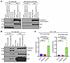 Figure 4
TRAILshort does not induce apoptosis, necroptosis, or NF-κB or NFAT activation in T cells. (A) WT or DR5-deficient Jurkat cells were treated with TRAILshort peptide or super killer TRAIL (SKT) for 30 or 60 minutes. DR5 immunoprecipitated lysates were immunoblotted for FADD, caspase 8, and DR5. FADD recruitment was detected with SKT but not TRAILshort. (B) Jurkat cells treated with SKT, TRAILshort, or both were subjected to DR5 immunoprecipitation. Caspase 8, FADD, and RIPK1 associated with DR5 following SKT but not TRAILshort treatment. (C) NF-κB and NFAT reporter activities were measured in primary CD3+ T cells following TRAILshort treatment. CD3/CD28 stimulation served as a positive control. *P < 0.05 and **P < 0.01, by 2-tailed, paired t test with Holm adjustment. Paired t test analysis showed a significant increase between untreated and TRAILshort-treated groups compared with CD3/CD28-stimulated groups (*P < 0.05). TRAILshort alone slightly reduced NFAT reporter activity. Data are presented as the mean ± SEM.
TRAILshort binding to DR5 recruits SHP-1. To identify mechanisms by which TRAILshort impairs TCR signaling, we performed Fc fusion pull-down assays using TRAILshort-Fc, Fas-Fc, and DR5-Fc constructs, followed by mass spectrometry. ZAP-70, CD3ζ, and SHP-1 (also known as protein tyrosine phosphatase nonreceptor type 6 [PTPN6]) were uniquely identified as TRAILshort-interacting proteins (Figure 5, A–C, and Supplemental Table 3). DR5 was detected in both the TRAILshort-Fc and DR5-Fc pull-downs; its presence in the TRAILshort-Fc assay further supported preferential TRAILshort-DR5 binding (22, 26), while in the DR5-Fc assay, it likely reflected the input fusion protein.
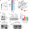 Figure 5
TRAILshort treatment induces the recruitment and activation of SHP-1 at the DR5 receptor. (A) CD3+ T cell lysates were immunoprecipitated with FAS-Fc, DR5-Fc, or TRAILshort-Fc, resolved by SDS-PAGE, and analyzed by in-gel digestion and proteomics. (B) Proteomics analysis identified DR5, ZAP-70, and SHP-1 as proteins pulled down by TRAILshort-Fc. (C) STRING (Search Tool for the Retrieval of Interacting Genes/Proteins) analysis revealed interactions among DR5, SHP-1, and TCR signaling molecules associated with TRAILshort-Fc. (D) Western blot showing full-length TRAIL and TRAILshort in EVs isolated from 293T cells transfected with TRAIL or TRAILshort constructs. (E) Western blot analysis of T cells from 2 healthy donors treated with purified EVs (D) at a 1:1,000 cell-to-EV ratio and probed for total SHP-1 and p–SHP-1 (Y536). (F) Western blot band quantification demonstrated increased p–SHP-1 expression in cells treated with TRAILshort EV (*P < 0.05, by paired, 2-tailed t test with Holm adjustment). Data are presented as the mean ± SEM. (G) Jurkat cells were treated with no stimulus, BSA, or TRAILshort, followed by immunoprecipitation with DR5 or isotype control antibodies. Immunoblotting for p–SHP-1 and total SHP-1 showed increased DR5-associated p–SHP-1 after TRAILshort treatment. (H) DR5-KO 293T cells were transfected with DR5 alone, DR5 plus Flag-tagged WT SHP-1, or Flag-tagged mutant SHP-1 (D419A). Cells were treated with TRAILshort peptide or BSA, followed by anti-Flag immunoprecipitation. Immunoblotting shows mutant SHP-1 (D419A) associated with DR5 under both conditions, whereas WT SHP-1 interacted with DR5 only after TRAILshort treatment, consistent with TRAILshort-induced SHP-1 recruitment and activation through DR5.
SHP-1, also known as PTPN6, is a cytoplasmic tyrosine phosphatase expressed predominantly in hematopoietic cells that acts as a negative regulator of T cell, B cell, and NK cell signaling (40). In T cells, SHP-1 dephosphorylates ZAP-70, CD3ζ, and linker for activation of T cells (LAT), thereby dampening TCR signaling, whereas SHP-1–deficient thymocytes display prolonged TCR-induced phosphorylation, enhanced proliferation, and increased ZAP-70 activity (41). SHP-1 is autoinhibited in its basal state through intramolecular N-terminal SH2-phosphatase domain interactions (42) and is activated upon engagement with immunoreceptor tyrosine-based inhibitory motif-containing receptors or immunoreceptor tyrosine-based switch motif-containing receptors, leading to autophosphorylation at Y536 (43). Death receptors of the TNF receptor (TNFR) family, including Fas, can recruit and activate SHP-1 via cytoplasmic phosphotyrosine motifs (44, 45), although DR5-specific SHP-1 engagement in human T cells had not been fully defined. Our identification of TRAILshort–DR5–SHP-1 complex formation (Figure 5, A–C) led us to hypothesize that TRAILshort binding to DR5 recruits and activates SHP-1, thereby antagonizing T cell receptor (TCR) signaling.
Both TRAILshort peptide (100–5000 ng/mL) and TRAILshort EVs induced SHP-1 phosphorylation in T cells at Y536 (Supplemental Figure 4, C and D). To model a physiological immune microenvironment containing TRAILshort and full-length TRAIL, we generated EVs from 293T cells transfected with TRAILshort, full-length TRAIL, or both (Figure 5D). Primary T cells treated with all 3 EV conditions exhibited significant SHP-1 phosphorylation (Figure 5, E and F; P < 0.05), demonstrating that TRAILshort-driven SHP-1 activation was preserved in the presence of full-length TRAIL. Notably, EVs containing full-length TRAIL alone were also sufficient to induce SHP-1 phosphorylation, indicating that SHP-1 activation via DR5 was not exclusive to TRAILshort but rather represents a shared signaling property of the TRAIL/DR5 axis.
Fas recruits and activates SHP-1 via a cytoplasmic YxxL motif (44). Since DR5 harbors a similar YxxL motif at position 383, we hypothesized that TRAILshort binding to DR5 recruits SHP-1 to this domain, triggering its activation. To test this idea, Jurkat T cells were treated with TRAILshort peptide, DR5 was immunoprecipitated, and the precipitate was probed for total and p–SHP-1 (Y536) levels. SHP-1–DR5 interaction was detected exclusively in TRAILshort-treated cells, and the associated SHP-1 was predominantly in the phosphorylated (activated) form, indicating that TRAILshort engagement of DR5 recruited and activated SHP-1 (Figure 5G).
To further assess the putative interaction of SHP-1 with TRAILshort and DR5, we constructed expression vectors containing the Flag-tagged WT SHP-1 and a mutant SHP-1 with an alanine substitution at aspartic acid 419 (D419A). This substitution stabilizes the interaction of SHP-1 with its binding partners and is referred to as a substrate-trapping mutant (46). These constructs were coexpressed with the DR5 in 293T cells lacking the DR5 gene, and the cells were subsequently treated with TRAILshort peptide and subjected to immunoprecipitation using an anti-Flag antibody. Our results revealed that the substrate-trapping mutant of SHP-1 was associated with DR5 in both BSA- and TRAILshort-treated cells, but that WT SHP-1 was associated with DR5 only when pretreated with TRAILshort (Figure 5H).
Blocking SHP-1 reverts the TRAILshort-induced inhibition of TCR signaling. Next, we evaluated whether SHP-1 inhibition could reverse TRAILshort-mediated suppression of TCR signaling using 3 independent approaches: (a) a pharmacologic SHP-1 inhibitor, (b) CRISPR-mediated SHP-1 KO, and (c) siRNA-mediated SHP-1 knockdown. All 3 strategies restored ZAP-70 phosphorylation following TCR ligation in the presence of TRAILshort (Figure 6, A–D, and Supplemental Figure 4, E and F). Moreover, SHP-1 inhibition increased IFN-γ secretion by T cells in ELISpot assays (Figure 6E) and enhanced both the frequency of activated CD69+CD40L+CD4+ T cells and IFN-γ secretion in TRAILshort-treated T cells exposed to EBV antigen (Figure 6, F and G). Additionally, SHP-1 inhibition promoted the proliferation of CD3+ T cells in response to TRAILshort EVs (Supplemental Figure 4G). The pharmacological inhibition of SHP-1 produced only a modest reversal of TRAILshort-induced suppression of ZAP-70 phosphorylation, in contrast to the more pronounced effects observed with CRISPR-mediated KO or siRNA knockdown of SHP-1. This difference is most consistent with the limited potency and specificity of the small-molecule inhibitor, which targets SHP-1 as well as related phosphatases such as SHP-2, thereby constraining its ability to fully restore TCR signaling and effector functions (47). These findings collectively underscore the pivotal role of SHP-1 in modulating the effects of TRAILshort on T cell function.
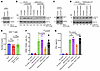 Figure 6
SHP-1 inhibition reverses TRAILshort-mediated suppression of T cell function. (A–D) Primary CD3+ T cells were transfected with nontargeting (NT) or SHP-1 siRNA to reduce SHP-1 expression. Cells were then pretreated with scrambled peptide or TRAILshort peptide and stimulated with CD3/CD28 beads (1:2 bead-to-cell ratio) for 30 minutes. (A and B). Alternatively, SHP-1 was knocked out using CRISPR/Cas9 and under the same experimental conditions (C and D). Western blotting was performed to assess total and phosphorylated ZAP-70. (E) Primary CD3+ T cells were analyzed by IFN-γ ELISpot analysis in response to recall antigen tetanus toxoid, either alone, in the presence of TRAILshort, or with TRAILshort plus the SHP-1 inhibitor (0.5 μM), showing that SHP-1 inhibition restored IFN-γ spot formation suppressed by TRAILshort. **P < 0.01 and ****P < 0.0001, by 1-sample t test against the normalized control value of 100%) for No EV versus TRAILshort EV, and 2-tailed, paired t test for TRAILshort EV versus TRAILshort EV plus the SHP-1 inhibitor. P values were adjusted for multiple comparisons using the Holm method. (F and G) PBMCs were exposed to EBV gp350 for 5 days and then cocultured with syngeneic EBV antigen–loaded macrophages and treated with either TRAILshort or TRAILshortΔTm EVs, alone or with the SHP-1 inhibitor. The proportion of CD4+CD69+CD40L+ T cells was assessed by flow cytometry (F), and IFN-γ secretion was measured by ELISA (G), demonstrating that SHP-1 inhibition reversed TRAILshort-mediated reductions in T cell activation and cytokine production. *P < 0.05, **P < 0.01, and ****P < 0.001, by Wilcoxon matched-pairs, signed-rank test for comparisons between 2 groups. data are presented as the mean ± SEM.
MEFs derived from TRAILshort-knockin mice are resistant to TRAIL-induced apoptosis but remain sensitive to Fas-mediated cell death. Since murine TRAIL genomic organization precludes TRAILshort generation, we generated a TRAILshort-knockin mouse using a Cre-LoxP strategy (Supplemental Figure 5A). MEFs derived from WT or homozygous TRAILshort embryos were transformed with SV40 large T antigen and transduced with a luciferase-GFP vector; TRAILshort expression and EV-associated secretion were confirmed by immunofluorescence and Western blotting (Supplemental Figure 5, B and C). When treated with murine super killer TRAIL or anti-Fas antibody and monitored by live-cell caspase 3/-7 imaging over 40 hours, we observed that TRAILshort MEFs were resistant to TRAIL-induced apoptosis (P < 0.01) but remained sensitive to Fas-mediated apoptosis (Supplemental Figure 5, D and E).
Evaluating the effect of TRAILshort in vivo. To evaluate the effect of TRAILshort on human T cell function in vivo, we used 2 complementary mouse models with transformed MEFs derived from either WT or TRAILshort-knockin mice. In this system, tumors required several weeks to reach a reliably measurable size, allowing differences driven by partial, sustained impairment of T cell function — rather than complete immune failure — to emerge and accumulate progressively over time. In Hu-CD34+ NOD scid gamma mice bearing a functional human immune system and engrafted with luciferase+ MEFs, WT MEFs were progressively controlled, whereas TRAILshort-expressing MEFs showed greater early expansion and higher bioluminescent signal, indicating that MEF-derived TRAILshort dampened human T cell–mediated clearance (Figure 7, A and B, and Supplemental Figure 6, A–C).
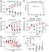 Figure 7
Effect of TRAILshort in vivo. (A) Luciferase-expressing immortalized WT or TRAILshort MEFs were implanted s.c. into Hu-CD34+ NSG mice (6 mice/group), and tumor growth was monitored weekly by bioluminescence imaging, with flux measured in photons/second (p/s). P values were calculated using 2-sample t tests on log-transformed flux at each prespecified time point. (B) Survival of humanized CD34+ mice implanted with WT or TRAILshort MEFs. WT MEFs showed limited expansion, whereas TRAILshort MEFs displayed increased and sustained bioluminescent signal. P < 0.001, by log-rank (Mantel-Cox) test. (C and D) NSG mice were s.c. implanted on both flanks with WT or TRAILshort MEFs (non-luciferase-expressing), followed by i.v. injection of luciferase-expressing JeKo-1 cells. Mice were treated with CD19 CAR-T cells, and JeKo-1 tumor growth was monitored weekly by bioluminescence imaging. TRAILshort MEFs impaired CD19 CAR-T–mediated control of both WT and DR5-KO JeKo-1 tumors compared with WT MEFs. P values were calculated using (C) 2-sample t tests on log-transformed flux at each prespecified time point and (D) Tukey’s honestly significant difference (HSD) pairwise comparisons on log-transformed flux at the final time point. (E and F) WT and TRAIL/TRAILshort-KO L428 cells were implanted s.c. into Hu-CD34+ NSG mice (6 mice/group), and tumor growth was monitored for 38 days by bioluminescence imaging. TRAIL/TRAILshort-deficient L428 tumors showed lower total flux than did WT L428 tumors at days 7 and 28 (E), and caliper measurements beginning on day 25 (F) likewise demonstrated reduced tumor volume in the TRAIL/TRAILshort-KO group. *P < 0.05, **P < 0.01, and ***P < 0.001, by 2-sample t test on log-transformed flux at each time point (E) and Wilcoxon rank-sum test on tumor volume at each time point (F). Data are presented as the mean ± SEM.
We next adapted an established NSG CD19 CAR-T/JeKo-1 mantle cell lymphoma model (48) by s.c. preimplanting WT or TRAILshort MEFs (not expressing luciferase) prior to i.v. injection of luciferase-labeled CD19+ JeKo-1 cells, followed by CD19 CAR-T cells (Supplemental Figure 6D). This model allowed us to determine if TRAILshort supplied by MEFs affect CAR-T control of tumors, and by implanting the tumors and MEFs only, it allowed us to determine if TRAILshort-expressing MEFs directly affect tumor cells, independent of their effects on T cells. Because TRAILshort MEFs release TRAILshort-containing EVs, any immunosuppressive effects of TRAILshort in this setting would be expected to act systemically rather than being confined to the local implantation site. In this system, WT MEFs did not impair CAR-T–mediated control of WT JeKo-1 cells, nor did TRAILshort MEFs alter JeKo-1 growth in the absence of CAR-T cells (Figure 7C and Supplemental Figure 6, E and F), indicating that MEF-derived TRAILshort did not affect lymphoma cell outgrowth and that WT MEF did not impair CAR-T control of tumors.
As expected, CD19 CAR-T cells efficiently eliminated WT JeKo-1 tumors in the presence of WT MEFs, and because CAR-T–mediated killing is in part driven by TRAIL-DR5 interactions (49, 50), this killing was attenuated when DR5 was deleted in JeKo-1 cells (Figure 7D). In the presence of TRAILshort MEFs, CD19 CAR-T–mediated killing of WT JeKo-1 cells was reduced compared with WT MEFs (P = 0.0012), and CD19 CAR-T–mediated killing of DR5-KO JeKo-1 cells was likewise diminished in mice bearing TRAILshort MEFs compared with mice with WT MEFs (Figure 7D; P < 0.01). Together, these results indicate that MEF-derived TRAILshort impaired CAR-T effector function rather than acting by making JeKo-1 tumor cells themselves less sensitive to DR5-dependent cytotoxicity.
To assess the effect of TRAILshort using an in vivo humanized tumor model, we used L428 Hodgkin lymphoma cells, which endogenously express high levels of TRAILshort (Supplemental Figure 7), in the same Hu-CD34 NSG model used in Figure 7, A and B, to mimic a native human immune system. Using CRISPR/Cas9, we generated TRAIL-KO L428 cells, in which both full-length TRAIL and TRAILshort were deleted, as TRAILshort is encoded within the same gene and cannot be selectively ablated. Equal numbers of WT L428 cells and TRAIL- and TRAILshort-deficient L428 cells were implanted on day 1, and tumor burden was quantified longitudinally by bioluminescence every 7 days. Over the course of 4 weeks, TRAIL/TRAILshort-deficient L428 tumors maintained lower total flux values than did WT L428 tumors at days 7 and 28 (Figure 7, E and F; P < 0.05), reflecting impaired tumor expansion in the absence of the immunosuppressive effects of TRAILshort. By day 28, both WT and TRAIL/TRAILshort-deficient tumors were controlled, suggesting that TRAILshort primarily delays, rather than prevents, immune-mediated tumor clearance in this setting. It is not surprising that deletion of TRAILshort alone did not lead to tumor eradication, as additional oncogenic and tumor-intrinsic survival pathways remained intact.


Copyright © 2026 American Society for Clinical Investigation
ISSN: 0021-9738 (print), 1558-8238 (online)