
Advertisement
Research ArticleGeneticsOncology
Open Access |  10.1172/JCI193280
FGFR3-driven gene regulatory network analysis reveals a protumoral role for p63 in luminal bladder tumors
Aura Moreno-Vega,1 Macarena Zambrano,2 Lilia Estrada-Virrueta,1,3 Xiangyu Meng,1,4 Julia Puig,5 Helene Neyret-Kahn,1 Mingjun Shi,1,6 Florent Dufour,1 Guerric Gilbert,1,7 Ke Li,1,7 Clarice Groeneveld,1 Jacqueline Fontugne,7,8,9 Mercedes Pérez-Escavy,10 Wajdi Dhifli,5 Clément Hua,1 Luc Cabel,1 Clémentine Krucker,1,7 Laura Tanguy,1,7 Sia Viborg Lindskrog,11,12 Claire Beraud,13 Yanina V. Langle,2 Tao Ye,14 Fariza Tahi,3 Irwin Davidson,14 Jesus M. Paramio,10 Lars Dyrskjøt,11,12 Yves Allory,1,7,8,9 Philippe Lluel,13 Ana Maria Eiján,2,15 Mohamed Elati,5 François Radvanyi,1,7 Catalina Lodillinsky,2,15 and Isabelle Bernard-Pierrot1,7
1Institut Curie, Centre National de Recherche Scientifique (CNRS), UMR144, Molecular Oncology team, PSL Research University, Sorbonne Université UPMC Université Paris 6, Paris, France.
2Universidad de Buenos Aires, Facultad de Medicina, Instituto de Oncología Ángel H. Roffo, Área de Investigación, Buenos Aires, Argentina.
3Université de Paris-Saclay, Université d’Evry, Genopole, Evry, France.
4Health Science Center, Hubei Minzu University, Enshi, China.
5Univ. Lille, CNRS, INSERM, CHU Lille, UMR9020 – UMR-1277 - Canther, F-59000 Lille, France.
6Department of Urology, Beijing Friendship Hospital, Capital Medical University, Beijing, China.
7Institut Curie, CNRS UMR9029, INSERM U1353, IMPACT team, Paris, France.
8Department of Pathology, Institut Curie, Saint-Cloud, France.
9Université Versailles St-Quentin, Université Paris-Saclay, F-78180, Montigny-le-Bretonneux, France.
10Molecular and Translational Oncology Division, Centro de Investigaciones Energéticas, Medioambientales y Tecnológicas (CIEMAT); Cell and Molecular Oncology, Instituto de Investigación Hospital “12 de Octubre”; and CIBER Cancer (CIBERONC), Madrid, Spain.
11Department of Molecular Medicine, Aarhus University Hospital, Aarhus, Denmark.
12Department of Clinical Medicine, Aarhus University, Aarhus, Denmark.
13Urosphere, Toulouse, France.
14Institut de Génétique et de Biologie Moléculaire et Cellulaire (IGBMC), INSERM U1258, CNRS UMR7104, Université de Strasbourg, Illkirch, France.
15Consejo Nacional de Investigaciones Científicas y Técnicas (CONICET), Buenos Aires, Argentina.
Address correspondence to: Isabelle Bernard-Pierrot, Institut Curie, UMR9029/U1353 BDD, 11 rue Pierre et Marie Curie, 75005 Paris, France. Phone: 33.1.42.34.63.40; Email: isabelle.bernard-pierrot@curie.fr.
Authorship note: AMV, MZ, LEV, XM, and JP have been designated as co–first authors. AME, ME, FR, CL, and IBP have been designated as co–last authors.
Find articles by Moreno-Vega, A. in: PubMed | Google Scholar
1Institut Curie, Centre National de Recherche Scientifique (CNRS), UMR144, Molecular Oncology team, PSL Research University, Sorbonne Université UPMC Université Paris 6, Paris, France.
2Universidad de Buenos Aires, Facultad de Medicina, Instituto de Oncología Ángel H. Roffo, Área de Investigación, Buenos Aires, Argentina.
3Université de Paris-Saclay, Université d’Evry, Genopole, Evry, France.
4Health Science Center, Hubei Minzu University, Enshi, China.
5Univ. Lille, CNRS, INSERM, CHU Lille, UMR9020 – UMR-1277 - Canther, F-59000 Lille, France.
6Department of Urology, Beijing Friendship Hospital, Capital Medical University, Beijing, China.
7Institut Curie, CNRS UMR9029, INSERM U1353, IMPACT team, Paris, France.
8Department of Pathology, Institut Curie, Saint-Cloud, France.
9Université Versailles St-Quentin, Université Paris-Saclay, F-78180, Montigny-le-Bretonneux, France.
10Molecular and Translational Oncology Division, Centro de Investigaciones Energéticas, Medioambientales y Tecnológicas (CIEMAT); Cell and Molecular Oncology, Instituto de Investigación Hospital “12 de Octubre”; and CIBER Cancer (CIBERONC), Madrid, Spain.
11Department of Molecular Medicine, Aarhus University Hospital, Aarhus, Denmark.
12Department of Clinical Medicine, Aarhus University, Aarhus, Denmark.
13Urosphere, Toulouse, France.
14Institut de Génétique et de Biologie Moléculaire et Cellulaire (IGBMC), INSERM U1258, CNRS UMR7104, Université de Strasbourg, Illkirch, France.
15Consejo Nacional de Investigaciones Científicas y Técnicas (CONICET), Buenos Aires, Argentina.
Address correspondence to: Isabelle Bernard-Pierrot, Institut Curie, UMR9029/U1353 BDD, 11 rue Pierre et Marie Curie, 75005 Paris, France. Phone: 33.1.42.34.63.40; Email: isabelle.bernard-pierrot@curie.fr.
Authorship note: AMV, MZ, LEV, XM, and JP have been designated as co–first authors. AME, ME, FR, CL, and IBP have been designated as co–last authors.
Find articles by Zambrano, M. in: PubMed | Google Scholar
1Institut Curie, Centre National de Recherche Scientifique (CNRS), UMR144, Molecular Oncology team, PSL Research University, Sorbonne Université UPMC Université Paris 6, Paris, France.
2Universidad de Buenos Aires, Facultad de Medicina, Instituto de Oncología Ángel H. Roffo, Área de Investigación, Buenos Aires, Argentina.
3Université de Paris-Saclay, Université d’Evry, Genopole, Evry, France.
4Health Science Center, Hubei Minzu University, Enshi, China.
5Univ. Lille, CNRS, INSERM, CHU Lille, UMR9020 – UMR-1277 - Canther, F-59000 Lille, France.
6Department of Urology, Beijing Friendship Hospital, Capital Medical University, Beijing, China.
7Institut Curie, CNRS UMR9029, INSERM U1353, IMPACT team, Paris, France.
8Department of Pathology, Institut Curie, Saint-Cloud, France.
9Université Versailles St-Quentin, Université Paris-Saclay, F-78180, Montigny-le-Bretonneux, France.
10Molecular and Translational Oncology Division, Centro de Investigaciones Energéticas, Medioambientales y Tecnológicas (CIEMAT); Cell and Molecular Oncology, Instituto de Investigación Hospital “12 de Octubre”; and CIBER Cancer (CIBERONC), Madrid, Spain.
11Department of Molecular Medicine, Aarhus University Hospital, Aarhus, Denmark.
12Department of Clinical Medicine, Aarhus University, Aarhus, Denmark.
13Urosphere, Toulouse, France.
14Institut de Génétique et de Biologie Moléculaire et Cellulaire (IGBMC), INSERM U1258, CNRS UMR7104, Université de Strasbourg, Illkirch, France.
15Consejo Nacional de Investigaciones Científicas y Técnicas (CONICET), Buenos Aires, Argentina.
Address correspondence to: Isabelle Bernard-Pierrot, Institut Curie, UMR9029/U1353 BDD, 11 rue Pierre et Marie Curie, 75005 Paris, France. Phone: 33.1.42.34.63.40; Email: isabelle.bernard-pierrot@curie.fr.
Authorship note: AMV, MZ, LEV, XM, and JP have been designated as co–first authors. AME, ME, FR, CL, and IBP have been designated as co–last authors.
Find articles by Estrada-Virrueta, L. in: PubMed | Google Scholar
1Institut Curie, Centre National de Recherche Scientifique (CNRS), UMR144, Molecular Oncology team, PSL Research University, Sorbonne Université UPMC Université Paris 6, Paris, France.
2Universidad de Buenos Aires, Facultad de Medicina, Instituto de Oncología Ángel H. Roffo, Área de Investigación, Buenos Aires, Argentina.
3Université de Paris-Saclay, Université d’Evry, Genopole, Evry, France.
4Health Science Center, Hubei Minzu University, Enshi, China.
5Univ. Lille, CNRS, INSERM, CHU Lille, UMR9020 – UMR-1277 - Canther, F-59000 Lille, France.
6Department of Urology, Beijing Friendship Hospital, Capital Medical University, Beijing, China.
7Institut Curie, CNRS UMR9029, INSERM U1353, IMPACT team, Paris, France.
8Department of Pathology, Institut Curie, Saint-Cloud, France.
9Université Versailles St-Quentin, Université Paris-Saclay, F-78180, Montigny-le-Bretonneux, France.
10Molecular and Translational Oncology Division, Centro de Investigaciones Energéticas, Medioambientales y Tecnológicas (CIEMAT); Cell and Molecular Oncology, Instituto de Investigación Hospital “12 de Octubre”; and CIBER Cancer (CIBERONC), Madrid, Spain.
11Department of Molecular Medicine, Aarhus University Hospital, Aarhus, Denmark.
12Department of Clinical Medicine, Aarhus University, Aarhus, Denmark.
13Urosphere, Toulouse, France.
14Institut de Génétique et de Biologie Moléculaire et Cellulaire (IGBMC), INSERM U1258, CNRS UMR7104, Université de Strasbourg, Illkirch, France.
15Consejo Nacional de Investigaciones Científicas y Técnicas (CONICET), Buenos Aires, Argentina.
Address correspondence to: Isabelle Bernard-Pierrot, Institut Curie, UMR9029/U1353 BDD, 11 rue Pierre et Marie Curie, 75005 Paris, France. Phone: 33.1.42.34.63.40; Email: isabelle.bernard-pierrot@curie.fr.
Authorship note: AMV, MZ, LEV, XM, and JP have been designated as co–first authors. AME, ME, FR, CL, and IBP have been designated as co–last authors.
Find articles by Meng, X. in: PubMed | Google Scholar
1Institut Curie, Centre National de Recherche Scientifique (CNRS), UMR144, Molecular Oncology team, PSL Research University, Sorbonne Université UPMC Université Paris 6, Paris, France.
2Universidad de Buenos Aires, Facultad de Medicina, Instituto de Oncología Ángel H. Roffo, Área de Investigación, Buenos Aires, Argentina.
3Université de Paris-Saclay, Université d’Evry, Genopole, Evry, France.
4Health Science Center, Hubei Minzu University, Enshi, China.
5Univ. Lille, CNRS, INSERM, CHU Lille, UMR9020 – UMR-1277 - Canther, F-59000 Lille, France.
6Department of Urology, Beijing Friendship Hospital, Capital Medical University, Beijing, China.
7Institut Curie, CNRS UMR9029, INSERM U1353, IMPACT team, Paris, France.
8Department of Pathology, Institut Curie, Saint-Cloud, France.
9Université Versailles St-Quentin, Université Paris-Saclay, F-78180, Montigny-le-Bretonneux, France.
10Molecular and Translational Oncology Division, Centro de Investigaciones Energéticas, Medioambientales y Tecnológicas (CIEMAT); Cell and Molecular Oncology, Instituto de Investigación Hospital “12 de Octubre”; and CIBER Cancer (CIBERONC), Madrid, Spain.
11Department of Molecular Medicine, Aarhus University Hospital, Aarhus, Denmark.
12Department of Clinical Medicine, Aarhus University, Aarhus, Denmark.
13Urosphere, Toulouse, France.
14Institut de Génétique et de Biologie Moléculaire et Cellulaire (IGBMC), INSERM U1258, CNRS UMR7104, Université de Strasbourg, Illkirch, France.
15Consejo Nacional de Investigaciones Científicas y Técnicas (CONICET), Buenos Aires, Argentina.
Address correspondence to: Isabelle Bernard-Pierrot, Institut Curie, UMR9029/U1353 BDD, 11 rue Pierre et Marie Curie, 75005 Paris, France. Phone: 33.1.42.34.63.40; Email: isabelle.bernard-pierrot@curie.fr.
Authorship note: AMV, MZ, LEV, XM, and JP have been designated as co–first authors. AME, ME, FR, CL, and IBP have been designated as co–last authors.
Find articles by Puig, J. in: PubMed | Google Scholar
1Institut Curie, Centre National de Recherche Scientifique (CNRS), UMR144, Molecular Oncology team, PSL Research University, Sorbonne Université UPMC Université Paris 6, Paris, France.
2Universidad de Buenos Aires, Facultad de Medicina, Instituto de Oncología Ángel H. Roffo, Área de Investigación, Buenos Aires, Argentina.
3Université de Paris-Saclay, Université d’Evry, Genopole, Evry, France.
4Health Science Center, Hubei Minzu University, Enshi, China.
5Univ. Lille, CNRS, INSERM, CHU Lille, UMR9020 – UMR-1277 - Canther, F-59000 Lille, France.
6Department of Urology, Beijing Friendship Hospital, Capital Medical University, Beijing, China.
7Institut Curie, CNRS UMR9029, INSERM U1353, IMPACT team, Paris, France.
8Department of Pathology, Institut Curie, Saint-Cloud, France.
9Université Versailles St-Quentin, Université Paris-Saclay, F-78180, Montigny-le-Bretonneux, France.
10Molecular and Translational Oncology Division, Centro de Investigaciones Energéticas, Medioambientales y Tecnológicas (CIEMAT); Cell and Molecular Oncology, Instituto de Investigación Hospital “12 de Octubre”; and CIBER Cancer (CIBERONC), Madrid, Spain.
11Department of Molecular Medicine, Aarhus University Hospital, Aarhus, Denmark.
12Department of Clinical Medicine, Aarhus University, Aarhus, Denmark.
13Urosphere, Toulouse, France.
14Institut de Génétique et de Biologie Moléculaire et Cellulaire (IGBMC), INSERM U1258, CNRS UMR7104, Université de Strasbourg, Illkirch, France.
15Consejo Nacional de Investigaciones Científicas y Técnicas (CONICET), Buenos Aires, Argentina.
Address correspondence to: Isabelle Bernard-Pierrot, Institut Curie, UMR9029/U1353 BDD, 11 rue Pierre et Marie Curie, 75005 Paris, France. Phone: 33.1.42.34.63.40; Email: isabelle.bernard-pierrot@curie.fr.
Authorship note: AMV, MZ, LEV, XM, and JP have been designated as co–first authors. AME, ME, FR, CL, and IBP have been designated as co–last authors.
Find articles by
Neyret-Kahn, H.
in:
PubMed
|
Google Scholar
|

1Institut Curie, Centre National de Recherche Scientifique (CNRS), UMR144, Molecular Oncology team, PSL Research University, Sorbonne Université UPMC Université Paris 6, Paris, France.
2Universidad de Buenos Aires, Facultad de Medicina, Instituto de Oncología Ángel H. Roffo, Área de Investigación, Buenos Aires, Argentina.
3Université de Paris-Saclay, Université d’Evry, Genopole, Evry, France.
4Health Science Center, Hubei Minzu University, Enshi, China.
5Univ. Lille, CNRS, INSERM, CHU Lille, UMR9020 – UMR-1277 - Canther, F-59000 Lille, France.
6Department of Urology, Beijing Friendship Hospital, Capital Medical University, Beijing, China.
7Institut Curie, CNRS UMR9029, INSERM U1353, IMPACT team, Paris, France.
8Department of Pathology, Institut Curie, Saint-Cloud, France.
9Université Versailles St-Quentin, Université Paris-Saclay, F-78180, Montigny-le-Bretonneux, France.
10Molecular and Translational Oncology Division, Centro de Investigaciones Energéticas, Medioambientales y Tecnológicas (CIEMAT); Cell and Molecular Oncology, Instituto de Investigación Hospital “12 de Octubre”; and CIBER Cancer (CIBERONC), Madrid, Spain.
11Department of Molecular Medicine, Aarhus University Hospital, Aarhus, Denmark.
12Department of Clinical Medicine, Aarhus University, Aarhus, Denmark.
13Urosphere, Toulouse, France.
14Institut de Génétique et de Biologie Moléculaire et Cellulaire (IGBMC), INSERM U1258, CNRS UMR7104, Université de Strasbourg, Illkirch, France.
15Consejo Nacional de Investigaciones Científicas y Técnicas (CONICET), Buenos Aires, Argentina.
Address correspondence to: Isabelle Bernard-Pierrot, Institut Curie, UMR9029/U1353 BDD, 11 rue Pierre et Marie Curie, 75005 Paris, France. Phone: 33.1.42.34.63.40; Email: isabelle.bernard-pierrot@curie.fr.
Authorship note: AMV, MZ, LEV, XM, and JP have been designated as co–first authors. AME, ME, FR, CL, and IBP have been designated as co–last authors.
Find articles by Shi, M. in: PubMed | Google Scholar
1Institut Curie, Centre National de Recherche Scientifique (CNRS), UMR144, Molecular Oncology team, PSL Research University, Sorbonne Université UPMC Université Paris 6, Paris, France.
2Universidad de Buenos Aires, Facultad de Medicina, Instituto de Oncología Ángel H. Roffo, Área de Investigación, Buenos Aires, Argentina.
3Université de Paris-Saclay, Université d’Evry, Genopole, Evry, France.
4Health Science Center, Hubei Minzu University, Enshi, China.
5Univ. Lille, CNRS, INSERM, CHU Lille, UMR9020 – UMR-1277 - Canther, F-59000 Lille, France.
6Department of Urology, Beijing Friendship Hospital, Capital Medical University, Beijing, China.
7Institut Curie, CNRS UMR9029, INSERM U1353, IMPACT team, Paris, France.
8Department of Pathology, Institut Curie, Saint-Cloud, France.
9Université Versailles St-Quentin, Université Paris-Saclay, F-78180, Montigny-le-Bretonneux, France.
10Molecular and Translational Oncology Division, Centro de Investigaciones Energéticas, Medioambientales y Tecnológicas (CIEMAT); Cell and Molecular Oncology, Instituto de Investigación Hospital “12 de Octubre”; and CIBER Cancer (CIBERONC), Madrid, Spain.
11Department of Molecular Medicine, Aarhus University Hospital, Aarhus, Denmark.
12Department of Clinical Medicine, Aarhus University, Aarhus, Denmark.
13Urosphere, Toulouse, France.
14Institut de Génétique et de Biologie Moléculaire et Cellulaire (IGBMC), INSERM U1258, CNRS UMR7104, Université de Strasbourg, Illkirch, France.
15Consejo Nacional de Investigaciones Científicas y Técnicas (CONICET), Buenos Aires, Argentina.
Address correspondence to: Isabelle Bernard-Pierrot, Institut Curie, UMR9029/U1353 BDD, 11 rue Pierre et Marie Curie, 75005 Paris, France. Phone: 33.1.42.34.63.40; Email: isabelle.bernard-pierrot@curie.fr.
Authorship note: AMV, MZ, LEV, XM, and JP have been designated as co–first authors. AME, ME, FR, CL, and IBP have been designated as co–last authors.
Find articles by
Dufour, F.
in:
PubMed
|
Google Scholar
|
1Institut Curie, Centre National de Recherche Scientifique (CNRS), UMR144, Molecular Oncology team, PSL Research University, Sorbonne Université UPMC Université Paris 6, Paris, France.
2Universidad de Buenos Aires, Facultad de Medicina, Instituto de Oncología Ángel H. Roffo, Área de Investigación, Buenos Aires, Argentina.
3Université de Paris-Saclay, Université d’Evry, Genopole, Evry, France.
4Health Science Center, Hubei Minzu University, Enshi, China.
5Univ. Lille, CNRS, INSERM, CHU Lille, UMR9020 – UMR-1277 - Canther, F-59000 Lille, France.
6Department of Urology, Beijing Friendship Hospital, Capital Medical University, Beijing, China.
7Institut Curie, CNRS UMR9029, INSERM U1353, IMPACT team, Paris, France.
8Department of Pathology, Institut Curie, Saint-Cloud, France.
9Université Versailles St-Quentin, Université Paris-Saclay, F-78180, Montigny-le-Bretonneux, France.
10Molecular and Translational Oncology Division, Centro de Investigaciones Energéticas, Medioambientales y Tecnológicas (CIEMAT); Cell and Molecular Oncology, Instituto de Investigación Hospital “12 de Octubre”; and CIBER Cancer (CIBERONC), Madrid, Spain.
11Department of Molecular Medicine, Aarhus University Hospital, Aarhus, Denmark.
12Department of Clinical Medicine, Aarhus University, Aarhus, Denmark.
13Urosphere, Toulouse, France.
14Institut de Génétique et de Biologie Moléculaire et Cellulaire (IGBMC), INSERM U1258, CNRS UMR7104, Université de Strasbourg, Illkirch, France.
15Consejo Nacional de Investigaciones Científicas y Técnicas (CONICET), Buenos Aires, Argentina.
Address correspondence to: Isabelle Bernard-Pierrot, Institut Curie, UMR9029/U1353 BDD, 11 rue Pierre et Marie Curie, 75005 Paris, France. Phone: 33.1.42.34.63.40; Email: isabelle.bernard-pierrot@curie.fr.
Authorship note: AMV, MZ, LEV, XM, and JP have been designated as co–first authors. AME, ME, FR, CL, and IBP have been designated as co–last authors.
Find articles by Gilbert, G. in: PubMed | Google Scholar
1Institut Curie, Centre National de Recherche Scientifique (CNRS), UMR144, Molecular Oncology team, PSL Research University, Sorbonne Université UPMC Université Paris 6, Paris, France.
2Universidad de Buenos Aires, Facultad de Medicina, Instituto de Oncología Ángel H. Roffo, Área de Investigación, Buenos Aires, Argentina.
3Université de Paris-Saclay, Université d’Evry, Genopole, Evry, France.
4Health Science Center, Hubei Minzu University, Enshi, China.
5Univ. Lille, CNRS, INSERM, CHU Lille, UMR9020 – UMR-1277 - Canther, F-59000 Lille, France.
6Department of Urology, Beijing Friendship Hospital, Capital Medical University, Beijing, China.
7Institut Curie, CNRS UMR9029, INSERM U1353, IMPACT team, Paris, France.
8Department of Pathology, Institut Curie, Saint-Cloud, France.
9Université Versailles St-Quentin, Université Paris-Saclay, F-78180, Montigny-le-Bretonneux, France.
10Molecular and Translational Oncology Division, Centro de Investigaciones Energéticas, Medioambientales y Tecnológicas (CIEMAT); Cell and Molecular Oncology, Instituto de Investigación Hospital “12 de Octubre”; and CIBER Cancer (CIBERONC), Madrid, Spain.
11Department of Molecular Medicine, Aarhus University Hospital, Aarhus, Denmark.
12Department of Clinical Medicine, Aarhus University, Aarhus, Denmark.
13Urosphere, Toulouse, France.
14Institut de Génétique et de Biologie Moléculaire et Cellulaire (IGBMC), INSERM U1258, CNRS UMR7104, Université de Strasbourg, Illkirch, France.
15Consejo Nacional de Investigaciones Científicas y Técnicas (CONICET), Buenos Aires, Argentina.
Address correspondence to: Isabelle Bernard-Pierrot, Institut Curie, UMR9029/U1353 BDD, 11 rue Pierre et Marie Curie, 75005 Paris, France. Phone: 33.1.42.34.63.40; Email: isabelle.bernard-pierrot@curie.fr.
Authorship note: AMV, MZ, LEV, XM, and JP have been designated as co–first authors. AME, ME, FR, CL, and IBP have been designated as co–last authors.
Find articles by Li, K. in: PubMed | Google Scholar
1Institut Curie, Centre National de Recherche Scientifique (CNRS), UMR144, Molecular Oncology team, PSL Research University, Sorbonne Université UPMC Université Paris 6, Paris, France.
2Universidad de Buenos Aires, Facultad de Medicina, Instituto de Oncología Ángel H. Roffo, Área de Investigación, Buenos Aires, Argentina.
3Université de Paris-Saclay, Université d’Evry, Genopole, Evry, France.
4Health Science Center, Hubei Minzu University, Enshi, China.
5Univ. Lille, CNRS, INSERM, CHU Lille, UMR9020 – UMR-1277 - Canther, F-59000 Lille, France.
6Department of Urology, Beijing Friendship Hospital, Capital Medical University, Beijing, China.
7Institut Curie, CNRS UMR9029, INSERM U1353, IMPACT team, Paris, France.
8Department of Pathology, Institut Curie, Saint-Cloud, France.
9Université Versailles St-Quentin, Université Paris-Saclay, F-78180, Montigny-le-Bretonneux, France.
10Molecular and Translational Oncology Division, Centro de Investigaciones Energéticas, Medioambientales y Tecnológicas (CIEMAT); Cell and Molecular Oncology, Instituto de Investigación Hospital “12 de Octubre”; and CIBER Cancer (CIBERONC), Madrid, Spain.
11Department of Molecular Medicine, Aarhus University Hospital, Aarhus, Denmark.
12Department of Clinical Medicine, Aarhus University, Aarhus, Denmark.
13Urosphere, Toulouse, France.
14Institut de Génétique et de Biologie Moléculaire et Cellulaire (IGBMC), INSERM U1258, CNRS UMR7104, Université de Strasbourg, Illkirch, France.
15Consejo Nacional de Investigaciones Científicas y Técnicas (CONICET), Buenos Aires, Argentina.
Address correspondence to: Isabelle Bernard-Pierrot, Institut Curie, UMR9029/U1353 BDD, 11 rue Pierre et Marie Curie, 75005 Paris, France. Phone: 33.1.42.34.63.40; Email: isabelle.bernard-pierrot@curie.fr.
Authorship note: AMV, MZ, LEV, XM, and JP have been designated as co–first authors. AME, ME, FR, CL, and IBP have been designated as co–last authors.
Find articles by Groeneveld, C. in: PubMed | Google Scholar
1Institut Curie, Centre National de Recherche Scientifique (CNRS), UMR144, Molecular Oncology team, PSL Research University, Sorbonne Université UPMC Université Paris 6, Paris, France.
2Universidad de Buenos Aires, Facultad de Medicina, Instituto de Oncología Ángel H. Roffo, Área de Investigación, Buenos Aires, Argentina.
3Université de Paris-Saclay, Université d’Evry, Genopole, Evry, France.
4Health Science Center, Hubei Minzu University, Enshi, China.
5Univ. Lille, CNRS, INSERM, CHU Lille, UMR9020 – UMR-1277 - Canther, F-59000 Lille, France.
6Department of Urology, Beijing Friendship Hospital, Capital Medical University, Beijing, China.
7Institut Curie, CNRS UMR9029, INSERM U1353, IMPACT team, Paris, France.
8Department of Pathology, Institut Curie, Saint-Cloud, France.
9Université Versailles St-Quentin, Université Paris-Saclay, F-78180, Montigny-le-Bretonneux, France.
10Molecular and Translational Oncology Division, Centro de Investigaciones Energéticas, Medioambientales y Tecnológicas (CIEMAT); Cell and Molecular Oncology, Instituto de Investigación Hospital “12 de Octubre”; and CIBER Cancer (CIBERONC), Madrid, Spain.
11Department of Molecular Medicine, Aarhus University Hospital, Aarhus, Denmark.
12Department of Clinical Medicine, Aarhus University, Aarhus, Denmark.
13Urosphere, Toulouse, France.
14Institut de Génétique et de Biologie Moléculaire et Cellulaire (IGBMC), INSERM U1258, CNRS UMR7104, Université de Strasbourg, Illkirch, France.
15Consejo Nacional de Investigaciones Científicas y Técnicas (CONICET), Buenos Aires, Argentina.
Address correspondence to: Isabelle Bernard-Pierrot, Institut Curie, UMR9029/U1353 BDD, 11 rue Pierre et Marie Curie, 75005 Paris, France. Phone: 33.1.42.34.63.40; Email: isabelle.bernard-pierrot@curie.fr.
Authorship note: AMV, MZ, LEV, XM, and JP have been designated as co–first authors. AME, ME, FR, CL, and IBP have been designated as co–last authors.
Find articles by Fontugne, J. in: PubMed | Google Scholar
1Institut Curie, Centre National de Recherche Scientifique (CNRS), UMR144, Molecular Oncology team, PSL Research University, Sorbonne Université UPMC Université Paris 6, Paris, France.
2Universidad de Buenos Aires, Facultad de Medicina, Instituto de Oncología Ángel H. Roffo, Área de Investigación, Buenos Aires, Argentina.
3Université de Paris-Saclay, Université d’Evry, Genopole, Evry, France.
4Health Science Center, Hubei Minzu University, Enshi, China.
5Univ. Lille, CNRS, INSERM, CHU Lille, UMR9020 – UMR-1277 - Canther, F-59000 Lille, France.
6Department of Urology, Beijing Friendship Hospital, Capital Medical University, Beijing, China.
7Institut Curie, CNRS UMR9029, INSERM U1353, IMPACT team, Paris, France.
8Department of Pathology, Institut Curie, Saint-Cloud, France.
9Université Versailles St-Quentin, Université Paris-Saclay, F-78180, Montigny-le-Bretonneux, France.
10Molecular and Translational Oncology Division, Centro de Investigaciones Energéticas, Medioambientales y Tecnológicas (CIEMAT); Cell and Molecular Oncology, Instituto de Investigación Hospital “12 de Octubre”; and CIBER Cancer (CIBERONC), Madrid, Spain.
11Department of Molecular Medicine, Aarhus University Hospital, Aarhus, Denmark.
12Department of Clinical Medicine, Aarhus University, Aarhus, Denmark.
13Urosphere, Toulouse, France.
14Institut de Génétique et de Biologie Moléculaire et Cellulaire (IGBMC), INSERM U1258, CNRS UMR7104, Université de Strasbourg, Illkirch, France.
15Consejo Nacional de Investigaciones Científicas y Técnicas (CONICET), Buenos Aires, Argentina.
Address correspondence to: Isabelle Bernard-Pierrot, Institut Curie, UMR9029/U1353 BDD, 11 rue Pierre et Marie Curie, 75005 Paris, France. Phone: 33.1.42.34.63.40; Email: isabelle.bernard-pierrot@curie.fr.
Authorship note: AMV, MZ, LEV, XM, and JP have been designated as co–first authors. AME, ME, FR, CL, and IBP have been designated as co–last authors.
Find articles by Pérez-Escavy, M. in: PubMed | Google Scholar
1Institut Curie, Centre National de Recherche Scientifique (CNRS), UMR144, Molecular Oncology team, PSL Research University, Sorbonne Université UPMC Université Paris 6, Paris, France.
2Universidad de Buenos Aires, Facultad de Medicina, Instituto de Oncología Ángel H. Roffo, Área de Investigación, Buenos Aires, Argentina.
3Université de Paris-Saclay, Université d’Evry, Genopole, Evry, France.
4Health Science Center, Hubei Minzu University, Enshi, China.
5Univ. Lille, CNRS, INSERM, CHU Lille, UMR9020 – UMR-1277 - Canther, F-59000 Lille, France.
6Department of Urology, Beijing Friendship Hospital, Capital Medical University, Beijing, China.
7Institut Curie, CNRS UMR9029, INSERM U1353, IMPACT team, Paris, France.
8Department of Pathology, Institut Curie, Saint-Cloud, France.
9Université Versailles St-Quentin, Université Paris-Saclay, F-78180, Montigny-le-Bretonneux, France.
10Molecular and Translational Oncology Division, Centro de Investigaciones Energéticas, Medioambientales y Tecnológicas (CIEMAT); Cell and Molecular Oncology, Instituto de Investigación Hospital “12 de Octubre”; and CIBER Cancer (CIBERONC), Madrid, Spain.
11Department of Molecular Medicine, Aarhus University Hospital, Aarhus, Denmark.
12Department of Clinical Medicine, Aarhus University, Aarhus, Denmark.
13Urosphere, Toulouse, France.
14Institut de Génétique et de Biologie Moléculaire et Cellulaire (IGBMC), INSERM U1258, CNRS UMR7104, Université de Strasbourg, Illkirch, France.
15Consejo Nacional de Investigaciones Científicas y Técnicas (CONICET), Buenos Aires, Argentina.
Address correspondence to: Isabelle Bernard-Pierrot, Institut Curie, UMR9029/U1353 BDD, 11 rue Pierre et Marie Curie, 75005 Paris, France. Phone: 33.1.42.34.63.40; Email: isabelle.bernard-pierrot@curie.fr.
Authorship note: AMV, MZ, LEV, XM, and JP have been designated as co–first authors. AME, ME, FR, CL, and IBP have been designated as co–last authors.
Find articles by Dhifli, W. in: PubMed | Google Scholar
1Institut Curie, Centre National de Recherche Scientifique (CNRS), UMR144, Molecular Oncology team, PSL Research University, Sorbonne Université UPMC Université Paris 6, Paris, France.
2Universidad de Buenos Aires, Facultad de Medicina, Instituto de Oncología Ángel H. Roffo, Área de Investigación, Buenos Aires, Argentina.
3Université de Paris-Saclay, Université d’Evry, Genopole, Evry, France.
4Health Science Center, Hubei Minzu University, Enshi, China.
5Univ. Lille, CNRS, INSERM, CHU Lille, UMR9020 – UMR-1277 - Canther, F-59000 Lille, France.
6Department of Urology, Beijing Friendship Hospital, Capital Medical University, Beijing, China.
7Institut Curie, CNRS UMR9029, INSERM U1353, IMPACT team, Paris, France.
8Department of Pathology, Institut Curie, Saint-Cloud, France.
9Université Versailles St-Quentin, Université Paris-Saclay, F-78180, Montigny-le-Bretonneux, France.
10Molecular and Translational Oncology Division, Centro de Investigaciones Energéticas, Medioambientales y Tecnológicas (CIEMAT); Cell and Molecular Oncology, Instituto de Investigación Hospital “12 de Octubre”; and CIBER Cancer (CIBERONC), Madrid, Spain.
11Department of Molecular Medicine, Aarhus University Hospital, Aarhus, Denmark.
12Department of Clinical Medicine, Aarhus University, Aarhus, Denmark.
13Urosphere, Toulouse, France.
14Institut de Génétique et de Biologie Moléculaire et Cellulaire (IGBMC), INSERM U1258, CNRS UMR7104, Université de Strasbourg, Illkirch, France.
15Consejo Nacional de Investigaciones Científicas y Técnicas (CONICET), Buenos Aires, Argentina.
Address correspondence to: Isabelle Bernard-Pierrot, Institut Curie, UMR9029/U1353 BDD, 11 rue Pierre et Marie Curie, 75005 Paris, France. Phone: 33.1.42.34.63.40; Email: isabelle.bernard-pierrot@curie.fr.
Authorship note: AMV, MZ, LEV, XM, and JP have been designated as co–first authors. AME, ME, FR, CL, and IBP have been designated as co–last authors.
Find articles by Hua, C. in: PubMed | Google Scholar
1Institut Curie, Centre National de Recherche Scientifique (CNRS), UMR144, Molecular Oncology team, PSL Research University, Sorbonne Université UPMC Université Paris 6, Paris, France.
2Universidad de Buenos Aires, Facultad de Medicina, Instituto de Oncología Ángel H. Roffo, Área de Investigación, Buenos Aires, Argentina.
3Université de Paris-Saclay, Université d’Evry, Genopole, Evry, France.
4Health Science Center, Hubei Minzu University, Enshi, China.
5Univ. Lille, CNRS, INSERM, CHU Lille, UMR9020 – UMR-1277 - Canther, F-59000 Lille, France.
6Department of Urology, Beijing Friendship Hospital, Capital Medical University, Beijing, China.
7Institut Curie, CNRS UMR9029, INSERM U1353, IMPACT team, Paris, France.
8Department of Pathology, Institut Curie, Saint-Cloud, France.
9Université Versailles St-Quentin, Université Paris-Saclay, F-78180, Montigny-le-Bretonneux, France.
10Molecular and Translational Oncology Division, Centro de Investigaciones Energéticas, Medioambientales y Tecnológicas (CIEMAT); Cell and Molecular Oncology, Instituto de Investigación Hospital “12 de Octubre”; and CIBER Cancer (CIBERONC), Madrid, Spain.
11Department of Molecular Medicine, Aarhus University Hospital, Aarhus, Denmark.
12Department of Clinical Medicine, Aarhus University, Aarhus, Denmark.
13Urosphere, Toulouse, France.
14Institut de Génétique et de Biologie Moléculaire et Cellulaire (IGBMC), INSERM U1258, CNRS UMR7104, Université de Strasbourg, Illkirch, France.
15Consejo Nacional de Investigaciones Científicas y Técnicas (CONICET), Buenos Aires, Argentina.
Address correspondence to: Isabelle Bernard-Pierrot, Institut Curie, UMR9029/U1353 BDD, 11 rue Pierre et Marie Curie, 75005 Paris, France. Phone: 33.1.42.34.63.40; Email: isabelle.bernard-pierrot@curie.fr.
Authorship note: AMV, MZ, LEV, XM, and JP have been designated as co–first authors. AME, ME, FR, CL, and IBP have been designated as co–last authors.
Find articles by Cabel, L. in: PubMed | Google Scholar
1Institut Curie, Centre National de Recherche Scientifique (CNRS), UMR144, Molecular Oncology team, PSL Research University, Sorbonne Université UPMC Université Paris 6, Paris, France.
2Universidad de Buenos Aires, Facultad de Medicina, Instituto de Oncología Ángel H. Roffo, Área de Investigación, Buenos Aires, Argentina.
3Université de Paris-Saclay, Université d’Evry, Genopole, Evry, France.
4Health Science Center, Hubei Minzu University, Enshi, China.
5Univ. Lille, CNRS, INSERM, CHU Lille, UMR9020 – UMR-1277 - Canther, F-59000 Lille, France.
6Department of Urology, Beijing Friendship Hospital, Capital Medical University, Beijing, China.
7Institut Curie, CNRS UMR9029, INSERM U1353, IMPACT team, Paris, France.
8Department of Pathology, Institut Curie, Saint-Cloud, France.
9Université Versailles St-Quentin, Université Paris-Saclay, F-78180, Montigny-le-Bretonneux, France.
10Molecular and Translational Oncology Division, Centro de Investigaciones Energéticas, Medioambientales y Tecnológicas (CIEMAT); Cell and Molecular Oncology, Instituto de Investigación Hospital “12 de Octubre”; and CIBER Cancer (CIBERONC), Madrid, Spain.
11Department of Molecular Medicine, Aarhus University Hospital, Aarhus, Denmark.
12Department of Clinical Medicine, Aarhus University, Aarhus, Denmark.
13Urosphere, Toulouse, France.
14Institut de Génétique et de Biologie Moléculaire et Cellulaire (IGBMC), INSERM U1258, CNRS UMR7104, Université de Strasbourg, Illkirch, France.
15Consejo Nacional de Investigaciones Científicas y Técnicas (CONICET), Buenos Aires, Argentina.
Address correspondence to: Isabelle Bernard-Pierrot, Institut Curie, UMR9029/U1353 BDD, 11 rue Pierre et Marie Curie, 75005 Paris, France. Phone: 33.1.42.34.63.40; Email: isabelle.bernard-pierrot@curie.fr.
Authorship note: AMV, MZ, LEV, XM, and JP have been designated as co–first authors. AME, ME, FR, CL, and IBP have been designated as co–last authors.
Find articles by Krucker, C. in: PubMed | Google Scholar
1Institut Curie, Centre National de Recherche Scientifique (CNRS), UMR144, Molecular Oncology team, PSL Research University, Sorbonne Université UPMC Université Paris 6, Paris, France.
2Universidad de Buenos Aires, Facultad de Medicina, Instituto de Oncología Ángel H. Roffo, Área de Investigación, Buenos Aires, Argentina.
3Université de Paris-Saclay, Université d’Evry, Genopole, Evry, France.
4Health Science Center, Hubei Minzu University, Enshi, China.
5Univ. Lille, CNRS, INSERM, CHU Lille, UMR9020 – UMR-1277 - Canther, F-59000 Lille, France.
6Department of Urology, Beijing Friendship Hospital, Capital Medical University, Beijing, China.
7Institut Curie, CNRS UMR9029, INSERM U1353, IMPACT team, Paris, France.
8Department of Pathology, Institut Curie, Saint-Cloud, France.
9Université Versailles St-Quentin, Université Paris-Saclay, F-78180, Montigny-le-Bretonneux, France.
10Molecular and Translational Oncology Division, Centro de Investigaciones Energéticas, Medioambientales y Tecnológicas (CIEMAT); Cell and Molecular Oncology, Instituto de Investigación Hospital “12 de Octubre”; and CIBER Cancer (CIBERONC), Madrid, Spain.
11Department of Molecular Medicine, Aarhus University Hospital, Aarhus, Denmark.
12Department of Clinical Medicine, Aarhus University, Aarhus, Denmark.
13Urosphere, Toulouse, France.
14Institut de Génétique et de Biologie Moléculaire et Cellulaire (IGBMC), INSERM U1258, CNRS UMR7104, Université de Strasbourg, Illkirch, France.
15Consejo Nacional de Investigaciones Científicas y Técnicas (CONICET), Buenos Aires, Argentina.
Address correspondence to: Isabelle Bernard-Pierrot, Institut Curie, UMR9029/U1353 BDD, 11 rue Pierre et Marie Curie, 75005 Paris, France. Phone: 33.1.42.34.63.40; Email: isabelle.bernard-pierrot@curie.fr.
Authorship note: AMV, MZ, LEV, XM, and JP have been designated as co–first authors. AME, ME, FR, CL, and IBP have been designated as co–last authors.
Find articles by Tanguy, L. in: PubMed | Google Scholar
1Institut Curie, Centre National de Recherche Scientifique (CNRS), UMR144, Molecular Oncology team, PSL Research University, Sorbonne Université UPMC Université Paris 6, Paris, France.
2Universidad de Buenos Aires, Facultad de Medicina, Instituto de Oncología Ángel H. Roffo, Área de Investigación, Buenos Aires, Argentina.
3Université de Paris-Saclay, Université d’Evry, Genopole, Evry, France.
4Health Science Center, Hubei Minzu University, Enshi, China.
5Univ. Lille, CNRS, INSERM, CHU Lille, UMR9020 – UMR-1277 - Canther, F-59000 Lille, France.
6Department of Urology, Beijing Friendship Hospital, Capital Medical University, Beijing, China.
7Institut Curie, CNRS UMR9029, INSERM U1353, IMPACT team, Paris, France.
8Department of Pathology, Institut Curie, Saint-Cloud, France.
9Université Versailles St-Quentin, Université Paris-Saclay, F-78180, Montigny-le-Bretonneux, France.
10Molecular and Translational Oncology Division, Centro de Investigaciones Energéticas, Medioambientales y Tecnológicas (CIEMAT); Cell and Molecular Oncology, Instituto de Investigación Hospital “12 de Octubre”; and CIBER Cancer (CIBERONC), Madrid, Spain.
11Department of Molecular Medicine, Aarhus University Hospital, Aarhus, Denmark.
12Department of Clinical Medicine, Aarhus University, Aarhus, Denmark.
13Urosphere, Toulouse, France.
14Institut de Génétique et de Biologie Moléculaire et Cellulaire (IGBMC), INSERM U1258, CNRS UMR7104, Université de Strasbourg, Illkirch, France.
15Consejo Nacional de Investigaciones Científicas y Técnicas (CONICET), Buenos Aires, Argentina.
Address correspondence to: Isabelle Bernard-Pierrot, Institut Curie, UMR9029/U1353 BDD, 11 rue Pierre et Marie Curie, 75005 Paris, France. Phone: 33.1.42.34.63.40; Email: isabelle.bernard-pierrot@curie.fr.
Authorship note: AMV, MZ, LEV, XM, and JP have been designated as co–first authors. AME, ME, FR, CL, and IBP have been designated as co–last authors.
Find articles by Lindskrog, S. in: PubMed | Google Scholar
1Institut Curie, Centre National de Recherche Scientifique (CNRS), UMR144, Molecular Oncology team, PSL Research University, Sorbonne Université UPMC Université Paris 6, Paris, France.
2Universidad de Buenos Aires, Facultad de Medicina, Instituto de Oncología Ángel H. Roffo, Área de Investigación, Buenos Aires, Argentina.
3Université de Paris-Saclay, Université d’Evry, Genopole, Evry, France.
4Health Science Center, Hubei Minzu University, Enshi, China.
5Univ. Lille, CNRS, INSERM, CHU Lille, UMR9020 – UMR-1277 - Canther, F-59000 Lille, France.
6Department of Urology, Beijing Friendship Hospital, Capital Medical University, Beijing, China.
7Institut Curie, CNRS UMR9029, INSERM U1353, IMPACT team, Paris, France.
8Department of Pathology, Institut Curie, Saint-Cloud, France.
9Université Versailles St-Quentin, Université Paris-Saclay, F-78180, Montigny-le-Bretonneux, France.
10Molecular and Translational Oncology Division, Centro de Investigaciones Energéticas, Medioambientales y Tecnológicas (CIEMAT); Cell and Molecular Oncology, Instituto de Investigación Hospital “12 de Octubre”; and CIBER Cancer (CIBERONC), Madrid, Spain.
11Department of Molecular Medicine, Aarhus University Hospital, Aarhus, Denmark.
12Department of Clinical Medicine, Aarhus University, Aarhus, Denmark.
13Urosphere, Toulouse, France.
14Institut de Génétique et de Biologie Moléculaire et Cellulaire (IGBMC), INSERM U1258, CNRS UMR7104, Université de Strasbourg, Illkirch, France.
15Consejo Nacional de Investigaciones Científicas y Técnicas (CONICET), Buenos Aires, Argentina.
Address correspondence to: Isabelle Bernard-Pierrot, Institut Curie, UMR9029/U1353 BDD, 11 rue Pierre et Marie Curie, 75005 Paris, France. Phone: 33.1.42.34.63.40; Email: isabelle.bernard-pierrot@curie.fr.
Authorship note: AMV, MZ, LEV, XM, and JP have been designated as co–first authors. AME, ME, FR, CL, and IBP have been designated as co–last authors.
Find articles by Beraud, C. in: PubMed | Google Scholar
1Institut Curie, Centre National de Recherche Scientifique (CNRS), UMR144, Molecular Oncology team, PSL Research University, Sorbonne Université UPMC Université Paris 6, Paris, France.
2Universidad de Buenos Aires, Facultad de Medicina, Instituto de Oncología Ángel H. Roffo, Área de Investigación, Buenos Aires, Argentina.
3Université de Paris-Saclay, Université d’Evry, Genopole, Evry, France.
4Health Science Center, Hubei Minzu University, Enshi, China.
5Univ. Lille, CNRS, INSERM, CHU Lille, UMR9020 – UMR-1277 - Canther, F-59000 Lille, France.
6Department of Urology, Beijing Friendship Hospital, Capital Medical University, Beijing, China.
7Institut Curie, CNRS UMR9029, INSERM U1353, IMPACT team, Paris, France.
8Department of Pathology, Institut Curie, Saint-Cloud, France.
9Université Versailles St-Quentin, Université Paris-Saclay, F-78180, Montigny-le-Bretonneux, France.
10Molecular and Translational Oncology Division, Centro de Investigaciones Energéticas, Medioambientales y Tecnológicas (CIEMAT); Cell and Molecular Oncology, Instituto de Investigación Hospital “12 de Octubre”; and CIBER Cancer (CIBERONC), Madrid, Spain.
11Department of Molecular Medicine, Aarhus University Hospital, Aarhus, Denmark.
12Department of Clinical Medicine, Aarhus University, Aarhus, Denmark.
13Urosphere, Toulouse, France.
14Institut de Génétique et de Biologie Moléculaire et Cellulaire (IGBMC), INSERM U1258, CNRS UMR7104, Université de Strasbourg, Illkirch, France.
15Consejo Nacional de Investigaciones Científicas y Técnicas (CONICET), Buenos Aires, Argentina.
Address correspondence to: Isabelle Bernard-Pierrot, Institut Curie, UMR9029/U1353 BDD, 11 rue Pierre et Marie Curie, 75005 Paris, France. Phone: 33.1.42.34.63.40; Email: isabelle.bernard-pierrot@curie.fr.
Authorship note: AMV, MZ, LEV, XM, and JP have been designated as co–first authors. AME, ME, FR, CL, and IBP have been designated as co–last authors.
Find articles by Langle, Y. in: PubMed | Google Scholar
1Institut Curie, Centre National de Recherche Scientifique (CNRS), UMR144, Molecular Oncology team, PSL Research University, Sorbonne Université UPMC Université Paris 6, Paris, France.
2Universidad de Buenos Aires, Facultad de Medicina, Instituto de Oncología Ángel H. Roffo, Área de Investigación, Buenos Aires, Argentina.
3Université de Paris-Saclay, Université d’Evry, Genopole, Evry, France.
4Health Science Center, Hubei Minzu University, Enshi, China.
5Univ. Lille, CNRS, INSERM, CHU Lille, UMR9020 – UMR-1277 - Canther, F-59000 Lille, France.
6Department of Urology, Beijing Friendship Hospital, Capital Medical University, Beijing, China.
7Institut Curie, CNRS UMR9029, INSERM U1353, IMPACT team, Paris, France.
8Department of Pathology, Institut Curie, Saint-Cloud, France.
9Université Versailles St-Quentin, Université Paris-Saclay, F-78180, Montigny-le-Bretonneux, France.
10Molecular and Translational Oncology Division, Centro de Investigaciones Energéticas, Medioambientales y Tecnológicas (CIEMAT); Cell and Molecular Oncology, Instituto de Investigación Hospital “12 de Octubre”; and CIBER Cancer (CIBERONC), Madrid, Spain.
11Department of Molecular Medicine, Aarhus University Hospital, Aarhus, Denmark.
12Department of Clinical Medicine, Aarhus University, Aarhus, Denmark.
13Urosphere, Toulouse, France.
14Institut de Génétique et de Biologie Moléculaire et Cellulaire (IGBMC), INSERM U1258, CNRS UMR7104, Université de Strasbourg, Illkirch, France.
15Consejo Nacional de Investigaciones Científicas y Técnicas (CONICET), Buenos Aires, Argentina.
Address correspondence to: Isabelle Bernard-Pierrot, Institut Curie, UMR9029/U1353 BDD, 11 rue Pierre et Marie Curie, 75005 Paris, France. Phone: 33.1.42.34.63.40; Email: isabelle.bernard-pierrot@curie.fr.
Authorship note: AMV, MZ, LEV, XM, and JP have been designated as co–first authors. AME, ME, FR, CL, and IBP have been designated as co–last authors.
Find articles by
Ye, T.
in:
PubMed
|
Google Scholar
|
1Institut Curie, Centre National de Recherche Scientifique (CNRS), UMR144, Molecular Oncology team, PSL Research University, Sorbonne Université UPMC Université Paris 6, Paris, France.
2Universidad de Buenos Aires, Facultad de Medicina, Instituto de Oncología Ángel H. Roffo, Área de Investigación, Buenos Aires, Argentina.
3Université de Paris-Saclay, Université d’Evry, Genopole, Evry, France.
4Health Science Center, Hubei Minzu University, Enshi, China.
5Univ. Lille, CNRS, INSERM, CHU Lille, UMR9020 – UMR-1277 - Canther, F-59000 Lille, France.
6Department of Urology, Beijing Friendship Hospital, Capital Medical University, Beijing, China.
7Institut Curie, CNRS UMR9029, INSERM U1353, IMPACT team, Paris, France.
8Department of Pathology, Institut Curie, Saint-Cloud, France.
9Université Versailles St-Quentin, Université Paris-Saclay, F-78180, Montigny-le-Bretonneux, France.
10Molecular and Translational Oncology Division, Centro de Investigaciones Energéticas, Medioambientales y Tecnológicas (CIEMAT); Cell and Molecular Oncology, Instituto de Investigación Hospital “12 de Octubre”; and CIBER Cancer (CIBERONC), Madrid, Spain.
11Department of Molecular Medicine, Aarhus University Hospital, Aarhus, Denmark.
12Department of Clinical Medicine, Aarhus University, Aarhus, Denmark.
13Urosphere, Toulouse, France.
14Institut de Génétique et de Biologie Moléculaire et Cellulaire (IGBMC), INSERM U1258, CNRS UMR7104, Université de Strasbourg, Illkirch, France.
15Consejo Nacional de Investigaciones Científicas y Técnicas (CONICET), Buenos Aires, Argentina.
Address correspondence to: Isabelle Bernard-Pierrot, Institut Curie, UMR9029/U1353 BDD, 11 rue Pierre et Marie Curie, 75005 Paris, France. Phone: 33.1.42.34.63.40; Email: isabelle.bernard-pierrot@curie.fr.
Authorship note: AMV, MZ, LEV, XM, and JP have been designated as co–first authors. AME, ME, FR, CL, and IBP have been designated as co–last authors.
Find articles by Tahi, F. in: PubMed | Google Scholar
1Institut Curie, Centre National de Recherche Scientifique (CNRS), UMR144, Molecular Oncology team, PSL Research University, Sorbonne Université UPMC Université Paris 6, Paris, France.
2Universidad de Buenos Aires, Facultad de Medicina, Instituto de Oncología Ángel H. Roffo, Área de Investigación, Buenos Aires, Argentina.
3Université de Paris-Saclay, Université d’Evry, Genopole, Evry, France.
4Health Science Center, Hubei Minzu University, Enshi, China.
5Univ. Lille, CNRS, INSERM, CHU Lille, UMR9020 – UMR-1277 - Canther, F-59000 Lille, France.
6Department of Urology, Beijing Friendship Hospital, Capital Medical University, Beijing, China.
7Institut Curie, CNRS UMR9029, INSERM U1353, IMPACT team, Paris, France.
8Department of Pathology, Institut Curie, Saint-Cloud, France.
9Université Versailles St-Quentin, Université Paris-Saclay, F-78180, Montigny-le-Bretonneux, France.
10Molecular and Translational Oncology Division, Centro de Investigaciones Energéticas, Medioambientales y Tecnológicas (CIEMAT); Cell and Molecular Oncology, Instituto de Investigación Hospital “12 de Octubre”; and CIBER Cancer (CIBERONC), Madrid, Spain.
11Department of Molecular Medicine, Aarhus University Hospital, Aarhus, Denmark.
12Department of Clinical Medicine, Aarhus University, Aarhus, Denmark.
13Urosphere, Toulouse, France.
14Institut de Génétique et de Biologie Moléculaire et Cellulaire (IGBMC), INSERM U1258, CNRS UMR7104, Université de Strasbourg, Illkirch, France.
15Consejo Nacional de Investigaciones Científicas y Técnicas (CONICET), Buenos Aires, Argentina.
Address correspondence to: Isabelle Bernard-Pierrot, Institut Curie, UMR9029/U1353 BDD, 11 rue Pierre et Marie Curie, 75005 Paris, France. Phone: 33.1.42.34.63.40; Email: isabelle.bernard-pierrot@curie.fr.
Authorship note: AMV, MZ, LEV, XM, and JP have been designated as co–first authors. AME, ME, FR, CL, and IBP have been designated as co–last authors.
Find articles by Davidson, I. in: PubMed | Google Scholar
1Institut Curie, Centre National de Recherche Scientifique (CNRS), UMR144, Molecular Oncology team, PSL Research University, Sorbonne Université UPMC Université Paris 6, Paris, France.
2Universidad de Buenos Aires, Facultad de Medicina, Instituto de Oncología Ángel H. Roffo, Área de Investigación, Buenos Aires, Argentina.
3Université de Paris-Saclay, Université d’Evry, Genopole, Evry, France.
4Health Science Center, Hubei Minzu University, Enshi, China.
5Univ. Lille, CNRS, INSERM, CHU Lille, UMR9020 – UMR-1277 - Canther, F-59000 Lille, France.
6Department of Urology, Beijing Friendship Hospital, Capital Medical University, Beijing, China.
7Institut Curie, CNRS UMR9029, INSERM U1353, IMPACT team, Paris, France.
8Department of Pathology, Institut Curie, Saint-Cloud, France.
9Université Versailles St-Quentin, Université Paris-Saclay, F-78180, Montigny-le-Bretonneux, France.
10Molecular and Translational Oncology Division, Centro de Investigaciones Energéticas, Medioambientales y Tecnológicas (CIEMAT); Cell and Molecular Oncology, Instituto de Investigación Hospital “12 de Octubre”; and CIBER Cancer (CIBERONC), Madrid, Spain.
11Department of Molecular Medicine, Aarhus University Hospital, Aarhus, Denmark.
12Department of Clinical Medicine, Aarhus University, Aarhus, Denmark.
13Urosphere, Toulouse, France.
14Institut de Génétique et de Biologie Moléculaire et Cellulaire (IGBMC), INSERM U1258, CNRS UMR7104, Université de Strasbourg, Illkirch, France.
15Consejo Nacional de Investigaciones Científicas y Técnicas (CONICET), Buenos Aires, Argentina.
Address correspondence to: Isabelle Bernard-Pierrot, Institut Curie, UMR9029/U1353 BDD, 11 rue Pierre et Marie Curie, 75005 Paris, France. Phone: 33.1.42.34.63.40; Email: isabelle.bernard-pierrot@curie.fr.
Authorship note: AMV, MZ, LEV, XM, and JP have been designated as co–first authors. AME, ME, FR, CL, and IBP have been designated as co–last authors.
Find articles by Paramio, J. in: PubMed | Google Scholar
1Institut Curie, Centre National de Recherche Scientifique (CNRS), UMR144, Molecular Oncology team, PSL Research University, Sorbonne Université UPMC Université Paris 6, Paris, France.
2Universidad de Buenos Aires, Facultad de Medicina, Instituto de Oncología Ángel H. Roffo, Área de Investigación, Buenos Aires, Argentina.
3Université de Paris-Saclay, Université d’Evry, Genopole, Evry, France.
4Health Science Center, Hubei Minzu University, Enshi, China.
5Univ. Lille, CNRS, INSERM, CHU Lille, UMR9020 – UMR-1277 - Canther, F-59000 Lille, France.
6Department of Urology, Beijing Friendship Hospital, Capital Medical University, Beijing, China.
7Institut Curie, CNRS UMR9029, INSERM U1353, IMPACT team, Paris, France.
8Department of Pathology, Institut Curie, Saint-Cloud, France.
9Université Versailles St-Quentin, Université Paris-Saclay, F-78180, Montigny-le-Bretonneux, France.
10Molecular and Translational Oncology Division, Centro de Investigaciones Energéticas, Medioambientales y Tecnológicas (CIEMAT); Cell and Molecular Oncology, Instituto de Investigación Hospital “12 de Octubre”; and CIBER Cancer (CIBERONC), Madrid, Spain.
11Department of Molecular Medicine, Aarhus University Hospital, Aarhus, Denmark.
12Department of Clinical Medicine, Aarhus University, Aarhus, Denmark.
13Urosphere, Toulouse, France.
14Institut de Génétique et de Biologie Moléculaire et Cellulaire (IGBMC), INSERM U1258, CNRS UMR7104, Université de Strasbourg, Illkirch, France.
15Consejo Nacional de Investigaciones Científicas y Técnicas (CONICET), Buenos Aires, Argentina.
Address correspondence to: Isabelle Bernard-Pierrot, Institut Curie, UMR9029/U1353 BDD, 11 rue Pierre et Marie Curie, 75005 Paris, France. Phone: 33.1.42.34.63.40; Email: isabelle.bernard-pierrot@curie.fr.
Authorship note: AMV, MZ, LEV, XM, and JP have been designated as co–first authors. AME, ME, FR, CL, and IBP have been designated as co–last authors.
Find articles by Dyrskjøt, L. in: PubMed | Google Scholar
1Institut Curie, Centre National de Recherche Scientifique (CNRS), UMR144, Molecular Oncology team, PSL Research University, Sorbonne Université UPMC Université Paris 6, Paris, France.
2Universidad de Buenos Aires, Facultad de Medicina, Instituto de Oncología Ángel H. Roffo, Área de Investigación, Buenos Aires, Argentina.
3Université de Paris-Saclay, Université d’Evry, Genopole, Evry, France.
4Health Science Center, Hubei Minzu University, Enshi, China.
5Univ. Lille, CNRS, INSERM, CHU Lille, UMR9020 – UMR-1277 - Canther, F-59000 Lille, France.
6Department of Urology, Beijing Friendship Hospital, Capital Medical University, Beijing, China.
7Institut Curie, CNRS UMR9029, INSERM U1353, IMPACT team, Paris, France.
8Department of Pathology, Institut Curie, Saint-Cloud, France.
9Université Versailles St-Quentin, Université Paris-Saclay, F-78180, Montigny-le-Bretonneux, France.
10Molecular and Translational Oncology Division, Centro de Investigaciones Energéticas, Medioambientales y Tecnológicas (CIEMAT); Cell and Molecular Oncology, Instituto de Investigación Hospital “12 de Octubre”; and CIBER Cancer (CIBERONC), Madrid, Spain.
11Department of Molecular Medicine, Aarhus University Hospital, Aarhus, Denmark.
12Department of Clinical Medicine, Aarhus University, Aarhus, Denmark.
13Urosphere, Toulouse, France.
14Institut de Génétique et de Biologie Moléculaire et Cellulaire (IGBMC), INSERM U1258, CNRS UMR7104, Université de Strasbourg, Illkirch, France.
15Consejo Nacional de Investigaciones Científicas y Técnicas (CONICET), Buenos Aires, Argentina.
Address correspondence to: Isabelle Bernard-Pierrot, Institut Curie, UMR9029/U1353 BDD, 11 rue Pierre et Marie Curie, 75005 Paris, France. Phone: 33.1.42.34.63.40; Email: isabelle.bernard-pierrot@curie.fr.
Authorship note: AMV, MZ, LEV, XM, and JP have been designated as co–first authors. AME, ME, FR, CL, and IBP have been designated as co–last authors.
Find articles by
Allory, Y.
in:
PubMed
|
Google Scholar
|
1Institut Curie, Centre National de Recherche Scientifique (CNRS), UMR144, Molecular Oncology team, PSL Research University, Sorbonne Université UPMC Université Paris 6, Paris, France.
2Universidad de Buenos Aires, Facultad de Medicina, Instituto de Oncología Ángel H. Roffo, Área de Investigación, Buenos Aires, Argentina.
3Université de Paris-Saclay, Université d’Evry, Genopole, Evry, France.
4Health Science Center, Hubei Minzu University, Enshi, China.
5Univ. Lille, CNRS, INSERM, CHU Lille, UMR9020 – UMR-1277 - Canther, F-59000 Lille, France.
6Department of Urology, Beijing Friendship Hospital, Capital Medical University, Beijing, China.
7Institut Curie, CNRS UMR9029, INSERM U1353, IMPACT team, Paris, France.
8Department of Pathology, Institut Curie, Saint-Cloud, France.
9Université Versailles St-Quentin, Université Paris-Saclay, F-78180, Montigny-le-Bretonneux, France.
10Molecular and Translational Oncology Division, Centro de Investigaciones Energéticas, Medioambientales y Tecnológicas (CIEMAT); Cell and Molecular Oncology, Instituto de Investigación Hospital “12 de Octubre”; and CIBER Cancer (CIBERONC), Madrid, Spain.
11Department of Molecular Medicine, Aarhus University Hospital, Aarhus, Denmark.
12Department of Clinical Medicine, Aarhus University, Aarhus, Denmark.
13Urosphere, Toulouse, France.
14Institut de Génétique et de Biologie Moléculaire et Cellulaire (IGBMC), INSERM U1258, CNRS UMR7104, Université de Strasbourg, Illkirch, France.
15Consejo Nacional de Investigaciones Científicas y Técnicas (CONICET), Buenos Aires, Argentina.
Address correspondence to: Isabelle Bernard-Pierrot, Institut Curie, UMR9029/U1353 BDD, 11 rue Pierre et Marie Curie, 75005 Paris, France. Phone: 33.1.42.34.63.40; Email: isabelle.bernard-pierrot@curie.fr.
Authorship note: AMV, MZ, LEV, XM, and JP have been designated as co–first authors. AME, ME, FR, CL, and IBP have been designated as co–last authors.
Find articles by Lluel, P. in: PubMed | Google Scholar
1Institut Curie, Centre National de Recherche Scientifique (CNRS), UMR144, Molecular Oncology team, PSL Research University, Sorbonne Université UPMC Université Paris 6, Paris, France.
2Universidad de Buenos Aires, Facultad de Medicina, Instituto de Oncología Ángel H. Roffo, Área de Investigación, Buenos Aires, Argentina.
3Université de Paris-Saclay, Université d’Evry, Genopole, Evry, France.
4Health Science Center, Hubei Minzu University, Enshi, China.
5Univ. Lille, CNRS, INSERM, CHU Lille, UMR9020 – UMR-1277 - Canther, F-59000 Lille, France.
6Department of Urology, Beijing Friendship Hospital, Capital Medical University, Beijing, China.
7Institut Curie, CNRS UMR9029, INSERM U1353, IMPACT team, Paris, France.
8Department of Pathology, Institut Curie, Saint-Cloud, France.
9Université Versailles St-Quentin, Université Paris-Saclay, F-78180, Montigny-le-Bretonneux, France.
10Molecular and Translational Oncology Division, Centro de Investigaciones Energéticas, Medioambientales y Tecnológicas (CIEMAT); Cell and Molecular Oncology, Instituto de Investigación Hospital “12 de Octubre”; and CIBER Cancer (CIBERONC), Madrid, Spain.
11Department of Molecular Medicine, Aarhus University Hospital, Aarhus, Denmark.
12Department of Clinical Medicine, Aarhus University, Aarhus, Denmark.
13Urosphere, Toulouse, France.
14Institut de Génétique et de Biologie Moléculaire et Cellulaire (IGBMC), INSERM U1258, CNRS UMR7104, Université de Strasbourg, Illkirch, France.
15Consejo Nacional de Investigaciones Científicas y Técnicas (CONICET), Buenos Aires, Argentina.
Address correspondence to: Isabelle Bernard-Pierrot, Institut Curie, UMR9029/U1353 BDD, 11 rue Pierre et Marie Curie, 75005 Paris, France. Phone: 33.1.42.34.63.40; Email: isabelle.bernard-pierrot@curie.fr.
Authorship note: AMV, MZ, LEV, XM, and JP have been designated as co–first authors. AME, ME, FR, CL, and IBP have been designated as co–last authors.
Find articles by Eiján, A. in: PubMed | Google Scholar
1Institut Curie, Centre National de Recherche Scientifique (CNRS), UMR144, Molecular Oncology team, PSL Research University, Sorbonne Université UPMC Université Paris 6, Paris, France.
2Universidad de Buenos Aires, Facultad de Medicina, Instituto de Oncología Ángel H. Roffo, Área de Investigación, Buenos Aires, Argentina.
3Université de Paris-Saclay, Université d’Evry, Genopole, Evry, France.
4Health Science Center, Hubei Minzu University, Enshi, China.
5Univ. Lille, CNRS, INSERM, CHU Lille, UMR9020 – UMR-1277 - Canther, F-59000 Lille, France.
6Department of Urology, Beijing Friendship Hospital, Capital Medical University, Beijing, China.
7Institut Curie, CNRS UMR9029, INSERM U1353, IMPACT team, Paris, France.
8Department of Pathology, Institut Curie, Saint-Cloud, France.
9Université Versailles St-Quentin, Université Paris-Saclay, F-78180, Montigny-le-Bretonneux, France.
10Molecular and Translational Oncology Division, Centro de Investigaciones Energéticas, Medioambientales y Tecnológicas (CIEMAT); Cell and Molecular Oncology, Instituto de Investigación Hospital “12 de Octubre”; and CIBER Cancer (CIBERONC), Madrid, Spain.
11Department of Molecular Medicine, Aarhus University Hospital, Aarhus, Denmark.
12Department of Clinical Medicine, Aarhus University, Aarhus, Denmark.
13Urosphere, Toulouse, France.
14Institut de Génétique et de Biologie Moléculaire et Cellulaire (IGBMC), INSERM U1258, CNRS UMR7104, Université de Strasbourg, Illkirch, France.
15Consejo Nacional de Investigaciones Científicas y Técnicas (CONICET), Buenos Aires, Argentina.
Address correspondence to: Isabelle Bernard-Pierrot, Institut Curie, UMR9029/U1353 BDD, 11 rue Pierre et Marie Curie, 75005 Paris, France. Phone: 33.1.42.34.63.40; Email: isabelle.bernard-pierrot@curie.fr.
Authorship note: AMV, MZ, LEV, XM, and JP have been designated as co–first authors. AME, ME, FR, CL, and IBP have been designated as co–last authors.
Find articles by Elati, M. in: PubMed | Google Scholar
1Institut Curie, Centre National de Recherche Scientifique (CNRS), UMR144, Molecular Oncology team, PSL Research University, Sorbonne Université UPMC Université Paris 6, Paris, France.
2Universidad de Buenos Aires, Facultad de Medicina, Instituto de Oncología Ángel H. Roffo, Área de Investigación, Buenos Aires, Argentina.
3Université de Paris-Saclay, Université d’Evry, Genopole, Evry, France.
4Health Science Center, Hubei Minzu University, Enshi, China.
5Univ. Lille, CNRS, INSERM, CHU Lille, UMR9020 – UMR-1277 - Canther, F-59000 Lille, France.
6Department of Urology, Beijing Friendship Hospital, Capital Medical University, Beijing, China.
7Institut Curie, CNRS UMR9029, INSERM U1353, IMPACT team, Paris, France.
8Department of Pathology, Institut Curie, Saint-Cloud, France.
9Université Versailles St-Quentin, Université Paris-Saclay, F-78180, Montigny-le-Bretonneux, France.
10Molecular and Translational Oncology Division, Centro de Investigaciones Energéticas, Medioambientales y Tecnológicas (CIEMAT); Cell and Molecular Oncology, Instituto de Investigación Hospital “12 de Octubre”; and CIBER Cancer (CIBERONC), Madrid, Spain.
11Department of Molecular Medicine, Aarhus University Hospital, Aarhus, Denmark.
12Department of Clinical Medicine, Aarhus University, Aarhus, Denmark.
13Urosphere, Toulouse, France.
14Institut de Génétique et de Biologie Moléculaire et Cellulaire (IGBMC), INSERM U1258, CNRS UMR7104, Université de Strasbourg, Illkirch, France.
15Consejo Nacional de Investigaciones Científicas y Técnicas (CONICET), Buenos Aires, Argentina.
Address correspondence to: Isabelle Bernard-Pierrot, Institut Curie, UMR9029/U1353 BDD, 11 rue Pierre et Marie Curie, 75005 Paris, France. Phone: 33.1.42.34.63.40; Email: isabelle.bernard-pierrot@curie.fr.
Authorship note: AMV, MZ, LEV, XM, and JP have been designated as co–first authors. AME, ME, FR, CL, and IBP have been designated as co–last authors.
Find articles by Radvanyi, F. in: PubMed | Google Scholar
1Institut Curie, Centre National de Recherche Scientifique (CNRS), UMR144, Molecular Oncology team, PSL Research University, Sorbonne Université UPMC Université Paris 6, Paris, France.
2Universidad de Buenos Aires, Facultad de Medicina, Instituto de Oncología Ángel H. Roffo, Área de Investigación, Buenos Aires, Argentina.
3Université de Paris-Saclay, Université d’Evry, Genopole, Evry, France.
4Health Science Center, Hubei Minzu University, Enshi, China.
5Univ. Lille, CNRS, INSERM, CHU Lille, UMR9020 – UMR-1277 - Canther, F-59000 Lille, France.
6Department of Urology, Beijing Friendship Hospital, Capital Medical University, Beijing, China.
7Institut Curie, CNRS UMR9029, INSERM U1353, IMPACT team, Paris, France.
8Department of Pathology, Institut Curie, Saint-Cloud, France.
9Université Versailles St-Quentin, Université Paris-Saclay, F-78180, Montigny-le-Bretonneux, France.
10Molecular and Translational Oncology Division, Centro de Investigaciones Energéticas, Medioambientales y Tecnológicas (CIEMAT); Cell and Molecular Oncology, Instituto de Investigación Hospital “12 de Octubre”; and CIBER Cancer (CIBERONC), Madrid, Spain.
11Department of Molecular Medicine, Aarhus University Hospital, Aarhus, Denmark.
12Department of Clinical Medicine, Aarhus University, Aarhus, Denmark.
13Urosphere, Toulouse, France.
14Institut de Génétique et de Biologie Moléculaire et Cellulaire (IGBMC), INSERM U1258, CNRS UMR7104, Université de Strasbourg, Illkirch, France.
15Consejo Nacional de Investigaciones Científicas y Técnicas (CONICET), Buenos Aires, Argentina.
Address correspondence to: Isabelle Bernard-Pierrot, Institut Curie, UMR9029/U1353 BDD, 11 rue Pierre et Marie Curie, 75005 Paris, France. Phone: 33.1.42.34.63.40; Email: isabelle.bernard-pierrot@curie.fr.
Authorship note: AMV, MZ, LEV, XM, and JP have been designated as co–first authors. AME, ME, FR, CL, and IBP have been designated as co–last authors.
Find articles by Lodillinsky, C. in: PubMed | Google Scholar
1Institut Curie, Centre National de Recherche Scientifique (CNRS), UMR144, Molecular Oncology team, PSL Research University, Sorbonne Université UPMC Université Paris 6, Paris, France.
2Universidad de Buenos Aires, Facultad de Medicina, Instituto de Oncología Ángel H. Roffo, Área de Investigación, Buenos Aires, Argentina.
3Université de Paris-Saclay, Université d’Evry, Genopole, Evry, France.
4Health Science Center, Hubei Minzu University, Enshi, China.
5Univ. Lille, CNRS, INSERM, CHU Lille, UMR9020 – UMR-1277 - Canther, F-59000 Lille, France.
6Department of Urology, Beijing Friendship Hospital, Capital Medical University, Beijing, China.
7Institut Curie, CNRS UMR9029, INSERM U1353, IMPACT team, Paris, France.
8Department of Pathology, Institut Curie, Saint-Cloud, France.
9Université Versailles St-Quentin, Université Paris-Saclay, F-78180, Montigny-le-Bretonneux, France.
10Molecular and Translational Oncology Division, Centro de Investigaciones Energéticas, Medioambientales y Tecnológicas (CIEMAT); Cell and Molecular Oncology, Instituto de Investigación Hospital “12 de Octubre”; and CIBER Cancer (CIBERONC), Madrid, Spain.
11Department of Molecular Medicine, Aarhus University Hospital, Aarhus, Denmark.
12Department of Clinical Medicine, Aarhus University, Aarhus, Denmark.
13Urosphere, Toulouse, France.
14Institut de Génétique et de Biologie Moléculaire et Cellulaire (IGBMC), INSERM U1258, CNRS UMR7104, Université de Strasbourg, Illkirch, France.
15Consejo Nacional de Investigaciones Científicas y Técnicas (CONICET), Buenos Aires, Argentina.
Address correspondence to: Isabelle Bernard-Pierrot, Institut Curie, UMR9029/U1353 BDD, 11 rue Pierre et Marie Curie, 75005 Paris, France. Phone: 33.1.42.34.63.40; Email: isabelle.bernard-pierrot@curie.fr.
Authorship note: AMV, MZ, LEV, XM, and JP have been designated as co–first authors. AME, ME, FR, CL, and IBP have been designated as co–last authors.
Find articles by Bernard-Pierrot, I. in: PubMed | Google Scholar
Published August 3, 2026 - More info
J Clin Invest. 2026;136(15):e193280. https://doi.org/10.1172/JCI193280.
© 2026 Moreno-Vega et al. This work is licensed under the Creative Commons Attribution 4.0 International License. To view a copy of this license, visit http://creativecommons.org/licenses/by/4.0/.
Received: March 14, 2025; Accepted: June 4, 2026

-
Results
BLCa GRN of TFs and coTFs in FGFR3-mutated tumors. To minimize bias associated with heterogeneous transcriptomic data from bladder tumors, particularly due to stromal contamination, we reconstructed the GRN using transcriptomic data from the 36 BLCa-derived cell lines included in the Cancer Cell Line Encyclopedia (CCLE) 2019Q1 database (see Methods). We applied the CoRegNet package, which implements the H-LICORN algorithm, to infer the GRN and further refined it by integrating protein-protein interactions (PPIs) and transcriptional regulatory interactions (transcription factor–binding sites; TFBS) (see Methods). The resulting GRN comprised 720 TFs/coTFs, 6,374 target genes, and 31,003 regulatory interactions, which were significantly enriched for validated PPIs (P value = 6.34 × 10–127) and TFBS (P value < 1 × 10–100). Based on shared targets between pairs of TFs/coTFs, the GRN was transformed into a cooperativity network (referred to as the coregulatory BLCa-GRN) (Figure 1A).
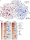 Figure 1
Transcriptional coregulatory network in FGFR3-altered bladder cancer cells and tumors. (A) The cooperativity network inferred from 36 bladder cancer–derived cell lines (BLCa-GRN) transcriptomes and TFs’ activity calculated only in FGFR3-altered cell lines (*FGFR3, n = 5) (Supplemental Table 2). Nodes represent transcription factors and cofactors (TFs/coTFs), with coregulatory interactions shown in gray (H-LICORN algorithm), blue (published protein-protein interactions; PPIs), and red arrows (transcriptional regulation). Node color (red = high, blue = low) indicates TF/coTF activity in *FGFR3 cells, with node size proportional to the number of targets and color intensity reflecting activation level. (B) Heatmap showing the 40 most active TFs/coTFs in *FGFR3 BLCa cell lines and *FGFR3 tumors across NMIBC (n = 261; UROMOL, UROMOL, a European multicenter cohort of patients with NMIBC) and MIBC (n = 66; TCGA) and across molecular subtypes.
To identify the transcriptional programs active in an FGFR3-mutated context, we assessed the activity of each TF/coTF from the expression of their target genes using the CoRegNet algorithm (Supplemental Table 1; supplemental material available online with this article; https://doi.org/10.1172/JCI193280DS1) and CCLE transcriptomic data from 5 FGFR3-dependent BLCa cell lines expressing FGFR3 mutants (fusion proteins or activating point mutations): RT112 (FGFR3-TACC3), RT112-84 (FGFR3-TACC3), RT4 (FGFR3-TACC3), SW-780 (FGFR3-BAIAP2L1), and UM-UC-14 (FGFR3-S249C) (Supplemental Table 2). The computed activity scores were then mapped onto the inferred BLCa-GRN (Figure 1A).
We also calculated the activity of the TFs/coTFs using transcriptomic data from 2 independent cohorts of FGFR3-mutated tumors: 261 NMIBCs from UROMOL (4) and 66 MIBCs from The Cancer Genome Atlas (TCGA) (6) (Supplemental Table 2). We focused on the 40 most active TFs/coTFs in the FGFR3-dependent BLCa cell lines and found that they were also active in the FGFR3-mutated NMIBC and MIBC tumors (36/40 and 40/40, respectively) (Figure 1B). However, when stratifying tumors by molecular subtype, these same TFs were consistently active in FGFR3-mutated LumP MIBCs and class 1 NMIBCs (40/40), whereas other subtypes, particularly classes 2a and 2b, displayed more divergent activation patterns.
These differences are likely because the analyzed cell lines are mainly derived from MIBC and classified as LumP (16, 17) and the luminal-like differentiation of class 1 NMIBCs (4, 5). Supporting the biological relevance of our reconstructed network, we identified several previously described BLCa driver genes, including master regulators known to drive luminal cell identity in LumP tumors. These included GATA3, PPARG, FOXA1, KLF5, TRIM29, and NOTCH3 (Figure 1B) (15, 17–21).
Our results highlighted both subtype-specific and shared transcriptional programs within the FGFR3-mutated tumors, with common TFs/coTFs across FGFR3-mutated subtypes likely representing core regulators of the FGFR3 oncogenic signaling pathway (Figure 1B). TF activity patterns were highly similar across the 5 FGFR3-dependent cell lines from CCLE and the MGH-U3 cell line (FGFR3-Y375C), regardless of whether FGFR3 alterations were translocations leading to expression of fusion proteins or point mutations (Supplemental Figure 1A), though mutated cell lines clustered together based on cosine similarity (Supplemental Figure 1B).
Dependency on FGFR3-driven TFs in FGFR3-mutated BLCa cells, with a focus on p63. Among the TFs activated in FGFR3-mutated tumors and cell lines (Supplemental Table 2), we sought to identify those directly regulated by FGFR3. We took advantage of published transcriptomic data obtained after inhibition of FGFR3 with the pan-FGFR inhibitor AZD4547 for 24 hours in 2 FGFR3-dependent human BLCa cell lines (22), RT112 and MGH-U3. We also used our previously published transcriptomic data from urothelial hyperplasia and bladder tumors of mice overexpressing FGFR3-S249C, together with normal urothelium from littermate controls (23). In this model, the expression of human FGFR3IIIb carrying the S249C mutation was specifically targeted to the urothelium using the murine uroplakin II promoter, leading to the development of hyperplastic lesions and low-grade papillary tumors from 6 and 18 months of age, respectively.
Cross-species analysis of transcriptomic data from human and mouse FGFR3-mutated tumors showed highly concordant transcriptomic programs (23), supporting the relevance of this model to decipher the FGFR3-driven signaling pathway. We assessed TF/coTF activity in each dataset and identified TFs with significantly altered activities (either up- or downregulated) after FGFR3 inhibition in cell lines or in FGFR3-induced mouse tumors compared with normal urothelium. This analysis identified 25 regulators displaying opposite activation patterns upon FGFR3 inhibition in BLCa cell lines or FGFR3-S249C overexpression in mouse bladder tumors (Supplemental Figure 1C). Notably, compared with normal urothelium, the TFs’ and coTFs’ activities (13 activated and 12 repressed by FGFR3) were not changed in FGFR3-S249C–induced hyperplasia but were specifically altered in tumors (Figure 2A), suggesting a role in malignant cell transformation.
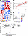 Figure 2
Identification of FGFR3-regulated TFs and discovery of TP63 as a key viability gene. (A) Heatmap showing the activity of 25 FGFR3-regulated TFs/coTFs identified by integrating transcriptomic data from FGFR3 inhibition in bladder cancer cell lines and FGFR3-S249C–induced mouse bladder tumors (Supplemental Figure 1C). TF activity in mouse samples: normal urothelium, hyperplasic urothelium, and bladder tumors. (B) Impact of CRISPR/Cas9 KO (DepMap, CERES scores) of the 25 FGFR3-regulated TFs on cell viability in FGFR3-dependent (*FGFR3, red) versus non-FGFR3-dependent bladder cancer cell lines. (C and D) Effect of CRISPR/Cas9 KO of TP63 on cell viability in bladder cancer cell lines according to FGFR3 status (C) and FGFR3 dependency (D). Box plots show the interquartile range, median (line), and minimum and maximum (whiskers). Molecular subtypes were determined using our previously established single cell–derived classifier (16, 17). TPM, transcripts per million.
To further assess the functional relevance of these regulators, we analyzed publicly available data from genome-wide gene dependency screening (Broad Institute, AVANA CRISPR-Cas9 dataset 2019Q3, https://depmap.org/portal/download/all/?release=DepMap+Public+19Q3). We calculated median CERES (Computational Estimation of gene dependencies from large-scale RNAi and CRISPR Essentiality Screens) dependency scores for each of these 25 genes in 3 FGFR3-dependent cell lines expressing FGFR3 mutants and compared them with scores from 26 BLCa cell lines expressing wild-type FGFR3 (Figure 2B). Only TP63, FOXM1, and NKX2-5 were identified as important for viability of FGFR3-dependent cells, based on a CERES dependency score below –0.3 as the threshold. Notably, the knockout of TP63 (which encodes p63) had the strongest effect on the viability of FGFR3-dependent BLCa cells. This effect was even more pronounced than that in wild-type FGFR3 cells (Figure 2, B and C). However, consistent with the well-established role of p63 in squamous/basal tumors in general and basal MIBC in particular (24–26), basal BLCa cell lines were TP63 dependent in this screen (Figure 2, C and D), and 3 wild-typeFGFR3 luminal cell lines were also TP63 dependent (Figure 2C) but independent of FGFR3 expression for their viability (Figure 2D). Given this unexpected link between FGFR3 activation, classically associated with luminal differentiation and p63 activation/dependency, we focused our study on deciphering the regulatory connection between FGFR3 and p63 and elucidating the functional role of p63 in FGFR3-mutated tumors.
p63 activation is associated with FGFR3 pathway activity and basal-like cell states within luminal tumors. Although p63 emerged as a highly active TF in FGFR3-mutated tumors in both MIBC and NMIBC cohorts (Figure 1B), FGFR3 activation mechanisms are not restricted to these genetic alterations. To further characterize the relationship between FGFR3 signaling and p63 activation, and to determine whether FGFR3 mutation status or FGFR3 pathway activity is associated with p63 activation, we analyzed p63 activity relative to FGFR3 signaling across UROMOL and TCGA cohorts using our previously established FGFR3 pathway signature derived from siRNA-mediated FGFR3 knockdown in FGFR3-dependent cell lines (23).
In MIBC, p63 regulon activity positively correlated with FGFR3 pathway activation, independently of FGFR3 mutation status (Figure 3A). LumP tumors showed the highest concordance between the FGFR3 pathway and p63 activity, whereas basal/squamous tumors presented elevated p63 activity with low FGFR3 pathway activation, consistent with lineage-specific p63 regulation. Similar results were observed in NMIBC, where p63 activity correlates with FGFR3 pathway activation in both FGFR3-mutated and wild-type FGFR3 tumors (Figure 3B). Class 1 and 3 NMIBC showed strong FGFR3–p63 concordance, while class 2 tumors displayed greater heterogeneity.
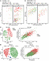 Figure 3
p63 activation correlates with FGFR3 pathway activity and basal-like cell states within luminal bladder tumors. (A and B) Correlation between TP63 GRN-regulon activity and FGFR3 pathway activation signature in individual MIBC tumors from TCGA cohort (A) and individual NMIBC tumors from the UROMOL cohort (Supplemental Table 2) (B), stratified by FGFR3 mutation status and molecular subtype. (C and D) Single-cell RNA-seq analysis of tumor cells from an FGFR3-S249C PDX (F659) (C) and of MGH-U3 cells (D). Uniform manifold approximation and projection (UMAP) representation showing clusters with high or low TP63 expression (left). Correlation between TP63 activity and basal/squamous cell differentiation scores (right). (A–D) Correlation analyses were performed using Spearman’s rank correlation coefficient.
To determine whether this variability reflects intratumoral heterogeneity within FGFR3-driven luminal tumors, we analyzed single-cell RNA-seq data from an FGFR3-S249C PDX (F659) (Figure 3C) and MGH-U3 cells (Figure 3D). In both models, unsupervised clustering identified 2 tumor cell populations defined by high or low TP63 mRNA expression and p63 regulon activity. FGFR3 pathway activation was observed across clusters (Supplemental Figure 2, A and B). Integration of p63 activity scores with basal lineage-specific transcriptional signature revealed that p63-high cells exhibited increased basal/squamous differentiation compared with p63-low cells, despite arising within an overall luminal FGFR3-driven tumor context. p63 activity correlated with basal score (Figure 3, C and D) but not with proliferation (Supplemental Figure 2, C and D), indicating that heterogeneity primarily reflects differences in differentiation state rather than cell cycle. Consistently, despite their distinct p63 activity scores, both MGH-U3 cell clusters exhibited a comparable antiproliferative response to FGFR treatment, indicating that baseline p63 activity does not predict sensitivity to FGFR3-dependent growth inhibition (Supplemental Figure 2C).
Despite heterogeneous p63 activation in FGFR3-mutated tumors and its association with basal-like cell states — a feature generally linked to poor prognosis in BLCa — p63 activity did not stratify clinical outcomes among FGFR3-mutated tumors. Specifically, no differences in overall survival were observed between TP63-high and TP63-low FGFR3-mutated tumors in TCGA MIBC cohort (Supplemental Figure 3A). Similarly, p63 activity did not predict progression-free or recurrence-free survival in FGFR3-mutated NMIBCs from the UROMOL cohort (Supplemental Figure 3, B and C).
Together, these data demonstrated that FGFR3 pathway activation is associated with heterogeneous p63 activation and the emergence of basal-like cell states within luminal tumors. These observations led us to investigate the molecular mechanisms by which FGFR3 regulates p63 activity and p63 function in FGFR3-driven luminal tumors.
FGFR3 regulates p63 activity through TP63 transcriptomic regulation. Based on GRN-derived activity scores, we initially predicted that p63 activity decreases upon FGFR3 inhibition using a pan-FGFR inhibitor in FGFR3-dependent BLCa cell lines and increases in FGFR3-S249C–induced mouse bladder tumors (Supplemental Figure 2C and Figure 2A). To independently validate this regulatory link and ensure specificity for FGFR3, we analyzed our previously obtained list of differentially expressed genes (Affymetrix arrays) after FGFR3 knockdown (KD) using siRNAs in RT112, MGH-U3, and UM-UC-14 cells (9, 27) and comparing FGFR3-S249C–induced mouse bladder tumors’ transcriptomic data with those from normal urothelium (23).
Upstream regulator analysis using Ingenuity Pathway Analysis (IPA; QIAGEN) identified TP63 as an inhibited upstream regulator following FGFR3 KD and as an activated regulator in FGFR3-induced mouse tumors (Figure 4A). We observed that FGFR3 inhibition was associated with a significant reduction in TP63 mRNA expression in MGH-U3 and RT112 cells, indicating transcriptional regulation of TP63 by FGFR3 (Figure 4A). A similar decrease in TP63 mRNA expression associated with a decrease of FGFR3 pathway activity was observed in both TP63-high and TP63-low populations in single-cell RNA-seq data from MGH-U3 cells treated with the FGFR inhibitor erdafitinib (Supplemental Figure 4A). Because TP63 encodes functionally distinct TAp63 and ΔNp63 isoforms, we next investigated which isoform was regulated downstream of FGFR3. Although our Affymetrix and single-cell RNA-seq data did not allow direct discrimination between isoforms, analysis of our previously published H3K4me3 (trimethylation of histone H3 at lysine 4) and H3K27ac (acetylation of histone H3 at lysine 27), marks of active promoters and enhancers, respectively, ChIP-seq data from BLCa cell lines and tumors (17) revealed active chromatin marks exclusively at the ΔNp63 promoter and enhancer regions (Supplemental Figure 4B). Consistently, RNA-seq–based isoform analysis showed that ΔNp63 was the predominant isoform expressed in BLCa cell lines and human tumors from TCGA cohort, with almost no detectable TAp63 expression (Supplemental Figure 4C). Taken together, these data suggested that FGFR3-driven regulation of p63 activity is ΔNp63 dependent. Accordingly, ΔNp63 mRNA levels decreased after FGFR3 inhibition using a pan-FGFR inhibitor in FGFR3-dependent cell lines (Figure 4B) and in an FGFR3-S249C PDX (F659) (9–11), supporting the relevance of the results in human tumors (Figure 4B). Western blot analysis confirmed reduced p63 protein levels after FGFR3 KD or FGFR3 pharmacological inhibition in a large panel of FGFR3-mutated cell lines (Figure 4C) and in the PDX model (Figure 4C). Long exposures revealed that ΔNp63 was the predominant isoform expressed, though FGFR3 expression or activation also affected very low TAp63 protein levels (Supplemental Figure 4D and Figure 4D). Consistent with the lack of association between p63 activity and FGFR3 pathway activation in basal tumors (Figure 3A), and with the absence of codependency between TP63 and FGFR3 in FGFR3 wild-type luminal cells (Figure 2D), p63 expression was not affected by FGFR inhibition in VM-CUB-1 and UM-UC-5 cells, which represent FGFR3 wild-type, FGFR3-independent basal and luminal models, respectively (Figure 4C). In contrast, p63 expression decreased following FGFR inhibition in UM-UC-1 cells, a luminal FGFR3 wild-type line that shows partial FGFR3 dependency but is also dependent on FGFR2 signaling (Figure 2D and Supplemental Figure 5B; DepMap analysis). Time-course experiments in RT112 cells showed that p63 downregulation occurred only at later time points following FGFR inhibition (≥24 hours) (Figure 4D), supporting transcriptional regulation as the primary mechanism rather than protein stabilization or degradation.
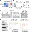 Figure 4
FGFR3 regulates p63 activity through ΔNp63 transcriptional regulation. (A) Upstream regulator analysis using Ingenuity Pathway Analysis software (IPA) identifying p63 as inhibited following FGFR3 knockdown in FGFR3-dependent cell lines and activated in FGFR3-S249C–induced mouse bladder tumors. siFGFR3, FGFR3 siRNA; hFGFR3-S249C OE, human FGFR3-S249C overexpression. (B) ΔNp63 mRNA expression following FGFR3 inhibition in FGFR3-dependent bladder cancer cell lines (PD173074) for 48 hours (n = 3 independent experiments) (left) and in an FGFR3-S249C PDX (BGJ398, 30 mg/kg/d, 4 days) (n = 5 mice/group) (right). Statistical comparisons were performed using an unpaired 2-tailed t test. (C) Western blot analysis of p63 levels following siRNA-mediated FGFR3 knockdown (48 hours) or pharmacological inhibition in FGFR3-mutated cell lines (PD173074, 100 nM, 40 hours) (left) and PDX tumors (BGJ398, 30 mg/kg/d, 4 days) (right). (D) Time-course analysis of p63 protein expression following FGFR inhibition in RT112 cells (PD173074, 500 nM). (E) Spearman’s correlation between TP63 mRNA expression and FGFR3 pathway activity scores across UROMOL NMIBC and TCGA MIBC cohort, respectively. FGFR3 mutation status and molecular subtype for each individual tumor are indicated.
Finally, consistent with these results, analysis of human bladder tumor datasets showed significantly higher TP63 mRNA expression in FGFR3-mutated NMIBCs and MIBCs compared with FGFR3 wild-type tumors in UROMOL and TCGA cohorts, respectively (Supplemental Figure 4E). Moreover, as observed for p63 activity (Figure 3, A and B), TP63 mRNA expression was elevated in FGFR3-active NMIBCs and MIBCs independently of the molecular subtypes (Figure 4E).
Although we did not aim here to comprehensively decipher the upstream signaling mechanisms controlling TP63 transcription by FGFR3 but rather focus on studying its unexpected role in a luminal context, we examined whether pathways that we previously demonstrated as implicated in FGFR3-dependent MYC regulation (9) might also contribute to p63 regulation. Western blot analysis of protein lysates from FGFR3-dependent cells treated with the p38 inhibitor (SB203580) or the PI3K inhibitor (LY294002) revealed reduced p63 expression, suggesting that these FGFR3-activated pathways also contribute to the regulation of TP63 expression, whereas the ERK inhibitor (PD98059) had no effect (Supplemental Figure 4F).
p63 regulates the growth and migration of FGFR3-dependent BLCa cells. To investigate the functional role of p63 in FGFR3-driven luminal tumors, we first sought to validate the dependency of FGFR3-mutated BLCa cells on p63 using siRNA-mediated knockdown, as suggested by our analysis of public CRISPR/Cas9 screening data (Figure 2, C and D). We extended these analyses to 2 additional FGFR3-dependent cell lines, RT4 and MGH-U3, expressing FGFR3-S249C and FGFR3-TACC3, respectively. TP63 was silenced after treatment by 3 siRNAs (Supplemental Figure 5A), resulting in a significant decrease in cell viability across all FGFR3-dependent cell lines tested, 72 hours or 96 hours after transfection (Figure 5A). Consistent with CRISPR-KO–based results (Figure 2D), TP63 knockdown also reduced viability in basal VM-CUB-1 and L1207 cells but had no effect in luminal FGFR3 wild-type, FGFR3-independent cell lines UM-UC-7 and UM-UC-9, except in UM-UC-1 cells (Supplemental Figure 5B).
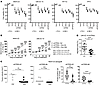 Figure 5
p63 promotes growth and migration of FGFR3-dependent bladder cancer cells. (A) Cell viability assessed by CellTiter-Glo following TP63 siRNA knockdown in FGFR3-dependent bladder cancer cell lines expressing FGFR3-mutated proteins (MGH-U3, UM-UC-4) or FGFR3-fusion protein (RT112 and RT4). Results are from 3 independent experiments, and statistical comparisons were performed using an unpaired 2-tailed t test. *P ≤ 0.05; **P ≤ 0.01. (B) 3D cellular spheroids were established from MGH-U3 and UM-UC-14 cells stably transduced with Dox-inducible shTP63 (shTP63i). Spheroids were grown after a transient (4 days) or with stable (30 days) Dox-induced TP63 knockdown. Statistical comparisons were performed using 2-way ANOVA. Representative images are shown in Supplemental Figure 5D. ††††P ≤ 0.0001 (shTP63i Ctrl vs. Dox 30d); ****P ≤ 0.0001 (shTP63i Ctrl vs. Dox 4d); #P ≤ 0.05 (shTP63i Dox 4d vs. Dox 30d); ####P ≤ 0.0001 (shTP63i Dox 4d vs. Dox 30d). (C) Xenograft tumors. MGH-U3 cells expressing a Dox-inducible shRNA targeting TP63 (shTP63i#1, shTP63i#4) were implanted in mice, with or without Dox in drinking water (1 g/L) for 30 days (n = 6 to 8 mice per group). Left: Tumor growth was measured twice a week and final tumor volumes were compared. Right: Proliferation was assessed via PCNA immunostaining in tumors. Representative images are shown in Supplemental Figure 5E. Comparisons were performed using Wilcoxon’s test. (D) Wound healing assay: migration of UM-UC-14 shTP63i#4 cells after Dox-induced TP63 knockdown. Migration areas relative to untreated controls are represented. Representative images are shown in Supplemental Figure 5F. Data are expressed as mean ± SDs. Statistical differences were defined by an unpaired 2-tailed t test.
To further evaluate the role of p63 in FGFR3-dependent cells in 3D culture and in vivo, we generated stable clones of MGH-U3 and UM-UC-14 cells expressing a doxycycline-inducible (Dox-inducible) shRNA targeting TP63 (shTP63i). Western blot analysis revealed that Dox treatment induced an approximately 50% reduction in p63 protein levels in both cell lines (Supplemental Figure 5C). Western blot analysis confirmed efficient TP63 KD upon Dox treatment (Supplemental Figure 5C). In 3D spheroid assays, both continuous and transient (4-day) TP63 silencing reduced spheroid growth (Figure 5B and Supplemental Figure 5D). Moreover, in vivo, Dox-treated xenografts derived from shTP63i–MGH-U3 cells displayed significantly reduced tumor growth compared with untreated controls (Figure 5C), accompanied by a marked reduction in proliferating cells as assessed by proliferating cell nuclear antigen (PCNA) staining (Figure 5C and Supplemental Figure 5E).
Since ΔNp63, the main TP63 isoform expressed in bladder tumors (Supplemental Figure 4, B and C), is known to regulate cell migration and invasion in several basal/squamous cancer models (28–31), we investigated whether p63 also contributes to FGFR3-induced migration. In a wound healing assay, Dox-induced TP63 KD in shTP63i UM-UC-14 cells significantly impaired cell migration, with effects comparable to those observed after FGFR3 inhibition via a pan-FGFR inhibitor (Figure 5D and Supplemental Figure 5F).
p63 directly regulates genes involved in proliferation and luminal differentiation and establishes an FGFR3-p63 feedback loop. To uncover the transcriptional program controlled by p63 in FGFR3-mutated luminal cells, and compare it with classical basal contexts, we integrated p63 ChIP-seq data with RNA-seq data after TP63 KD in MGH-U3, RT112, and VM-CUB-1 cells. Annotation of ChIP-seq peaks to the nearest genes identified 14,839 unique p63-bound genes, with 9,667 (65%) shared across all 3 cell lines (Figure 6A and Supplemental Figure 6, A and B). This highly significant 3-way overlap (hypergeometric test, P ≈ 5 × 10–131) indicated a conserved set of p63-bound regulatory regions across luminal and basal contexts. We next defined p63-dependent targets in FGFR3-driven tumors by integrating ChIP-seq binding with genes differentially expressed after TP63 KD. Among the 4,936 TP63-regulated genes (DESeq2 analysis, |log2FC| > 0.58) (Supplemental Table 3), 2,642 were direct p63 targets, including 1,542 upregulated and 1,111 downregulated genes (Figure 6B). Gene Ontology (GO) enrichment analysis in MGH-U3 revealed that direct p63 targets were enriched for pathways involved in cell differentiation, proliferation, and lipid metabolism (Figure 6B), consistent with processes regulated by p63 in VM-CUB-1 basal cells (Supplemental Figure 6C) and by FGFR3 (23).
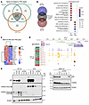 Figure 6
p63 directly regulates proliferation and differentiation genes and establishes an FGFR3–p63 feedback loop. (A) Overlap of p63-bound genes identified by ChIP-seq in luminal FGFR3-dependent cell lines (MGH-U3 and RT112) and in basal cell lines (VM-CUB-1). (B) Venn diagram comparing genes associated with a p63 peak and genes differentially regulated upon siRNA-mediated knockdown of TP63 in MGH-U3. The intersections are considered p63 direct targets (left). Gene Ontology enrichment analysis of directly repressed and activated p63 target genes in FGFR3-dependent luminal MGH-U3 cells (right). (C) Heatmap visualization of expression changes after TP63 KD for selected p63-regulated direct target genes: focus on luminal differentiation TFs and FGFR3. Lipo, Lipofectamine (transfection control). (D) TP63 and chromatin mark (H3K27ac, H3K4me3) occupancy at FGFR3 regulatory regions in bladder cancer cell lines. Basal cell lines are highlighted in red, luminal cell lines in green. (E) Western blot validation of FGFR3 downregulation following siRNA-mediated TP63 knockdown (48 hours) in RT112 and RT4 cells expressing FGFR3-TACC3 and in MGH-U3 cells (FGFR3-Y375C). β-Actin was used as a loading control.
In line with p63 association with basal features in FGFR3-driven tumors even in a luminal context (Figure 3), several key TFs for luminal differentiation, including FOXA1, GATA3, PPARG, and AHR (16, 17, 19), were upregulated following TP63 KD and identified as direct p63 targets (Figure 6C and Supplemental Table 3). Notably, FGFR3 itself was a direct and positively regulated p63 target (Figure 6C), revealing a positive feedback loop between FGFR3 signaling and p63. ChIP-seq for p63 and active chromatin marks in RT112 and MGH-U3 confirmed p63 binding at FGFR3 regulatory regions through p63 binding at enhancer regions (Figure 6D). Western blot analysis in RT112, MGH-U3, and RT4 validated decreased FGFR3 protein levels following TP63 KD (Figure 6E).
Together, these results identify p63 as a key transcriptional regulator in FGFR3-dependent luminal tumors, controlling genes involved in proliferation and differentiation and participating in a positive feedback loop reinforcing FGFR3 signaling.


Copyright © 2026 American Society for Clinical Investigation
ISSN: 0021-9738 (print), 1558-8238 (online)