Go to JCI Insight
  • About
  • Editors
  • Consulting Editors
  • For authors
  • Publication ethics
  • Publication alerts by email
  • Advertising
  • Job board
  • Contact
  • Clinical Research and Public Health
  • Current issue
  • Past issues
  • By specialty
    • COVID-19
    • Cardiology
    • Gastroenterology
    • Immunology
    • Metabolism
    • Nephrology
    • Neuroscience
    • Oncology
    • Pulmonology
    • Vascular biology
    • All ...
  • Videos
    • ASCI Milestone Awards
    • Video Abstracts
    • Conversations with Giants in Medicine
  • Reviews
    • View all reviews ...
    • The cGAS-STING pathway: DNA sensing in health and disease (Jun 2026)
    • Neurodegeneration (Mar 2026)
    • Clinical innovation and scientific progress in GLP-1 medicine (Nov 2025)
    • Pancreatic Cancer (Jul 2025)
    • Complement Biology and Therapeutics (May 2025)
    • Evolving insights into MASLD and MASH pathogenesis and treatment (Apr 2025)
    • Microbiome in Health and Disease (Feb 2025)
    • View all review series ...
  • Viewpoint
  • Collections
    • In-Press Preview
    • Clinical Research and Public Health
    • Research Letters
    • Letters to the Editor
    • Editorials
    • Commentaries
    • Editor's notes
    • Reviews
    • Viewpoints
    • 100th anniversary
    • Top read articles

  • Current issue
  • Past issues
  • Specialties
  • Reviews
  • Review series
  • ASCI Milestone Awards
  • Video Abstracts
  • Conversations with Giants in Medicine
  • In-Press Preview
  • Clinical Research and Public Health
  • Research Letters
  • Letters to the Editor
  • Editorials
  • Commentaries
  • Editor's notes
  • Reviews
  • Viewpoints
  • 100th anniversary
  • Top read articles
  • About
  • Editors
  • Consulting Editors
  • For authors
  • Publication ethics
  • Publication alerts by email
  • Advertising
  • Job board
  • Contact

Research Article

  • 25,923 Articles
  • 0 Posts
  • ← Previous
  • 1
  • 2
  • …
  • 325
  • 326
  • 327
  • …
  • 2592
  • 2593
  • Next →
Foxc1 and Foxc2 deletion causes abnormal lymphangiogenesis and correlates with ERK hyperactivation
Anees Fatima, Ying Wang, Yutaka Uchida, Pieter Norden, Ting Liu, Austin Culver, William H. Dietz, Ford Culver, Meredith Millay, Yoh-suke Mukouyama, Tsutomu Kume
View: Text | PDF

Foxc1 and Foxc2 deletion causes abnormal lymphangiogenesis and correlates with ERK hyperactivation

  • Text
  • PDF
Abstract

The lymphatic vasculature is essential for maintaining interstitial fluid homeostasis, and dysfunctional lymphangiogenesis contributes to various pathological processes, including inflammatory disease and tumor metastasis. Mutations in FOXC2 are dominantly associated with late-onset lymphedema; however, the precise role of FOXC2 and a closely related factor, FOXC1, in the lymphatic system remains largely unknown. Here we identified a molecular cascade by which FOXC1 and FOXC2 regulate ERK signaling in lymphatic vessel growth. In mice, lymphatic endothelial cell–specific (LEC-specific) deletion of Foxc1, Foxc2, or both resulted in increased LEC proliferation, enlarged lymphatic vessels, and abnormal lymphatic vessel morphogenesis. Compared with LECs from control animals, LECs from mice lacking both Foxc1 and Foxc2 exhibited aberrant expression of Ras regulators, and embryos with LEC-specific deletion of Foxc1 and Foxc2, alone or in combination, exhibited ERK hyperactivation. Pharmacological ERK inhibition in utero abolished the abnormally enlarged lymphatic vessels in FOXC-deficient embryos. Together, these results identify FOXC1 and FOXC2 as essential regulators of lymphangiogenesis and indicate a new potential mechanistic basis for lymphatic-associated diseases.

Authors

Anees Fatima, Ying Wang, Yutaka Uchida, Pieter Norden, Ting Liu, Austin Culver, William H. Dietz, Ford Culver, Meredith Millay, Yoh-suke Mukouyama, Tsutomu Kume

×

Uropathogenic Escherichia coli strain CFT073 disrupts NLRP3 inflammasome activation
Anna Waldhuber, Manoj Puthia, Andreas Wieser, Christine Cirl, Susanne Dürr, Silke Neumann-Pfeifer, Simone Albrecht, Franziska Römmler, Tina Müller, Yunji Zheng, Sören Schubert, Olaf Groß, Catharina Svanborg, Thomas Miethke
View: Text | PDF

Uropathogenic Escherichia coli strain CFT073 disrupts NLRP3 inflammasome activation

  • Text
  • PDF
Abstract

Successful bacterial pathogens produce an array of virulence factors that allow subversion of the immune system and persistence within the host. For example, uropathogenic Escherichia coli strains, such as CFT073, express Toll/IL-1 receptor–containing (TIR-containing) protein C (TcpC), which impairs TLR signaling, thereby suppressing innate immunity in the urinary tract and enhancing persistence in the kidneys. Here, we have reported that TcpC also reduces secretion of IL-1β by directly interacting with the NACHT leucin-rich repeat PYD protein 3 (NLRP3) inflammasome, which is crucial for recognition of pathogens within the cytosol. At a low MOI, IL-1β secretion was minimal in CFT073-infected macrophages; however, IL-1β release was markedly increased in macrophages infected with CFT073 lacking tcpC. Induction of IL-1β secretion by CFT073 and tcpC–deficient CFT073 required the NLRP3 inflammasome. TcpC attenuated activation of the NLRP3 inflammasome by binding both NLRP3 and caspase-1 and thereby preventing processing and activation of caspase-1. Moreover, in a murine urinary tract infection model, CFT073 infection rapidly induced expression of the NLRP3 inflammasome in the bladder mucosa; however, the presence of TcpC in WT CFT073 reduced IL-1β levels in the urine of infected mice. Together, these findings illustrate how uropathogenic E. coli use the multifunctional virulence factor TcpC to attenuate innate immune responses in the urinary tract.

Authors

Anna Waldhuber, Manoj Puthia, Andreas Wieser, Christine Cirl, Susanne Dürr, Silke Neumann-Pfeifer, Simone Albrecht, Franziska Römmler, Tina Müller, Yunji Zheng, Sören Schubert, Olaf Groß, Catharina Svanborg, Thomas Miethke

×

MicroRNA-140-5p and SMURF1 regulate pulmonary arterial hypertension
Alexander M.K. Rothman, Nadine D. Arnold, Josephine A. Pickworth, James Iremonger, Loredana Ciuclan, Robert Allen, Sabine Guth-Gundel, Mark Southwood, Nicholas W. Morrell, Matthew Thomas, Sheila E. Francis, David J. Rowlands, Allan Lawrie
View: Text | PDF

MicroRNA-140-5p and SMURF1 regulate pulmonary arterial hypertension

  • Text
  • PDF
Abstract

Loss of the growth-suppressive effects of bone morphogenetic protein (BMP) signaling has been demonstrated to promote pulmonary arterial endothelial cell dysfunction and induce pulmonary arterial smooth muscle cell (PASMC) proliferation, leading to the development of pulmonary arterial hypertension (PAH). MicroRNAs (miRs) mediate higher order regulation of cellular function through coordinated modulation of mRNA targets; however, miR expression is altered by disease development and drug therapy. Here, we examined treatment-naive patients and experimental models of PAH and identified a reduction in the levels of miR-140-5p. Inhibition of miR-140-5p promoted PASMC proliferation and migration in vitro. In rat models of PAH, nebulized delivery of miR-140-5p mimic prevented the development of PAH and attenuated the progression of established PAH. Network and pathway analysis identified SMAD-specific E3 ubiquitin protein ligase 1 (SMURF1) as a key miR-140-5p target and regulator of BMP signaling. Evaluation of human tissue revealed that SMURF1 is increased in patients with PAH. miR-140-5p mimic or SMURF1 knockdown in PASMCs altered BMP signaling, further supporting these factors as regulators of BMP signaling. Finally, Smurf1 deletion protected mice from PAH, demonstrating a critical role in disease development. Together, these studies identify both miR-140-5p and SMURF1 as key regulators of disease pathology and as potential therapeutic targets for the treatment of PAH.

Authors

Alexander M.K. Rothman, Nadine D. Arnold, Josephine A. Pickworth, James Iremonger, Loredana Ciuclan, Robert Allen, Sabine Guth-Gundel, Mark Southwood, Nicholas W. Morrell, Matthew Thomas, Sheila E. Francis, David J. Rowlands, Allan Lawrie

×

Paracellular epithelial sodium transport maximizes energy efficiency in the kidney
Lei Pei, Glenn Solis, Mien T.X. Nguyen, Nikhil Kamat, Lynn Magenheimer, Min Zhuo, Jiahua Li, Joshua Curry, Alicia A. McDonough, Timothy A. Fields, William J. Welch, Alan S.L. Yu
View: Text | PDF

Paracellular epithelial sodium transport maximizes energy efficiency in the kidney

  • Text
  • PDF
Abstract

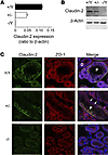
Efficient oxygen utilization in the kidney may be supported by paracellular epithelial transport, a form of passive diffusion that is driven by preexisting transepithelial electrochemical gradients. Claudins are tight-junction transmembrane proteins that act as paracellular ion channels in epithelial cells. In the proximal tubule (PT) of the kidney, claudin-2 mediates paracellular sodium reabsorption. Here, we used murine models to investigate the role of claudin-2 in maintaining energy efficiency in the kidney. We found that claudin-2–null mice conserve sodium to the same extent as WT mice, even during profound dietary sodium depletion, as a result of the upregulation of transcellular Na-K-2Cl transport activity in the thick ascending limb of Henle. We hypothesized that shifting sodium transport to transcellular pathways would lead to increased whole-kidney oxygen consumption. Indeed, compared with control animals, oxygen consumption in the kidneys of claudin-2–null mice was markedly increased, resulting in medullary hypoxia. Furthermore, tubular injury in kidneys subjected to bilateral renal ischemia-reperfusion injury was more severe in the absence of claudin-2. Our results indicate that paracellular transport in the PT is required for efficient utilization of oxygen in the service of sodium transport. We speculate that paracellular permeability may have evolved as a general strategy in epithelial tissues to maximize energy efficiency.

Authors

Lei Pei, Glenn Solis, Mien T.X. Nguyen, Nikhil Kamat, Lynn Magenheimer, Min Zhuo, Jiahua Li, Joshua Curry, Alicia A. McDonough, Timothy A. Fields, William J. Welch, Alan S.L. Yu

×

Transcription factor TLX1 controls retinoic acid signaling to ensure spleen development
Elisa Lenti, Diego Farinello, Kazunari K. Yokoyama, Dmitry Penkov, Laura Castagnaro, Giovanni Lavorgna, Kenly Wuputra, Lisa L. Sandell, Naomi E. Butler Tjaden, Francesca Bernassola, Nicoletta Caridi, Anna De Antoni, Michael Wagner, Katja Kozinc, Karen Niederreither, Francesco Blasi, Diego Pasini, Gregor Majdic, Giovanni Tonon, Paul A. Trainor, Andrea Brendolan
View: Text | PDF

Transcription factor TLX1 controls retinoic acid signaling to ensure spleen development

  • Text
  • PDF
Abstract

The molecular mechanisms that underlie spleen development and congenital asplenia, a condition linked to increased risk of overwhelming infections, remain largely unknown. The transcription factor TLX1 controls cell fate specification and organ expansion during spleen development, and Tlx1 deletion causes asplenia in mice. Deregulation of TLX1 expression has recently been proposed in the pathogenesis of congenital asplenia in patients carrying mutations of the gene-encoding transcription factor SF-1. Herein, we have shown that TLX1-dependent regulation of retinoic acid (RA) metabolism is critical for spleen organogenesis. In a murine model, loss of Tlx1 during formation of the splenic anlage increased RA signaling by regulating several genes involved in RA metabolism. Uncontrolled RA activity resulted in premature differentiation of mesenchymal cells and reduced vasculogenesis of the splenic primordium. Pharmacological inhibition of RA signaling in Tlx1-deficient animals partially rescued the spleen defect. Finally, spleen growth was impaired in mice lacking either cytochrome P450 26B1 (Cyp26b1), which results in excess RA, or retinol dehydrogenase 10 (Rdh10), which results in RA deficiency. Together, these findings establish TLX1 as a critical regulator of RA metabolism and provide mechanistic insights into the molecular determinants of human congenital asplenia.

Authors

Elisa Lenti, Diego Farinello, Kazunari K. Yokoyama, Dmitry Penkov, Laura Castagnaro, Giovanni Lavorgna, Kenly Wuputra, Lisa L. Sandell, Naomi E. Butler Tjaden, Francesca Bernassola, Nicoletta Caridi, Anna De Antoni, Michael Wagner, Katja Kozinc, Karen Niederreither, Francesco Blasi, Diego Pasini, Gregor Majdic, Giovanni Tonon, Paul A. Trainor, Andrea Brendolan

×

Stabilization of histone demethylase PHF8 by USP7 promotes breast carcinogenesis
Qian Wang, Shuai Ma, Nan Song, Xin Li, Ling Liu, Shangda Yang, Xiang Ding, Lin Shan, Xing Zhou, Dongxue Su, Yue Wang, Qi Zhang, Xinhua Liu, Na Yu, Kai Zhang, Yongfeng Shang, Zhi Yao, Lei Shi
View: Text | PDF

Stabilization of histone demethylase PHF8 by USP7 promotes breast carcinogenesis

  • Text
  • PDF
Abstract

The histone demethylase PHF8 has been implicated in multiple pathological disorders, including X-linked mental retardation and tumorigenesis. However, it is not clear how the abundance and function of PHF8 are regulated. Here, we report that PHF8 physically associates with the deubiquitinase USP7. Specifically, we demonstrated that USP7 promotes deubiquitination and stabilization of PHF8, leading to the upregulation of a group of genes, including cyclin A2, that are critical for cell growth and proliferation. The USP7-encoding gene was also transcriptionally regulated by PHF8, via positive feedback. USP7 was overexpressed in breast carcinomas, and the level of expression positively correlated with expression of PHF8 and cyclin A2 and with the histological grade of breast cancer. We showed that USP7 promotes breast carcinogenesis by stabilizing PHF8 and upregulating cyclin A2 and that the interaction between USP7 and PHF8 is augmented during DNA damage. Moreover, USP7-promoted PHF8 stabilization conferred cellular resistance to genotoxic insults and was required for the recruitment of BLM and KU70, which are both essential for DNA double-strand break repair. Our study mechanistically links USP7 to epigenetic regulation and DNA repair. Moreover, these data support the pursuit of USP7 and PHF8 as potential targets for breast cancer intervention, especially in combination with chemo- or radiotherapies.

Authors

Qian Wang, Shuai Ma, Nan Song, Xin Li, Ling Liu, Shangda Yang, Xiang Ding, Lin Shan, Xing Zhou, Dongxue Su, Yue Wang, Qi Zhang, Xinhua Liu, Na Yu, Kai Zhang, Yongfeng Shang, Zhi Yao, Lei Shi

×

Enzyme replacement with PEGylated cystathionine β-synthase ameliorates homocystinuria in murine model
Erez M. Bublil, Tomas Majtan, Insun Park, Richard S. Carrillo, Helena Hůlková, Jakub Krijt, Viktor Kožich, Jan P. Kraus
View: Text | PDF

Enzyme replacement with PEGylated cystathionine β-synthase ameliorates homocystinuria in murine model

  • Text
  • PDF
Abstract

Homocystinuria, which typically results from cystathionine β-synthase (CBS) deficiency, is the most common defect of sulfur amino acid metabolism. CBS condenses homocysteine and serine to cystathionine that is then converted to cysteine. Individuals with homocystinuria have markedly elevated plasma levels of homocysteine and methionine and reduced concentrations of cystathionine and cysteine. Clinical disease manifestations include thromboembolism and neuropsychiatric, ocular, and skeletal complications. Here, we have shown that administration of PEGylated CBS into the circulation of homocystinuria model mice alters the extra- and intracellular equilibrium of sulfur amino acids, resulting in a decrease of approximately 75% in plasma total homocysteine (tHcy) and normalization of cysteine concentrations. Moreover, the decrease in homocysteine and the normalization of cysteine in PEGylated CBS–treated model mice were accompanied by improvement of histopathological liver symptoms and increased survival. Together, these data suggest that CBS enzyme replacement therapy (ERT) is a promising approach for the treatment of homocystinuria and that ERT for metabolic diseases may not necessitate introduction of the deficient enzyme into its natural intracellular compartment.

Authors

Erez M. Bublil, Tomas Majtan, Insun Park, Richard S. Carrillo, Helena Hůlková, Jakub Krijt, Viktor Kožich, Jan P. Kraus

×

Inhibition of SHP2 ameliorates the pathogenesis of systemic lupus erythematosus
Jianxun Wang, Masayuki Mizui, Li-Fan Zeng, Roderick Bronson, Michele Finnell, Cox Terhorst, Vasileios C. Kyttaris, George C. Tsokos, Zhong-Yin Zhang, Maria I. Kontaridis
View: Text | PDF

Inhibition of SHP2 ameliorates the pathogenesis of systemic lupus erythematosus

  • Text
  • PDF
Abstract

Systemic lupus erythematosus (SLE) is a devastating multisystemic autoimmune disorder. However, the molecular mechanisms underlying its pathogenesis remain elusive. Some patients with Noonan syndrome, a congenital disorder predominantly caused by gain-of-function mutations in the protein tyrosine phosphatase SH2 domain–containing PTP (SHP2), have been shown to develop SLE, suggesting a functional correlation between phosphatase activity and systemic autoimmunity. To test this directly, we measured SHP2 activity in spleen lysates isolated from lupus-prone MRL/lpr mice and found it was markedly increased compared with that in control mice. Similar increases in SHP2 activity were seen in peripheral blood mononuclear cells isolated from lupus patients relative to healthy patients. To determine whether SHP2 alters autoimmunity and related immunopathology, we treated MRL/lpr mice with an SHP2 inhibitor and found increased life span, suppressed crescentic glomerulonephritis, reduced spleen size, and diminished skin lesions. SHP2 inhibition also reduced numbers of double-negative T cells, normalized ERK/MAPK signaling, and decreased production of IFN-γ and IL-17A/F, 2 cytokines involved in SLE-associated organ damage. Moreover, in cultured human lupus T cells, SHP2 inhibition reduced proliferation and decreased production of IFN-γ and IL-17A/F, further implicating SHP2 in lupus-associated immunopathology. Taken together, these data identify SHP2 as a critical regulator of SLE pathogenesis and suggest targeting of its activity as a potent treatment for lupus patients.

Authors

Jianxun Wang, Masayuki Mizui, Li-Fan Zeng, Roderick Bronson, Michele Finnell, Cox Terhorst, Vasileios C. Kyttaris, George C. Tsokos, Zhong-Yin Zhang, Maria I. Kontaridis

×

CD62L+ NKT cells have prolonged persistence and antitumor activity in vivo
Gengwen Tian, Amy N. Courtney, Bipulendu Jena, Andras Heczey, Daofeng Liu, Ekaterina Marinova, Linjie Guo, Xin Xu, Hiroki Torikai, Qianxing Mo, Gianpietro Dotti, Laurence J. Cooper, Leonid S. Metelitsa
View: Text | PDF

CD62L+ NKT cells have prolonged persistence and antitumor activity in vivo

  • Text
  • PDF
Abstract

Vα24-invariant natural killer T cells (NKTs) localize to tumors and have inherent antitumor properties, making them attractive chimeric antigen receptor (CAR) carriers for redirected cancer immunotherapy. However, clinical application of CAR-NKTs has been impeded, as mechanisms responsible for NKT expansion and the in vivo persistence of these cells are unknown. Here, we demonstrated that antigen-induced expansion of primary NKTs in vitro associates with the accumulation of a CD62L+ subset and exhaustion of CD62L– cells. Only CD62L+ NKTs survived and proliferated in response to secondary stimulation. When transferred to immune-deficient NSG mice, CD62L+ NKTs persisted 5 times longer than CD62L– NKTs. Moreover, CD62L+ cells transduced with a CD19-specific CAR achieved sustained tumor regression in a B cell lymphoma model. Proliferating CD62L+ cells downregulated or maintained CD62L expression when activated via T cell receptor alone or in combination with costimulatory receptors. We generated HLAnull K562 cell clones that were engineered to express CD1d and costimulatory ligands. Clone B-8-2 (HLAnullCD1dmedCD86high4-1BBLmedOX40Lhigh) induced the highest rates of NKT expansion and CD62L expression. B-8-2–expanded CAR-NKTs exhibited prolonged in vivo persistence and superior therapeutic activities in models of lymphoma and neuroblastoma. Therefore, we have identified CD62L as a marker of a distinct NKT subset endowed with high proliferative potential and have developed artificial antigen-presenting cells that generate CD62L-enriched NKTs for effective cancer immunotherapy.

Authors

Gengwen Tian, Amy N. Courtney, Bipulendu Jena, Andras Heczey, Daofeng Liu, Ekaterina Marinova, Linjie Guo, Xin Xu, Hiroki Torikai, Qianxing Mo, Gianpietro Dotti, Laurence J. Cooper, Leonid S. Metelitsa

×

Hotspot autoimmune T cell receptor binding underlies pathogen and insulin peptide cross-reactivity
David K. Cole, Anna M. Bulek, Garry Dolton, Andrea J. Schauenberg, Barbara Szomolay, William Rittase, Andrew Trimby, Prithiviraj Jothikumar, Anna Fuller, Ania Skowera, Jamie Rossjohn, Cheng Zhu, John J. Miles, Mark Peakman, Linda Wooldridge, Pierre J. Rizkallah, Andrew K. Sewell
View: Text | PDF | Corrigendum

Hotspot autoimmune T cell receptor binding underlies pathogen and insulin peptide cross-reactivity

  • Text
  • PDF
Abstract

The cross-reactivity of T cells with pathogen- and self-derived peptides has been implicated as a pathway involved in the development of autoimmunity. However, the mechanisms that allow the clonal T cell antigen receptor (TCR) to functionally engage multiple peptide–major histocompatibility complexes (pMHC) are unclear. Here, we studied multiligand discrimination by a human, preproinsulin reactive, MHC class-I–restricted CD8+ T cell clone (1E6) that can recognize over 1 million different peptides. We generated high-resolution structures of the 1E6 TCR bound to 7 altered peptide ligands, including a pathogen-derived peptide that was an order of magnitude more potent than the natural self-peptide. Evaluation of these structures demonstrated that binding was stabilized through a conserved lock-and-key–like minimal binding footprint that enables 1E6 TCR to tolerate vast numbers of substitutions outside of this so-called hotspot. Highly potent antigens of the 1E6 TCR engaged with a strong antipathogen-like binding affinity; this engagement was governed though an energetic switch from an enthalpically to entropically driven interaction compared with the natural autoimmune ligand. Together, these data highlight how T cell cross-reactivity with pathogen-derived antigens might break self-tolerance to induce autoimmune disease.

Authors

David K. Cole, Anna M. Bulek, Garry Dolton, Andrea J. Schauenberg, Barbara Szomolay, William Rittase, Andrew Trimby, Prithiviraj Jothikumar, Anna Fuller, Ania Skowera, Jamie Rossjohn, Cheng Zhu, John J. Miles, Mark Peakman, Linda Wooldridge, Pierre J. Rizkallah, Andrew K. Sewell

×
  • ← Previous
  • 1
  • 2
  • …
  • 325
  • 326
  • 327
  • …
  • 2592
  • 2593
  • Next →

No posts were found with this tag.

Advertisement

Copyright © 2026 American Society for Clinical Investigation
ISSN: 0021-9738 (print), 1558-8238 (online)

Sign up for email alerts