
Go to JCI Insight
  • About
  • Editors
  • Consulting Editors
  • For authors
  • Journal stats
  • Publication ethics
  • Publication alerts by email
  • Advertising
  • Job board
  • Contact
  • Clinical Research and Public Health
  • Current issue
  • Past issues
  • By specialty
    • COVID-19
    • Cardiology
    • Gastroenterology
    • Immunology
    • Metabolism
    • Nephrology
    • Neuroscience
    • Oncology
    • Pulmonology
    • Vascular biology
    • All ...
  • Videos
    • ASCI Milestone Awards
    • Video Abstracts
    • Conversations with Giants in Medicine
  • Reviews
    • View all reviews ...
    • Emerging therapeutic strategies in breast cancer (Oct 2026)
    • The cGAS-STING pathway: DNA sensing in health and disease (Jun 2026)
    • Neurodegeneration (Mar 2026)
    • Clinical innovation and scientific progress in GLP-1 medicine (Nov 2025)
    • Pancreatic Cancer (Jul 2025)
    • Complement Biology and Therapeutics (May 2025)
    • Evolving insights into MASLD and MASH pathogenesis and treatment (Apr 2025)
    • View all review series ...
  • Viewpoint
  • Collections
    • In-Press Preview
    • Clinical Research and Public Health
    • Research Letters
    • Letters to the Editor
    • Editorials
    • Commentaries
    • Editor's notes
    • Reviews
    • Viewpoints
    • 100th anniversary
    • Top read articles

  • Current issue
  • Past issues
  • Specialties
  • Reviews
  • Review series
  • ASCI Milestone Awards
  • Video Abstracts
  • Conversations with Giants in Medicine
  • In-Press Preview
  • Clinical Research and Public Health
  • Research Letters
  • Letters to the Editor
  • Editorials
  • Commentaries
  • Editor's notes
  • Reviews
  • Viewpoints
  • 100th anniversary
  • Top read articles
  • About
  • Editors
  • Consulting Editors
  • For authors
  • Journal stats
  • Publication ethics
  • Publication alerts by email
  • Advertising
  • Job board
  • Contact
Top
  • View PDF
  • Download citation information
  • Send a comment
  • Terms of use
  • Standard abbreviations
  • Need help? Email the journal
  • Top
  • Abstract
  • Shared distress creates a diversity of responses
  • A vasopressin gate for care
  • Reinforcing prosocial action
  • Unresolved questions
  • Conflict of interest
  • Funding support
  • Acknowledgments
  • Footnotes
  • References
  • Version history
  • Article usage
  • Citations to this article

Advertisement

Commentary Open Access | 10.1172/JCI210876

A vasopressin gate transforms social distress into caregiving

Jonathan P. Fadok1,2,3

1Department of Psychology, School of Science and Engineering, and

2Tulane Brain Institute, Tulane University, New Orleans, Louisiana, USA.

3Southeast Louisiana VA Healthcare System (SLVHCS), New Orleans, Louisiana, USA.

Address correspondence to: Jonathan P. Fadok, Department of Psychology, Tulane University, 208 Flower Hall, 6823 St. Charles Avenue, New Orleans, Louisiana, 70118, USA. Email: jfadok@tulane.edu.

Find articles by Fadok, J. in: PubMed | Google Scholar

Published October 1, 2026 - More info

Published in Volume 136, Issue 19 on October 1, 2026
J Clin Invest. 2026;136(19):e210876. https://doi.org/10.1172/JCI210876.
© 2026 Fadok et al. This work is licensed under the Creative Commons Attribution 4.0 International License. To view a copy of this license, visit http://creativecommons.org/licenses/by/4.0/.
Published October 1, 2026 - Version history
View PDF

Related article:

Hypothalamic-amygdala gating of caregiving-like prosocial behavior in mice
Kai-Wen Geng, Rui-Rui Wang, Yan Yang, Yan Wang, Ting He, Chun-Li Li, Chong-Shun Xu, Jun Chen
Research Article Cell biology Neuroscience

Hypothalamic-amygdala gating of caregiving-like prosocial behavior in mice

  • Text
  • PDF
Abstract

Distress sharing within an ingroup instinctively motivates caregiving-like prosocial behavior in social animals and humans, yet how aversive social stimuli paradoxically trigger such behavior remains unclear. Here, we show that stress-state matching in observer mice elicits caregiving-like behaviors (allogrooming and injury-targeted allolicking) toward cagemates in pain, resulting in synchronized social buffering. Importantly, we identify that the excitation of vasopressin receptor 1a–expressing (V1aR-expressing) neurons in the central medial amygdala (CeM) underlies both emotional-state matching and subsequent allogrooming/allolicking. This excitation is accomplished by arginine vasopressin (AVP) release from presynaptic terminals of the hypothalamic paraventricular nucleus (PVN) to act specifically on postsynaptic V1aR in the CeM. The CeMV1aR neurons then project to the ventral tegmental area (VTA) to drive caregiving-like actions through positive reinforcement, manifested as prosocial preference and recurrent allogrooming/allolicking, and to produce anxiolysis in the caregiver. Collectively, we unravel a dedicated trisynaptic PVNAVP-CeMV1aR-VTA circuit driving and gating caregiving-like behaviors and advance the mechanistic understanding of AVP-V1aR signaling in prosociality.

Authors

Kai-Wen Geng, Rui-Rui Wang, Yan Yang, Yan Wang, Ting He, Chun-Li Li, Chong-Shun Xu, Jun Chen

×

Abstract

Another individual’s distress can provoke withdrawal and avoidance behaviors, yet, in social species, it can also motivate approach and caregiving behavior. In this issue, Geng et al. identified a vasopressin-sensitive neuronal circuit that converts distress signals from a cagemate into caregiving-like behavior in mice. Using fiber photometry, electrophysiological recordings, and optogenetic activation to dissect this circuit, they showed that vasopressin neurons in the paraventricular hypothalamus recruit vasopressin 1a receptor–expressing (V1aR-expressing) neurons in the medial central amygdala, which engage the ventral tegmental area to promote allogrooming and injury-directed licking. The findings link emotional state matching, prosocial action, reinforcement, and stress relief, while raising important questions about circuit specificity, sex as a biological variable, and translation.

Shared distress creates a diversity of responses

The distress of another individual is itself a potent stimulus. It can capture attention, elevate arousal, and promote avoidance. In social species, however, the same signal can motivate approach, touch, and targeted care. Empathy is often discussed as the capacity to share or understand another’s emotional state, but shared distress alone does not necessarily lead to prosocial behavior. It must be coupled to motivational and motor systems that select an appropriate behavioral response (1–3). How the brain transforms another’s negative state into an organized action that benefits the distressed individual is a central question in social neuroscience.

Rodent models have made it possible to study this transformation in an experimental setting. Prairie voles and mice allogroom distressed partners, and mice can direct licking toward a site of injury (4–6). These behaviors should be described cautiously as caregiving-like rather than equated with the full cognitive experience of human empathy. Even so, they allow investigators to separate several processes that are difficult to disentangle in people, such as detection of social distress, emotional state matching, approach, action selection, and reinforcement. In this issue, Geng and colleagues used distress-evoked allogrooming in mice to study caregiving-like behaviors and define a subcortical pathway linking these components (7).

A vasopressin gate for care

Geng et al. modeled distress-induced allogrooming with pairs of mice: a “demonstrator” mouse and an “observer” mouse that was already familiar to the demonstrator. The authors injected melittin, the main pain-producing component of honeybee venom, into the nape of a demonstrator mouse, producing ongoing pain at a location the animal could not easily groom. After reunion, a familiar observer rapidly investigated the demonstrator and then performed allogrooming and injury-targeted allolicking. Blocking peripheral pain in the demonstrator reduced these responses, indicating that the observer was responding to the partner’s pain state rather than the injection procedure alone. A transparent divider that preserved visual, olfactory, and auditory contact while preventing touch increased circulating corticosterone in observers. Moreover, antagonizing corticotropin-releasing factor receptor 1 reduced caregiving-like behavior, and the amount of caregiving correlated negatively with corticosterone in both members of the pair. Together, these findings support a reciprocal model in which social stress drives caregiving, and caregiving then limits stress transmission. This interpretation is consistent with evidence that reducing affective arousal can impair helping in rats (8), although corticosterone measured at the end of an interaction cannot by itself resolve the moment-to-moment internal state of either animal.

Geng et al. next identified arginine vasopressin (AVP) signaling as a molecular gate for this transformation. In the central nervous system, AVP secretion has been linked to social behavior via its actions in limbic and mesolimbic pathways, which involve the amygdala, hypothalamus, and ventral tegmental area (VTA), among other regions. c-Fos mapping revealed increased c-Fos expression in the central amygdala of observer mice, while enriched V1aR expression in the medial central amygdala (CeM) identified this subregion as a candidate mediator of caregiving-like responses. Global deletion of V1aR, or local V1aR knockdown in CeM, impaired emotional state matching, prosocial preference, and caregiving-like behavior. By contrast, oxytocin-null mice showed intact behavior. This distinction is notable because AVP and oxytocin can engage neighboring but functionally distinct populations in the central amygdala (9, 10). Fiber photometry measured increased population calcium signals from CeM V1aR neurons during the observer mouse’s initial investigation of a demonstrator mouse in pain and again at the onset of allogrooming and allolicking. Ablating or inhibiting these neurons reduced caregiving, whereas their activation evoked social approach and allogrooming even toward an uninjured cagemate. Injury-directed licking remained contingent on a partner in pain, suggesting that CeM V1aR activity provides a permissive action signal, while sensory or cortical information preserves behavioral specificity.

The upstream source of AVP secretion in the CeM was the paraventricular nucleus of the hypothalamus (PVN), a region positioned to couple social information to physiological arousal (11). Monosynaptic tracing identified PVN AVP input to CeM V1aR neurons. Disrupting PVN AVP neurons reduced caregiving, whereas optogenetic activation of these neurons or their CeM terminals initiated the behavior. Electrophysiological recordings in brain slices provided an important mechanistic link: AVP depolarized CeM V1aR neurons, stimulation of PVN AVP terminals reproduced this effect, and a V1aR antagonist blocked it. The response persisted when ionotropic glutamate receptors were blocked. This unusually complete set of anatomical, physiological, pharmacological, and behavioral experiments strongly supports direct peptidergic recruitment of the CeM.

Reinforcing prosocial action

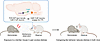
The circuit did not end with action initiation. CeM V1aR neurons projected to the VTA, and inhibiting their VTA terminals reduced spontaneous caregiving. Optogenetic activation of VTA terminals promoted allogrooming and allolicking, supported real-time place preference, and reduced corticosterone in observers exposed to a distressed cagemate behind a divider. These results place the VTA at the intersection of prosocial motivation, positive valence, and stress relief. The most important conceptual advance is the circuit architecture linking distinct brain areas to promote prosocial function: A negatively valenced social signal recruits PVN AVP input to a CeM gate for caregiving; CeM output engages the VTA, reinforcing an action that reduces stress in both animals (Figure 1). This feedback loop offers a biological explanation for why caregiving can recur despite being triggered by an aversive event. It also fits broader evidence that positive affect can reinforce prosocial behavior in humans (12).
A hypothalamic-amygdala-midbrain circuit transforms shared distress into caFigure 1

A hypothalamic-amygdala-midbrain circuit transforms shared distress into caregiving-like behavior. Geng et al.’s findings support a model wherein exposure to a familiar demonstrator in pain induces emotional state matching in an observer. Arginine vasopressin (AVP) released from paraventricular hypothalamic nucleus (PVN) terminals in the medial central amygdala (CeM) acts on vasopressin 1a receptor–expressing (V1aR-expressing) neurons. These neurons project to and engage the ventral tegmental area (VTA) to promote allogrooming and injury-targeted allolicking. VTA engagement provides positive reinforcement that may support recurrent caregiving-like behavior. Caregiving is accompanied by reduced corticosterone in both animals, consistent with reciprocal social buffering.
Unresolved questions

Several questions will determine how broadly this model applies. First, the VTA is not a unitary reward center; it contains molecularly and anatomically distinct dopamine, GABA, and glutamate populations with divergent outputs (13). The identity of the VTA neurons receiving CeM V1aR input, and the pathways through which they reinforce caregiving or regulate the hypothalamic-pituitary-adrenal axis, remain unknown. It will also be important to test whether motivation, motor patterning, positive valence, and stress relief are carried by the same CeM-to-VTA projection or by separable branches. Likewise, population photometry cannot determine whether single CeM V1aR neurons sequentially encode social distress and caregiving actions, or whether distinct ensembles are intermixed. Cell-resolved recordings across the full interaction could distinguish these possibilities.

Second, the pathway Geng et al. identified is best viewed as a core subcortical scaffold rather than a complete circuit for empathy. The sensory inputs that recruit PVN AVP neurons are unknown, and cortical systems likely provide context, experience, and action specificity. The anterior cingulate cortex, for example, has been implicated in selecting allogrooming versus injury-directed allolicking (6). Emotional state matching between individuals also requires further temporal resolution using simultaneous autonomic, hormonal, and neural measurements. Finally, most mechanistic experiments were performed in male mice, despite the strong dependence of AVP systems on sex and social context (11). Testing unfamiliar animals, different relationships and stressors, and both sexes will reveal whether this pathway is a dedicated caregiving circuit or a more general mechanism that converts social arousal into affiliative action.

Translation of these findings should therefore remain measured. Allogrooming in mice is not human empathy, and the intact behavior of oxytocin-null mice does not exclude oxytocin from other forms of care or from compensation during development (14). Nonetheless, the study suggests a useful organizing principle. Prosocial deficits could arise from failures to detect distress, share arousal, convert arousal into action, or experience reinforcement from helping. These are biologically distinct problems that may look similar at the behavioral level. Human studies linking amygdala responsiveness and VTA activity to empathic concern are consistent with this framework, although they do not establish circuit homology (15, 16). Geng et al. move the field beyond asking where empathy resides and toward a more tractable question: how does the brain transform another’s distress into care?

Conflict of interest

The author has declared that no conflict of interest exists.

Funding support
  • Merit Review Award # I01 BX006434 from the United States (U.S.) Department of Veterans Affairs, Biomedical Laboratory Research and Development Service.
Acknowledgments

The contents of this article do not represent the views of the U.S. Department of Veterans Affairs or the United States Government. The author used ChatGPT (OpenAI) to assist with editing the manuscript. All content was reviewed and revised by the author.

Address correspondence to: Jonathan P. Fadok, Department of Psychology, Tulane University, 208 Flower Hall, 6823 St. Charles Avenue, New Orleans, Louisiana, 70118, USA. Email: jfadok@tulane.edu.

Footnotes

Copyright: © 2026, Fadok et al. This is an open access article published under the terms of the Creative Commons Attribution 4.0 International License.

Reference information: J Clin Invest. 2026;136(19):e210876. https://doi.org/10.1172/JCI210876.

See the related article at Hypothalamic-amygdala gating of caregiving-like prosocial behavior in mice.

References
  1. de Waal FBM, Preston SD. Mammalian empathy: behavioural manifestations and neural basis. Nat Rev Neurosci. 2017;18(8):498–509.
    View this article via: PubMed CrossRef Google Scholar
  2. Wu YE, Hong W. Neural basis of prosocial behavior. Trends Neurosci. 2022;45(10):749–762.
    View this article via: CrossRef PubMed Google Scholar
  3. Keysers C, et al. Emotional contagion and prosocial behavior in rodents. Trends Cogn Sci. 2022;26(8):688–706.
    View this article via: CrossRef PubMed Google Scholar
  4. Burkett JP, et al. Oxytocin-dependent consolation behavior in rodents. Science. 2016;351(6271):375–378.
    View this article via: CrossRef PubMed Google Scholar
  5. Wu YE, et al. Neural control of affiliative touch in prosocial interaction. Nature. 2021;599(7884):262–267.
    View this article via: CrossRef PubMed Google Scholar
  6. Zhang M, et al. Cortical regulation of helping behaviour towards others in pain. Nature. 2024;626(7997):136–144.
    View this article via: CrossRef PubMed Google Scholar
  7. Geng KW, et al. Hypothalamic-amygdala gating of caregiving-like prosocial behavior in mice. J Clin Invest. 2026;136(19):e204317.
    View this article via: JCI CrossRef Google Scholar
  8. Ben-Ami Bartal I, et al. Anxiolytic treatment impairs helping behavior in rats. Front Psychol. 2016;7:850.
    View this article via: PubMed CrossRef Google Scholar
  9. Huber D, et al. Vasopressin and oxytocin excite distinct neuronal populations in the central amygdala. Science. 2005;308(5719):245–248.
    View this article via: CrossRef PubMed Google Scholar
  10. Fadok JP, et al. New perspectives on central amygdala function. Curr Opin Neurobiol. 2018;49:141–147.
    View this article via: CrossRef PubMed Google Scholar
  11. Neumann ID, Landgraf R. Balance of brain oxytocin and vasopressin: implications for anxiety, depression, and social behaviors. Trends Neurosci. 2012;35(11):649–659.
    View this article via: CrossRef PubMed Google Scholar
  12. Aknin LB, et al. Positive feelings reward and promote prosocial behavior. Curr Opin Psychol. 2018;20:55–59.
    View this article via: CrossRef PubMed Google Scholar
  13. Morales M, Margolis EB. Ventral tegmental area: cellular heterogeneity, connectivity and behaviour. Nat Rev Neurosci. 2017;18(2):73–85.
    View this article via: CrossRef PubMed Google Scholar
  14. Berendzen KM, et al. Oxytocin receptor is not required for social attachment in prairie voles. Neuron. 2023;111(6):787–796.
    View this article via: CrossRef PubMed Google Scholar
  15. Marsh AA, et al. Neural and cognitive characteristics of extraordinary altruists. Proc Natl Acad Sci U S A. 2014;111(42):15036–15041.
    View this article via: CrossRef PubMed Google Scholar
  16. FeldmanHall O, et al. Empathic concern drives costly altruism. Neuroimage. 2015;105:347–356.
    View this article via: CrossRef PubMed Google Scholar
Version history
  • Version 1 (October 1, 2026): Electronic publication

Article tools

  • View PDF
  • Download citation information
  • Send a comment
  • Terms of use
  • Standard abbreviations
  • Need help? Email the journal

Metrics

  • Article usage
  • Citations to this article

Go to

  • Top
  • Abstract
  • Shared distress creates a diversity of responses
  • A vasopressin gate for care
  • Reinforcing prosocial action
  • Unresolved questions
  • Conflict of interest
  • Funding support
  • Acknowledgments
  • Footnotes
  • References
  • Version history
Advertisement
Advertisement

Copyright © 2026 American Society for Clinical Investigation
ISSN: 0021-9738 (print), 1558-8238 (online)

Sign up for email alerts