
Go to JCI Insight
  • About
  • Editors
  • Consulting Editors
  • For authors
  • Journal stats
  • Publication ethics
  • Publication alerts by email
  • Advertising
  • Job board
  • Contact
  • Clinical Research and Public Health
  • Current issue
  • Past issues
  • By specialty
    • COVID-19
    • Cardiology
    • Gastroenterology
    • Immunology
    • Metabolism
    • Nephrology
    • Neuroscience
    • Oncology
    • Pulmonology
    • Vascular biology
    • All ...
  • Videos
    • ASCI Milestone Awards
    • Video Abstracts
    • Conversations with Giants in Medicine
  • Reviews
    • View all reviews ...
    • Emerging therapeutic strategies in breast cancer (Oct 2026)
    • The cGAS-STING pathway: DNA sensing in health and disease (Jun 2026)
    • Neurodegeneration (Mar 2026)
    • Clinical innovation and scientific progress in GLP-1 medicine (Nov 2025)
    • Pancreatic Cancer (Jul 2025)
    • Complement Biology and Therapeutics (May 2025)
    • Evolving insights into MASLD and MASH pathogenesis and treatment (Apr 2025)
    • View all review series ...
  • Viewpoint
  • Collections
    • In-Press Preview
    • Clinical Research and Public Health
    • Research Letters
    • Letters to the Editor
    • Editorials
    • Commentaries
    • Editor's notes
    • Reviews
    • Viewpoints
    • 100th anniversary
    • Top read articles

  • Current issue
  • Past issues
  • Specialties
  • Reviews
  • Review series
  • ASCI Milestone Awards
  • Video Abstracts
  • Conversations with Giants in Medicine
  • In-Press Preview
  • Clinical Research and Public Health
  • Research Letters
  • Letters to the Editor
  • Editorials
  • Commentaries
  • Editor's notes
  • Reviews
  • Viewpoints
  • 100th anniversary
  • Top read articles
  • About
  • Editors
  • Consulting Editors
  • For authors
  • Journal stats
  • Publication ethics
  • Publication alerts by email
  • Advertising
  • Job board
  • Contact
Top
  • View PDF
  • Download citation information
  • Send a comment
  • Terms of use
  • Standard abbreviations
  • Need help? Email the journal
  • Top
  • Abstract
  • Introduction
  • Making sense of the cardiac macrophage diversity
  • Cell-specific effects of type I IFN signaling in the infarcted myocardium
  • Conclusion and future directions
  • Conflict of interest
  • Funding support
  • Footnotes
  • References
  • Version history
  • Article usage
  • Citations to this article

Advertisement

Commentary Open Access | 10.1172/JCI209791

Type I IFN signaling shapes subset-specific monocyte fates in the injured myocardium

Ecem Tugba Sakalli,1 Giuseppe Rizzo,1 Alma Zernecke,1 and Gustavo Campos Ramos2

1Institute of Experimental Biomedicine, University Hospital Würzburg, Würzburg, Germany.

2Department of Internal Medicine I / Comprehensive Heart Failure Centre, University Hospital Würzburg, Würzburg, Germany.

Address correspondence to: Gustavo Campos Ramos, Immunocardiology Laboratory, Comprehensive Heart Failure Centre (CHFC), Am Schwarzenberg 15, 97078 Würzburg, Würzburg, Germany. Phone: 49.931.201.46477; Email: Ramos_g@ukw.de.

Find articles by Sakalli, E. in: PubMed | Google Scholar

1Institute of Experimental Biomedicine, University Hospital Würzburg, Würzburg, Germany.

2Department of Internal Medicine I / Comprehensive Heart Failure Centre, University Hospital Würzburg, Würzburg, Germany.

Address correspondence to: Gustavo Campos Ramos, Immunocardiology Laboratory, Comprehensive Heart Failure Centre (CHFC), Am Schwarzenberg 15, 97078 Würzburg, Würzburg, Germany. Phone: 49.931.201.46477; Email: Ramos_g@ukw.de.

Find articles by Rizzo, G. in: PubMed | Google Scholar

1Institute of Experimental Biomedicine, University Hospital Würzburg, Würzburg, Germany.

2Department of Internal Medicine I / Comprehensive Heart Failure Centre, University Hospital Würzburg, Würzburg, Germany.

Address correspondence to: Gustavo Campos Ramos, Immunocardiology Laboratory, Comprehensive Heart Failure Centre (CHFC), Am Schwarzenberg 15, 97078 Würzburg, Würzburg, Germany. Phone: 49.931.201.46477; Email: Ramos_g@ukw.de.

Find articles by Zernecke, A. in: PubMed | Google Scholar |

1Institute of Experimental Biomedicine, University Hospital Würzburg, Würzburg, Germany.

2Department of Internal Medicine I / Comprehensive Heart Failure Centre, University Hospital Würzburg, Würzburg, Germany.

Address correspondence to: Gustavo Campos Ramos, Immunocardiology Laboratory, Comprehensive Heart Failure Centre (CHFC), Am Schwarzenberg 15, 97078 Würzburg, Würzburg, Germany. Phone: 49.931.201.46477; Email: Ramos_g@ukw.de.

Find articles by Campos Ramos, G. in: PubMed | Google Scholar

Published September 1, 2026 - More info

Published in Volume 136, Issue 17 on September 1, 2026
J Clin Invest. 2026;136(17):e209791. https://doi.org/10.1172/JCI209791.
© 2026 Sakalli et al. This work is licensed under the Creative Commons Attribution 4.0 International License. To view a copy of this license, visit http://creativecommons.org/licenses/by/4.0/.
Published September 1, 2026 - Version history
View PDF

Related article:

Fate-mapping infiltrating monocytes following experimental myocardial infarction reveals differentiation trajectories in the infarcted heart
Andrew L. Koenig, Farid F. Kadyrov, Junedh M. Amrute, Steven Yang, Carla J. Weinheimer, Jessica M. Nigro, Attila Kovacs, Wenjun Li, Gabriella B. Smith, Lance Yeh, Daniel Kreisel, Kory J. Lavine
In myocardial infarction and heart failure, monocytes differentiate into distinct macrophage subsets, influencing inflammation and tissue repair and offering potential therapeutic targets through fate modulation.
Research Article Cardiology Immunology

Fate-mapping infiltrating monocytes following experimental myocardial infarction reveals differentiation trajectories in the infarcted heart

  • Text
  • PDF
Abstract

Inflammation contributes to the pathogenesis of myocardial infarction and heart failure and represents a viable therapeutic target. Monocytes and their progeny are highly abundant and display striking functional diversity, serving as key determinants of myocardial inflammation and tissue repair. Much remains to be learned regarding mechanisms and signaling events that instruct monocyte fate decisions. We devised a genetic lineage tracing strategy using Ccr2crERT2Rosa26LSL–tdTomato mice in combination with single cell RNA-seq to map the differentiation trajectories of monocytes that infiltrate the heart after reperfused myocardial infarction. Monocytes were recruited to the heart early after injury and gave rise to transcriptionally distinct and spatially restricted macrophage and dendritic cell–like subsets that were specified prior to extravasation and chronically persisted within the myocardium. Pseudotime analysis predicted 2 differentiation trajectories of monocyte-derived macrophages that are partitioned into the border and infarct zones, respectively. Among these trajectories, we demonstrated that macrophages expressing a type I interferon–responsive signature were an intermediate population that gave rise to MHC-IIhi macrophages, were localized within the border zone, induce regulatory T cells, and promote myocardial protection. Collectively, these data uncover complexities of monocyte differentiation in the infarcted heart and suggest that modulating monocyte fate decisions may have clinical implications.

Authors

Andrew L. Koenig, Farid F. Kadyrov, Junedh M. Amrute, Steven Yang, Carla J. Weinheimer, Jessica M. Nigro, Attila Kovacs, Wenjun Li, Gabriella B. Smith, Lance Yeh, Daniel Kreisel, Kory J. Lavine

×

Abstract

Monocytes and macrophages promote tissue repair following myocardial infarction, but the mechanisms tuning their effector functions remain elusive. While macrophages are essential in clearing debris and resolving inflammation, they can also contribute to uncontrolled inflammation and provoke additional damage. Thus, factors that influence macrophage differentiation trajectories and phenotypes play an important role in cardiac repair outcomes. By combining genetic lineage tracing with cell-specific targeting, the study from Koenig et al. sheds light on key signaling events that shape monocyte fate decisions in the injured myocardium, establishing a differentiation hierarchy among monocyte-macrophage subsets. The findings also reveal that macrophages with an IFN response signature give rise to MHCIIhi macrophages, which, in turn, contribute to regulatory T cell generation and cardioprotection. This work underscores the importance of understanding cardiac macrophage phenotypic plasticity within a broader framework of lineage relationships.

Introduction

Macrophages, though initially defined by their phagocytic capacity (1), can display broad phenotypic and functional plasticity and contribute to both homeostatic and pathological processes (2). In the healthy myocardium, macrophages are the most abundant immune cell type and contribute to important housekeeping functions, including facilitating electrical conduction (3). Following myocardial infarction (MI), the local pool of tissue-resident macrophages is further complemented by infiltrating monocytes, which give rise to a broad range of cardiac macrophage subsets that shape tissue responses to acute injury (2). On the one hand, macrophages are important for clearing necrotic cell debris and for mediating acute inflammation, an indispensable step toward tissue repair. Yet, if uncontrolled, inflammatory responses can cause collateral damage and further cardiomyocyte death (4). This trade-off can be particularly costly for the adult mammalian heart, an organ with negligible regenerative capacity and low tolerance for disease (5). This Janus-faced cardio-immune crosstalk underscores the need for a deeper understanding of how macrophage phenotypic plasticity is regulated in the injured myocardium.

Studies over the past decade have shown that specialized subsets of cardiac macrophages can be distinguished into embryonic and adult hematopoietic lineages that are broadly defined by the surface expression of C-C chemokine receptor type 2 (CCR2), which is required for monocyte recruitment into inflamed tissues (6, 7). Cardiac-resident macrophages lacking CCR2 expression (and characterized by expression of CX3CR1) are enriched for cells of embryonic origin that play crucial roles in maintaining tissue homeostasis (2) and orchestrating tissue repair after MI (8). In contrast, CCR2+ monocytes are typically recruited to the injured myocardium and their progeny contribute to in situ inflammation (7). Several transcriptionally distinct monocyte and macrophage states have been identified in the healthy and infarcted heart (9–11), but the differentiation trajectories and relationships among different cardiac macrophage lineages remain poorly understood.

Making sense of the cardiac macrophage diversity

In a recent study, Koenig et al. shed light on key events shaping macrophage differentiation in the infarcted myocardium by combining murine genetic models for lineage tracing and cell-specific ablation tools. The authors used Ccr2creERT2Rosa26LSL-tdTomato mice to specifically label monocytes and monocyte-derived macrophages, combined with single-cell RNA sequencing (scRNA-seq) of genetically labeled cells that were purified from the heart at different time points after MI. This approach enabled the authors to dissect the temporal dynamics of recruited monocytes, establish their differentiation trajectories, and uncover lineage relationships among their progeny (12). During the acute stage, monocyte-derived Arg1+ macrophages and type I IFN–activated macrophages were identified as intermediate states that give rise to Trem2+ macrophages and MHCIIhi macrophages, respectively, which are conserved in humans and, consistent with previous literature, represent terminally differentiated states (9, 10). Furthermore, spatial transcriptomics showed that Arg1+ and Trem2+ macrophages were localized in the infarct region, whereas type I IFN–activated macrophages and MHCIIhi macrophages were primarily found at the infarct border zone (12). Using a pulse-chase strategy in Cx3cr1CreERT2Rosa26LSL-tdTomato mice and specific labeling of Cx3cr1+ resident macrophages, Koenig et al. could further confirm that resident macrophages did not contribute to these macrophage subsets (12). Previously, a monocyte and monocyte-derived macrophage depletion approach using an anti-CCR2 antibody showed that the diverse macrophage subsets in the infarcted heart were derived from recruited monocytes rather than from homeostatic tissue-resident macrophages (10). These observations provide an innovative framework for establishing lineage relationships among the various cardiac macrophage subsets and delineating the hierarchy of transient and terminally differentiated states (Figure 1A).
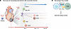
Monocyte-derived type I IFN-activated macrophages localize near the infarctFigure 1

Monocyte-derived type I IFN-activated macrophages localize near the infarct border zone, giving rise to MHCIIhi macrophages that promote Treg development during cardiac remodeling after MI. (A) Koenig et al. (12) showed that monocytes differentiated into several macrophage subpopulations within days of injury, with a hierarchical relationship among subsets. Type I IFN–activated macrophages represent an intermediate state that localized near the infarct border zone and terminally differentiated into MHCII+ macrophages that persisted in the heart through 28 days after injury. (B) Additionally, MHCII+ macrophages contributed to cardiac repair after MI by promoting Treg development in the ischemic heart.

A particularly interesting finding in the study by Koenig et al. emerged from their examination of the early fate decisions underlying monocyte-macrophage differentiation already during tissue extravasation (12). Intravenous anti-CD45 antibody labeling 5 minutes prior to termination to label intravascular leukocytes, combined with flushing of cells that have not adhered to the endothelium at harvest, further revealed that monocyte-to-macrophage differentiation had already begun during monocyte extravasation into the infarcted tissue (12). This observation introduces the concept that macrophage cell-state specification does not occur solely within the infarcted heart but begins during extravasation. For future research, it would be highly interesting to further investigate how extravasation affects specific monocyte differentiation trajectories (13, 14) and, in particular, how the biological niche and interactions between immune and stromal cells, such as endothelial cells and pericytes, regulate monocyte-to-macrophage fate determination.
Cell-specific effects of type I IFN signaling in the infarcted myocardium

Although the importance of the IFN response in immunity has been well established in the context of antiviral defense and autoimmunity (15), its complex roles in cardiovascular diseases remain to be fully elucidated (14, 16–18). In both human and experimental MI models, ischemic injury has been shown to trigger an IFN response in monocyte and neutrophil progenitors already in the bone marrow (14). Moreover, the systemic effects of a type I IFN response in the context of MI were investigated, demonstrating that global suppression of IFN regulatory factor 3 (Irf3) improved cardiac remodeling and survival after MI in mice (16). To further examine the cell-specific roles of the type I IFN response, the effects of selective Irf3 deletion across different cell types, including cardiomyocytes, fibroblasts, macrophages, neutrophils, and endothelial cells, were studied, and indicated that only cardiomyocyte-specific Irf3 deficiency reduced IFN-stimulated gene expression and colonies of IFN-induced cells, while deletion in leukocytes did not exert any effects (17).

Koenig et al. further investigated the cell-specific effect of the IFN response by selectively deleting Ifnar1, encoding a subunit of the cell-surface receptor for type I interferons in recruited monocytes. This deletion led to a significant reduction in the MHCIIhi macrophage population, indicating that type I IFN–activated macrophages and type I IFN signaling are the major contributors to this population (12). Unexpectedly, the authors observed a worsened cardiac adverse remodeling in mice with monocyte and monocyte-derived macrophage specific Ifnar1 ablation, demonstrating that type I IFN signaling in infiltrating monocytes and their progeny confers a protective effect on MI remodeling (12). Mechanistically, this phenotype was associated with enhanced infiltration of Tregs into the myocardium, (12) with MHCII expression by monocyte-derived macrophages mediating this protective response following MI. Furthermore, among cardiac macrophages, type I IFN–activated macrophages expressed PD-L1 (programmed death-ligand 1), an immune checkpoint ligand that promotes immune tolerance by engaging PD-1 on T cells, which may have contributed to Treg differentiation. By linking Type I IFN macrophages to Treg priming, these observations provide what we believe to be the first evidence that type I IFN–activated macrophages can foster Treg expansion associated with cardioprotective effects. Previous studies have shown that myocardial Treg cells can suppress myocardial inflammation and contribute to myocardial protection (19). Along these lines, Treg ablation leads to increased proinflammatory myeloid cell recruitment and overt myocardial inflammation (20, 21). The study by Koenig et al. expands this picture by highlighting that Treg-macrophage crosstalk is bidirectional (Figure 1B), opening interesting avenues for immunoregulation.

Conclusion and future directions

Because inflammatory mechanisms can exert both beneficial and detrimental effects on healing after MI, future approaches are required to target inflammatory mechanisms linked to collateral damage without disturbing immune cells’ critical roles in promoting tissue repair. The study by Koenig et al. contributes to an in-depth understanding of the cardiac macrophage dynamics by describing distinct transcriptional and spatially resolved states over the course of repair after MI. In addition, they report that key events defining monocyte fate decisions already begin during their extravasation and infiltration into the infarcted tissue. Importantly, these findings reveal the complex nature of the type I IFN response and its diverging cell-specific effects, which, in specific circumstances, can also contribute to cardiac repair by fostering Treg differentiation.

Conflict of interest

GCR is coinventor on a pending patent (EP26192875.8) application with focus on engineered T cells. The remaining authors have declared that no conflict of interest exists.

Funding support
  • The German Research Foundation (Collaborative Research Centre 1525 ‘Cardio-Immune Interfaces’, grant #453989101, projects A3/ B6 to GCR, A1/ C6 to AZ, and PS2 to GR).
  • The Heisenberg Program, German Research Foundation (grant #517001338 to GCR).
Footnotes

Copyright: © 2026, Sakalli et al. This is an open access article published under the terms of the Creative Commons Attribution 4.0 International License.

Reference information: J Clin Invest. 2026;136(17):e209791. https://doi.org/10.1172/JCI209791.

See the related article at Fate-mapping infiltrating monocytes following experimental myocardial infarction reveals differentiation trajectories in the infarcted heart.

References
  1. Metchnikoff É. Lectures on the Comparative Pathology of Inflammation. Kegan Paul, Trench, Trübner; 1893.
  2. Zaman R, Epelman S. Resident cardiac macrophages: Heterogeneity and function in health and disease. Immunity. 2022;55(9):1549–1563.
    View this article via: CrossRef PubMed Google Scholar
  3. Hulsmans M, et al. Macrophages facilitate electrical conduction in the heart. Cell. 2017;169(3):510–522.
    View this article via: CrossRef PubMed Google Scholar
  4. Hilgendorf I, et al. Repair of the infarcted heart: cellular effectors, molecular mechanisms and therapeutic opportunities. Circ Res. 2024;134(12):1718–1751.
    View this article via: CrossRef PubMed Google Scholar
  5. Medzhitov R, et al. Disease tolerance as a defense strategy. Science. 2012;335(6071):936–941.
    View this article via: CrossRef PubMed Google Scholar
  6. Bajpai G, et al. The human heart contains distinct macrophage subsets with divergent origins and functions. Nat Med. 2018;24(8):1234–1245.
    View this article via: CrossRef PubMed Google Scholar
  7. Bajpai G, et al. Tissue resident CCR2– and CCR2+ cardiac macrophages differentially orchestrate monocyte recruitment and fate specification following myocardial injury. Circ Res. 2019;124(2):263–278.
    View this article via: CrossRef PubMed Google Scholar
  8. Zuo W, et al. Macrophage-driven cardiac inflammation and healing: insights from homeostasis and myocardial infarction. Cell Mol Biol Lett. 2023;28(1):81.
    View this article via: CrossRef PubMed Google Scholar
  9. Koenig AL, et al. Single-cell transcriptomics reveals cell-type-specific diversification in human heart failure. Nat Cardiovasc Res. 2022;1(3):263–280.
    View this article via: CrossRef PubMed Google Scholar
  10. Rizzo G, et al. Dynamics of monocyte-derived macrophage diversity in experimental myocardial infarction. Cardiovasc Res. 2023;119(3):772–785.
    View this article via: CrossRef PubMed Google Scholar
  11. Kasam RK, et al. Selective expansion of cardiac macrophage subtypes distinguishes their functional roles in disease and homeostasis. J Clin Invest. 2026;136(15):e200194.
    View this article via: JCI CrossRef PubMed Google Scholar
  12. Koenig et al. Genetic mapping of monocyte fate decisions following myocardial infarction. J Clin Invest. 2026;136(17):e189684.
    View this article via: JCI PubMed Google Scholar
  13. Ikeda N, et al. Emergence of immunoregulatory Ym1+Ly6Chi monocytes during recovery phase of tissue injury. Sci Immunol. 2018;3(28):eaat0207.
    View this article via: CrossRef PubMed Google Scholar
  14. Calcagno DM, et al. The myeloid type I interferon response to myocardial infarction begins in bone marrow and is regulated by Nrf2-activated macrophages. Sci Immunol. 2020;5(51):eaaz1974.
    View this article via: CrossRef PubMed Google Scholar
  15. Bondet V, et al. Differential levels of IFNα subtypes in autoimmunity and viral infection. Cytokine. 2021;144:155533.
    View this article via: CrossRef PubMed Google Scholar
  16. King KR, et al. IRF3 and type I interferons fuel a fatal response to myocardial infarction. Nat Med. 2017;23(12):1481–1487.
    View this article via: CrossRef PubMed Google Scholar
  17. Ninh VK, et al. Spatially clustered type I interferon responses at injury borderzones. Nature. 2024;633(8028):174–181.
    View this article via: CrossRef PubMed Google Scholar
  18. Tran DT, et al. Interferons and interferon-related pathways in heart disease. Front Cardiovasc Med. 2024;11:1357343.
    View this article via: CrossRef PubMed Google Scholar
  19. Rieckmann M, et al. Myocardial infarction triggers cardioprotective antigen-specific T helper cell responses. J Clin Invest. 2019;129(11):4922–4936.
    View this article via: JCI CrossRef PubMed Google Scholar
  20. Weirather J, et al. Foxp3+ CD4+ T cells improve healing after myocardial infarction by modulating monocyte/macrophage differentiation. Circ Res. 2014;115(1):55–67.
    View this article via: CrossRef PubMed Google Scholar
  21. Alshoubaki YK, et al. Tregs delivered post-myocardial infarction adopt an injury-specific phenotype promoting cardiac repair via macrophages in mice. Nat Commun. 2024;15(1):6480.
    View this article via: CrossRef PubMed Google Scholar
Version history
  • Version 1 (September 1, 2026): Electronic publication

Article tools

  • View PDF
  • Download citation information
  • Send a comment
  • Terms of use
  • Standard abbreviations
  • Need help? Email the journal

Metrics

  • Article usage
  • Citations to this article

Go to

  • Top
  • Abstract
  • Introduction
  • Making sense of the cardiac macrophage diversity
  • Cell-specific effects of type I IFN signaling in the infarcted myocardium
  • Conclusion and future directions
  • Conflict of interest
  • Funding support
  • Footnotes
  • References
  • Version history
Advertisement
Advertisement

Copyright © 2026 American Society for Clinical Investigation
ISSN: 0021-9738 (print), 1558-8238 (online)

Sign up for email alerts