
Advertisement
Commentary
Open Access |
10.1172/JCI209169
Department of Cell Biology and Physiology, The University of North Carolina at Chapel Hill, Chapel Hill, North Carolina, USA.
Address correspondence to: Kathleen M. Caron, 111 Mason Farm Road, MBRB, CB 7545, The University of North Carolina at Chapel Hill, Chapel Hill, North Carolina 27599, USA. Phone: 919.966.5215; Email: kathleen_caron@med.unc.edu.
Find articles by Tian, Y. in: PubMed | Google Scholar
Department of Cell Biology and Physiology, The University of North Carolina at Chapel Hill, Chapel Hill, North Carolina, USA.
Address correspondence to: Kathleen M. Caron, 111 Mason Farm Road, MBRB, CB 7545, The University of North Carolina at Chapel Hill, Chapel Hill, North Carolina 27599, USA. Phone: 919.966.5215; Email: kathleen_caron@med.unc.edu.
Find articles by Caron, K. in: PubMed | Google Scholar
Published August 3, 2026 - More info
Enhanced TGF-β signaling caused by mutations in Fibrillin-1 (FBN1) in patients with Marfan syndrome (MFS) leads to myxomatous degeneration of the mitral valve (MDMV). MDMV can result in mitral valve prolapse, severe regurgitation, and sudden cardiac death. However, it remains unknown whether lymphatic vessel (LV) dysfunction contributes to MDMV development in MFS. Here, we show that lymphangiogenesis in murine mitral valves (MVs) begins postnatally. However, this process is inhibited in a mouse MFS model, Fbn1 mutant (Fbn1C1039G/+) mice, accompanied by disrupted lymphatic cell-cell junctions, impaired lymphatic drainage, and an abnormally widespread distribution of MHCII+ infiltrating macrophages. Treatment of Fbn1 mutant mice with VEGF-C156S, a selective VEGFR3 agonist, stimulates the ERK and Akt pathways, increases LV density in MVs, and ameliorates MDMV. Fbn1 mutant MVs display disorganized valvular endothelial cells (VECs) and decreased expression of the antiinflammatory modulator Zfp36 (zinc finger protein 36) in VECs and immune cells. Treatment with FTY720 (fingolimod), a ZFP36 activator and S1P antagonist, rescues MDMV phenotypes in Fbn1 mutant mice by reducing immune cell infiltration and restoring lymphatic cell junctions and drainage. These findings suggest that the Fbn1 mutation causes LV hypoplasia and defective lymphatic drainage in MVs, driven in part by proinflammatory VECs, leading to MFS-related MDMV.
Can Tan, Ziyou Ren, Shreya Kurup, Xianpeng Liu, Zhi-Dong Ge, Shodai Suzuki, Pritika Jakka, Cheryl Tang, M. Luisa Iruela-Arispe, Tsutomu Kume
Myxomatous degeneration of the mitral valve (MDMV) is a common cardiovascular manifestation of Marfan syndrome (MFS), yet the role of lymphatic vessels in the disease progression remains unknown. In this Commentary, we discuss the study by Tan, Kume, and colleagues, which identifies defective lymphangiogenesis as a previously unrecognized driver of MDMV. Their work demonstrates that impaired lymphatic development and drainage promote valve inflammation through reduced ZFP36-mediated antiinflammatory signaling, whereas restoration of lymphatic function or pharmacological activation of ZFP36 with the FDA-approved drug FTY720 ameliorates disease progression. These findings establish lymphatic vessels as critical regulators of mitral valve homeostasis and support exploration of lymphatic-targeted therapeutic opportunities for MFS-associated valvular disease.
Marfan syndrome (MFS) was named after Antoine Marfan, who first reported a child with distinguishing skeletal features in 1896 and later reported 150 additional cases. In 1955, Victor McKusick described the cardiovascular features of MFS using echocardiography (1). MFS is a multisystem connective tissue disorder defined by ocular abnormalities (lens dislocation and myopia), skeletal features (dolichostenomelia and joint laxity), and, most importantly, cardiovascular pathology, including aortic root dilatation, dissection, mitral valve prolapse (MVP), and valvular insufficiency (2).
MFS is most commonly caused by pathogenic variants in the FBN1 gene, which encodes Fibrillin-1 (3, 4). Approximately 75% of affected individuals inherit the disorder in an autosomal manner, while 25% of cases arise from de novo mutations. Nearly 3,000 rare FBN1 variants have been identified, contributing to phenotypic variability.
FBN1 is a large ECM glycoprotein that assembles into microfibrils, providing structural integrity, elasticity, and resilience to tissues such as arteries, cardiac valves, lung, and skin. Beyond structural support, FBN1-containing microfibrils regulate growth factor signaling by sequestering TGF-β family members, including TGF-β1 and bone morphogenetic proteins. Pathogenic variants in FBN1 disrupt microfibril assembly and growth factor sequestration, leading to excessive TGF-β accumulation, a primary driver of aortic disease in patients with MFS (4, 5).
Pathophysiology of myxomatous degeneration of the mitral valve. Myxomatous degeneration of the mitral valve (MDMV) and MVP are common manifestations of MFS, affecting approximately 16.1% of patients with MFS (6). Disease progression is characterized by thickened, elongated, and mechanically weakened valve leaflets due to ECM remodeling, often resulting in mitral regurgitation. According to the Ghent II criteria, MVP is one of the clinical features for the diagnosis of MFS (3). Although the prevalence and severity vary, valvular disease is especially severe in neonatal MFS, where MVP and regurgitation are major causes of morbidity and mortality (7, 8). Like aortic pathology, MDMV is largely driven by FBN1 mutations, with increased TGF-β/SMAD signaling observed in both human tissues and animal models (9–13). Recent studies have expanded this paradigm, identifying integrin-mediated mTOR signaling as a contributor to TGF-β overactivity due to FBN1 mutations (14). Additional mechanisms include altered mechanotransduction and inflammatory signaling. Reduced endothelial KLF2/4 expression, increased SMAD signaling, and enhanced monocyte recruitment have been observed in both experimental models and MDMVs (15).
Immune cell infiltration has emerged as another important contributor to the progression of MDMV in MFS. Notably, increased accumulation of proinflammatory macrophages has been demonstrated in myxomatous valves from FBN1 mutant mice, sheep, pigs, and human patients (16). These macrophages are derived predominantly from CCR2+ classical monocytes (16), implicating monocyte recruitment and inflammation as key drivers of disease progression. Collectively, dysregulated ECM signaling, impaired mechanosensing, and chronic inflammation cooperate to drive mitral valve degeneration. In this issue of the JCI, Tan, Kume, and colleagues extended this framework by identifying a previously unrecognized link between disrupted lymphangiogenesis and MDMV progression in Fbn1 mutant mice (17).
Lymphatic vessels are essential for maintaining fluid homeostasis, mediating immune cell trafficking, and resolving inflammation. In the heart, the lymphatic network plays an increasingly recognized role in tissue remodeling and immune regulation under both physiological and pathological conditions. Evidence for lymphatic involvement in valvular disease has been reported in rheumatic heart valve disease and infective endocarditis, where lymphatic vessels are present within diseased leaflets (18, 19). Previous studies show lymphatics localized to ECM-rich regions, with increased abundance in inflammatory conditions such as infective endocarditis (18). Lymphangiogenesis is also associated with cardiac valvular disorders, such as rheumatic heart valve disease (19). Other recent work by the Kume group suggests that lymphatic vessels help maintain mitral valve structure by regulating interstitial fluid balance and ECM turnover (20). Despite these observations, the developmental origin and functional significance of valve lymphatics remain poorly understood. In particular, it has been unclear whether FBN1 mutations impair lymphatic formation and function or whether restoring lymphatic activity could provide therapeutic benefit.
As mentioned above, previous studies by the Kume group demonstrated lymphatic vessels beneath valvular endothelial cells on the atrial side of mouse mitral valves (20). In the current study, Tan and colleagues examined lymphatic development in WT and Fbn1 heterozygous (Fbn1C1039G/+) mice. Whole-mount staining revealed that VEGFR3+ lymphatic vessels expanded postnatally into the mitral annulus at P3, sprouting from regions above the anterior commissure. Lymphatic growth continued into both leaflets through P14 in WT mice. In contrast, Fbn1 mutant mice exhibited reduced lymphatic proliferation, with decreased vessel density and branching at P14, along with abnormal valve enlargement. These defects were likely driven by elevated TGF-β signaling suppressing lymphangiogenesis. Structural abnormalities were also observed in lymphatic endothelial cell junctions. Whereas normal lymphatic vessels displayed discontinuous button-like junctions in capillaries and continuous zipper-like junctions in collecting vessels, mutant mice exhibited predominantly button-like junctions throughout. These abnormalities impaired lymphatic drainage, leading to fluid accumulation and increased valve thickness in MFS mice.
To test causality, the authors used VEGF-C156S, a specific VEGFR3 ligand, to promote lymphangiogenesis and rescue MDMV phenotypes in Fbn1 mutant mice. Treatment restored VEGFC/VEGFR3 signaling via increased ERK1/2 and Akt phosphorylation, increased lymphatic vessel density, and significantly reduced mitral valve thickening. These findings establish defective lymphangiogenesis as a key contributor to MDMV pathogenesis, suggesting that restoring lymphatic function can ameliorate MDMV in MFS.
Tan et al. also observed that lymphatic dysfunction in Fbn1 mutant valves was associated with disrupted endothelial organization and increased macrophage infiltration. Reanalysis of scRNA-seq data identified ZFP36 as a key regulator of MDMV progression.
ZFP36 (tristetraprolin) is an antiinflammatory protein that promotes degradation of proinflammatory mRNAs by binding AU-rich elements in their 3′ untranslated regions. In Fbn1 mutant valves, ZFP36 expression was reduced across multiple cell types, including endothelial cells, macrophages, and DCs, indicating loss of antiinflammatory regulation. Endothelium-specific deletion of Zfp36 (Cdh5-CreERT2 Zfp36fl/fl mice) resulted in increased immune cell infiltration and valve thickening, recapitulating MDMV phenotypes.
To restore ZFP36 function, the authors used FTY720 (fingolimod), an FDA-approved drug for multiple sclerosis (21). Although best known as a modulator of sphingosine-1-phosphate receptor (S1PR) signaling, FTY720 activates protein phosphatase 2A, which enhances ZFP36 activity by promoting its dephosphorylation and stabilization (22–24). Daily oral administration of FTY720 from P1 to P59 significantly reduced CD45+ immune cell infiltration in Fbn1 mutant valves in mice. Mitral valve abnormalities, including leaflet elongation and thickening, were attenuated, and cardiac function was improved, with restored ejection fraction and fractional shortening, by FTY720 administration in Fbn1 mutant mice.
FTY720 also influenced lymphatic vessels in unexpected ways. While FTY720 reduced lymphatic density in WT valves, it did not alter the lymphatic density in Fbn1 mutant valves. FTY720 binds S1PRs, promoting receptor internalization and degradation, and decreasing S1PR immunostaining in lymphatic endothelial cells of WT mice. In a mechanism that remains yet unclear, the Fbn1 mutant mitral valves did not show a decrease in S1PR levels with FTY720 treatment. Interestingly, however, in Fbn1 mutant mice, FTY720 alleviated MDMV phenotypes by promoting continuous zipper-like junctions in the lymphatic endothelial cells, reducing the number of reticular adherens junctions in valvular endothelial cells, enhancing lymphatic drainage, and reducing immune cell accumulation. These findings demonstrate that improving lymphatic function, even without increasing vessel number, can mitigate disease.
This study advances our understanding of MDMV in MFS by identifying lymphatic dysfunction and impaired antiinflammatory regulation as central contributors. Restoration of lymphangiogenesis using VEGF-C156S and pharmacological activation of ZFP36 using FTY720 both significantly improved valve pathology in Fbn1 mutant mice (Figure 1). Current MFS management relies primarily on surgical repair and pharmacologic treatments such as beta blockers and angiotensin receptor blockers, including losartan. Losartan inhibits TGF-β signaling and has been shown to prevent aortic aneurysm formation in Fbn1C1041G/+ mice, with efficacy comparable with TGF-β–neutralizing antibodies and superior to β-blockade alone (25). However, the clinical efficacy of the add-on effect of losartan with beta blockers is inconsistent (26). The findings presented here highlight therapeutic avenues beyond conventional treatments. Targeting lymphatic vessel growth and function, along with modulation of inflammatory pathways via ZFP36, offers promising strategies for preventing and treating mitral valve disease in MFS. Additional studies are needed to determine whether the pathways identified by Tan et al. and lymphatic-targeted therapies can be utilized to alter the clinical course of mitral valvular disease in patients with MFS.
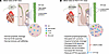
Figure 1Implicating defective lymphatic vessel development in MFS-associated valvular disease. Tan et al. (17) compared the development of VEGFR3+ lymphatic vessels (LVs) in the mitral valve (MV) of WT and FbnC1039G/+ mice, a model of MFS. (A) In WT mice, LVs emerged from the ventricle and septum and progressed from the anterior triangle (appearance at P0) to the anterior leaflet (aL) (invasion by P7), with posterior leaflet (pL) branching by P14. Normal lymphatic endothelial cells (LECs) displayed discontinuous VE-cadherin junctions in capillaries and continuous zipper junctions in collecting vessels, structures that are essential for maintaining LV function and MV morphology. (B) FbnC1039G/+ MVs exhibited disrupted VE-cadherin junctions, which corresponded with reduced LV density, impaired lymphatic drainage, and immune cell accumulation, leading to MDMV. VEGF-C156S restored VEGFC/VEGFR3 signaling, promoted lymphangiogenic extension, and corrected LV and MDMV defects. Mutant MVs also showed reduced ZFP36, and FDA-approved FTY720 enhanced ZFP36 activity via PP2A, stabilizing VE-cadherin junctions, improving lymphatic function, and mitigating MDMV. VEC, valvular endothelial cell.
Author contributions: KMC and YT drafted, edited, and reviewed the manuscript. YT generated the figure using an academic institutional BioRender license (CU29KSQ8UB).
The authors have declared that no conflict of interest exists.
This work is the result of NIH funding, in whole or in part, and is subject to the NIH Public Access Policy. Through acceptance of this federal funding, the NIH has been given a right to make the work publicly available in PubMed Central.
The authors thank members of the Caron lab for helpful discussion and review.
Address correspondence to: Kathleen M. Caron, 111 Mason Farm Road, MBRB, CB 7545, The University of North Carolina at Chapel Hill, Chapel Hill, North Carolina 27599, USA. Phone: 919.966.5215; Email: kathleen_caron@med.unc.edu.
Copyright: © 2026, Tian et al. This is an open access article published under the terms of the Creative Commons Attribution 4.0 International License.
Reference information: J Clin Invest. 2026;136(15):e209169. https://doi.org/10.1172/JCI209169.
See the related article at Lymphatic dysfunction and ZFP36 deficiency contribute to myxomatous valve degeneration in Marfan syndrome mice.
