
Advertisement
Research ArticleAgingCell biologyDevelopment
Open Access |  10.1172/JCI206733
A rare TMEM175 mutation drives Parkinson’s disease pathology by disrupting proton gating and lysosomal acidification
Xiaotong Yang,1,2 Ping Li,1 Man Lv,1 Lingyong Wang,3 Ruyi Shan,1 Zikai Jiang,1 Danzhen Chen,1 Fangqian Huang,1 Weijie Cai,1 Liding Zhao,1 Zuoyou Guo,1 Xinhui Chen,2 Zhidong Cen,2 Yichang Wang,1 Wei Yang,4 Xinran Li,1,2 Wei Luo,2 Lin Yang,3 Meiqin Hu,1,2,5 and Haoxing Xu1,2,4,6,7
1New Cornerstone Science Laboratory & Liangzhu Laboratory, the Second Affiliated Hospital & School of Basic Medical Sciences, Zhejiang University, Hangzhou, China.
2Department of Neurology, the Second Affiliated Hospital and
3Departments of Pharmacology and Pathology, Sir Run Shaw Hospital, Zhejiang University School of Medicine, Hangzhou, China.
4Department of Biophysics, the School of Basic Medical Sciences, Zhejiang University, Hangzhou, China.
5Zhejiang Key Laboratory of Pain Perception and Neuromodulation, Hangzhou, China.
6Institute of Fundamental and Transdisciplinary Research & The State Key Laboratory of Brain-Machine Intelligence, Zhejiang University, Hangzhou, China.
7Department of Molecular, Cellular, and Developmental Biology, University of Michigan, Ann Arbor, Michigan, USA.
Authorship note: XY, PL, and MH contributed equally to this work.
Find articles by Yang, X. in: PubMed | Google Scholar
1New Cornerstone Science Laboratory & Liangzhu Laboratory, the Second Affiliated Hospital & School of Basic Medical Sciences, Zhejiang University, Hangzhou, China.
2Department of Neurology, the Second Affiliated Hospital and
3Departments of Pharmacology and Pathology, Sir Run Shaw Hospital, Zhejiang University School of Medicine, Hangzhou, China.
4Department of Biophysics, the School of Basic Medical Sciences, Zhejiang University, Hangzhou, China.
5Zhejiang Key Laboratory of Pain Perception and Neuromodulation, Hangzhou, China.
6Institute of Fundamental and Transdisciplinary Research & The State Key Laboratory of Brain-Machine Intelligence, Zhejiang University, Hangzhou, China.
7Department of Molecular, Cellular, and Developmental Biology, University of Michigan, Ann Arbor, Michigan, USA.
Authorship note: XY, PL, and MH contributed equally to this work.
Find articles by Li, P. in: PubMed | Google Scholar
1New Cornerstone Science Laboratory & Liangzhu Laboratory, the Second Affiliated Hospital & School of Basic Medical Sciences, Zhejiang University, Hangzhou, China.
2Department of Neurology, the Second Affiliated Hospital and
3Departments of Pharmacology and Pathology, Sir Run Shaw Hospital, Zhejiang University School of Medicine, Hangzhou, China.
4Department of Biophysics, the School of Basic Medical Sciences, Zhejiang University, Hangzhou, China.
5Zhejiang Key Laboratory of Pain Perception and Neuromodulation, Hangzhou, China.
6Institute of Fundamental and Transdisciplinary Research & The State Key Laboratory of Brain-Machine Intelligence, Zhejiang University, Hangzhou, China.
7Department of Molecular, Cellular, and Developmental Biology, University of Michigan, Ann Arbor, Michigan, USA.
Authorship note: XY, PL, and MH contributed equally to this work.
Find articles by Lv, M. in: PubMed | Google Scholar
1New Cornerstone Science Laboratory & Liangzhu Laboratory, the Second Affiliated Hospital & School of Basic Medical Sciences, Zhejiang University, Hangzhou, China.
2Department of Neurology, the Second Affiliated Hospital and
3Departments of Pharmacology and Pathology, Sir Run Shaw Hospital, Zhejiang University School of Medicine, Hangzhou, China.
4Department of Biophysics, the School of Basic Medical Sciences, Zhejiang University, Hangzhou, China.
5Zhejiang Key Laboratory of Pain Perception and Neuromodulation, Hangzhou, China.
6Institute of Fundamental and Transdisciplinary Research & The State Key Laboratory of Brain-Machine Intelligence, Zhejiang University, Hangzhou, China.
7Department of Molecular, Cellular, and Developmental Biology, University of Michigan, Ann Arbor, Michigan, USA.
Authorship note: XY, PL, and MH contributed equally to this work.
Find articles by Wang, L. in: PubMed | Google Scholar
1New Cornerstone Science Laboratory & Liangzhu Laboratory, the Second Affiliated Hospital & School of Basic Medical Sciences, Zhejiang University, Hangzhou, China.
2Department of Neurology, the Second Affiliated Hospital and
3Departments of Pharmacology and Pathology, Sir Run Shaw Hospital, Zhejiang University School of Medicine, Hangzhou, China.
4Department of Biophysics, the School of Basic Medical Sciences, Zhejiang University, Hangzhou, China.
5Zhejiang Key Laboratory of Pain Perception and Neuromodulation, Hangzhou, China.
6Institute of Fundamental and Transdisciplinary Research & The State Key Laboratory of Brain-Machine Intelligence, Zhejiang University, Hangzhou, China.
7Department of Molecular, Cellular, and Developmental Biology, University of Michigan, Ann Arbor, Michigan, USA.
Authorship note: XY, PL, and MH contributed equally to this work.
Find articles by Shan, R. in: PubMed | Google Scholar
1New Cornerstone Science Laboratory & Liangzhu Laboratory, the Second Affiliated Hospital & School of Basic Medical Sciences, Zhejiang University, Hangzhou, China.
2Department of Neurology, the Second Affiliated Hospital and
3Departments of Pharmacology and Pathology, Sir Run Shaw Hospital, Zhejiang University School of Medicine, Hangzhou, China.
4Department of Biophysics, the School of Basic Medical Sciences, Zhejiang University, Hangzhou, China.
5Zhejiang Key Laboratory of Pain Perception and Neuromodulation, Hangzhou, China.
6Institute of Fundamental and Transdisciplinary Research & The State Key Laboratory of Brain-Machine Intelligence, Zhejiang University, Hangzhou, China.
7Department of Molecular, Cellular, and Developmental Biology, University of Michigan, Ann Arbor, Michigan, USA.
Authorship note: XY, PL, and MH contributed equally to this work.
Find articles by Jiang, Z. in: PubMed | Google Scholar
1New Cornerstone Science Laboratory & Liangzhu Laboratory, the Second Affiliated Hospital & School of Basic Medical Sciences, Zhejiang University, Hangzhou, China.
2Department of Neurology, the Second Affiliated Hospital and
3Departments of Pharmacology and Pathology, Sir Run Shaw Hospital, Zhejiang University School of Medicine, Hangzhou, China.
4Department of Biophysics, the School of Basic Medical Sciences, Zhejiang University, Hangzhou, China.
5Zhejiang Key Laboratory of Pain Perception and Neuromodulation, Hangzhou, China.
6Institute of Fundamental and Transdisciplinary Research & The State Key Laboratory of Brain-Machine Intelligence, Zhejiang University, Hangzhou, China.
7Department of Molecular, Cellular, and Developmental Biology, University of Michigan, Ann Arbor, Michigan, USA.
Authorship note: XY, PL, and MH contributed equally to this work.
Find articles by Chen, D. in: PubMed | Google Scholar
1New Cornerstone Science Laboratory & Liangzhu Laboratory, the Second Affiliated Hospital & School of Basic Medical Sciences, Zhejiang University, Hangzhou, China.
2Department of Neurology, the Second Affiliated Hospital and
3Departments of Pharmacology and Pathology, Sir Run Shaw Hospital, Zhejiang University School of Medicine, Hangzhou, China.
4Department of Biophysics, the School of Basic Medical Sciences, Zhejiang University, Hangzhou, China.
5Zhejiang Key Laboratory of Pain Perception and Neuromodulation, Hangzhou, China.
6Institute of Fundamental and Transdisciplinary Research & The State Key Laboratory of Brain-Machine Intelligence, Zhejiang University, Hangzhou, China.
7Department of Molecular, Cellular, and Developmental Biology, University of Michigan, Ann Arbor, Michigan, USA.
Authorship note: XY, PL, and MH contributed equally to this work.
Find articles by Huang, F. in: PubMed | Google Scholar
1New Cornerstone Science Laboratory & Liangzhu Laboratory, the Second Affiliated Hospital & School of Basic Medical Sciences, Zhejiang University, Hangzhou, China.
2Department of Neurology, the Second Affiliated Hospital and
3Departments of Pharmacology and Pathology, Sir Run Shaw Hospital, Zhejiang University School of Medicine, Hangzhou, China.
4Department of Biophysics, the School of Basic Medical Sciences, Zhejiang University, Hangzhou, China.
5Zhejiang Key Laboratory of Pain Perception and Neuromodulation, Hangzhou, China.
6Institute of Fundamental and Transdisciplinary Research & The State Key Laboratory of Brain-Machine Intelligence, Zhejiang University, Hangzhou, China.
7Department of Molecular, Cellular, and Developmental Biology, University of Michigan, Ann Arbor, Michigan, USA.
Authorship note: XY, PL, and MH contributed equally to this work.
Find articles by Cai, W. in: PubMed | Google Scholar
1New Cornerstone Science Laboratory & Liangzhu Laboratory, the Second Affiliated Hospital & School of Basic Medical Sciences, Zhejiang University, Hangzhou, China.
2Department of Neurology, the Second Affiliated Hospital and
3Departments of Pharmacology and Pathology, Sir Run Shaw Hospital, Zhejiang University School of Medicine, Hangzhou, China.
4Department of Biophysics, the School of Basic Medical Sciences, Zhejiang University, Hangzhou, China.
5Zhejiang Key Laboratory of Pain Perception and Neuromodulation, Hangzhou, China.
6Institute of Fundamental and Transdisciplinary Research & The State Key Laboratory of Brain-Machine Intelligence, Zhejiang University, Hangzhou, China.
7Department of Molecular, Cellular, and Developmental Biology, University of Michigan, Ann Arbor, Michigan, USA.
Authorship note: XY, PL, and MH contributed equally to this work.
Find articles by Zhao, L. in: PubMed | Google Scholar
1New Cornerstone Science Laboratory & Liangzhu Laboratory, the Second Affiliated Hospital & School of Basic Medical Sciences, Zhejiang University, Hangzhou, China.
2Department of Neurology, the Second Affiliated Hospital and
3Departments of Pharmacology and Pathology, Sir Run Shaw Hospital, Zhejiang University School of Medicine, Hangzhou, China.
4Department of Biophysics, the School of Basic Medical Sciences, Zhejiang University, Hangzhou, China.
5Zhejiang Key Laboratory of Pain Perception and Neuromodulation, Hangzhou, China.
6Institute of Fundamental and Transdisciplinary Research & The State Key Laboratory of Brain-Machine Intelligence, Zhejiang University, Hangzhou, China.
7Department of Molecular, Cellular, and Developmental Biology, University of Michigan, Ann Arbor, Michigan, USA.
Authorship note: XY, PL, and MH contributed equally to this work.
Find articles by Guo, Z. in: PubMed | Google Scholar
1New Cornerstone Science Laboratory & Liangzhu Laboratory, the Second Affiliated Hospital & School of Basic Medical Sciences, Zhejiang University, Hangzhou, China.
2Department of Neurology, the Second Affiliated Hospital and
3Departments of Pharmacology and Pathology, Sir Run Shaw Hospital, Zhejiang University School of Medicine, Hangzhou, China.
4Department of Biophysics, the School of Basic Medical Sciences, Zhejiang University, Hangzhou, China.
5Zhejiang Key Laboratory of Pain Perception and Neuromodulation, Hangzhou, China.
6Institute of Fundamental and Transdisciplinary Research & The State Key Laboratory of Brain-Machine Intelligence, Zhejiang University, Hangzhou, China.
7Department of Molecular, Cellular, and Developmental Biology, University of Michigan, Ann Arbor, Michigan, USA.
Authorship note: XY, PL, and MH contributed equally to this work.
Find articles by Chen, X. in: PubMed | Google Scholar
1New Cornerstone Science Laboratory & Liangzhu Laboratory, the Second Affiliated Hospital & School of Basic Medical Sciences, Zhejiang University, Hangzhou, China.
2Department of Neurology, the Second Affiliated Hospital and
3Departments of Pharmacology and Pathology, Sir Run Shaw Hospital, Zhejiang University School of Medicine, Hangzhou, China.
4Department of Biophysics, the School of Basic Medical Sciences, Zhejiang University, Hangzhou, China.
5Zhejiang Key Laboratory of Pain Perception and Neuromodulation, Hangzhou, China.
6Institute of Fundamental and Transdisciplinary Research & The State Key Laboratory of Brain-Machine Intelligence, Zhejiang University, Hangzhou, China.
7Department of Molecular, Cellular, and Developmental Biology, University of Michigan, Ann Arbor, Michigan, USA.
Authorship note: XY, PL, and MH contributed equally to this work.
Find articles by Cen, Z. in: PubMed | Google Scholar
1New Cornerstone Science Laboratory & Liangzhu Laboratory, the Second Affiliated Hospital & School of Basic Medical Sciences, Zhejiang University, Hangzhou, China.
2Department of Neurology, the Second Affiliated Hospital and
3Departments of Pharmacology and Pathology, Sir Run Shaw Hospital, Zhejiang University School of Medicine, Hangzhou, China.
4Department of Biophysics, the School of Basic Medical Sciences, Zhejiang University, Hangzhou, China.
5Zhejiang Key Laboratory of Pain Perception and Neuromodulation, Hangzhou, China.
6Institute of Fundamental and Transdisciplinary Research & The State Key Laboratory of Brain-Machine Intelligence, Zhejiang University, Hangzhou, China.
7Department of Molecular, Cellular, and Developmental Biology, University of Michigan, Ann Arbor, Michigan, USA.
Authorship note: XY, PL, and MH contributed equally to this work.
Find articles by Wang, Y. in: PubMed | Google Scholar
1New Cornerstone Science Laboratory & Liangzhu Laboratory, the Second Affiliated Hospital & School of Basic Medical Sciences, Zhejiang University, Hangzhou, China.
2Department of Neurology, the Second Affiliated Hospital and
3Departments of Pharmacology and Pathology, Sir Run Shaw Hospital, Zhejiang University School of Medicine, Hangzhou, China.
4Department of Biophysics, the School of Basic Medical Sciences, Zhejiang University, Hangzhou, China.
5Zhejiang Key Laboratory of Pain Perception and Neuromodulation, Hangzhou, China.
6Institute of Fundamental and Transdisciplinary Research & The State Key Laboratory of Brain-Machine Intelligence, Zhejiang University, Hangzhou, China.
7Department of Molecular, Cellular, and Developmental Biology, University of Michigan, Ann Arbor, Michigan, USA.
Authorship note: XY, PL, and MH contributed equally to this work.
Find articles by
Yang, W.
in:
PubMed
|
Google Scholar
|

1New Cornerstone Science Laboratory & Liangzhu Laboratory, the Second Affiliated Hospital & School of Basic Medical Sciences, Zhejiang University, Hangzhou, China.
2Department of Neurology, the Second Affiliated Hospital and
3Departments of Pharmacology and Pathology, Sir Run Shaw Hospital, Zhejiang University School of Medicine, Hangzhou, China.
4Department of Biophysics, the School of Basic Medical Sciences, Zhejiang University, Hangzhou, China.
5Zhejiang Key Laboratory of Pain Perception and Neuromodulation, Hangzhou, China.
6Institute of Fundamental and Transdisciplinary Research & The State Key Laboratory of Brain-Machine Intelligence, Zhejiang University, Hangzhou, China.
7Department of Molecular, Cellular, and Developmental Biology, University of Michigan, Ann Arbor, Michigan, USA.
Authorship note: XY, PL, and MH contributed equally to this work.
Find articles by Li, X. in: PubMed | Google Scholar
1New Cornerstone Science Laboratory & Liangzhu Laboratory, the Second Affiliated Hospital & School of Basic Medical Sciences, Zhejiang University, Hangzhou, China.
2Department of Neurology, the Second Affiliated Hospital and
3Departments of Pharmacology and Pathology, Sir Run Shaw Hospital, Zhejiang University School of Medicine, Hangzhou, China.
4Department of Biophysics, the School of Basic Medical Sciences, Zhejiang University, Hangzhou, China.
5Zhejiang Key Laboratory of Pain Perception and Neuromodulation, Hangzhou, China.
6Institute of Fundamental and Transdisciplinary Research & The State Key Laboratory of Brain-Machine Intelligence, Zhejiang University, Hangzhou, China.
7Department of Molecular, Cellular, and Developmental Biology, University of Michigan, Ann Arbor, Michigan, USA.
Authorship note: XY, PL, and MH contributed equally to this work.
Find articles by Luo, W. in: PubMed | Google Scholar
1New Cornerstone Science Laboratory & Liangzhu Laboratory, the Second Affiliated Hospital & School of Basic Medical Sciences, Zhejiang University, Hangzhou, China.
2Department of Neurology, the Second Affiliated Hospital and
3Departments of Pharmacology and Pathology, Sir Run Shaw Hospital, Zhejiang University School of Medicine, Hangzhou, China.
4Department of Biophysics, the School of Basic Medical Sciences, Zhejiang University, Hangzhou, China.
5Zhejiang Key Laboratory of Pain Perception and Neuromodulation, Hangzhou, China.
6Institute of Fundamental and Transdisciplinary Research & The State Key Laboratory of Brain-Machine Intelligence, Zhejiang University, Hangzhou, China.
7Department of Molecular, Cellular, and Developmental Biology, University of Michigan, Ann Arbor, Michigan, USA.
Authorship note: XY, PL, and MH contributed equally to this work.
Find articles by Yang, L. in: PubMed | Google Scholar
1New Cornerstone Science Laboratory & Liangzhu Laboratory, the Second Affiliated Hospital & School of Basic Medical Sciences, Zhejiang University, Hangzhou, China.
2Department of Neurology, the Second Affiliated Hospital and
3Departments of Pharmacology and Pathology, Sir Run Shaw Hospital, Zhejiang University School of Medicine, Hangzhou, China.
4Department of Biophysics, the School of Basic Medical Sciences, Zhejiang University, Hangzhou, China.
5Zhejiang Key Laboratory of Pain Perception and Neuromodulation, Hangzhou, China.
6Institute of Fundamental and Transdisciplinary Research & The State Key Laboratory of Brain-Machine Intelligence, Zhejiang University, Hangzhou, China.
7Department of Molecular, Cellular, and Developmental Biology, University of Michigan, Ann Arbor, Michigan, USA.
Authorship note: XY, PL, and MH contributed equally to this work.
Find articles by
Hu, M.
in:
PubMed
|
Google Scholar
|
1New Cornerstone Science Laboratory & Liangzhu Laboratory, the Second Affiliated Hospital & School of Basic Medical Sciences, Zhejiang University, Hangzhou, China.
2Department of Neurology, the Second Affiliated Hospital and
3Departments of Pharmacology and Pathology, Sir Run Shaw Hospital, Zhejiang University School of Medicine, Hangzhou, China.
4Department of Biophysics, the School of Basic Medical Sciences, Zhejiang University, Hangzhou, China.
5Zhejiang Key Laboratory of Pain Perception and Neuromodulation, Hangzhou, China.
6Institute of Fundamental and Transdisciplinary Research & The State Key Laboratory of Brain-Machine Intelligence, Zhejiang University, Hangzhou, China.
7Department of Molecular, Cellular, and Developmental Biology, University of Michigan, Ann Arbor, Michigan, USA.
Authorship note: XY, PL, and MH contributed equally to this work.
Find articles by Xu, H. in: PubMed | Google Scholar
Published November 2, 2026 - More info
J Clin Invest. 2026;136(21):e206733. https://doi.org/10.1172/JCI206733.
© 2026 Yang et al. This work is licensed under the Creative Commons Attribution 4.0 International License. To view a copy of this license, visit http://creativecommons.org/licenses/by/4.0/.
Received: March 10, 2026; Accepted: September 3, 2026


-
Results
Electrophysiological analyses identified a gating-deficiency TMEM175 mutation in a local PD cohort. To investigate the role of TMEM175 in PD pathogenesis, we performed whole-genome/exon sequencing on more than 800 cases in a local PD cohort and identified several rare variants (minor allele frequency < 0.01) in the protein-coding region of TMEM175 (Supplemental Table 1; supplemental material available online with this article; https://doi.org/10.1172/JCI206733DS1). As heterologously overexpressed TMEM175 proteins can ectopically traffic to the plasma membrane (11), we first studied these variants using whole-cell patch clamp in HEK293T cells transfected with WT and mutant TMEM175 (Figure 1A). Under this recording configuration, the extracellular side is topologically equivalent to the luminal lysosome side, such that the pH of the external/bath solution (pHE/L) can vary. At pHE/L 4.6, which mimics the physiological acidity of the lysosomal lumen (11), large H+ currents (IH) with positive reversal potentials were seen in cells transfected with WT TMEM175, but not in nontransfected control cells (Figure 1, B and C). Among the mutant variants analyzed, a leucine–to–arginine (L-to-R) substitution (L275R) in the pore-helix resulted in the almost complete loss of IH, yet with normal protein expression (Figure 1, B and C, and Supplemental Figure 1, A–K). A dramatic difference in IH between TMEM175WT and TMEM175L275R was also seen at pHE/L 3.0 (Supplemental Figure 2, A and B). Nevertheless, although small, whole-cell IH through TMEM175L275R was significant, especially at pHE/L 3.0 (Figure 1, B and C, and Supplemental Figure 2, A–D). Two other substitutions at the same residue — L275F and L275P — were present in public databases (GnomAD v4.1.0: 4-955871-C-T, 4-955872-T-C). TMEM175L275P also showed a loss of H+ conductance (Supplemental Figure 2, E and F, and Supplemental Figure 1K). As TMEM175 is a proton-activated proton-permeable channel (11), the reduction in IH could be caused by a deficit in H+ permeability and/or activation gating. However, 4-(2-Butyl-6,7-dichloro-2-cyclopentyl-indan-1-on-5-yl) oxobutyric acid (DCPIB) and arachidonic acid, chemical agonists of TMEM175 (11), failed to further stimulate H+ currents through TMEM175L275R at pHE/L 4.6 or 3.0 (Figure 1, D and E, Supplemental Figure 2, G and H). When we coexpressed mCherry-TMEM175L275R with EGFP-TMEM175WT, we observed a roughly 50% reduction in whole-cell H+ current amplitude relative to cells coexpressing mCherry-TMEM175WT and EGFP-TMEM175WT (Supplemental Figure 2, I and J). When channel activity was assayed in the physiological setting of lysosomes using the whole-endolysosome [late endosomes and lysosomes (LEL)] patch-clamp technique (19), TMEM175L275R failed to produce detectable whole-LEL H+ currents (Supplemental Figure 3, A–D), yet with normal lysosomal expression and trafficking (Supplemental Figure 3, E–G). Thus, L275R, as a mutation in the pore-helix (20, 21), may specifically abolish channel activation gating.
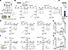 Figure 1
The PD patient–derived L275R mutation selectively disrupts the activation gating of the TMEM175 channel. (A) Diagram of whole-cell patch-clamp recordings of H+ currents through TMEM175 channels carrying PD patient–derived rare variants (MAF < 0.01) in a heterologous overexpression system. TMEM175 rare variants were identified from a WES/WGS database for a local PD cohort. (B and C) Whole-cell H+ currents recorded in HEK293T cells overexpressing TMEM175WT or TMEM175L275R with cytosolic pH (pHC) set to 7.2, and extracellular/luminal pH (pHE/L) varied as indicated. Nontransfected cells served as the negative control (Ctrl). Unless otherwise indicated, all recording solutions contained NMDG+ and MSA– as the major ions; n = 6 (Ctrl); 16 (WT); 27 (L275R) cells. (D and E) Whole-cell H+ currents recorded in HEK293T cells overexpressing TMEM175WT or TMEM175L275R in response to DCPIB (10 μM); n = 3–4 cells. Each dashed line represents the same cell before and after DCPIB application. (F and G) Whole-cell Cs+ currents recorded in HEK293T cells overexpressing TMEM175WT or TMEM175L275R. The recording solutions contained 140 mM Cs or Na-MSA (pH 7.2); n = 4–5 cells. Unless otherwise indicated, data are means ± SEM with dots representing samples from ≥ 3 biological replicates. ***P < 0.001, *P < 0.05; by 1-way ANOVA test.
TMEM175 was initially characterized as a constitutively active (“leaky”) K+/Cs+-permeable channel (14). Luminal/extracellular K+ was recently reported to inhibit TMEM175’s H+ conductance, suggesting that K+, but not H+, is responsible for the lysosomal functions of TMEM175 (22). However, we found that extracellular/luminal K+ (1–10 mM) did not significantly inhibit whole-cell and whole-LEL TMEM175-mediated H+ currents (Supplemental Figure 4, A, D, and G–I). The rapid accumulation of local H+ on the cytosolic side during continuous whole-cell recordings of proton channels such as those of TMEM175 and otopetrins tends to quickly “erode” the transmembrane H+ gradient, resulting in the “artifactual” inhibition of H+ currents (17, 23) (Supplemental Figure 4, B–F). On the other hand, consistent with previous findings (11, 14), at neutral pHE/L, basal TMEM175-specific whole-cell K+/Cs+ currents could be detected (Figure 1, F and G). Remarkably, basal K+/Cs+ currents were comparable for TMEM175WT and TMEM175L275R. However, DCPIB failed to further stimulate IK/Cs in TMEM175L275R-transfected cells, in sharp contrast to the dramatic (up to 100-fold) activation seen with TMEM175WT (Figure 1, F and G). Thus, the L275R mutation results in defective gating without compromising the channel’s cationic (H+/Cs+/K+) permeability/selectivity (Supplemental Figure 4J).
Endogenous TMEM175 generates only negligible amounts of H+ currents under the physiological setting of luminal pH (pHL 4.6), potentially due to lysosome-associated membrane protein 1/2 (LAMP1/2) inhibition (24). Hence, pHL 4.6 is proposed as the “threshold” pH for endogenous TMEM175 activation to prevent lysosomal over acidification (11). However, upon further acidification below pHL 4.6 or in the presence of chemical agonists, TMEM175 may mediate profound H+ “leakage” activity to counteract V-ATPase–mediated acidification (10, 18). On the other hand, a high level of TMEM175 overexpression can produce significant H+ “leakage” even under physiological conditions (i.e., pHL 4.6) (11). Although TMEM175L275R retains its proton permeability, the lack of a proton activation effect may eliminate its H+ “leakage” activity in lysosomes. Indeed, lysosomal acidity, roughly estimated by LysoTracker staining, revealed that, unlike TMEM175WT, TMEM175L275R overexpression failed to induce intralysosomal deacidification (Supplemental Figure 4K). Hence, the naturally occurring gating-deficiency L275R variant may be a unique tool for the differentiation of the controversial roles of constitutive K+ “leakage” and H+-gated H+ “leakage” in the regulation of lysosomal pH homeostasis, as well as PD pathology in patients carrying this variant.
The monoallelic L275R mutation diminishes lysosomal H+ conductance in cells derived from patients with PD. A patient carrying a monoallelic TMEM175 p.L275R mutation (TMEM175+/L275R; case #37) was diagnosed with early onset PD in a local hospital at the age of 45 years based on clinical evaluations (Supplemental Figure 5A) and dopamine transporter positron emission tomography (DAT-PET) (25, 26) examination, which showed the bilateral loss of dopaminergic neurons, akin to other sporadic PD cases (Figure 2A). We collected skin fibroblasts from the patient (Figure 2, B–D) and generated iPSCs (Figure 2, E–G) and iDA neurons (Figure 2, H–K) using established protocols (27, 28). Successful neuronal differentiation was verified using immunostaining and functional assays (Figure 2I and Supplemental Figure 5, B and C). Whole-LEL recordings demonstrated that agonist-activated (DCPIB-activated) and proton-activated (pHL 3.0–activated) lysosomal H+ currents were reduced by 40%–70% in TMEM175+/L275R cells relative to those in control TMEM175+/+ cells derived from an age-matched healthy individual (Figure 2, B–K, and Supplemental Figure 5, D and E; Supplemental Figure 6). The nonphysiological pHL of 3.0 was chosen as a “suprathreshold” stimulus to improve the experimental detection of endogenous IH [see (11)]. Other lysosomal channel conductances, including TRPML1, did not differ between TMEM175+/+ and TMEM175+/L275R fibroblasts (Supplemental Figure 5, F and G), nor was TMEM175 mRNA expression (Supplemental Figure 5H).
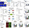 Figure 2
The monoallelic L275R mutation is responsible for the diminished lysosomal H+ currents in PD patient–derived fibroblasts, iPSCs, and iDA neurons. (A) Brain DAT-PET images of (i) a female (F) PD case (#37) carrying the monoallelic p.L275R variant (TMEM175+/L275R), (ii) 2 sporadic PD cases, and (iii) 2 healthy controls (TMEM175+/+). M, male. (B) Genotyping of human skin fibroblasts (HFs) from PD case #37 and an age-matched healthy individual. (C) Whole-LEL H+ currents from TMEM175+/+ and TMEM175+/L275R fibroblasts (DCPIB: 100 μM, pHL: 4.6, pHC: 7.2). (D) Summary of whole-LEL H+ currents (at –120 mV, Vm = VCytosol – VLumen) from experiments as in C; n = 4–22 LELs. (E) Whole-LEL H+ currents from healthy control (+/+), patient-derived +/L275R), “mutated” (+/L275RMut), and “corrected” WT TMEM175 (+/+Cor) iPSCs (DCPIB: 100 μM, pHL: 3.0, pHC: 7.2). Mutated (+/L275RMut) and corrected (+/+Cor) TMEM175 iPSCs were generated from healthy control (+/+) and patient-derived (+/L275R) TMEM175 parental cells, respectively. (F and G) Summary of whole-LEL H+ currents (at –120 mV) from each group shown in E; n = 3–9 LELs. (H) Generation of iDA neurons from iPSCs using indicated differentiation factors. (I) Immunostaining of tyrosine hydroxylase (TH) and MAP2 in iDA neurons. Scale bar: 100 μm. (J and K) Summary of whole-LEL H+ currents (at –120 mV) from TMEM175 (+/+), (+/L275R), (+/L275RMut), and (+/+Cor) iDA neurons (DCPIB: 100 μM, pHL: 4.6/3.0, pHC: 7.2); n = 3–16 LELs. See Supplemental Figure 6 for the representative traces. Data are presented as means ± SEM, and dots represent samples from ≥ 3 biological replicates. ***P < 0.001, **P < 0.01, *P < 0.05; by 1-way ANOVA test.
In a converse effort, we introduced a monoallelic L275R mutation (+/L275RMut) into control (+/+) iPSCs using CRISPR/Cas9 technology (29). These +/L275RMut cells also exhibited greater than 50% reduction in IH compared with the parental control cells (Figure 2, E–K). When we corrected the mutant allele in patient-derived +/L275R iPSCs using genome editing to generate isogenic “+/+” iPSCs (+/+Cor), IH was fully restored (Figure 2, E–G, and Supplemental Figure 6, A and B). Similar results were seen with iDA neurons (Figure 2, H–K, and Supplemental Figure 6, C–J). Taken together, these results suggest that the monoallelic expression of the L275R variant plays a causative role in the observed IH reduction.
The monoallelic L275R mutation disrupts lysosomal acidification and degradation in human cells. TMEM175 mediates fast H+ leakage in lysosomes, and its deficiency causes lysosomal hyperacidification in cells (refs. 11, 13–15, 24, 30; but see ref. 22 for a contrary view). We examined lysosomal acidity in TMEM175+/L275R fibroblasts using 2 ratiometric lysosomal pH indicators: Oregon Green-488 dextran and CQ-Lyso (12, 31). When the fluorescence ratios were calibrated to pH values (Figure 3A), the average lysosomal pH in TMEM175+/L275R fibroblasts was 4.18 ± 0.02, 0.38 units lower than that in TMEM175+/+ cells (pHL= 4.56 ± 0.02; Figure 3, B–D). An optimal lysosomal pH (4.5–5.0) is required for efficient lysosomal degradation, such that lysosomal hyperacidification (pH < 4.5) may counterintuitively decrease the activities of most lysosomal hydrolases (11, 12, 32, 33). The overall degradative capability, assayed by dye-quenched BSA (DQ-BSA) staining (34); the cathepsin B activity, assayed by Magic-Red staining (35); and the GCase activity, evaluated by PFB-FDGlu staining (36), were reduced in TMEM175+/L275R fibroblasts compared with TMEM175+/+ cells (Figure 3, E–H, and Supplemental Figure 7, A and B). Consistent with these degradation defects, the accumulation of autofluorescence lipofuscin materials, often seen in fibroblasts affected by lysosomal storage disease (19, 37), was observed in the lysosomes of TMEM175+/L275R fibroblasts (Supplemental Figure 7, C and D). When lysosomal hyperacidification was pharmacologically “corrected” using a low dose (1–3 nM) of the V-ATPase inhibitor bafilomycin-A1 (11) (Figure 3, B–D), lysosomal degradation was restored (Figure 3, E–H, and Supplemental Figure 7, A–D) without affecting lysosomal enzyme expression (Supplemental Figure 7E). Hence, lysosomal hyperacidity caused by reduced lysosomal H+ leakage is primarily responsible for degradation impairment in patient-derived (+/L275R) fibroblasts.
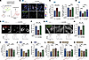 Figure 3
The monoallelic L275R mutation impairs lysosomal acidification and hydrolytic degradation in PD patient–derived cells. (A) Schematic of lysosomal pH determination using ratiometric pH indicators (CQ-Lyso and Oregon-Green-dextran). (B) Lysosomal luminal acidity in TMEM175+/+ and patient-derived (TMEM175+/L275R) fibroblasts (HFs). A low dose (3 nM) of bafilomycin-A1 (Baf-A1; Baf[3]) was used to correct lysosomal pH abnormality in patient-derived (TMEM175+/L275R) fibroblasts. Scale bar: 20 μm. (C and D) Lysosomal pHs in HFs with the indicated genotypes and treatments. pH values were calibrated based on fluorescence ratios in C; n = 10–14 images. A low Baf-A1 dose (1 or 3 nM; Baf[1] or Baf[3]) was used for lysosomal pH normalization. (E and F) Lysosomal cathepsin B activity, assayed by Magic Red staining, in TMEM175+/+ and TMEM175+/L275R fibroblasts. Scale bar: 20 μm; n = 4 replicates. (G and H) Lysosomal GCase activity, assayed by PFB-FDGlu green staining, in TMEM175+/+ and TMEM175+/L275R fibroblasts. Scale bar: 20 μm; n = 4 replicates. (I–N) Lysosomal luminal acidity (CQ-Lyso staining), cathepsin B activity (MagicRed staining), and GCase activity (PFB-FDGlu staining) in iPSCs (n = 5–12 images) and iDA neurons (n = 8–17 images) of various genotypes. Data are presented as means ± SEM, with dots representing biological replicates or indicated samples from ≥ 3 biological replicates. ***P < 0.001, **P < 0.01, *P < 0.05, by 1-way ANOVA test.
We also observed lysosomal hyperacidity and degradation impairment in +/L275RMut iPSCs and iDA neurons compared with their parental control cells (Figure 3, I–N). Discernible “rescue” effects were seen in genetically corrected isogenic WT (+/+Cor) iPSCs and iDA neurons (Figure 3, I–N). Thus, the monoallelic L275R mutation likely impairs lysosomal acidification and degradation across all 3 patient-derived (+/L275R) cell types.
Pharmacological TMEM175 activation using potent small-molecule agonists rescues lysosome dysfunction in patient-derived cells. Human TMEM175 proteins form homodimeric channels (20). Although the monoallelic L275R mutation causes haploinsufficiency or has a partial dominant-negative effect on lysosomal H+ conductance (Figure 2D), the expression of the single-copy WT allele resulted in intermediate-sized H+ currents that could be enhanced by chemical agonists. At pHL 3.0, DCPIB upmodulated lysosomal H+ currents in TMEM175+/L275R fibroblasts, to a level comparable to those in control TMEM175+/+ cells without agonist treatment (Figure 2D). We reasoned that an appropriate dose of DCPIB could be used to “counteract” lysosomal pH abnormality (Figure 4, A and B), restoring the lysosomal degradation capability in TMEM175+/L275R cells (Figure 4C and Supplemental Figure 7, A and B). Indeed, DCPIB treatment readily normalized the activities of cathepsin B and GCase in TMEM175+/L275R fibroblasts, but not in Tmem175 KO mouse embryonic fibroblasts (MEFs; Supplemental Figure 7F). Remarkably, prolonged (> 2 days) DCPIB treatment also mitigated lipofuscin accumulation without boosting the expression of relevant hydrolases (Supplemental Figure 7, C–E).
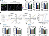 Figure 4
Small-molecule agonists of TMEM175 rescue lysosomal functions in patient-derived TMEM175+/L275R fibroblasts. (A and B) Lysosomal pHs in TMEM175+/+ and patient-derived TMEM175+/L275R fibroblasts (HFs) with or without DCPIB (30 μΜ) treatment (Oregon Green dextran assay). Scale bar: 10 μm; n = 4–6 images. (C) Summary of lysosomal cathepsin B activity (Magic Red staining) and GCase activity (PFB-FDGlu staining) in TMEM175+/+ and TMEM175+/L275R fibroblasts; n = 3–4 replicates. (D) Activation effects of DCPIB (LHA001) and LHA016 on whole-cell TMEM175 Cs+ currents. All solutions contained 140 mM Cs-MSA (pH 7.2). Left: chemical structures; right: dose-response curves, n = 3 or 4 cells. (E and F) Effects of LHA016 on lysosomal pH (n = 4–6 images) and cathepsin B activity (Magic Red assay, n = 4–7 images) in TMEM175+/+, TMEM175+/L275R, and TMEM175–/– iNeurons. Data are presented as means ± SEM, with dots representing biological replicates or indicated samples from ≥ 3 biological replicates. ***P < 0.001, **P < 0.01, *P < 0.05; by 1-way ANOVA test.
To reduce potential off-target effects of DCPIB, we conducted several rounds of structure-activity relationship analysis to increase the compound potency by 10-fold from a half-maximal effective concentration of 11.7 μM for DCPIB [LysoHAgonist (LHA) 001] to 1.1 μM for LHA016 (Figure 4D and Supplemental Figure 8A). Solid-supported membrane–based (SSM-based) electrophysiology, used to measure ionic currents in native lysosomal membranes without LEL enlargement manipulation required for whole-LEL recordings (38, 39), confirmed LHA016’s specificity and potency for TMEM175 (Supplemental Figure 8B). The activation effect of LHA016 on TMEM175 was specific, as it evoked IH in WT, but not TMEM175-KO cells (Supplemental Figure 8C). Lysosomal pH assays (11, 12) revealed that LHA016 at concentrations as low as 1–3 μM, which produced minimal inhibition on DCPIB-sensitive VRAC channels (11) (Supplemental Figure 8D), significantly abridged lysosomal hyperacidification in TMEM175+/L275R but not TMEM175–/– iNeurons (40) (Figure 4E). Consistently, LHA016’s rescue effect on lysosomal degradation was seen in TMEM175+/L275R, but not TMEM175–/–, iNeurons (Figure 4F), suggesting on-target specificity.
Small-molecule agonists of TMEM175 prevent pathological α-synuclein aggregation in TMEM175+/L275R neurons. Lysosomal degradation, mediated by cathepsins B and D, is a primary route for the clearance of pathological α-synuclein aggregates (3, 4). To maximally facilitate α-synuclein aggregation in iPSC-derived neurons, doxycycline-inducible (Dox-inducible) expression of exogenous α-synucleinA53T was combined with α-synuclein preformed fibril (α-syn pff) seeding (40, 41) (Figure 5A). Patient-derived (+/L275R) and mutated (+/L275RMut) iDA neurons exhibited more pathologically phosphorylated α-synuclein (p-α-syn) fluorescence puncta than did control (+/+) and corrected (+/+Cor) iDA neurons (Figure 5B). More p-α-syn aggregation puncta and detergent-resistant insoluble p-α-syn aggregates were observed in patient-derived (+/L275R) and mutated (+/L275RMut) iNeurons than in control (+/+) and corrected (+/+Cor) neurons (Figure 5, C and D, and Supplemental Figure 8E). Taken together, these results suggest that the p.L275R mutation is likely a disease-causing variant promoting PD-like synucleinopathy.
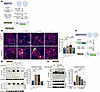 Figure 5
Pharmacological TMEM175 activation averts pathological α-synuclein aggregation in induced TMEM175+/L275R neurons. (A) Diagram of generation of iPSC-differentiated iDA neurons, in which α-synuclein A53T (PB-SNCAA53T) expression was induced by Dox to facilitate p-α-syn aggregation. (B) Immunostaining of p-α-syn aggregation in TMEM175 (+/+), (+/L275R), (+/L275RMut), and (+/+Cor) iDA neurons after α-syn pff (3 μg/ml) seeding for 14 days. β-tubulin III (β-tub III) served as a neuronal marker. Scale bar: 50 μm, n = 13–20 images. (C) Diagram of rapid transdifferentiation of iPSCs into iNeurons, in which Dox-inducible coexpression of neurogenin 2 (NGN2) and α-synuclein A53T (PB-SNCAA53T–sfGFP-P2A-NGN2). (D) Western blots of p-α-syn aggregation (detergent-resistant insoluble fractions) in iNeurons with the indicated genotypes after α-syn pff (3 μg/ml) seeding for 15 days. β-Tub III served as a neuronal loading control. Soluble fractions of p-α-syn are shown for comparison; n = 5 replicates. (E) Effects of LHA016 on p-α-syn aggregation in TMEM175+/+ and TMEM175+/L275R pff-seeded (10 days) iNeurons; n = 4 replicates. Data are presented as means ± SEM, with dots representing biological replicates or indicated samples from ≥ 3 biological replicates. ***P < 0.001, **P < 0.01, *P < 0.05; by 1-way ANOVA test.
Remarkably, treatment with LHA016 at concentrations as low as 1 μM significantly reduced pathological α-synuclein aggregation in TMEM175+/L275R iNeurons (Figure 5E), but not in TMEM175–/– iNeurons (Supplemental Figure 8, F–H). Thus, pharmacological upregulation of TMEM175 H+ channel activity readily restores lysosomal pH homeostasis to mitigate synucleinopathy.
Primary mouse neurons carrying the Tmem175 p.L272R mutation exhibit lysosomal hyperacidity and degradation inefficiency. To investigate the role of the L275R mutation in vivo, we generated knock-in mice with the p.L272R mutation, equivalent to human p.L275R (Figure 6A and Supplemental Figure 9A). IH through mouse TMEM175L272R was markedly reduced in multiple primary mouse cells (Supplemental Figure 9, B and C), including hippocampal neurons, astrocytes, and bone marrow–derived macrophages (BMDMs; Figure 6, B and C, and Supplemental Figure 9, D–F). Whereas heterozygotic (Tmem175+/L272R) primary neurons and astrocytes retained roughly 30%–40% of lysosomal H+ currents, those from homozygotic (Tmem175 L272R/L272R) mice lost their lysosomal H+ conductance completely, resembling cells from KO (Tmem175–/–) mice (Figure 6, B and C, and Supplemental Figure 9, D–F). Accordingly, Tmem175+/L272R and Tmem175L272R/L272R primary neurons exhibited lysosomal hyperacidification and degradation impairment; the defects observed in homozygotic cells (Tmem175 L272R/L272R) were comparable to those in Tmem175–/– neurons (Figure 6, D–F, and Supplemental Figure 9, G and H).
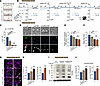 Figure 6
Lysosomal H+ currents, acidification, and hydrolytic degradation are compromised in primary neurons isolated from Tmem175L272R knock-in mice. (A) Genotypes of Tmem175+/+, Tmem175+/L272R, and Tmem175L272R/L272R mice. (B and C) Whole-LEL H+ currents in cultured mouse hippocampal neurons of the indicated genotypes in response to DCPIB (100 μM), with pHL = 4.6 and cytosolic pH (pHC) = 7.2. Genotyping results for Tmem175–/– mice are shown on the bottom right. Summary of current densities (at –120 mV) is shown in C; n = 5 (+/+); 4 (+/L272R); 3 (L272R/L272R); 5 (-/-) LELs. (D–F) Lysosomal acidity (Oregon Green dextran assay) and cathepsin B activity (MagicRed assay) in primary hippocampal neurons isolated from mice of indicated genotypes. Scale bar: 5 μm. Summaries are shown in E and F; n = 3 or 4 replicates. (G and H) Immunostaining of p-α-syn aggregation in primary neurons with the indicated genotypes after pff seeding for 7 days. β-tubulin III (β-tub III) served as the neuronal marker. Scale bar: 50 μm, n = 3 mice. (I and J) Western blots of p-α-syn aggregation in primary hippocampal neurons isolated from mice of indicated genotypes after pff seeding for 7 days. β-tub III served as the neuronal loading control. The thin black dividing line indicates splicing of noncontiguous lanes of the same blot. For panels H and J, results from separate experiments conducted with Tmem175–/– neurons are shown for comparison. Data are presented as means ± SEM, with dots representing biological replicates or indicated samples from ≥ 3 biological replicates. Statistical significance was determined using a 2-tailed Student’s t test for H and J and 1-way ANOVA test for C, E, and F, with ***P < 0.001, **P < 0.01.
Mice carrying the Tmem175 p.L272R mutation show PD-like synucleinopathy and motor deficits. As in Tmem175–/– neurons (Supplemental Figure 10, A and B), pathological α-synuclein aggregation was increased significantly in Tmem175+/L272R and Tmem175L272R/L272R neurons (Figure 6, G–J). To investigate the in-vivo roles of the L272R mutation, we performed striatal α-syn pff injection of Tmem175+/L272R and Tmem175L272R/L272R mice according to an established protocol (41). Three months after injection, cryosections and detergent-resistant insoluble proteins were collected from striatal tissues for immunostaining and immunoblotting analyses. In both assays, the levels of p-α-synuclein were significantly higher in Tmem175+/L272R and Tmem175L272R/L272R mice than in their Tmem175+/+ littermates (Figure 7, A–C). Moreover, Tmem175+/L272R and Tmem175 L272R/L272R mice exhibited certain degrees of motor deficits (Figure 7D), with the homozygous mutants resembling Tmem175 –/– mice (Supplemental Figure 10, C–E). To determine the extent of dopaminergic neuron loss, we performed brain DAT-PET imaging (26) of genetically modified transgenic mice. All mice that received α-syn pff injections (PD modeling) showed significant reductions in 18F-FP-CIT intensity around the injection areas, suggesting a general loss of dopaminergic neurons (Figure 7, E and F). This reduction was most obvious in Tmem175L272R/L272R mice, with intermediate phenotypes observed for Tmem175+/L272R mice (Figure 7, E and F). Consistent with the reported transneuronal α-synuclein “spreading” effect observed in pff-seeding mouse models (42), the uptake of 18F-FP-CIT was reduced on the pff- and vehicle-injected sides (Figure 7, E and F). Collectively, our data from cellular and animal models suggest that the L272R knock-in mutation promotes toxic α-synuclein aggregation, causing the progressive loss of dopaminergic neurons and increasing the susceptibility to PD pathology.
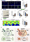 Figure 7
Tmem175L272R knock-in mice exhibit synuclein aggregation, dopaminergic neuron loss, and motor impairment. (A and B) Immunostaining of p-α-syn aggregation in striatal sections from mice of indicated genotypes after a single intrastriatal injection (magenta arrows) of α-syn pff (2.5 μg) for 3 months. White arrows: contralateral vehicle injection. Alphabetically labeled boxed regions are shown at higher magnification below. DAPI staining served as the nuclear control. Scale bar: 100 μm, n = 5–6 replicates. (C) Western blots of p-α-syn aggregation in brain tissues from Tmem175+/+, Tmem175+/L272R, and Tmem175L272R/L272R mice receiving single intrastriatal injections of α-syn pff (2.5 μg) for 3 months. Neuronal β-tubulin III (β-tub III) served as the loading control; n = 7 replicates. (D) Rotarod and hanging test results for Tmem175+/+, Tmem175+/L272R, and Tmem175L272R/L272R mice after a single intrastriatal injection of α-syn pff (2.5 μg) for 3 months; n = 5–6 mice. (E and F) Representative brain DAT-PET images from Tmem175+/+, Tmem175+/L272R, and Tmem175L272R/L272R mice after a single intrastriatal injection of α-syn pff (2.5 μg) for 5 months. Mice that received no pff injection served as negative controls. White and black arrowheads indicate the vehicle and α-syn pff injection sites, respectively; n = 6–7 mice. Data are presented as means ± SEM, with dots representing individual animals. **P < 0.01, *P < 0.05, by a 2-tailed Student’s t test. (G) Maintaining optimal lysosomal pH via V-ATPase (H+ influx) and TMEM175 (H+ efflux/leakage) is required for efficient hydrolytic degradation and clearance of α-synuclein aggregates in healthy dopaminergic neurons. Monoallelic expression of the TMEM175 p.L275R mutation, which disrupts the proton gating effect on TMEM175, reduces lysosomal H+ leakage to cause lysosomal hyper-acidification and hydrolytic impairment. Subsequently, pathogenic α-synuclein aggregates accumulate in dopaminergic neurons, ultimately promoting the progressive loss of midbrain dopaminergic neurons and increasing PD susceptibility.


Copyright © 2026 American Society for Clinical Investigation
ISSN: 0021-9738 (print), 1558-8238 (online)