
Advertisement
Research ArticleCell biologyNeuroscience
Open Access |  10.1172/JCI202787
UA-30 ameliorates motor deficits through RalA-mediated mitophagy in ALS mice
Bingge Zhang,1 Ye He,1,2 Ting Su,2 Xiaomei Li,2 Xiufen Zhang,2 Ruijuan Liu,1,2 Xiao Han,2 Ruiming Zhang,3 Chao Yang,3 Xinlei Liu,3 Qinghua Hou,4 Zaijun Zhang,5 Yongmei Xie,3 Gongping Liu,1,6 and Xifei Yang2
1Department of Pathophysiology, School of Basic Medicine and the Collaborative Innovation Center for Brain Science, Key Laboratory of Ministry of Education of China/Hubei Province for Neurological Disorders, Tongji Medical College, Huazhong University of Science and Technology, Wuhan, China.
2Shenzhen Key Laboratory of Modern Toxicology, Shenzhen Medical Key Discipline of Health Toxicology (2020–2024), Shenzhen Center for Disease Control and Prevention, Shenzhen, China.
3State Key Laboratory of Biotherapy, West China Hospital, Sichuan University and Collaborative Innovation Center for Biotherapy, Chengdu, China.
4Department of Neurology, Clinical Neuroscience Center, The Seventh Affiliated Hospital, Sun Yat-sen University, Shenzhen, China.
5State Key Laboratory of Bioactive Molecules and Druggability Assessment, Guangdong Basic Research Center of Excellence for Natural Bioactive Molecules and Discovery of Innovative Drugs, Institute of New Drug Research, College of Pharmacy, Jinan University, Guangzhou, China.
6Co-innovation Center of Neuroregeneration, Nantong University, Nantong, China.
Address correspondence to: Xifei Yang, Shenzhen Key Laboratory of Modern Toxicology, Shenzhen Center for Disease Control and Prevention, No. 8, Longyuan Road, Nanshan District, Shenzhen 518055, China. Email: xifeiyang@gmail.com. Or to: Gongping Liu, Department of Pathophysiology, School of Basic Medicine, Key Laboratory of Ministry of Education of China/Hubei Province for Neurological Disorders, Tongji Medical College, Huazhong University of Science and Technology, Wuhan 430030, China. Email: liugp111@mail.hust.edu.cn. Or to: Yongmei Xie, State Key Laboratory of Biotherapy and Cancer Center, West China Hospital, Sichuan University and Collaborative Innovation Center of Biotherapy, Chengdu 610041, China. Email: xieym@scu.edu.cn.
Authorship note: BZ, YH, and TS contributed equally to this work.
Find articles by Zhang, B. in: PubMed | Google Scholar
1Department of Pathophysiology, School of Basic Medicine and the Collaborative Innovation Center for Brain Science, Key Laboratory of Ministry of Education of China/Hubei Province for Neurological Disorders, Tongji Medical College, Huazhong University of Science and Technology, Wuhan, China.
2Shenzhen Key Laboratory of Modern Toxicology, Shenzhen Medical Key Discipline of Health Toxicology (2020–2024), Shenzhen Center for Disease Control and Prevention, Shenzhen, China.
3State Key Laboratory of Biotherapy, West China Hospital, Sichuan University and Collaborative Innovation Center for Biotherapy, Chengdu, China.
4Department of Neurology, Clinical Neuroscience Center, The Seventh Affiliated Hospital, Sun Yat-sen University, Shenzhen, China.
5State Key Laboratory of Bioactive Molecules and Druggability Assessment, Guangdong Basic Research Center of Excellence for Natural Bioactive Molecules and Discovery of Innovative Drugs, Institute of New Drug Research, College of Pharmacy, Jinan University, Guangzhou, China.
6Co-innovation Center of Neuroregeneration, Nantong University, Nantong, China.
Address correspondence to: Xifei Yang, Shenzhen Key Laboratory of Modern Toxicology, Shenzhen Center for Disease Control and Prevention, No. 8, Longyuan Road, Nanshan District, Shenzhen 518055, China. Email: xifeiyang@gmail.com. Or to: Gongping Liu, Department of Pathophysiology, School of Basic Medicine, Key Laboratory of Ministry of Education of China/Hubei Province for Neurological Disorders, Tongji Medical College, Huazhong University of Science and Technology, Wuhan 430030, China. Email: liugp111@mail.hust.edu.cn. Or to: Yongmei Xie, State Key Laboratory of Biotherapy and Cancer Center, West China Hospital, Sichuan University and Collaborative Innovation Center of Biotherapy, Chengdu 610041, China. Email: xieym@scu.edu.cn.
Authorship note: BZ, YH, and TS contributed equally to this work.
Find articles by He, Y. in: PubMed | Google Scholar
1Department of Pathophysiology, School of Basic Medicine and the Collaborative Innovation Center for Brain Science, Key Laboratory of Ministry of Education of China/Hubei Province for Neurological Disorders, Tongji Medical College, Huazhong University of Science and Technology, Wuhan, China.
2Shenzhen Key Laboratory of Modern Toxicology, Shenzhen Medical Key Discipline of Health Toxicology (2020–2024), Shenzhen Center for Disease Control and Prevention, Shenzhen, China.
3State Key Laboratory of Biotherapy, West China Hospital, Sichuan University and Collaborative Innovation Center for Biotherapy, Chengdu, China.
4Department of Neurology, Clinical Neuroscience Center, The Seventh Affiliated Hospital, Sun Yat-sen University, Shenzhen, China.
5State Key Laboratory of Bioactive Molecules and Druggability Assessment, Guangdong Basic Research Center of Excellence for Natural Bioactive Molecules and Discovery of Innovative Drugs, Institute of New Drug Research, College of Pharmacy, Jinan University, Guangzhou, China.
6Co-innovation Center of Neuroregeneration, Nantong University, Nantong, China.
Address correspondence to: Xifei Yang, Shenzhen Key Laboratory of Modern Toxicology, Shenzhen Center for Disease Control and Prevention, No. 8, Longyuan Road, Nanshan District, Shenzhen 518055, China. Email: xifeiyang@gmail.com. Or to: Gongping Liu, Department of Pathophysiology, School of Basic Medicine, Key Laboratory of Ministry of Education of China/Hubei Province for Neurological Disorders, Tongji Medical College, Huazhong University of Science and Technology, Wuhan 430030, China. Email: liugp111@mail.hust.edu.cn. Or to: Yongmei Xie, State Key Laboratory of Biotherapy and Cancer Center, West China Hospital, Sichuan University and Collaborative Innovation Center of Biotherapy, Chengdu 610041, China. Email: xieym@scu.edu.cn.
Authorship note: BZ, YH, and TS contributed equally to this work.
Find articles by Su, T. in: PubMed | Google Scholar
1Department of Pathophysiology, School of Basic Medicine and the Collaborative Innovation Center for Brain Science, Key Laboratory of Ministry of Education of China/Hubei Province for Neurological Disorders, Tongji Medical College, Huazhong University of Science and Technology, Wuhan, China.
2Shenzhen Key Laboratory of Modern Toxicology, Shenzhen Medical Key Discipline of Health Toxicology (2020–2024), Shenzhen Center for Disease Control and Prevention, Shenzhen, China.
3State Key Laboratory of Biotherapy, West China Hospital, Sichuan University and Collaborative Innovation Center for Biotherapy, Chengdu, China.
4Department of Neurology, Clinical Neuroscience Center, The Seventh Affiliated Hospital, Sun Yat-sen University, Shenzhen, China.
5State Key Laboratory of Bioactive Molecules and Druggability Assessment, Guangdong Basic Research Center of Excellence for Natural Bioactive Molecules and Discovery of Innovative Drugs, Institute of New Drug Research, College of Pharmacy, Jinan University, Guangzhou, China.
6Co-innovation Center of Neuroregeneration, Nantong University, Nantong, China.
Address correspondence to: Xifei Yang, Shenzhen Key Laboratory of Modern Toxicology, Shenzhen Center for Disease Control and Prevention, No. 8, Longyuan Road, Nanshan District, Shenzhen 518055, China. Email: xifeiyang@gmail.com. Or to: Gongping Liu, Department of Pathophysiology, School of Basic Medicine, Key Laboratory of Ministry of Education of China/Hubei Province for Neurological Disorders, Tongji Medical College, Huazhong University of Science and Technology, Wuhan 430030, China. Email: liugp111@mail.hust.edu.cn. Or to: Yongmei Xie, State Key Laboratory of Biotherapy and Cancer Center, West China Hospital, Sichuan University and Collaborative Innovation Center of Biotherapy, Chengdu 610041, China. Email: xieym@scu.edu.cn.
Authorship note: BZ, YH, and TS contributed equally to this work.
Find articles by Li, X. in: PubMed | Google Scholar
1Department of Pathophysiology, School of Basic Medicine and the Collaborative Innovation Center for Brain Science, Key Laboratory of Ministry of Education of China/Hubei Province for Neurological Disorders, Tongji Medical College, Huazhong University of Science and Technology, Wuhan, China.
2Shenzhen Key Laboratory of Modern Toxicology, Shenzhen Medical Key Discipline of Health Toxicology (2020–2024), Shenzhen Center for Disease Control and Prevention, Shenzhen, China.
3State Key Laboratory of Biotherapy, West China Hospital, Sichuan University and Collaborative Innovation Center for Biotherapy, Chengdu, China.
4Department of Neurology, Clinical Neuroscience Center, The Seventh Affiliated Hospital, Sun Yat-sen University, Shenzhen, China.
5State Key Laboratory of Bioactive Molecules and Druggability Assessment, Guangdong Basic Research Center of Excellence for Natural Bioactive Molecules and Discovery of Innovative Drugs, Institute of New Drug Research, College of Pharmacy, Jinan University, Guangzhou, China.
6Co-innovation Center of Neuroregeneration, Nantong University, Nantong, China.
Address correspondence to: Xifei Yang, Shenzhen Key Laboratory of Modern Toxicology, Shenzhen Center for Disease Control and Prevention, No. 8, Longyuan Road, Nanshan District, Shenzhen 518055, China. Email: xifeiyang@gmail.com. Or to: Gongping Liu, Department of Pathophysiology, School of Basic Medicine, Key Laboratory of Ministry of Education of China/Hubei Province for Neurological Disorders, Tongji Medical College, Huazhong University of Science and Technology, Wuhan 430030, China. Email: liugp111@mail.hust.edu.cn. Or to: Yongmei Xie, State Key Laboratory of Biotherapy and Cancer Center, West China Hospital, Sichuan University and Collaborative Innovation Center of Biotherapy, Chengdu 610041, China. Email: xieym@scu.edu.cn.
Authorship note: BZ, YH, and TS contributed equally to this work.
Find articles by Zhang, X. in: PubMed | Google Scholar
1Department of Pathophysiology, School of Basic Medicine and the Collaborative Innovation Center for Brain Science, Key Laboratory of Ministry of Education of China/Hubei Province for Neurological Disorders, Tongji Medical College, Huazhong University of Science and Technology, Wuhan, China.
2Shenzhen Key Laboratory of Modern Toxicology, Shenzhen Medical Key Discipline of Health Toxicology (2020–2024), Shenzhen Center for Disease Control and Prevention, Shenzhen, China.
3State Key Laboratory of Biotherapy, West China Hospital, Sichuan University and Collaborative Innovation Center for Biotherapy, Chengdu, China.
4Department of Neurology, Clinical Neuroscience Center, The Seventh Affiliated Hospital, Sun Yat-sen University, Shenzhen, China.
5State Key Laboratory of Bioactive Molecules and Druggability Assessment, Guangdong Basic Research Center of Excellence for Natural Bioactive Molecules and Discovery of Innovative Drugs, Institute of New Drug Research, College of Pharmacy, Jinan University, Guangzhou, China.
6Co-innovation Center of Neuroregeneration, Nantong University, Nantong, China.
Address correspondence to: Xifei Yang, Shenzhen Key Laboratory of Modern Toxicology, Shenzhen Center for Disease Control and Prevention, No. 8, Longyuan Road, Nanshan District, Shenzhen 518055, China. Email: xifeiyang@gmail.com. Or to: Gongping Liu, Department of Pathophysiology, School of Basic Medicine, Key Laboratory of Ministry of Education of China/Hubei Province for Neurological Disorders, Tongji Medical College, Huazhong University of Science and Technology, Wuhan 430030, China. Email: liugp111@mail.hust.edu.cn. Or to: Yongmei Xie, State Key Laboratory of Biotherapy and Cancer Center, West China Hospital, Sichuan University and Collaborative Innovation Center of Biotherapy, Chengdu 610041, China. Email: xieym@scu.edu.cn.
Authorship note: BZ, YH, and TS contributed equally to this work.
Find articles by Liu, R. in: PubMed | Google Scholar
1Department of Pathophysiology, School of Basic Medicine and the Collaborative Innovation Center for Brain Science, Key Laboratory of Ministry of Education of China/Hubei Province for Neurological Disorders, Tongji Medical College, Huazhong University of Science and Technology, Wuhan, China.
2Shenzhen Key Laboratory of Modern Toxicology, Shenzhen Medical Key Discipline of Health Toxicology (2020–2024), Shenzhen Center for Disease Control and Prevention, Shenzhen, China.
3State Key Laboratory of Biotherapy, West China Hospital, Sichuan University and Collaborative Innovation Center for Biotherapy, Chengdu, China.
4Department of Neurology, Clinical Neuroscience Center, The Seventh Affiliated Hospital, Sun Yat-sen University, Shenzhen, China.
5State Key Laboratory of Bioactive Molecules and Druggability Assessment, Guangdong Basic Research Center of Excellence for Natural Bioactive Molecules and Discovery of Innovative Drugs, Institute of New Drug Research, College of Pharmacy, Jinan University, Guangzhou, China.
6Co-innovation Center of Neuroregeneration, Nantong University, Nantong, China.
Address correspondence to: Xifei Yang, Shenzhen Key Laboratory of Modern Toxicology, Shenzhen Center for Disease Control and Prevention, No. 8, Longyuan Road, Nanshan District, Shenzhen 518055, China. Email: xifeiyang@gmail.com. Or to: Gongping Liu, Department of Pathophysiology, School of Basic Medicine, Key Laboratory of Ministry of Education of China/Hubei Province for Neurological Disorders, Tongji Medical College, Huazhong University of Science and Technology, Wuhan 430030, China. Email: liugp111@mail.hust.edu.cn. Or to: Yongmei Xie, State Key Laboratory of Biotherapy and Cancer Center, West China Hospital, Sichuan University and Collaborative Innovation Center of Biotherapy, Chengdu 610041, China. Email: xieym@scu.edu.cn.
Authorship note: BZ, YH, and TS contributed equally to this work.
Find articles by Han, X. in: PubMed | Google Scholar
1Department of Pathophysiology, School of Basic Medicine and the Collaborative Innovation Center for Brain Science, Key Laboratory of Ministry of Education of China/Hubei Province for Neurological Disorders, Tongji Medical College, Huazhong University of Science and Technology, Wuhan, China.
2Shenzhen Key Laboratory of Modern Toxicology, Shenzhen Medical Key Discipline of Health Toxicology (2020–2024), Shenzhen Center for Disease Control and Prevention, Shenzhen, China.
3State Key Laboratory of Biotherapy, West China Hospital, Sichuan University and Collaborative Innovation Center for Biotherapy, Chengdu, China.
4Department of Neurology, Clinical Neuroscience Center, The Seventh Affiliated Hospital, Sun Yat-sen University, Shenzhen, China.
5State Key Laboratory of Bioactive Molecules and Druggability Assessment, Guangdong Basic Research Center of Excellence for Natural Bioactive Molecules and Discovery of Innovative Drugs, Institute of New Drug Research, College of Pharmacy, Jinan University, Guangzhou, China.
6Co-innovation Center of Neuroregeneration, Nantong University, Nantong, China.
Address correspondence to: Xifei Yang, Shenzhen Key Laboratory of Modern Toxicology, Shenzhen Center for Disease Control and Prevention, No. 8, Longyuan Road, Nanshan District, Shenzhen 518055, China. Email: xifeiyang@gmail.com. Or to: Gongping Liu, Department of Pathophysiology, School of Basic Medicine, Key Laboratory of Ministry of Education of China/Hubei Province for Neurological Disorders, Tongji Medical College, Huazhong University of Science and Technology, Wuhan 430030, China. Email: liugp111@mail.hust.edu.cn. Or to: Yongmei Xie, State Key Laboratory of Biotherapy and Cancer Center, West China Hospital, Sichuan University and Collaborative Innovation Center of Biotherapy, Chengdu 610041, China. Email: xieym@scu.edu.cn.
Authorship note: BZ, YH, and TS contributed equally to this work.
Find articles by Zhang, R. in: PubMed | Google Scholar
1Department of Pathophysiology, School of Basic Medicine and the Collaborative Innovation Center for Brain Science, Key Laboratory of Ministry of Education of China/Hubei Province for Neurological Disorders, Tongji Medical College, Huazhong University of Science and Technology, Wuhan, China.
2Shenzhen Key Laboratory of Modern Toxicology, Shenzhen Medical Key Discipline of Health Toxicology (2020–2024), Shenzhen Center for Disease Control and Prevention, Shenzhen, China.
3State Key Laboratory of Biotherapy, West China Hospital, Sichuan University and Collaborative Innovation Center for Biotherapy, Chengdu, China.
4Department of Neurology, Clinical Neuroscience Center, The Seventh Affiliated Hospital, Sun Yat-sen University, Shenzhen, China.
5State Key Laboratory of Bioactive Molecules and Druggability Assessment, Guangdong Basic Research Center of Excellence for Natural Bioactive Molecules and Discovery of Innovative Drugs, Institute of New Drug Research, College of Pharmacy, Jinan University, Guangzhou, China.
6Co-innovation Center of Neuroregeneration, Nantong University, Nantong, China.
Address correspondence to: Xifei Yang, Shenzhen Key Laboratory of Modern Toxicology, Shenzhen Center for Disease Control and Prevention, No. 8, Longyuan Road, Nanshan District, Shenzhen 518055, China. Email: xifeiyang@gmail.com. Or to: Gongping Liu, Department of Pathophysiology, School of Basic Medicine, Key Laboratory of Ministry of Education of China/Hubei Province for Neurological Disorders, Tongji Medical College, Huazhong University of Science and Technology, Wuhan 430030, China. Email: liugp111@mail.hust.edu.cn. Or to: Yongmei Xie, State Key Laboratory of Biotherapy and Cancer Center, West China Hospital, Sichuan University and Collaborative Innovation Center of Biotherapy, Chengdu 610041, China. Email: xieym@scu.edu.cn.
Authorship note: BZ, YH, and TS contributed equally to this work.
Find articles by Yang, C. in: PubMed | Google Scholar
1Department of Pathophysiology, School of Basic Medicine and the Collaborative Innovation Center for Brain Science, Key Laboratory of Ministry of Education of China/Hubei Province for Neurological Disorders, Tongji Medical College, Huazhong University of Science and Technology, Wuhan, China.
2Shenzhen Key Laboratory of Modern Toxicology, Shenzhen Medical Key Discipline of Health Toxicology (2020–2024), Shenzhen Center for Disease Control and Prevention, Shenzhen, China.
3State Key Laboratory of Biotherapy, West China Hospital, Sichuan University and Collaborative Innovation Center for Biotherapy, Chengdu, China.
4Department of Neurology, Clinical Neuroscience Center, The Seventh Affiliated Hospital, Sun Yat-sen University, Shenzhen, China.
5State Key Laboratory of Bioactive Molecules and Druggability Assessment, Guangdong Basic Research Center of Excellence for Natural Bioactive Molecules and Discovery of Innovative Drugs, Institute of New Drug Research, College of Pharmacy, Jinan University, Guangzhou, China.
6Co-innovation Center of Neuroregeneration, Nantong University, Nantong, China.
Address correspondence to: Xifei Yang, Shenzhen Key Laboratory of Modern Toxicology, Shenzhen Center for Disease Control and Prevention, No. 8, Longyuan Road, Nanshan District, Shenzhen 518055, China. Email: xifeiyang@gmail.com. Or to: Gongping Liu, Department of Pathophysiology, School of Basic Medicine, Key Laboratory of Ministry of Education of China/Hubei Province for Neurological Disorders, Tongji Medical College, Huazhong University of Science and Technology, Wuhan 430030, China. Email: liugp111@mail.hust.edu.cn. Or to: Yongmei Xie, State Key Laboratory of Biotherapy and Cancer Center, West China Hospital, Sichuan University and Collaborative Innovation Center of Biotherapy, Chengdu 610041, China. Email: xieym@scu.edu.cn.
Authorship note: BZ, YH, and TS contributed equally to this work.
Find articles by Liu, X. in: PubMed | Google Scholar
1Department of Pathophysiology, School of Basic Medicine and the Collaborative Innovation Center for Brain Science, Key Laboratory of Ministry of Education of China/Hubei Province for Neurological Disorders, Tongji Medical College, Huazhong University of Science and Technology, Wuhan, China.
2Shenzhen Key Laboratory of Modern Toxicology, Shenzhen Medical Key Discipline of Health Toxicology (2020–2024), Shenzhen Center for Disease Control and Prevention, Shenzhen, China.
3State Key Laboratory of Biotherapy, West China Hospital, Sichuan University and Collaborative Innovation Center for Biotherapy, Chengdu, China.
4Department of Neurology, Clinical Neuroscience Center, The Seventh Affiliated Hospital, Sun Yat-sen University, Shenzhen, China.
5State Key Laboratory of Bioactive Molecules and Druggability Assessment, Guangdong Basic Research Center of Excellence for Natural Bioactive Molecules and Discovery of Innovative Drugs, Institute of New Drug Research, College of Pharmacy, Jinan University, Guangzhou, China.
6Co-innovation Center of Neuroregeneration, Nantong University, Nantong, China.
Address correspondence to: Xifei Yang, Shenzhen Key Laboratory of Modern Toxicology, Shenzhen Center for Disease Control and Prevention, No. 8, Longyuan Road, Nanshan District, Shenzhen 518055, China. Email: xifeiyang@gmail.com. Or to: Gongping Liu, Department of Pathophysiology, School of Basic Medicine, Key Laboratory of Ministry of Education of China/Hubei Province for Neurological Disorders, Tongji Medical College, Huazhong University of Science and Technology, Wuhan 430030, China. Email: liugp111@mail.hust.edu.cn. Or to: Yongmei Xie, State Key Laboratory of Biotherapy and Cancer Center, West China Hospital, Sichuan University and Collaborative Innovation Center of Biotherapy, Chengdu 610041, China. Email: xieym@scu.edu.cn.
Authorship note: BZ, YH, and TS contributed equally to this work.
Find articles by Hou, Q. in: PubMed | Google Scholar
1Department of Pathophysiology, School of Basic Medicine and the Collaborative Innovation Center for Brain Science, Key Laboratory of Ministry of Education of China/Hubei Province for Neurological Disorders, Tongji Medical College, Huazhong University of Science and Technology, Wuhan, China.
2Shenzhen Key Laboratory of Modern Toxicology, Shenzhen Medical Key Discipline of Health Toxicology (2020–2024), Shenzhen Center for Disease Control and Prevention, Shenzhen, China.
3State Key Laboratory of Biotherapy, West China Hospital, Sichuan University and Collaborative Innovation Center for Biotherapy, Chengdu, China.
4Department of Neurology, Clinical Neuroscience Center, The Seventh Affiliated Hospital, Sun Yat-sen University, Shenzhen, China.
5State Key Laboratory of Bioactive Molecules and Druggability Assessment, Guangdong Basic Research Center of Excellence for Natural Bioactive Molecules and Discovery of Innovative Drugs, Institute of New Drug Research, College of Pharmacy, Jinan University, Guangzhou, China.
6Co-innovation Center of Neuroregeneration, Nantong University, Nantong, China.
Address correspondence to: Xifei Yang, Shenzhen Key Laboratory of Modern Toxicology, Shenzhen Center for Disease Control and Prevention, No. 8, Longyuan Road, Nanshan District, Shenzhen 518055, China. Email: xifeiyang@gmail.com. Or to: Gongping Liu, Department of Pathophysiology, School of Basic Medicine, Key Laboratory of Ministry of Education of China/Hubei Province for Neurological Disorders, Tongji Medical College, Huazhong University of Science and Technology, Wuhan 430030, China. Email: liugp111@mail.hust.edu.cn. Or to: Yongmei Xie, State Key Laboratory of Biotherapy and Cancer Center, West China Hospital, Sichuan University and Collaborative Innovation Center of Biotherapy, Chengdu 610041, China. Email: xieym@scu.edu.cn.
Authorship note: BZ, YH, and TS contributed equally to this work.
Find articles by Zhang, Z. in: PubMed | Google Scholar
1Department of Pathophysiology, School of Basic Medicine and the Collaborative Innovation Center for Brain Science, Key Laboratory of Ministry of Education of China/Hubei Province for Neurological Disorders, Tongji Medical College, Huazhong University of Science and Technology, Wuhan, China.
2Shenzhen Key Laboratory of Modern Toxicology, Shenzhen Medical Key Discipline of Health Toxicology (2020–2024), Shenzhen Center for Disease Control and Prevention, Shenzhen, China.
3State Key Laboratory of Biotherapy, West China Hospital, Sichuan University and Collaborative Innovation Center for Biotherapy, Chengdu, China.
4Department of Neurology, Clinical Neuroscience Center, The Seventh Affiliated Hospital, Sun Yat-sen University, Shenzhen, China.
5State Key Laboratory of Bioactive Molecules and Druggability Assessment, Guangdong Basic Research Center of Excellence for Natural Bioactive Molecules and Discovery of Innovative Drugs, Institute of New Drug Research, College of Pharmacy, Jinan University, Guangzhou, China.
6Co-innovation Center of Neuroregeneration, Nantong University, Nantong, China.
Address correspondence to: Xifei Yang, Shenzhen Key Laboratory of Modern Toxicology, Shenzhen Center for Disease Control and Prevention, No. 8, Longyuan Road, Nanshan District, Shenzhen 518055, China. Email: xifeiyang@gmail.com. Or to: Gongping Liu, Department of Pathophysiology, School of Basic Medicine, Key Laboratory of Ministry of Education of China/Hubei Province for Neurological Disorders, Tongji Medical College, Huazhong University of Science and Technology, Wuhan 430030, China. Email: liugp111@mail.hust.edu.cn. Or to: Yongmei Xie, State Key Laboratory of Biotherapy and Cancer Center, West China Hospital, Sichuan University and Collaborative Innovation Center of Biotherapy, Chengdu 610041, China. Email: xieym@scu.edu.cn.
Authorship note: BZ, YH, and TS contributed equally to this work.
Find articles by Xie, Y. in: PubMed | Google Scholar
1Department of Pathophysiology, School of Basic Medicine and the Collaborative Innovation Center for Brain Science, Key Laboratory of Ministry of Education of China/Hubei Province for Neurological Disorders, Tongji Medical College, Huazhong University of Science and Technology, Wuhan, China.
2Shenzhen Key Laboratory of Modern Toxicology, Shenzhen Medical Key Discipline of Health Toxicology (2020–2024), Shenzhen Center for Disease Control and Prevention, Shenzhen, China.
3State Key Laboratory of Biotherapy, West China Hospital, Sichuan University and Collaborative Innovation Center for Biotherapy, Chengdu, China.
4Department of Neurology, Clinical Neuroscience Center, The Seventh Affiliated Hospital, Sun Yat-sen University, Shenzhen, China.
5State Key Laboratory of Bioactive Molecules and Druggability Assessment, Guangdong Basic Research Center of Excellence for Natural Bioactive Molecules and Discovery of Innovative Drugs, Institute of New Drug Research, College of Pharmacy, Jinan University, Guangzhou, China.
6Co-innovation Center of Neuroregeneration, Nantong University, Nantong, China.
Address correspondence to: Xifei Yang, Shenzhen Key Laboratory of Modern Toxicology, Shenzhen Center for Disease Control and Prevention, No. 8, Longyuan Road, Nanshan District, Shenzhen 518055, China. Email: xifeiyang@gmail.com. Or to: Gongping Liu, Department of Pathophysiology, School of Basic Medicine, Key Laboratory of Ministry of Education of China/Hubei Province for Neurological Disorders, Tongji Medical College, Huazhong University of Science and Technology, Wuhan 430030, China. Email: liugp111@mail.hust.edu.cn. Or to: Yongmei Xie, State Key Laboratory of Biotherapy and Cancer Center, West China Hospital, Sichuan University and Collaborative Innovation Center of Biotherapy, Chengdu 610041, China. Email: xieym@scu.edu.cn.
Authorship note: BZ, YH, and TS contributed equally to this work.
Find articles by Liu, G. in: PubMed | Google Scholar
1Department of Pathophysiology, School of Basic Medicine and the Collaborative Innovation Center for Brain Science, Key Laboratory of Ministry of Education of China/Hubei Province for Neurological Disorders, Tongji Medical College, Huazhong University of Science and Technology, Wuhan, China.
2Shenzhen Key Laboratory of Modern Toxicology, Shenzhen Medical Key Discipline of Health Toxicology (2020–2024), Shenzhen Center for Disease Control and Prevention, Shenzhen, China.
3State Key Laboratory of Biotherapy, West China Hospital, Sichuan University and Collaborative Innovation Center for Biotherapy, Chengdu, China.
4Department of Neurology, Clinical Neuroscience Center, The Seventh Affiliated Hospital, Sun Yat-sen University, Shenzhen, China.
5State Key Laboratory of Bioactive Molecules and Druggability Assessment, Guangdong Basic Research Center of Excellence for Natural Bioactive Molecules and Discovery of Innovative Drugs, Institute of New Drug Research, College of Pharmacy, Jinan University, Guangzhou, China.
6Co-innovation Center of Neuroregeneration, Nantong University, Nantong, China.
Address correspondence to: Xifei Yang, Shenzhen Key Laboratory of Modern Toxicology, Shenzhen Center for Disease Control and Prevention, No. 8, Longyuan Road, Nanshan District, Shenzhen 518055, China. Email: xifeiyang@gmail.com. Or to: Gongping Liu, Department of Pathophysiology, School of Basic Medicine, Key Laboratory of Ministry of Education of China/Hubei Province for Neurological Disorders, Tongji Medical College, Huazhong University of Science and Technology, Wuhan 430030, China. Email: liugp111@mail.hust.edu.cn. Or to: Yongmei Xie, State Key Laboratory of Biotherapy and Cancer Center, West China Hospital, Sichuan University and Collaborative Innovation Center of Biotherapy, Chengdu 610041, China. Email: xieym@scu.edu.cn.
Authorship note: BZ, YH, and TS contributed equally to this work.
Find articles by Yang, X. in: PubMed | Google Scholar
Published September 15, 2026 - More info
J Clin Invest. 2026;136(18):e202787. https://doi.org/10.1172/JCI202787.
© 2026 Zhang et al. This work is licensed under the Creative Commons Attribution 4.0 International License. To view a copy of this license, visit http://creativecommons.org/licenses/by/4.0/.
Received: November 24, 2025; Accepted: July 14, 2026

-
Results
UA-30 improved motor performance and extended lifespan in SOD1G93A mice. To evaluate the CNS distribution of UA-30, pharmacokinetic analysis was performed after a single oral dose in mice. UA-30 was detected in both brain and spinal cord. The Cmax in spinal cord reached 6,293 ± 1,022 ng/g at 0.625 h after dose, while the Cmax in brain reached 16,490 ± 2,204 ng/g at 0.875 h after dose. UA-30 also showed favorable CNS distribution (brain/plasma, >2.5; spinal cord/plasma, >0.7), together with relatively prolonged tissue half-lives (brain t1/2 = 3.44 h; spinal cord t1/2 = 3.67 h) (Supplemental Figure 2 and Supplemental Table 1).
Next, we evaluated the effects of UA-30 on ALS disease progression and survival in SOD1G93A mice. SOD1G93A transgenic mice at 13 weeks of age received oral administration of UA-30 (25, 50, and 100 mg/kg) or riluzole (10 mg/kg) once daily for 6 weeks (Figure 1A). Baseline behavioral assessments before treatment initiation showed comparable motor function across all experimental groups (Supplemental Figure 3A). Longitudinal behavioral assessments such as pole climbing, wire hanging, and grip strength were performed at weeks 2, 4, and 6 during the 6-week treatment period to evaluate the effects of UA-30 on motor function (Figure 1, B–D, Supplemental Figure 2, B and C, and Supplemental Figure 4, A–C). At the week 6 endpoint, motor coordination and balance were further evaluated using the rotarod test (Figure 1E and Supplemental Figure 4D), and locomotor patterns were assessed by gait analysis (Figure 1F, Supplemental Figure 4E, and Supplemental Videos 1 and 2). Compared with control mice, SOD1G93A mice exhibited impaired climbing ability, endurance, and grip strength as the disease advanced, with males showing a faster rate of disease progression than females (Figure 1, B–D, Supplemental Figure 3, B and C, and Supplemental Figure 4, A–C). UA-30 treatment attenuated the decline in motor performance in both sexes across multiple behavioral endpoints. In male mice, UA-30 reduced pole descent time, prolonged wire-hang latency, increased grip strength, and improved rotarod performance (Figure 1, B–E). Female mice also showed improvement in these behavioral measures (Supplemental Figure 4, A–D). Gait analysis showed that SOD1G93A mice had substantially reduced stride lengths compared with WT controls, while high-dose UA-30 treatment increased the average stride length in male mice (Figure 1F and Supplemental Video 1) and improved specific stride parameters in female mice (Supplemental Figure 4E and Supplemental Video 2). Specifically, compared with the low dose, the high dose improved performance by 19.36% in the pole-climbing test and increased hanging latency by 22.11%, grip strength by 2.30%, and rotarod latency by 8.39% (Figure 1, B–E). Notably, high-dose UA-30 demonstrated superior efficacy to riluzole treatment in improving most ALS-associated motor deficits, shown as more pronounced improvements in pole descent time (reducing time by 13.7% in females and 10.6% in males), wire hanging endurance (increasing time by 39.7% in females and 64.5% in males), and rotarod fall latency (increasing latency by 29.7% in females and 14.9% in males) (Figure 1, B, C, and E, and Supplemental Figure 4, A, B, and D).
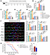 Figure 1
UA-30 treatment improved motor dysfunction and increased lifespan in male SOD1G93A mice. (A) Schematic of the experimental protocol and administration schedule. (B–F) Motor performance and muscular strength assessments in male SOD1G93A mice treated with different doses of UA-30 or saline for 6 weeks. Tests performed: pole climbing (B), wire hanging (C), grip strength (D), rotarod (E), and gait (F). Representative marker paw prints in gait analysis and statistics on left/right forelimb and left/right hind limb stride length. n = 8 male mice per group. (G) Kaplan-Meier survival curves for male SOD1G93A mice treated with saline, UA-30 (100 mg/kg), or riluzole (10 mg/kg). Statistical summaries show overall lifespan for each cohort. n = 17 SOD1G93A male mice. n = 14 SOD1G93A + UA-30 male mice, n = 13 SOD1G93A + riluzole male mice. Data are expressed as mean ± SEM. *P < 0.05, **P < 0.01, ***P < 0.001, and ****P < 0.0001. B–F, 1-way ANOVA followed by Tukey’s multiple-comparison test; G, log-rank (Mantel-Cox) test for survival curves and 1-way ANOVA followed by Tukey’s multiple-comparison test for overall lifespan summary.
To further determine whether treatment responses differed by sex, we performed 2-way ANOVA using sex and treatment as factors for the week 6 behavioral endpoints. No significant sex × treatment interaction was detected for pole climbing (P = 0.3222), wire hanging (P = 0.5612), grip strength (P = 0.8287), or rotarod performance (P = 0.5821) (Supplemental Figure 5, A–D), indicating that there was no statistical evidence for sex-specific treatment responses in these assays. Significant main effects of treatment were observed for all 4 behavioral endpoints, whereas sex main effects were detected for wire hanging (P < 0.0001), grip strength (P = 0.0326), and rotarod performance (P = 0.0118), but not for pole climbing (P = 0.3674) (Supplemental Figure 5, A–D), reflecting endpoint-dependent overall differences between males and females rather than differential treatment responses.
To address whether the structural modification of UA-30 confers a therapeutic advantage over its parent compound, we performed a head-to-head comparison at equimolar doses in SOD1G93A mice. Functional assessment in SOD1G93A mice treated with UA-30 revealed improvements in motor performance. UA-30 demonstrated greater improvements in pole-climbing descent time, hanging endurance, and rotarod performance (Supplemental Figure 6, A–C), whereas grip strength did not differ significantly between the 2 groups (Supplemental Figure 6D).
To investigate the effect of UA-30 on survival in SOD1G93A mice, we established cohorts treated with UA-30, riluzole, or vehicle control. Compared with untreated SOD1G93A mice, UA-30–treated SOD1G93A mice showed a longer mean overall lifespan (171.5 ± 8.7 days vs. 161.9 ± 8.1 days, P = 0.027) and an 8.5-day increase in median survival. Although the difference in survival distribution between UA-30 and riluzole groups did not reach statistical significance (P = 0.107), the UA-30 group showed a trend toward longer survival, as indicated by a greater mean overall lifespan (171.5 ± 8.7 vs. 164.5 ± 14.2 days) and median survival (170.5 vs. 163 days) (Figure 1G). Survival was also analyzed in the female cohort. However, under the present study conditions, the survival benefit of UA-30 did not reach statistical significance (Supplemental Figure 4F).
UA-30 reduced muscle damage and fibrosis in the gastrocnemius muscle of SOD1G93A mice. At the end of the 6-week treatment period, biochemical and histopathological analyses were performed on the same cohort of mice used for behavioral assessments. Oral administration of different doses of UA-30 did not affect the body weight of SOD1G93A mice (Supplemental Figure 7A). Compared with control mice, SOD1G93A mice exhibited a statistically significant reduction in gastrocnemius muscle weight (P < 0.0001). Notably, UA-30 treatment increased gastrocnemius muscle weight in female SOD1G93A mice (Supplemental Figure 7B). Furthermore, UA-30 reduced serum creatine kinase and lactate dehydrogenase activities (Figure 2A) and normalized the reduced blood glucose levels observed in SOD1G93A mice (Supplemental Figure 8, A and B). Riluzole treatment was associated with increased serum alanine aminotransferase and aspartate aminotransferase levels in SOD1G93A mice (Supplemental Figure 8A).
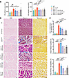 Figure 2
UA-30 treatment alleviated muscle damage and suppressed muscle pathologic deterioration of male SOD1G93A mice. Samples were collected at the experimental endpoint after 6 weeks of treatment (19 weeks of age) from the same cohort of mice used for behavioral assessments. (A) Blood biochemical analysis of creatine kinase (CK) and lactate dehydrogenase (LDH) activity in SOD1G93A male mice treated with UA-30 or saline in each group. n = 8 male per group. (B and C) Representative H&E, Masson’s, and Sirius red staining of gastrocnemius muscle from male SOD1G93A mice after UA-30 treatment, with corresponding quantification of inflammatory area, collagen fiber content, and collagen deposition. Scale bars: 50 μm (H&E), 100 μm (Masson), 200 μm (Sirius red). n = 4 male per group. Data are expressed as mean ± SEM. *P < 0.05, **P < 0.01 ***P < 0.001, and ****P < 0.0001. 1-way ANOVA followed by Dunnett’s multiple-comparison test.
In ALS, atrophy of the gastrocnemius muscle is frequently accompanied by fibrosis (21). Treatment with UA-30 reduced the inflammatory area and collagen fiber hyperplasia in the gastrocnemius muscle of SOD1G93A mice (Figure 2, B and C). Masson’s and Sirius red staining revealed substantially higher levels of muscular fibrosis in untreated SOD1G93A mice compared with WT mice. UA-30 administration attenuated gastrocnemius fibrosis in this model, which was particularly evident at the high dose of UA-30. Compared with riluzole, high-dose UA-30 showed numerically lower fibrotic areas (Masson-positive area: 6.7% ± 3.1% vs. 9.0% ± 2.2%; Sirius red–positive area: 4.7% ± 1.1% vs. 9.2% ± 2.8%), although no statistically significant difference was detected between the 2 groups. In WT mice, synaptophysin-positive presynaptic terminals showed extensive overlap with α-bungarotoxin–labeled motor end plates, indicating intact neuromuscular junction (NMJ) innervation. By contrast, SOD1G93A mice exhibited evident NMJ denervation. UA-30 treatment partially restored NMJ innervation in SOD1G93A mice (Supplemental Figure 9).
UA-30–attenuated motor neuron loss and glial cell proliferation in SOD1G93A mice. The principal neuropathological hallmark of ALS is the progressive loss of motor neurons (1). To assess motor neuron survival within the anterior horn of the L4-L5 lumbar spinal cord, neurons and motor neurons were specifically labeled using NeuN and choline acetyltransferase immunohistochemistry, respectively. SOD1G93A mice showed a statistically significant reduction in the number of spinal motor neurons compared with controls (P < 0.0001). UA-30 administration ameliorated loss of spinal motor neurons in the ALS model (Figure 3, A and B, and Supplemental Figure 10A). We also found that UA-30 treatment attenuated the increase in both astrocyte and microglial cell numbers observed in the spinal cord of male SOD1G93A mice (Figure 3, C and D). Similar results were observed in the spinal cord of female SOD1G93A mice (Supplemental Figure 10A). High-dose UA-30 treatment proved superior to riluzole, exhibiting a 20.3% greater preservation of neurons in male mice and a 9.5% greater preservation in female mice compared with the riluzole-treated group.
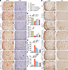 Figure 3
UA-30 treatment attenuated motor neuron loss and neuroinflammation in male SOD1G93A mice. (A and B) Representative images (A) and statistical analysis (B) of immunohistochemical staining of neurons (NeuN) and cholinergic neurons labeled with choline acetyltransferase (ChAT) of L4-L5 spinal cord from saline- or UA-30–treated male SOD1G93A mice. Scale bar: 200 μm. (C and D) Representative images (C) and quantification (D) of astrocytes (GFAP) and microglia (Iba1) immunohistochemical staining in spinal cord after UA-30 treatment in male SOD1G93A mice. Scale bar: 200 μm. n = 3 male per group. Data are expressed as mean ± SEM. *P < 0.05, **P < 0.01 ***P < 0.001, and ****P < 0.0001. 1-way ANOVA followed by Dunnett’s multiple-comparison test.
To comprehensively evaluate the pathogenesis of ALS, human SOD1 (hSOD1) levels were quantified in the medulla oblongata and spinal cord of SOD1G93A mice (Supplemental Figure 10B). We found that high-dose UA-30 administration reduced hSOD1 levels in both the medulla and spinal cord of SOD1G93A mice. These results indicate that UA-30 treatment mitigates the extent of cellular pathology associated with ALS.
UA-30 modified the protein expression profile in SOD1G93A mice. To elucidate the therapeutic mechanism of UA-30, we performed proteomic analyses of the medulla oblongata and spinal cord in SOD1G93A mice. A total of 6,266 proteins were identified and quantified in spinal cord samples, while 5,893 proteins were detected in the medulla oblongata. Compared with WT mice, SOD1G93A mice exhibited 1,364 differentially expressed (DE) proteins in the spinal cord (Supplemental Figure 11A). Bioinformatic analysis revealed that downregulated proteins were enriched in biological processes including mitochondrial ATP synthesis, aerobic respiration, TCA cycle, mitochondrial respiratory chain assembly, and mitochondrial electron transport (Supplemental Figure 11B). In the medulla oblongata, 653 DE proteins were identified in SOD1G93A mice compared with WT controls (Supplemental Figure 11C), with downregulated DE proteins primarily involved in endocytosis, protein transport, mitochondrial ATP synthesis, aerobic respiration, and mitochondrial respiratory chain assembly (Supplemental Figure 11D). These results indicate marked energy metabolism dysregulation and mitochondrial dysfunction in ALS model mice, suggesting that improving mitochondrial function represents a potential therapeutic strategy for ALS.
Gene Ontology analysis of DE proteins in the medulla oblongata demonstrated that high-dose UA-30 treatment enriched biological processes related to actin cytoskeleton organization regulation, canonical glycolysis, ATP metabolism, mitochondrial organization, oxidative stress response, and autophagosome assembly in SOD1G93A mice compared with untreated model mice (Figure 4A). KEGG pathway analysis of the relevant proteins implicated pathways primarily associated with ALS, autophagy, lysosomes, and glycolysis (Figure 4B). Subsequent unsupervised hierarchical clustering was performed on the 269 DE proteins identified in the spinal cord to group proteins based on their normalized expression profiles across all experimental conditions. One cluster displaying an ameliorated expression pattern following UA-30 treatment was identified (Figure 4C). A heatmap of the 72 proteins within this cluster is shown (Figure 4D). Gene enrichment analysis of these 72 proteins revealed their involvement in biological processes including PI3K activity regulation, GTPase activity regulation, and mitochondrial translation (Figure 4E). KEGG pathway analysis of this cluster identified enriched pathways including galactose metabolism, FOXO signaling, and PI3K/AKT signaling (Figure 4F).
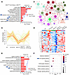 Figure 4
Proteomics analysis of the spinal cord and medulla oblongata in male SOD1G93A mice after UA-30 treatment. (A and B) Corresponding biological processes (A) and KEGG pathways (B) of DE protein enrichment in medulla oblongata proteomics under high-dose UA-30 treatment, with P value of enrichment less than 0.05. (C) An ameliorated expression pattern in the spinal cord proteome identified by unsupervised hierarchical clustering of DE proteins based on their normalized expression profiles across experimental groups. The red line indicates the mean expression trend of this cluster. (D) Heatmap showing the expression profiles of 72 DE proteins belonging to the ameliorated expression cluster shown in (C), which decreased in SOD1G93A mice and was restored toward WT levels after UA-30 treatment. Rows represent individual proteins, columns represent experimental groups, and colors indicate row-wise z score–normalized protein abundance (red, higher expression; blue, lower expression). (E and F) Biological processes (E) and KEGG pathways (F) enriched among the DE proteins within this expression pattern. Enrichment was defined as P value < 0.05. Arrows indicate terms/pathways with high enrichment scores and greater biological relevance.
UA-30 ameliorated ALS pathology by enhancing mitochondrial function and mitophagy. Proteomic analysis in this study indicated that UA-30 partially alleviated ALS-related changes by modulating mitochondrial and intracellular homeostasis. To determine the impact of UA-30 on mitochondria, we analyzed mitochondrial oxidative phosphorylation proteins in the spinal cord and medulla oblongata of SOD1G93A mice. UA-30 treatment increased levels of ETC complex III (UQCRFS1) and complex IV (COX5b) in the spinal cord (Figure 5, A and B) and elevated levels of complexes I (Ndufs1), III, and IV in the medulla oblongata (Supplemental Figure 12, A and B). Consistent with these findings, UA-30 improved levels of ETC complexes III and IV in PC12 cells transfected with hSOD1G93A (an ALS cellular model; Supplemental Figure 13, A–C) (22). Mitochondria were categorized into 3 types based on the degree of cristae loss (23). Transmission electron microscopy (TEM) revealed that spinal cord neurons of SOD1G93A mice exhibited rounded, swollen mitochondria with disorganized or dissipated cristae. UA-30 intervention effectively mitigated these mitochondrial morphological defects (Figure 5, C and D). Furthermore, UA-30 reversed the decrease in ATP levels and the increase in malondialdehyde (MDA) levels observed in the spinal cord, medulla oblongata, and gastrocnemius muscle of SOD1G93A mice (Figure 5, E and F, and Supplemental Figure 12, C and D). Consistently, UA-30 increased ATP levels, restored mitochondrial membrane potential (MMP), and reduced ROS and mitochondrial ROS (mitoROS) levels in SOD1G93A cells (Supplemental Figure 13, D–F).
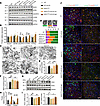 Figure 5
UA-30 treatment improved mitochondrial damage and mitophagy defects in male SOD1G93A mice. (A and B) Expression and quantification of mitochondrial respiratory complex I (Ndufs1), II (SDHB), III (UQCRFS1), IV (COX5b), and V (ATP5a) in the spinal cord from saline- or UA-30–treated mice. n = 4 male per group. (C and D) Mitochondria morphology in spinal cord neurons of SOD1G93A mice was observed using TEM. Representative mitochondrial images are shown in C. Scale bar: 1 μm. Mitochondria were classified into 3 categories based on cristae morphology (green, intact; orange, mild cristae loss; purple, severe cristae loss). The proportion of each mitochondria category was quantified separately (D), and data are presented as frequency distributions. At least 150 mitochondria from 3 male mice were analyzed in each group. (E and F) Steady-state ATP content and lipid peroxidation levels were measured in the spinal cord of SOD1G93A mice exposed to different treatments. n = 5 male per group. (G and H) Western blot (G) and subsequent quantitation (H) for levels of PINK1 and Parkin in spinal cord after UA-30 treatment in SOD1G93A mice. n = 4 male per group. (I) RT-qPCR analysis of Pink1 and Prkn mRNA levels in spinal cord of UA-30–treated SOD1G93A mice. n = 5 male per group. (J) Co-immunofluorescence staining of LC3 (green) and Tomm40 (red) and co-immunofluorescence staining of Lamp2 (green) and Tomm20 (red) in the spinal cord of male mice treated with UA-30. Scale bars: 50 μm. Data are expressed as mean ± SEM. *P < 0.05, **P < 0.01 ***P < 0.001, and ****P < 0.0001. 1-way ANOVA followed by Dunnett’s multiple-comparison test.
Based on proteomic data suggesting UA-30 involvement in autophagy pathways, we first assessed LC3 and p62 levels in the medulla oblongata. High-dose UA-30 treatment reduced the elevated levels of LC3 (by ~99%) and p62 (by ~60%) in SOD1G93A mice (Supplemental Figure 14, A and B). We therefore next focused on the mitochondrial clearance pathway. In both the spinal cord and medulla oblongata, PINK1 and Parkin levels were markedly reduced in SOD1G93A mice, and this decline was reversed by UA-30 treatment (Figure 5, G and H, and Supplemental Figure 14, A and B). Similarly, UA-30 increased PINK1 and Parkin levels in SOD1G93A cells (by ~100% and ~50%, respectively) (Supplemental Figure 14, C and D). Real-time qPCR analysis further confirmed that UA-30 ameliorated the decrease in Pink1 and Prkn mRNA levels in the spinal cord of SOD1G93A mice (Figure 5I). In the mitochondrial fraction, both PINK1 and Parkin were markedly decreased in SOD1G93A mice compared with WT controls, whereas UA-30 treatment restored their mitochondrial accumulation (Supplemental Figure 15, A and B). In addition, immunofluorescence staining showed that p-Ubiquitin and p-Parkin signals in the ventral horn of the spinal cord were markedly reduced in SOD1G93A mice but restored after UA-30 treatment (Supplemental Figure 15, C and D). Together, these data indicate that UA-30 not only increases PINK1 and Parkin expression, but also promotes mitochondrial recruitment and activation of the PINK1/Parkin mitophagy machinery. Immunofluorescence analysis further demonstrated reduced colocalization of LC3 (autophagosome marker) with Tomm40 (mitochondrial marker) and of Lamp2 (lysosome marker) with Tomm20 (mitochondrial marker) in the ventral horn of the spinal cord of SOD1G93A mice compared with controls. Both colocalization events increased following UA-30 treatment (Figure 5J). UA-30 also promoted an increase in LC3 puncta and improved mitochondrial integrity in SOD1G93A-transfected cells (Supplemental Figure 14E). Taken together, these findings indicate that UA-30 promotes PINK1/Parkin-mediated mitophagy-related processes and mitochondrial quality control in the SOD1G93A model.
UA-30 stabilized FOXO3a protein by inhibiting the RalA/ERK/FOXO3a signaling axis. RalA was identified as a molecular target of UA-30 in complementary target identification experiments. In these studies, thermal proteome profiling, cellular thermal shift assay, and surface plasmon resonance collectively supported the interaction between UA-30 and RalA. Specifically, thermal proteome profiling nominated RalA as a candidate target, cellular thermal shift assay demonstrated a UA-30–dependent thermal shift of RalA in cells, and surface plasmon resonance confirmed direct binding between UA-30 and RalA. Our results indicate that UA-30 did not affect RalA protein levels or RalA mRNA levels (Figure 6, A–C). As a Ras-related small GTPase that cycles between a GTP-bound active state and a GDP-bound inactive state, RalA activates downstream effectors when GTP bound (24). Utilizing a small G protein activation assay (G-LISA), we detected elevated RalA-GTP levels in the spinal cord of SOD1G93A mice. UA-30 treatment dose-dependently reduced RalA-GTP levels, with higher doses producing more pronounced inhibitory effects (Figure 6D).
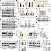 Figure 6
UA-30 treatment regulated the ERK/FOXO3a pathway by inhibiting RalA activity. (A and B) Western blot and quantification of RalA in spinal cord of different treatment groups. n = 4 male per group. (C) Relative Rala mRNA levels in spinal cord of UA-30 treatment groups. n = 5 male per group. (D) Analysis of active RalA in spinal cord by G-LISA assay. The level of RalA activity was expressed as RalA-GTP levels. n = 7 male per group. (E and F) Western blot analysis of p-ERK, ERK, p-FOXO3a (Ser294), and FOXO3a expression in spinal cord after UA-30 treatment in SOD1G93A mice. n = 4 male per group. (G and H) Western blot and quantitative analysis of RalA, p-ERK, ERK, p-FOXO3a (Ser294), and FOXO3a in SOD1G93A cells treated with vehicle or UA-30. n = 3 per group. (I) Relative Foxo3a mRNA levels in SOD1G93A cells of UA-30 treatment. n = 4 per group. (J and K) Western blot analysis of FOXO3a expression at 0, 4, 8, and 12 h in SOD1G93A cells after UA-30 or cycloheximide (CHX) treatment. n = 3 per group. (L and M) Immunoprecipitation analysis of ubiquitination levels of FOXO3a in male SOD1G93A mice after 100 mg/kg UA-30 treatment (L) or PC12 cells transfected with SOD1G93A after 100 μM UA-30 treatment (M). n = 3 per group. (N and O) Immunoblots and quantitation of FOXO3a in nuclear or cytoplasmic fraction in spinal cord after UA-30 treatment in male SOD1G93A mice (N) or SOD1G93A cells after 100 μM UA-30 administration. n = 3 per group. Data are expressed as mean ± SEM. *P < 0.05, **P < 0.01, and ***P < 0.001. B–D, F, H, I, L, N, and O, 1-way ANOVA followed by Dunnett’s multiple-comparison test; K, 2-way ANOVA followed by Bonferroni’s multiple-comparison test; M, unpaired 2-tailed Student’s t test.
One of the most prominent alterations in the spinal cord proteome of UA-30–treated SOD1G93A mice was associated with the FOXO signaling pathway. FOXO signaling, particularly involving FOXO3a, serves as a critical transcriptional hub regulating mitophagy (25–27). Ral regulates the activation of various transcription factors primarily through the MAPK pathway, wherein ERK acts as a key effector of Ral (28, 29). In both SOD1G93A mice and SOD1G93A-transfected cells, UA-30 treatment decreased the levels of phosphorylated ERK (p-ERK/ERK) and phosphorylated FOXO3a (p-FOXO3a/FOXO3a), while concurrently increasing the total FOXO3a protein expression level (Figure 6, E–H).
To elucidate the mechanism by which UA-30 increases FOXO3a protein expression, protein degradation assays were performed in UA-30–treated SOD1G93A cells. UA-30 treatment did not detectably alter Foxo3a mRNA levels (Figure 6I). Following inhibition of protein synthesis with cycloheximide, UA-30 suppressed FOXO3a degradation, resulting in higher FOXO3a expression than in untreated SOD1G93A cells (Figure 6, J and K). Previous studies have reported that ERK phosphorylates FOXO3a at Ser294, thereby regulating its degradation via the ubiquitin-proteasome pathway and preventing its nuclear translocation (30, 31). Co-IP assays revealed that UA-30 administration inhibited FOXO3a ubiquitination compared with the vehicle-treated SOD1G93A group, by approximately 50% in mice (P < 0.01) and 45% in cells (P < 0.01) (Figure 6, L and M). These findings confirm that UA-30 inhibits FOXO3a protein degradation through the ubiquitin-proteasome pathway. Furthermore, both in vivo and in vitro experiments revealed that UA-30 promoted an increase in FOXO3a levels within the nuclear fraction (by ~110% and ~55%, respectively, compared with SOD1G93A), while not affecting FOXO3a levels in the cytoplasmic fraction (Figure 6, N and O).
Overexpression of active RalA reversed UA-30’s effects on mitophagy via the RalA/ERK/FOXO3a pathway. To further investigate whether UA-30 improves mitophagy and mitochondrial function through the RalA/ERK/FOXO3a signaling pathway, we overexpressed constitutively active RalA (RalAG23V) (32) in UA-30–treated SOD1G93A cells. To determine whether RalAG23V itself causes mitochondrial impairment under unstressed conditions, we also expressed RalAG23V in WT PC12 cells. RalAG23V expression did not affect cell viability, ATP levels, MDA levels, total SOD activity, or mitoROS signals in WT PC12 cells (Supplemental Figure 16, A–E). Overexpression of active RalA reversed the UA-30–induced suppression of the ERK/FOXO3a signaling pathway. Specifically, it increased the p-ERK/ERK and p-FOXO3a/FOXO3a ratios while decreasing total FOXO3a protein levels (Figure 7, A and B). Furthermore, overexpression of active RalA enhanced the ubiquitination of FOXO3a and counteracted the UA-30–mediated increase in FOXO3a nuclear translocation in cells (Figure 7, C and D).
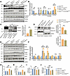 Figure 7
Overexpression of active RalA abolished the impact of UA-30 on mitophagy and the ERK/FOXO3a pathway. (A and B) After RalAG23V overexpression, immunoblot analysis and quantitation of hSOD1, RalA, p-ERK, ERK, p-FOXO3a (Ser294), and FOXO3a in SOD1G93A cells treated with 100 μM UA-30. n = 3 per group. (C) The immunoprecipitation analysis of ubiquitination levels of FOXO3a in PC12 cells transfected with SOD1G93A after 100 μM UA-30 or RalAG23V overexpression treatment. n = 3 per group. (D) Western blot analysis of FOXO3a expression in nuclear or cytoplasmic fraction of SOD1G93A cells after 100 μM UA-30 and RalAG23V overexpression treatment. n = 3 per group. (E and F) Western blot analysis of the levels of mitochondrial respiratory complex, PINK1, and Parkin in SOD1G93A cells overexpressing RalAG23V. n = 3 per group. (G–J) Assessment of ATP level (G), MMP level (JC-1 staining red/green fluorescence intensity) (H), ROS production (I), and mitoROS level (J) in SOD1G93A cells treated with 100 μM UA-30 and overexpressing RalAG23V. n = 4–5 per group. Data are expressed as mean ± SEM. *P < 0.05, **P < 0.01 ***P < 0.001, and ****P < 0.0001. B and F–J, 1-way ANOVA followed by Dunnett’s multiple-comparison test; C and D, unpaired 2-tailed Student’s t test.
Subsequently, overexpression of active RalA reduced the UA-30–induced protein levels of ETC complexes I, III, and IV, as well as PINK1 and Parkin, in SOD1G93A cells (Figure 7, E and F). Furthermore, active RalA overexpression in SOD1G93A cells attenuated the ameliorative effects of UA-30 on mitochondrial dysfunction, as evidenced by decreased ATP levels and MMP and increased levels of total cellular ROS and mitoROS (Figure 7, G–J).
Collectively, these results demonstrate that UA-30 ameliorates mitochondrial dysfunction in the SOD1G93A model by enhancing mitophagy, mediated through the targeted suppression of the RalA/ERK/FOXO3a signaling pathway.
Mdivi-1 abrogated the UA-30–induced improvement in mitochondrial function and neuroprotection in SOD1G93A mice. To corroborate the involvement of mitophagy in UA-30’s therapeutic mechanism against ALS, Mdivi-1 was used to pharmacologically interfere with mitophagy-related signaling (33, 34). SOD1G93A mice received combined treatment with Mdivi-1 and UA-30. Spinal cord tissues from Mdivi-1–treated mice showed reduced levels of PINK1 and Parkin proteins (Figure 8, A and B), indicating that Mdivi-1 interfered with mitophagy-related signaling.
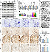 Figure 8
Mdivi-1 eliminated the beneficial effects of UA-30 on motor neuron loss and neuroinflammation. (A and B) Western blot analysis and quantification of PINK1, Parkin, and mitochondrial respiratory complex protein levels in spinal cord of SOD1G93A mice treated with UA-30 or Mdivi-1. n = 4 male per group. (C and D) Representative mitochondrial images (C) were observed in the spinal cord neurons of male SOD1G93A mice from different treatment groups using TEM. Scale bar: 1 μm. Mitochondria were classified into 3 categories based on cristae morphology (green, intact; orange, mild cristae loss; purple, severe cristae loss). The proportion of each mitochondria category was quantified separately (D), and data are presented as frequency distributions. At least 150 mitochondria from 3 mice were analyzed in each group. (E and F) Measurement of ATP level and lipid peroxidation level in spinal cord of SOD1G93A mice after UA-30 or Mdivi-1 treatment. n = 5 male per group. (G) Representative images of Nissl staining and neurons (NeuN), astrocytes (GFAP), and microglia (Iba1) immunohistochemical staining in spinal cord after UA-30 and Mdivi-1 treatment in male SOD1G93A mice. Scale bars: 100 μm (Nissl), 200 μm (NeuN), 200 μm (GFAP), 200 μm (Iba1). Data are expressed as mean ± SEM. *P < 0.05, **P < 0.01 ***P < 0.001, and ****P < 0.0001. 1-way ANOVA followed by Dunnett’s multiple-comparison test.
Notably, the expression of mitochondrial complex III (UQCRFS1) and complex IV (COX5b) in the spinal cord of UA-30–treated SOD1G93A mice was decreased upon Mdivi-1 treatment (Figure 8, A and B). TEM analysis revealed swollen, vacuolar mitochondria with an apparent loss of cristae in Mdivi-1–treated groups (Figure 8, C and D). Critically, the beneficial effects of UA-30 on increasing ATP levels and reducing MDA levels were completely abolished in the presence of Mdivi-1 (Figure 8, E and F).
Mdivi-1 eliminated the protective effect of UA-30 against motor neuron loss, as evidenced by reduced neuronal counts using both Nissl staining and NeuN immunohistochemistry (Figure 8G and Supplemental Figure 17, A and B). Furthermore, compared with SOD1G93A mice treated with UA-30 alone, the group receiving combined treatment of UA-30 and Mdivi-1 increased the numbers of astrocytes and microglia (Figure 8G and Supplemental Figure 17, C and D). Additionally, UA-30 treatment reduced muscle inflammation and fibrosis in SOD1G93A mice, which was also counteracted by Mdivi-1 (Figure 9A and Supplemental Figure 17, E and F).
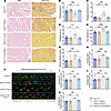 Figure 9
Mdivi-1 attenuated UA-30–induced improvement in motor function. (A) Representative histological H&E and Sirius red staining images of the gastrocnemius muscles of male SOD1G93A mice exposed to different treatments. Scale bars: 100 μm (H&E), 100 μm (Sirius red). (B–E) Motor performance of SOD1G93A mice treated with UA-30 or Mdivi-1 measured by climbing time on pole tests (B), hanging endurance time (C), retention time on rotarod (D), and grip strength (E). (F–K) Representative images of gait tracking via gait analysis (F) and quantified stride speed, average stride of left hind limbs and forelimbs or right hind limbs and forelimbs (G–K). n = 8 male per group. Data are expressed as mean ± SEM. *P < 0.05, **P < 0.01, ***P < 0.001, and ****P < 0.0001. 1-way ANOVA followed by Tukey’s multiple-comparison test.
Mdivi-1 abolished UA-30–induced improvement of motor deficit in SOD1G93A mice. Behavioral assessments showed that UA-30 reduced climbing time, increased fall latency in the wire hang and rotarod tests, and enhanced grip strength. However, these improvements were completely abolished by coadministration of mdivi-1 (Figure 9, B–E). Similar outcomes were observed in gait analysis (Figure 9, F–K, and Supplemental Video 3). Overall, these findings indicate that administration of mdivi-1 blocked improvement of motor deficit in SOD1G93A mice induced by UA-30.
-
Discussion
ALS is a lethal neurodegenerative disorder characterized by the progressive degeneration of motor neurons in the brain and spinal cord (1, 2). Despite extensive research, clinically effective pharmacological treatments for ALS remain unavailable. In this study, we evaluated the small-molecule compound UA-30, which has been shown to be safe and exhibits a favorable pharmacokinetic profile. UA-30 achieved CNS exposure after oral administration, ameliorated motor dysfunction, and extended both the median survival and overall lifespan in SOD1G93A mice. UA-30 treatment attenuated key ALS-associated pathological features. Specifically, it mitigated the loss of spinal motor neurons, decreased microgliosis and astrogliosis, and reduced the degree of gastrocnemius muscle fibrosis. Mechanistically, as an inhibitor of RalA GTPase, UA-30 suppressed RalA activation and modulated the ERK/FOXO3a signaling pathway. UA-30 inhibited FOXO3a degradation via the ubiquitin-proteasome pathway, enhanced FOXO3a stability, and supported transcription of mitophagy-related genes, including Pink1 and Prkn. Consistent with this upstream transcriptional support, UA-30 enhanced PINK1/Parkin-dependent mitophagy and improved mitochondrial dysfunction (Figure 10). Collectively, these results support UA-30 as a promising therapeutic candidate for ALS and provide a foundation for future preclinical evaluation.
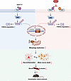 Figure 10
Proposed working model of UA-30 in SOD1G93A mice. In the pathogenesis of ALS, SOD1G93A mutation induces RalA activation, and aberrant activation of RalA triggers a downstream RalA/ERK signaling cascade, which promotes ubiquitin-proteasome–mediated degradation of the key transcription factor FOXO3a. Reduced ubiquitin-proteasome diminishes its nuclear translocation, leading to the downregulation of mitophagy-related genes and consequent impairment of mitophagy. Accumulation of damaged mitochondria contributes to neuroinflammation and ultimately drives motor neuron death, resulting in muscle fibrosis and atrophy. UA-30 exerts its therapeutic effects by directly targeting and inhibiting RalA activation. Suppression of the ERK signaling stabilizes FOXO3a protein levels. Restoration of FOXO3a function enhances mitophagy, clears dysfunctional mitochondria, alleviates neuroinflammation, and prevents motor neuron death, ultimately halting ALS progression. Created with BioRender.
RalA, a member of the Ras superfamily of small GTPases, functions as a molecular switch cycling between an active GTP-bound state and an inactive GDP-bound state (24). RalA translocates to mitochondria following depolarization, suggesting its potential involvement in mitophagy (35). Furthermore, aberrant RalA activation promotes excessive mitochondrial fission in adipocytes of high-fat diet–induced obese mice, impairing mitochondrial function and metabolic capacity (32). As a downstream effector of Ras, RalA regulates multiple signaling pathways, including the ERK/MAPK cascade (29, 36, 37). In our study, activation of RalA increased ERK phosphorylation at Thr202/Tyr204, supporting the involvement of the RalA/ERK axis under our experimental conditions. Activated ERK has been reported to phosphorylate FOXO3a at Ser294, Ser344, and Ser425, thereby enhancing its interaction with the E3 ubiquitin ligase MDM2 and promoting ubiquitin-proteasome–dependent degradation (30, 31). Thus, phosphorylation of FOXO3a at Ser294 is primarily associated with FOXO3a ubiquitination and reduced protein stability. By contrast, FOXO3a nuclear exclusion and cytoplasmic retention are more classically regulated by phosphorylation at sites such as Thr32, Ser253, and Ser315, which promotes 14-3-3 binding and retention of FOXO3a in the cytoplasm (38–40). Our results showed that UA-30 increased total FOXO3a protein levels and promoted FOXO3a nuclear accumulation, whereas overexpression of constitutively active RalA abolished the regulatory effects of UA-30 on the RalA/ERK/FOXO3a axis and impaired FOXO3a nuclear import. These findings suggest that UA-30 suppresses RalA/ERK signaling, reduces inhibitory FOXO3a phosphorylation and ubiquitination, and restores FOXO3a nuclear localization.
FOXO3a is a well-conserved transcription factor critical for stress resistance, longevity, and cellular homeostasis (41). Activation of FOXO3a in the C. elegans model has been shown to induce mitophagy, markedly ameliorating muscle aging and mitochondrial dysfunction (42). Previous studies further support a transcriptional role of FOXO3a in mitophagy regulation, as the genes encoding PINK1 and Parkin can be regulated by FOXO3a, and disruption of the FOXO3a/PINK1/Parkin axis impairs mitophagy restoration (27, 43). Consistent with this framework, our results support FOXO3a-dependent transcriptional regulation as an upstream component of UA-30–enhanced mitophagy. UA-30 increased FOXO3a nuclear localization and was associated with transcriptional upregulation of Pink1 and Prkn, together with enhanced PINK1/Parkin-mediated mitophagy. It should be noted that UA-30 leads to a drastic increase in mitophagy protein levels but has only mild effects on their transcripts. This suggests a posttranscriptional regulatory mechanism, independent of FOXO3a, which remains to be investigated in future studies. Overexpression of active RalA counteracted the UA-30–induced increase in FOXO3a nuclear localization and the associated enhancement of mitophagy. In addition, in the SOD1G93A cellular model where mitochondrial dysfunction is already pronounced, constitutively active RalA did not necessarily cause a further measurable decline in these mitochondrial parameters but was sufficient to abolish the mitochondrial improvements conferred by UA-30. These findings suggest that the pathological consequences of RalA activation are context dependent and become more evident in the setting of SOD1G93A-associated proteotoxic stress. Consistent with this interpretation, the protective effects of UA-30 are most apparent under disease-associated stress conditions. A similar pattern was also observed in the pharmacological intervention study involving Mdivi-1. In the SOD1G93A model, Mdivi-1 abolished the protective effects of UA-30 but did not further worsen baseline outcomes under the treatment window used here.
Because Mdivi-1 primarily inhibits Drp1-dependent mitochondrial fission, its effect on mitophagy should be regarded as indirect rather than direct. Nevertheless, inhibition of mitochondrial fission can still attenuate mitophagy-related signaling in the PINK1/Parkin pathway (44). In this context, the reduction in Parkin observed after Mdivi-1 treatment is more plausibly interpreted as a downstream consequence of attenuated mitophagy-related signaling, rather than direct suppression of transcription of the gene encoding Parkin or Parkin protein synthesis. Although previous studies have suggested that Mdivi-1 alone does not markedly alter behavioral performance in WT or control animals (45, 46), the effects of Mdivi-1 alone on motor function in WT mice and on survival in SOD1G93A mice were not systematically evaluated this study and remain as limitations to be addressed in future work.
Deficiencies in mitochondrial respiratory chain complex IV are widely observed in patients with sporadic ALS (47, 48). Furthermore, swollen and vacuolated mitochondria are markedly increased in spinal motor neurons of ALS patients (49, 50). Impairment of mitophagy leads to the accumulation of dysfunctional mitochondria, disrupting mitochondrial regulatory functions. Our findings demonstrate that UA-30 treatment reversed the decrease in ATP levels and the increase in MDA levels observed in the spinal cord, medulla oblongata, and gastrocnemius muscle of ALS mice. Additionally, UA-30 treatment increased the expression levels of mitochondrial complex III (UQCRFS1) and IV (COX5b) in the spinal cord and medulla oblongata. UA-30 also ameliorated mitochondrial damage in spinal cord neurons of ALS mice. Mdivi-1 treatment suppressed the expression of PINK1 and Parkin. This inhibition of mitophagy initiation blocked the beneficial effects of UA-30 on mitochondrial function. These results suggest that UA-30 improves mitochondrial dysfunction in ALS mice by enhancing mitophagy.
Mitochondrial dysfunction drives ALS progression through interrelated mechanisms, including bioenergetic deficits, oxidative stress, and neuroinflammation (51–53). This pathogenic framework underlies the key pathological features observed in ALS mice, including spinal motor neuron loss, microglial and astrocytic activation, muscle denervation and atrophy, as well as muscle fibrosis. Consistent with mitigating core pathologies, UA-30 treatment increased spinal motor neuron survival, reduced microgliosis and astrogliosis, and inhibited muscle fibrosis. These improvements collectively contributed to the enhanced motor performance observed in UA-30–treated ALS mice. While our study demonstrates that UA-30 reduces microgliosis in SOD1G93A mice, the functional phenotype of microglia is also likely to be important. In ALS, microglia undergo dynamic and stage-dependent changes, and their activation states may range from relatively protective to proinflammatory phenotypes rather than fitting strictly into a simple M1/M2 dichotomy (54–56). Therefore, the therapeutic effects of UA-30 may involve not only reducing overall microglial abundance, but also modulating microglial phenotype and inflammatory output. This possibility needs to be addressed in future studies using established polarization-related markers and pathways.
Additional assessment of NMJs showed that UA-30 only partially improved NMJ innervation in the gastrocnemius muscle of SOD1G93A mice. This finding may explain why UA-30 produced substantial improvements in muscle pathology and motor neuron survival, yet only a modest survival benefit. Given that treatment was initiated at 13 weeks of age, when denervation is already underway in this model, the therapeutic window for fully preserving distal neuromuscular connectivity may have been limited.
In an additional equimolar comparison, UA-30 improved several motor performance readouts more than UA under matched conditions, and together with its improved oral CNS exposure, these data support UA-30 as a more developable compound based on the UA scaffold for further evaluation. It should also be noted that behavioral outcomes did not exhibit a strictly dose-dependent pattern, with low and high doses often yielding comparable effects despite numerical separation in some measures. This pattern may reflect an in vivo plateau effect, whereby the lower dose is already sufficient to produce near-maximal efficacy for certain functional endpoints, and further dose escalation therefore yields only limited additional benefit. Furthermore, the invasive nature of delivery routes, intravenous for edaravone and intrathecal for tofersen, can substantially compromise the quality of life for ALS patients, further limiting their utility. In contrast, UA-30’s ability to achieve neuroprotection through oral administration makes it more accessible and less invasive. Here, UA-30 showed a trend toward improved efficacy compared with the first-tier drug riluzole, specifically in terms of improved motor performance in pole-climbing, hanging, and rotarod tests, prolonged median survival in ALS mice, and inhibition of muscle fibrosis. Given that UA-30 and riluzole act through distinct mechanisms, it is also important for future studies to determine whether UA-30 combined with riluzole can provide an additive or synergistic benefit.
Based on its oral convenience, favorable safety profile (LD50 = 1,992.5 mg/kg, NOAEL > 400 mg/kg), and the preclinical efficacy observed here, UA-30 represents a promising therapeutic candidate for ALS. As ALS diagnosis shifts toward earlier recognition (57), earlier intervention, such as presymptomatic administration of UA-30, may provide greater therapeutic benefit, further delay disease progression, and enhance neuroprotection. These preclinical findings provide a solid foundation for continued evaluation of UA-30.


Copyright © 2026 American Society for Clinical Investigation
ISSN: 0021-9738 (print), 1558-8238 (online)