
Advertisement
Research ArticleAgingCell biology
Open Access |  10.1172/JCI199854
LSD1-mediated demethylation of the DNA damage response factor ATM promotes senescence and organ aging
Yingying Zhang,1,2 Chen Yu,2 Xiaoqin Zhang,1,2 Linda Xiaoyan Li,1 Alice Shasha Cheng,1 and Xiaogang Li1,3
1Department of Internal Medicine, Mayo Clinic, Rochester, Minnesota, USA.
2Department of Nephrology, Shanghai Tongji Hospital, Tongji University School of Medicine, Shanghai, China.
3Department of Biochemistry and Molecular Biology, Mayo Clinic, Rochester, Minnesota, USA.
Address correspondence to: Xiaogang Li, Biochemistry and Molecular Biology, and Medicine, Mayo Clinic, 200 1st Street, SW, Rochester, Minnsesota, 55905, USA. Phone: 507.266.0110; Email: li.xiaogang@mayo.edu.
Authorship note: YZ and CY contributed equally to this work.
Find articles by Zhang, Y. in: PubMed | Google Scholar
1Department of Internal Medicine, Mayo Clinic, Rochester, Minnesota, USA.
2Department of Nephrology, Shanghai Tongji Hospital, Tongji University School of Medicine, Shanghai, China.
3Department of Biochemistry and Molecular Biology, Mayo Clinic, Rochester, Minnesota, USA.
Address correspondence to: Xiaogang Li, Biochemistry and Molecular Biology, and Medicine, Mayo Clinic, 200 1st Street, SW, Rochester, Minnsesota, 55905, USA. Phone: 507.266.0110; Email: li.xiaogang@mayo.edu.
Authorship note: YZ and CY contributed equally to this work.
Find articles by Yu, C. in: PubMed | Google Scholar
1Department of Internal Medicine, Mayo Clinic, Rochester, Minnesota, USA.
2Department of Nephrology, Shanghai Tongji Hospital, Tongji University School of Medicine, Shanghai, China.
3Department of Biochemistry and Molecular Biology, Mayo Clinic, Rochester, Minnesota, USA.
Address correspondence to: Xiaogang Li, Biochemistry and Molecular Biology, and Medicine, Mayo Clinic, 200 1st Street, SW, Rochester, Minnsesota, 55905, USA. Phone: 507.266.0110; Email: li.xiaogang@mayo.edu.
Authorship note: YZ and CY contributed equally to this work.
Find articles by Zhang, X. in: PubMed | Google Scholar
1Department of Internal Medicine, Mayo Clinic, Rochester, Minnesota, USA.
2Department of Nephrology, Shanghai Tongji Hospital, Tongji University School of Medicine, Shanghai, China.
3Department of Biochemistry and Molecular Biology, Mayo Clinic, Rochester, Minnesota, USA.
Address correspondence to: Xiaogang Li, Biochemistry and Molecular Biology, and Medicine, Mayo Clinic, 200 1st Street, SW, Rochester, Minnsesota, 55905, USA. Phone: 507.266.0110; Email: li.xiaogang@mayo.edu.
Authorship note: YZ and CY contributed equally to this work.
Find articles by Li, L. in: PubMed | Google Scholar
1Department of Internal Medicine, Mayo Clinic, Rochester, Minnesota, USA.
2Department of Nephrology, Shanghai Tongji Hospital, Tongji University School of Medicine, Shanghai, China.
3Department of Biochemistry and Molecular Biology, Mayo Clinic, Rochester, Minnesota, USA.
Address correspondence to: Xiaogang Li, Biochemistry and Molecular Biology, and Medicine, Mayo Clinic, 200 1st Street, SW, Rochester, Minnsesota, 55905, USA. Phone: 507.266.0110; Email: li.xiaogang@mayo.edu.
Authorship note: YZ and CY contributed equally to this work.
Find articles by Cheng, A. in: PubMed | Google Scholar
1Department of Internal Medicine, Mayo Clinic, Rochester, Minnesota, USA.
2Department of Nephrology, Shanghai Tongji Hospital, Tongji University School of Medicine, Shanghai, China.
3Department of Biochemistry and Molecular Biology, Mayo Clinic, Rochester, Minnesota, USA.
Address correspondence to: Xiaogang Li, Biochemistry and Molecular Biology, and Medicine, Mayo Clinic, 200 1st Street, SW, Rochester, Minnsesota, 55905, USA. Phone: 507.266.0110; Email: li.xiaogang@mayo.edu.
Authorship note: YZ and CY contributed equally to this work.
Find articles by Li, X. in: PubMed | Google Scholar
Published September 15, 2026 - More info
J Clin Invest. 2026;136(18):e199854. https://doi.org/10.1172/JCI199854.
© 2026 Zhang et al. This work is licensed under the Creative Commons Attribution 4.0 International License. To view a copy of this license, visit http://creativecommons.org/licenses/by/4.0/.
Received: September 3, 2025; Accepted: July 14, 2026


-
Results
The expression of LSD1 is increased in aged organs and senescent cells. To understand the roles of LSD1 in organ senescence and aging, first, we found that LSD1 protein was increased in aged organs, including kidneys, hearts, livers, spleens, lungs and intestines, collected from 20–22-month-old mice compared with young organs collected from 2-month-old mice (Figure 1, A–F), accompanied by an increase of the expression of p16ink4a (p16) and p21cip1(p21), 2 markers of senescence. However, we found that, in contrast with the upregulation of LSD1 protein as well as Cdkn2A (p16) and Cdkn1A (p21) mRNA, the expression of Lsd1 mRNA was not increased in aged organs compared with young organs (Figure 1, G–I).
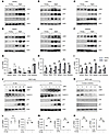 Figure 1
The expression of LSD1 is increased in aged organs and senescent cells. (A–F) Western blot analysis of the expression of LSD1, p16, and p21 in aged organs, including kidneys (A), hearts (B), livers (C), spleens (D), lungs (E), and intestines (F), collected from 20–22-months-old mice (Aged) compared with expression in young organs collected from 2-month-old mice (Young). n = 6. (G–I) qRT-PCR analysis of Lsd1 (G), p16 (H), and p21 (I) mRNA in mouse organs, including kidney, heart, liver, spleen, lung, and intestine, collected from young and aged mice (n = 6, per group; ns, not significant; **P < 0.01; unpaired, 2-tailed Student’s t test). (J) Western blot analysis of the expression of LSD1, Cyclin D, p16, and p21 from whole-cell lysates of mouse IMCD cells treated with D-galactose (D-Gal) and etoposide (ETO) for 48 hours with different doses. Represent data from 3 independent experiments are shown. (K) Western blot analysis of the expression of LSD1, Cyclin D, p16, and p21 in mouse IMCD cells cultured after 100% confluency for indicated days. Represent data from 3 independent experiments are shown. (L) qRT-PCR analysis of Lsd1 (L) mRNA in mouse IMCD cells treated with D-Gal (left panel) and ETO (right panel). All experiments were independently repeated at least 3 times; ns, not significant; unpaired, 2-tailed Student’s t-test (M and N) qRT-PCR analysis of p16 (M) and p21 (N) mRNAs in mouse IMCD cells treated with D-Gal or ETO. All experiments were independently repeated at least 3 times; **P < 0.01; unpaired, 2-tailed Student’s t-test.
Cellular senescence plays a crucial role in the progression of organ aging (8). We found that induction of senescence by the oxidative stress injury agent D-Galactose (D-gal) and the DNA damaging agent etoposide (ETO) also increased the expression of LSD1 protein in mouse inner medullary collecting duct (IMCD) cells (Figure 1J) and mouse embryonic fibroblasts (3T3 cells) (Supplemental Figure 1A; supplemental material available online with this article; https://doi.org/10.1172/JCI199854DS1). Treatment with ETO and D-Gal did not affect the expression of LSD1 mRNA in senescent cells compared with vehicle-treated controls (Figure 1L). The induction of senescence was indicated by the upregulation of p16 and p21 protein and mRNA levels in mouse IMCD cells (Figure 1, J, M, and N). In addition, LSD1 protein was not changed in quiescent cells induced by contact inhibition (Figure 1K). We further found that LSD1 protein was increased in naturally aged primary renal tubular epithelial cells isolated from 20–22-month-old mouse kidneys compared with primary kidney tubular epithelial cells isolated from 2-month-old mouse kidneys (Supplemental Figure 1B). These results support that the stability of LSD1 protein, rather than the regulation of Lsd1 mRNA, is associated with senescence and aging.
Targeting LSD1 decreases cellular senescence, and elimination of senescent cells leads to a decline of LSD1 protein in aged organs. To understand the role of LSD1 in organ senescence and aging, we focused on 3 key parenchymal organs: kidney, heart, and liver. We found that treatment with senolytic drugs Dasatinib plus Quercetin (D+Q), which could eliminate senescent cells (27), decreased LSD1 protein in kidneys, hearts, and livers in naturally aged 20–22-month-old mice, accompanied by a reduction of p16 and p21 proteins in those organs (Figure 2, A–C). Administration of Dasatinib (D), Quercetin (Q), or D+Q also decreased the expression of LSD1 protein as well as p16 and p21 in mouse IMCD cells (Figure 2D). In addition, D, Q, or D+Q also decreased the expression of p16 and p21 mRNA levels in mouse IMCD cells (Figure 2E). These results suggest that there is a positive feedback loop between the upregulation of LSD1 and cellular senescence in aged organs.
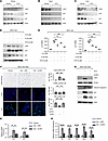 Figure 2
Targeting LSD1 decreases cellular senescence and elimination of senescent cells leads to a decline of LSD1 protein in aged organs. (A–C) Western blot analysis of the expression LSD1, p16, and p21 in organs, including kidneys (A), heart (B), and liver (C), collected from aged 20–22-month-old mice treated with senolytic drugs (Dasatinib 5 mg/Kg and Quercetin 50 mg/Kg, D + Q). (D) Western blot analysis of the expression of LSD1, p16 and p21 in mouse IMCD cells treated with Dasatinib (D) or Quercetin (Q), and cotreated with ETO (5 μM). Represent data from 3 independent experiments are shown. (E) qRT-PCR analysis of p16 and p21 mRNAs in mouse IMCD cells treated with Dasatinib (D) or Quercetin (Q) and cotreated with ETO (5 μM). All experiments were independently repeated at least 3 times; ns, no significant, **P < 0.01; 1-way ANOVA. (F) Representative images and quantification of and SA-β-gal staining (top), TUNEL assay (middle), and Ki67 staining (bottom) in mouse IMCD cells treated with LSD1 siRNA (siLSD1) and control siRNA (siNC) for 48 hours after ETO (5 μM) treatment. Scale bar: 50 μm. The percentage of positive cells was quantified using the same software in 10 randomly selected fields; ns, not significant, **P < 0.01; 1-way ANOVA. (G) Western blot analysis of the expression of LSD1, PCNA, cleaved caspase-3, p21, and p16 in mouse IMCD cells treated with ETO (5 μM) and cotreat with or without siLSD1 for 48 hours. n = 3. (H and I) qRT-PCR analysis of p16 and p21 (H), and SASP factors Il6, Il1b, Ccl2, Tgfb1, and Tnfa (I) mRNAs in mouse IMCD cells with or without LSD1 siRNA, 48 hours after ETO (5 μM) treatment. All experiments were repeated at least 3 times; ns, not significant, *P < 0.05, **P < 0.01; 1-way ANOVA.
To define the functional role of LSD1 in other cellular process, including proliferation and apoptosis, we knocked down Lsd1 with siRNA in etoposide-treated (ETO-treated) IMCD cells. ETO treatment not only induced senescence but also reduced proliferation with no effect on apoptosis at 48 hours. Knockdown of Lsd1 partially restored proliferation and reduced cell senescence, without affecting apoptosis (Figure 2, F and G). Consistent with this, qPCR analysis demonstrated that knockdown or inhibition of LSD1 with ORY-1001 markedly reduced the expression of senescence markers (p16 and p21) and senescence-associated secretory phenotype (SASP) factors, including IL-1β, TNF-α, IL-6, MCP-1, and TGF-β, in senescent IMCD cells (Figure 2, H and I, and Supplemental Figure 2, A and B). LSD1 overexpression further enhanced senescence and suppressed cell proliferation in ETO-treated IMCD cells (Supplemental Figure 3, A–C). Importantly, neither inhibition nor overexpression of LSD1 altered apoptosis in senescent IMCD cells, as assessed by TUNEL assay and the levels of cleaved caspase-3 (Figure 2, F and G, and Supplemental Figure 3, A and B). These results support a model in which upregulation of LSD1 is associated with, and may contribute to, the maintenance of senescence in renal epithelial cells.
Treatment with ORY-1001 decreases ionizing radiation and natural aging-induced DNA damage and senescence in mouse organs and has a protective effect against IR-induced hair graying. Ionizing radiation (IR) generates a high level of DNA damage, which often leads to the induction of cellular senescence and multiorgan aging (28). To determine the functional role of LSD1 in these processes, 10-week-old C57BL/6J mice were subjected to a single 7 Gy radiation dose, and then treated with LSD1 inhibitor ORY-1001 (0.04 mg/kg) or PBS by intraperitoneal injections every other day for a duration of 8 weeks (Figure 3A). IR exposure induced hair graying, which was attenuated by ORY-1001 treatment (Figure 3B). IR also increased LSD1 expression in kidney, heart, and liver, accompanied by elevated p16 and p21 levels. Notably, ORY-1001 treatment reduced both mRNA and protein levels of p16 and p21 in these tissues (Figure 3, C and D), indicating attenuation of IR-associated senescence markers. Additionally, ORY-1001 treatment reduced the levels of SASP factors, including IL-1β, TNF-α, IL-6, MCP-1, and TGF-β, in IR-treated organs (Figure 3E).
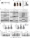 Figure 3
Treatment with ORY-1001 decreases IR-induced DNA damage in mouse organs and protects hair graying. (A) Schedule of the irradiation (IR) and ORY-1001 treatment in C57BL/6J mice. (B) Representative images of mice from the control group, IR group (7Gy single dose), and IR group plus ORY-1001 (0.04 mg/Kg) injection (60). Error bars represent the mean ± SEM values represent gray hairs numbers per 100 strands. n = 10; **P < 0.01; 1-way ANOVA. (C) Western blot analysis of the levels of LSD1, p16, p21, phospho-ATM (p-ATM), total of ATM, and γH2AX in mouse organs, including kidneys (left panel), heart (middle panel), and livers (right panel), collected from the mice treated with vehicle, IR, and IR plus ORY-1001. (D–E) qRT-PCR analysis of p16/p21 (D), and inflammatory/SASP factors Il6, Il1b, Ccl2, Tgfb1, and Tnfa. (E) mRNA levels in mouse organs. n = 6/group; **P < 0.01; 1-way ANOVA.
To assess the impact of LSD1 inhibition on organismal aging, 22-month-old mice were treated with the LSD1 inhibitor ORY-1001 (0.04 mg/kg) and PBS via intraperitoneal injection every other day until 28 months of age (Supplemental Figure 4A). ORY-1001–treated mice exhibited protection against hair graying compared with controls (Supplemental Figure 4B) and markedly attenuated senescence, as seen by decreased senescence-associated β-galactosidase (SA-β-gal) staining, reduced expression of p16 and p21, and suppression of SASP factors (TGF-β, IL-1β, TNF-α, IL-6, and MCP-1), as well as attenuated fibrosis in aged kidney, heart, and liver tissues (Supplemental Figure 4, C–G). These results provide in vivo evidence that LSD1 promotes age-associated tissue dysfunction and that its inhibition mitigates senescence and aging phenotypes.
LSD1-mediated regulation of senescence is independent of canonical histone methylation at p16/p21 loci. LSD1 is known to demethylate dimethylated H3 lysine residues (e.g., H3K4me2 or H3K9me2) to monomethylated forms (29, 30). Consistent with this activity, aged tissues (kidney, heart, and liver) and senescent cells exhibited decreased H3K4me2 and H3K9me2, accompanied by increased H3K4me1 and H3K9me1 (Supplemental Figure 5, A and B). H3K4me1 is generally associated with transcriptional activation, whereas H3K9me2 is linked to transcriptional repression. Given these changes and the increased expression of LSD1 in naturally aged and senescent conditions, we investigated the potential involvement of H3K4me1 and H3K9me2 in regulating the transcription of p16 and p21. Analysis of publicly available ChIP-seq databases (GSE106415) showed the binding of H3K4me1 on the promoter of p21 and p16 (Supplemental Figure 5, C and D). However, Analysis of ChIP-seq databases (GSE38442) revealed no significant changes in the occupancy or peak enrichment of H3K9me2 at these loci under senescent conditions (Supplemental Figure 5E). These results suggest that LSD1 promotes cellular senescence, at least in part, through canonical histone methylation at the p16/p21 loci.
LSD1 is involved in DNA damage–mediated senescence and DNA damage repair. ATM is a central kinase in the DNA damage response (DDR) to double-strand breaks (31). Upon DNA damage, ATM is rapidly phosphorylated and recruited to sites of DNA lesions, where it remains active for minutes to hours before undergoing progressive dephosphorylation as repair is completed (32). Given that cellular senescence is frequently driven by DNA damage, we investigated whether LSD1 is functionally linked to DNA damage response pathways. We found that increased LSD1 protein was associated with elevated ATM phosphorylation in IR-treated tissues and aged (28-month-old) organs, whereas treatment with the LSD1 inhibitor ORY-1001 reduced levels of phosphorylated ATM and γH2AX in kidney, heart, and liver under both conditions (Figure 3C and Supplemental Figure 4F).
To investigate the relationship between LSD1 and ATM following DNA damage, we examined whether LSD1 regulates ATM activation dynamics in IR-induced DDR and senescence. 3T3 cells were exposed to different doses of IR and analyzed over time. Low-dose IR (5 Gy) did not alter LSD1 expression until 48 hours after treatment, although it induced rapid ATM phosphorylation and γH2AX accumulation at 1 hour, which decreased to baseline by 12 hours. In contrast, high-dose IR (20 Gy) increased LSD1 expression at approximately 12 hours after irradiation and was associated with prolonged ATM phosphorylation and delayed resolution of γH2AX, persisting up to 24 hours (Figure 4A). Consistently, overexpression of GFP-tagged LSD1 under low-dose IR (5 Gy) prolonged ATM phosphorylation and delayed γH2AX resolution up to 48 hours, accompanied by increased p16 and p21 expression at 24 hours compared with controls (Figure 4B). Conversely, LSD1 knockdown under high-dose IR (20 Gy) accelerated ATM dephosphorylation and γH2AX clearance and reduced p16 and p21 expression (Figure 4C). Moreover, LSD1 knockdown enhanced DNA repair efficiency, as assessed by comet assay at 12 hours after IR (Figure 4D). These results indicate that LSD1 modulates the duration, rather than the initiation, of ATM signaling. While ATM phosphorylation is rapidly induced following DNA damage, LSD1 appears to contribute to the maintenance and amplification of ATM signaling, which is associated with sustained DNA damage response (DDR) activation and senescence. These results also provide insight into mechanistic prolonged ATM activation and suggest a role for LSD1 in limiting “checkpoint recovery” following genotoxic stress.
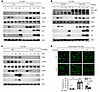 Figure 4
Increased LSD1 is involved in DNA damage–mediated senescence and DNA damage repair. (A) Western blot analysis of the levels of LSD1, phospho-ATM, and total of ATM, γH2AX. p16 and p21 in mouse 3T3 cells treated with either 5Gy- IR or 20Gy-IR at indicated time points. (B) Western blot analysis of the levels of GFP-LSD1, LSD1, phospho-ATM, and total of ATM, γH2AX, p16, and p21 in GFP-LSD1 and GFP-empty vector transfected 3T3 cells treated with IR (5 Gy) at indicated time points. (C) Western blot analysis of the levels of LSD1, phospho-ATM and total of ATM, γH2AX, p16, and p21 in Lsd1 knockdown (siLSD1) 3T3 cells treated with IR (20 Gy) compared with the control siRNA (siNC) at indicated time points. Representative data from 3 independent experiments are shown. (D) Representative images and statistical graph of comet assay in Lsd1 knockdown (siLSD1) 3T3 cells treated with IR at indicated time points. Error bars represent the SEM. Data were analyzed from 10 images in 3 independent experiments. Scale bar: 50 μm. ns, no significance, *P < 0.05, **P < 0.01; 1-way ANOVA.
LSD1 interacts with and demethylates ATM. To investigate how LSD1 regulates DNA damage repair, we found an interaction between LSD1 and ATM in 3T3 cells as examined with coimmunoprecipitation (co-IP) assay (Figure 5A). In addition, we found that GFP-tagged LSD1 could also pull-down ATM in HEK293T cells, as examined with anti-GFP antibody (Figure 5B). Notably, this interaction was markedly enhanced upon induction of DNA damage, particularly at 12 hours after IR, as demonstrated by co-IP assays at different time points of IR treatment in 3T3 cells (Figure 5C). We further found that the upregulation of LSD1 following IR treatment is accompanied by an increase of nuclear LSD1 where its interaction with ATM was also enhanced, as examined by nucleus separation assays (Figure 5D).
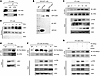 Figure 5
LSD1 interacts with and demethylates ATM. (A) Immunoprecipitation assay to detect the interaction between ATM and LSD1 in mouse 3T3 cells. Immunoglobulin G (IgG) was used as a negative control. (B) Immunoprecipitation assay to detect the interaction between GFP tagged LSD1 and ATM in HEK 293T cells transfected with GFP empty vector (EV) and GFP tagged LSD1 construct (GFP-LSD1). (C) Immunoprecipitation assay to detect the interactions between ATM and LSD1 in mouse 3T3 cells treated with IR (10 Gy) at indicated time points. (D) The nuclear fractions were isolated from mouse 3T3 cells treated with IR (10Gy) at indicated time points. Western blot analysis of the LSD1 expression in nuclear fractions. (E) Immunoprecipitation assay to detect the methylation of ATM in mouse 3T3 cells treated with ORY-1001 (5 μM) or vehicle. (F) Immunoprecipitation assay to detect the methylation of ATM in mouse 3T3 cells treated with IR (20 Gy) at indicated time points. (G) Immunoprecipitation assay to detect the methylation of ATM in mouse 3T3 cells treated with LSD1 siRNA or control siRNA (siNC) at 12 hours after IR (20 Gy) treatment.
Modification of ATM has been demonstrated to be essential for its activation upon DNA damage, such as its acetylation and phosphorylation (17). However, whether the methylation of ATM affects its activation remains unknown. Based on the interaction of LSD1 with ATM, we examined whether LSD1 affects the methylation levels of ATM in 3T3 cells by using anti-ATM antibody for immunoprecipitation, followed by blotting with antimethyl lysine antibody. We found that inhibition of LSD1 with ORY-1001 increased the methylation of ATM in 3T3 cells (Figure 5E). To explore the dynamic regulation of ATM methylation, we examined changes in ATM methylation in response to DNA damage. We observed that ATM methylation progressively decreased by 12 hours following IR (20 Gy) treatment, coinciding with increased LSD1 expression in 3T3 cells (Figure 5F), indicating that LSD1 contributes to ATM demethylation at this stage. Notably, depletion of LSD1 with siRNA prevented this decrease in methylation of ATM at 12 hours after IR (20 Gy) treatment, suggesting that LSD1 is responsible for the demethylation of ATM at 12 hours after IR (Figure 5G). Our results suggest that LSD1-mediated demethylation of ATM is associated with sustained ATM phosphorylation and occurs predominantly during the later stages of DNA damage response.
LSD1 demethylates ATM at lysine 3,016 (K3016). The lysines of ATM at K1992 and K3016 have been associated with ATM phosphorylation in previous studies, in that the serine site at S1981 of ATM that is close to K1992 can be phosphorylated following DNA damage and the acetylation of K3016 is a prerequisite for the phosphorylation of ATM at S1981 (33). To determine whether LSD1 demethylates ATM at K1992 and K3016 sites, we generated 2 mutant ATM constructs by replacing these two lysine (K) sites with arginine (R), respectively. We also generated ATM knockout (KO) 3T3 cells using CRISPR-Cas9 technology (Supplemental Figure 6, A and B). We transfected flag-tagged WT ATM and its lysine-to-arginine mutants, including ATM-K1992R and ATM-K3016R, into ATM-KO 3T3 cells. We found that inhibition of LSD1 recovered the methylation of WT ATM and mutant ATM-K1992R, but not methylation in mutant ATM-K3016R in ATM-KO 3T3 cells at 12 hours after IR exposure (Figure 6A). To further demonstrate whether ATM is methylated at K3016 by LSD1, we generated a polyclonal antibody against methylated K3016 peptide of ATM, ATM-K3016me. The specificity of ATM-K3016me antibody was characterized by ELISA and the site-directed mutagenesis (Supplemental Figure 7; see details in Methods). These results confirmed that the newly generated antibody specifically recognizes methylation at ATM-K3016.
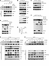 Figure 6
LSD1 demethylates ATM at K3016, and LSD1 mediated demethylation of ATM inhibits its interaction with WIP1. (A) Immunoprecipitation assay to detect the methylation of ATM in the indicated plasmids transfected ATM knockout 3T3 cells treated with LSD1 siRNA or control siRNA (siNC) at 12 hours post IR (10 Gy). (B) Western blot analysis of LSD1, ATM-K3016 methylation (ATM-K3016me), phospho-ATM (p-ATM), and total ATM at 12 hours after irradiation (IR), as well as p16 and p21 at 24 hours after IR, in 3T3 cells treated with the indicated irradiation doses. (C) Western blot analysis of the levels of ATM-K3016 methylation (ATM-K3016me), p-ATM, and total ATM in 3T3 cells treated with 10 Gy and harvest cells at indicated time points. (D) Western blot analysis of the levels of LSD1, methylated ATM at K3016, phospho-ATM, and total of ATM in 3T3 cells treated with LSD1 siRNA and control siRNA (siNC) at 12 hours after IR (10 Gy). (E and F) Western blot and statistical analysis of the levels of methylated ATM at K3016, Flag-tagged ATM, and GFP-tagged LSD1 in ATM knockout mouse 3T3 cells transfected with the indicated plasmids. All experiments were repeated at least 3 times; **P < 0.01; unpaired, 2-tailed Student’s t test. (G) Immunoprecipitation assay to detect the interaction between ATM and WIP1 in mouse 3T3 cells treated with LSD1 siRNA or control siRNA (siNC) at the indicated time points after IR (20 Gy). (H) Immunoprecipitation assay to detect the interaction between Flag-tagged ATM and WIP1 in ATM knockout 3T3 cells transfected with the indicated plasmids at indicated time points after 20 Gy irradiation injury.
To define the relationship between ATM phosphorylation and ATM-K3016 methylation following DNA damage, we performed dose- and time-dependent analyses. At 12 hours after IR, LSD1 expression increased in parallel with a dose-dependent reduction in ATM-K3016 methylation (Figure 6B). Notably, during the early phase (1 hour) after DNA damage, ATM phosphorylation was already detectable, whereas ATM-K3016 methylation remained unchanged (Figures 6C). In addition, we found that knockdown of LSD1 with siRNA reversed IR-induced ATM-K3016 demethylation and reduced ATM phosphorylation at 12 hours after IR (20 Gy) in 3T3 cells (Figure 6D). Conversely, overexpression of GFP-tagged LSD1 decreased ATM methylation in ATM-deficient (KO) 3T3 and mouse IMCD cells reconstituted with Flag-ATM (Figure 6, E and F, and Supplemental Figure 6C). Our results demonstrate that LSD1 demethylates ATM at K3016, thereby modulating the phosphorylation of ATM following DNA damage.
LSD1-mediated demethylation of ATM inhibits its interaction with WIP1. It has been reported that WIP1 (WT p53-induced phosphatase 1) can dephosphorylate ATM to attenuate DNA damage–induced stress response (34). We found that ATM interacted with WIP1 at late stages of DNA damage repair in 3T3 cells (Figure 6G), which led to ATM dephosphorylation and deactivation once DNA repair was completed (35). In LSD1-knockdown 3T3 cells, this interaction was markedly increased compared with control cells at 12 hours after IR exposure (Figure 6G). To investigate whether the methylation of ATM at lysine 3,016 affects its interaction with WIP1, we reintroduced either flag-tagged WT and K3016R mutant ATM into ATM-stable knockout 3T3 cells. We found that the interaction of flag-tagged WT ATM with WIP1 could be detected as early as 1 hour and was increased at 12 hours after IR treatment, whereas the interaction of flag-tagged mutant ATM-K3016R with WIP1 could be detected at 1 hour but was decreased at 12 hours after IR treatment in ATM-KO 3T3 cells (Figure 6H).
Previous studies have shown that acetylation at ATM-K3016 regulates ATM activation (36). To compare the roles of acetylation and methylation at this site, we generated an acetylation-mimetic mutant (K3016Q). At 12 hours after IR, K3016Q mutant showed modestly increased ATM phosphorylation and reduced interaction with WIP1 compared with the methylation-deficient K3016R mutant (Supplemental Figure 8A). Consistent with prior reports, acetylation at K3016 predominates during the early phase of the DNA damage response — 10 minutes to approximately 8 hours post-IR (17) — whereas our results indicate that methylation at this site becomes more prominent at later stages (approximately 12 hours) (Figure 6C). To further define this temporal switch, we enhanced acetylation using nicotinamide (NAM) or inhibited LSD1-mediated demethylation using ORY-1001 at 8 hours after IR for 4 hours. Enhancing acetylation at this stage had minimal effect on ATM phosphorylation, whereas inhibition of demethylation markedly reduced ATM phosphorylation (Supplemental Figure 8B). These results support a model in which LSD1-mediated demethylation of ATM at K3016 sustained ATM phosphorylation during the late phase of the DNA damage response, at least in part by limiting its interaction with WIP1, which further suggests a role for LSD1 in prolonging ATM signaling following genotoxic stress.
LSD1 is subjected to autophagosome-lysosome degradation during organ senescence and aging. Next, we investigate the mechanism of how LSD1 protein is increased during organ senescence and aging. We found that treatment with the proteasome inhibitor MG132 had no effect on the levels of LSD1 protein in senescent mouse IMCD cells (Figure 7A). We then tested whether lysosome-mediated degradation contributes to LSD1 protein upregulation. A central lysosomal-mediated process is autophagy, which is inhibited upon cellular senescence (37). We found that treatment with autophagy inhibitor Lys05 (a dimeric form of chloroquine) increased LSD1 protein in senescent mouse IMCD cells in a dose-dependent manner (Figure 7B), accompanying a decrease of the expression of LC3 and an increase of the expression of p62, a substrate of autophagy, whereas treatment with one of the autophagy inducers, rapamycin (Rapa), decreased LSD1 protein in senescent mouse IMCD cells in a dose-dependent manner, accompanying an increase of the expression of LC3 and a decrease of the expression of p62 (Figure 7C). Treatment with Lys05 or Rapa did not affect the levels of Lsd1 mRNA in mouse IMCD cells treated with ETO, as examined by qRT-PCR analysis (Figure 7D), indicating posttranslational regulation.
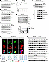 Figure 7
LSD1 is subjected to autophagosome-lysosome degradation during cellular senescence and organ aging. (A) Western blot analysis of the expression of LSD1 in mouse IMCD cells treated with ETO and MG132 with indicated doses. (B and C) Western blot analysis of the expression of LSD1 in mouse IMCD cells treated with ETO, and cotreated with Lys05 (B) or Rapamycin (C) with indicated doses. (D) qRT-PCR analysis of Lsd1 mRNA in mouse IMCD cells treated with ETO and cotreated with Lys05 or Rapamycin for 48 hours. All experiments were repeated at least 3 times; ns, not significant; 1-way ANOVA. (E) Western blot analysis of the expression of LSD1 in nuclear (N) and cytoplasmic (C) fractions isolated from mouse IMCD cells treated with ETO and then Lys05 (5 μM). All experiments were repeated at least 3 times; *P < 0.05; 1-way ANOVA. (F) Representative image and quantification of the immunofluorescence staining for LSD1 (green) and LAMP1 (red) in mouse ETO-induced IMCD cells treated with lys05 (5 μM) or rapamycin (1.5 μM). Scale bar: 25 μm. (G) Immunoprecipitation assay to detect the interaction between ATM and LSD1 in mouse IMCD cells treated with or without Lys05. (H) Western blot analysis of the levels of LSD1, phospho-ATM (p-ATM) and total ATM, p62, LC3B, H3K4me2, p16, and p21 in IR-induced mouse IMCD cells treated with rapamycin or vehicle at the indicated time points. ns, no significance, *P < 0.05.
Nuclear-cytoplasmic fractionation revealed that LSD1 is predominantly localized in the nucleus in senescent cells, with autophagy inhibition further enhancing its nuclear accumulation (Figure 7E). Immunofluorescence staining confirmed this dynamic regulation, showing increased nuclear LSD1 upon autophagy inhibition and reduced nuclear localization upon autophagy activation (Figure 7F). Coimmunoprecipitation experiments demonstrated that inhibition of autophagy sustained the interaction between LSD1 and ATM following DNA damage, accompanied by decreased ATM-K3016 methylation and prolonged ATM phosphorylation at 12 hours after IR (Figure 7G). Conversely, activation of autophagy with rapamycin reduced LSD1 protein levels in IR-treated IMCD cells, along with decreased ATM phosphorylation, γH2AX and senescence markers (p16 and p21), as well as reduced H3K4me2 (Figure 7H).
Importantly, upregulation of LSD1 was associated with a decrease of autophagy in aged organs, including kidney, heart, and liver, characterized by a decrease of the expression of LC3 but an increase of the expression of p62 (Supplemental Figure 9A). Pharmacological inhibition of autophagy with Lys05 increased LSD1 protein in young organs, including kidneys, hearts, and livers, compared with that in age-matched organs treated with vehicle, in which the inhibition of autophagy was confirmed by the accumulation of p62 in those organs (Supplemental Figure 9B). Notably, Lsd1 mRNA levels remained unchanged following Lys05 treatment in those organs (Supplemental Figure 9C). Activation of autophagy with rapamycin decreased LSD1 protein without affecting its mRNA in aged organs from 20–22-month-old mice compared with age-matched vehicle treated controls (Supplemental Figure 9, D and E). In sum, these results indicate that LSD1 is regulated posttranscriptionally by autophagy and support a dynamic interplay among DNA damage, autophagy, and LSD1-mediated regulation of cellular senescence both in vitro and in vivo.
LSD1 interacts with autophagy proteins LC3 and Beclin1. Binding to autophagy proteins is essential for the degradation of autophagy substrates (37). To determine whether LSD1 interacts with autophagy component(s), we transfected GFP-tagged constructs of autophagy components, including LC3, ATG5, ATG7, ATG13, Beclin1, and ULK1, into HEK293T cells and performed immunoprecipitation assay. The expression of GFP-tagged autophagy proteins was detected with Western blot analysis. We found that only GFP-tagged LC3 and Beclin1 could pull down LSD1 (Figure 8A). In addition, GST pull down using bacteria-expressed and purified GST-LSD1 and LC3/Beclin1 proteins showed that LSD1 directly binds to LC3 and Beclin1 (Figure 8B).
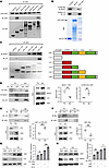 Figure 8
LSD1 interacts with autophagy protein LC3 and Beclin1. (A) Immunoprecipitation assay to detect the interaction of LSD1 with GFP-tagged constructs, including LC3, ATG5, ATG7, ULK1, ATG13, and Beclin1. (B) GST pull-down assay of bacteria-expressed LSD1 with recombination LC3 and Beclin1 protein. (C) Immunoprecipitation assay to detect the interaction of GFP-tagged LSD1 truncated constructs with LC3 or Beclin1. (D–F) Immunoprecipitation assay to detect the interactions between LSD1 and LC3 or Beclin1 in mouse IMCD cells treated with ETO or vehicle. Statistical analysis of the 3 independent expressions of IP/IB is shown in D–F. **P < 0.01; unpaired, 2-tailed Student’s t test. (G and H) Western blot analysis and quantitative data of the expression of LSD1 in mouse IMCD cells treated with LC3 siRNA (G) or Beclin1 siRNA (H) or control siRNA (siNC) at 48 hours. Statistical analysis of the 3 independent expression; ns, not significant, *P < 0.05, **P < 0.01; 1-way ANOVA.
Next, we defined the region within LSD1 that interacted with Beclin1 and LC3 by generating GFP-tagged LSD1 subdomain deletion constructs. The GFP pull-down assay showed that Beclin1 interacts with the AOL-N domain of LSD1, and LC3B interacts with the AOL-C domain of LSD1 (Figure 8C). The AOL domain has been defined as the catalytic center and is responsible for targeting substrate proteins (38). These results suggest that the dysregulation of LSD1 by autophagy is possibly regulated through the interaction of the AOL domain of LSD1 with Beclin1 and LC3.
LC3 is responsible for the lysosomal degradation of LSD1 in senescent cells. Our above results indicated that LSD1 interacted with LC3 and Beclin1 at the proliferating state, and we next investigated whether induction of senescence affected their interaction by performing co-IP assay with anti-LSD1, anti-LC3, and anti-Beclin1 antibodies. We found that the interaction of LSD1 with LC3 was at a similar level in proliferating and senescent mouse IMCD cells, whereas the interaction of LSD1 with Beclin1 was decreased in senescent mouse IMCD cells compared with the interaction in proliferating mouse IMCD cells (Figure 8, D–F). We further found that knockdown of Beclin1 could enhance the expression of LSD1 in proliferating cells but not expression in senescent cells, whereas knockdown of LC3 could augment the expression of LSD1 in both proliferating and senescent cells (Figure 8, G and H). These results suggested that,in the context of cellular proliferation and senescence, the degradation of LSD1 is regulated by distinct autophagy-related proteins, Beclin1 and LC3.
To further investigate the interaction of LSD1 with either Beclin1 or LC3, we assessed the affinity by performing the molecular docking experiments and molecular dynamic simulation assay (39). The Root Mean Square Deviation (RMSD) value of the protein backbone of LSD1 and LC3, as well as LSD1 and Beclin1, was calculated to determine the stability of protein structure during the period of simulation by protein docking test. We found an instability of the interaction of LSD1 and Beclin1 as well as LSD1 and LC3B in the initial 20 nanosecond (ns) of the simulation, whereas a stable interaction was observed from 20–50 nanoseconds by dynamics simulation. The plots of LSD1 and Beclin1 as well as LSD1 and LC3B exhibit average RMSD values of 18 Å and 15 Å, respectively (Supplemental Figure 10, A and B). We also utilized AlphaFoldmultimer (40) to predict the 3D structure and interaction between LSD1 and Beclin1 as well as LSD1 and LC3. The red box represents the predicted Aligned error (pAE) of protein-protein interactions. We found that the value of pAE, which was indicated in the red boxes, of LSD1 with LC3B is lower than that of LSD1 with Beclin1 (Supplemental Figure 10C). During the entire 50 nanoseconds of molecular dynamics (MD) simulation, LSD1 formed a maximum of 65 hydrogen bonds with Beclin1, while LSD1 formed a maximum of 700 hydrogen bonds with LC3, as shown by the frequency plots (Supplemental Figure 10D). These findings suggest that the interaction between LSD1 and LC3 exhibits a higher binding affinity than Beclin1. Collectively, our results suggest that LSD1 interacts with Beclin1/LC3 at the basal state. However, due to the reduced expression of Beclin1 during cellular senescence, LSD1 cannot be fully cleared. Meanwhile, LC3 exhibits enhanced binding affinity with LSD1 compared with Beclin1, supporting that LC3 is responsible for the degradation of LSD1 in senescent cells.
-
Discussion
Cellular senescence and DNA damage are central drivers of aging; however, how epigenetic mechanisms intersect with these processes remains incompletely understood. Here, we identify a role for LSD1 in regulating senescence and organ aging and define mechanisms governing its stability. We show that LSD1 is upregulated in senescent cells and aged organs, where it interacts with and demethylates ATM at K3016. This modification is associated with sustained ATM phosphorylation, impaired DNA damage checkpoint recovery, and persistent DDR signaling, features linked to senescence and tissue dysfunction. Inhibition or knockdown of LSD1 promotes ATM dephosphorylation via WIP1 and facilitates DNA repair (Figure 9). In vivo, pharmacologic inhibition of LSD1 with ORY-1001 attenuates age- and irradiation-induced tissue injury and reduces hair graying, supporting a functional role for LSD1 in aging phenotypes. Additionally, clearance of senescent cells reduces LSD1 protein levels, suggesting a feedback relationship between LSD1 and senescence. We further demonstrate that LSD1 is regulated posttranslationally through autophagy. LSD1 interacts with LC3 and Beclin1 and undergoes autophagosome-lysosome degradation. Impaired autophagy in senescent cells and aged tissues leads to LSD1 accumulation, whereas autophagy activation reduces LSD1 protein levels, demethylase activity, and downstream DDR signaling. Notably, LSD1 accumulates in the nucleus under senescent conditions, where reduced nuclear autophagy is associated with enhanced LSD1-ATM interaction. These findings link declining autophagic capacity during aging to sustain LSD1 activity and prolonged DDR signaling.
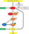 Figure 9
Working model. Cellular senescence is marked by a state of stable cell cycle arrest and plays a crucial role in aging and age-related diseases. DNA damage, a fundamental driver of cellular senescence, influences all hallmarks of aging. DNA damage response (DDR) is regulated either by histone modifications or by their interactions with and modifications of nonhistone substrates. In this study, we define the roles of lysine-specific histone demethylase 1 (LSD1) in senescence and aging and mechanistically address (a) a crosstalk of LSD1 and ATM activation, and (b) how the stability of LSD1 is regulated by autophagy during senescence and organ aging. Aging, irradiation or other factors led to increased expression of LSD1. In response to DNA damage, upregulation of LSD1 results in: (a) LSD1 interaction with and demethylation of ATM at its Lysine-3016 (K3016), (b) LSD1 mediated demethylation of ATM and inhibition of its interaction with WIP1, which blocks DNA damage repair and promotes cellular senescence, (c) aging-induced impairment of autophagy, leading to a reduced degradation of LSD1, (d) LC3 and Beclin1 interaction with LSD1, subjecting it to autophagy-mediated degradation. Thus, treatment with senolytic drugs (Dasatinib and Quercetin) to eliminate senescent cells reduces LSD1 expression, and inhibition of LSD1 by its specific inhibitor ORY-1001 ameliorates cellular senescence and organ aging.
Cellular senescence is triggered by diverse endogenous and exogenous stresses, including telomere dysfunction, oncogene activation, persistent DNA damage, epigenetic dysregulation, and mitochondrial impairment (6). Among these, DNA damage represents a central driver that influences multiple hallmarks of aging and promotes senescence across tissues (41). Posttranslational modifications are critical regulators of the DNA damage response (DDR) and DNA repair pathways, with coordinated inputs from phosphorylation, acetylation, and ubiquitylation shaping key outputs such as cell cycle arrest, DNA repair, apoptosis, and senescence (42, 43). In this context, the role of epigenetic enzymes that mediate methylation of nonhistone proteins in DDR regulation is an emerging area of investigation.
LSD1, the first identified lysine demethylase, regulates chromatin plasticity through the demethylation of histones and nonhistone substrates (22). LSD1 has also been implicated in the DNA damage response (DDR) through its recruitment to sites of DNA damage, accompanying local reduction of H3K4 demethylation (44). Although LSD1 demethylates H3K4me2 and H3K9me2 without affecting the corresponding monomethylated states (30), our data do not exclude the role of these monomethylated histones in mediating senescence in this context. Notably, we show that LSD1-mediated ATM demethylation operates predominantly in the context of DNA damage–induced senescence and aging, particularly at later stages of the response. This function is distinct from previously described chromatin- or metabolism-centered roles of LSD1, including HIF-1α signaling or Sirt4 induction, which are primarily linked to metabolic adaptation or stress responses (24, 25). Rather, our study defines a DNA damage–specific role of LSD1 in DNA damage–induced senescence and aging, which complements previously described metabolic and signaling pathways and links epigenetic regulation to sustained ATM activation and downstream effectors, including γH2AX, p16, p21, and SASP factors. These results underscore the versatility of LSD1 in coordinating diverse regulatory networks that govern cellular senescence and support a model in which context- and tissue-dependent LSD1 activities converge to drive aging-related phenotypes.
The DNA damage checkpoint transiently arrests cell cycle progression to allow time for DNA repair, relying on damage sensor proteins, such as ATM and ATR, to detect DNA damage and to initiate downstream signaling cascades (45). Upon completion of DNA repair, checkpoint signaling is terminated and cell cycle progression is resumed through a process termed checkpoint recovery (46). Persistent ATM activation is known to impair DNA repair and contributes to organ aging by promoting cellular senescence and tissue dysfunction (47). However, the mechanisms governing checkpoint recovery remain incompletely understood. We show that LSD1-mediated demethylation is dispensable for initial ATM activation but contributes to the persistence of ATM signaling. Notably, K3016, the site of LSD1-mediated demethylation, is also a known acetylation site linked to ATM activation (48), suggesting potential crosstalk between acetylation and methylation. Consistent with prior studies, K3016 acetylation predominates during the early phase of the DNA damage response (minutes to approximately 8 hours after IR) (48), whereas our results show that demethylation at this site becomes more prominent at later stages (approximately 12 hours after IR), coinciding with sustained ATM phosphorylation and delayed dephosphorylation and resulting in a defect of DNA damage repair. This study supports a temporally regulated model in which acetylation drives early ATM activation, while LSD1-mediated demethylation sustains ATM signaling at later stages, at least in part by modulating the ATM-WIP1 interaction. By prolonging ATM activity, LSD1 may limit checkpoint recovery, an essential step for restoring cell cycle progression following DNA repair (15), thereby contributing to persistent DDR signaling and cellular senescence.
Autophagy and DNA repair are essential for cellular homeostasis (49), and enhanced autophagy is associated with improved health span and longevity (50). This study identifies LSD1 as a substrate of autophagy, with its turnover dependent on core autophagy components, including LC3 and Beclin1, under both basal and senescent conditions. While LC3 is predominantly cytoplasmic under starvation conditions, it remains partially retained in the nucleus in senescent cells (37). Beclin1 functions in autophagy initiation (51), while LC3 promotes autophagosome formation, reflecting their distinct roles in autophagic flux and cellular proliferation and senescence (52). Notably, LC3 interacts more strongly with LSD1 than Beclin1, suggesting a primary role for LC3 in mediating LSD1 turnover. Although LSD1 is present in cytoplasmic puncta following IR treatment, it exhibits pronounced nuclear enrichment in senescent cells. Under senescent and aging conditions, reduced autophagy, accompanied by decreased LC3 and Beclin1, limits the degradation of LSD1 in both nuclear and cytoplasmic compartments, leading to its accumulation and enhanced LSD1-ATM interaction. Consistent with this model, pharmacologic activation of autophagy following DNA damage reduces LSD1 protein levels and demethylase activity following DNA damage, supporting posttranslational regulation of LSD1 by autophagy. Together, these findings indicate that impaired autophagy sustains LSD1 abundance and activity in senescent cells, particularly in the nucleus, thereby promoting persistent ATM signaling and reinforcing the temporal control of LSD1 during the DNA damage response.
Although we systematically define the role and mechanisms of LSD1 in senescence and aged organs, several limitations warrant consideration. First, the absence of a methylation-mimetic mutation precludes direct interrogation of the interplay between K3016 methylation and acetylation. Second, the lack of LSD1 catalytic mutants selectively targeting histone versus nonhistone substrates limits our ability to exclude additional contributing pathways. Third, although LSD1 inhibition attenuates hair graying, whether similar mechanisms extend to other aging phenotypes remains to be determined. Finally, as cellular senescence represents only one component of organismal aging, future genetic models and longitudinal studies are needed to establish the causal role of the LSD1-ATM axis in aging.
In conclusion, this study identifies LSD1 as a regulator of DNA damage-induced senescence and organ aging through modulation of ATM demethylation and signaling duration. LSD1 is controlled by autophagy-dependent degradation, linking impaired proteostasis to sustained DDR. This study suggests that targeting LSD1 may be a novel strategy to mitigate senescence-associated tissue dysfunction and aging-related phenotypes, such as hair graying, in elder populations.


Copyright © 2026 American Society for Clinical Investigation
ISSN: 0021-9738 (print), 1558-8238 (online)