
Advertisement
Research ArticleDermatologyHematologyOncology
Open Access |  10.1172/JCI199141
JAK1/2 inhibition promotes aggressive cutaneous carcinogenesis through COT kinase–mediated alternative MAPK pathway activation
Hélène Pasquer,1,2 Antoine Forget,3,4 Maxime Battistella,5 Raphael Marie,1 Léa Provent,1 Barouyr Baroudjian,6 Emmanuelle Latour,1 Cécile Culeux,1 Blandine Roux,1 Khansa Saadallah,1 Amandine Gouverneur,7 Maxime Demourgues,7 Fanélie Jouenne,8 Jérôme Lambert,9 Bérangère Lombard,10 Damarys Loew,10 Sophie Tartare-Deckert,11 Lionel Adès,12 Emmanuel Raffoux,12 Bruno Cassinat,1,13 Juliette Soret-Dulphy,2 Pernelle Noize,7 Samia Mourah,8 Camille Lobry,1 Jean-Jacques Kiladjian,1,2 Alexandre Puissant,1 Céleste Lebbé,6 and Lina Benajiba1,2
1INSERM UMR 1342, Institut de Recherche Saint-Louis, Paris, France.
2Université Paris Cité, INSERM Clinical Investigation Center (CIC) 1427, AP-HP, Hôpital Saint-Louis, Paris, France.
3Quantitative Biosciences Institute and
4Department of Cellular and Molecular Pharmacology, University of California, San Francisco, San Francisco, California, USA.
5Service de Pathologie, INSERM U976, Institut de Recherche Saint-Louis, and
6Dermato-oncology Unit and CIC, Cancer Institute AP-HP.Nord Paris Cité, INSERM U976, Université Paris Cité, AP-HP, Hôpital Saint-Louis, Paris, France.
7Université de Bordeaux, INSERM, BPH, U1219, AHeaD Team, CHU de Bordeaux, Pôle de Santé Publique, Service de Pharmacologie Médicale, Bordeaux, France.
8Service de Génomique des Tumeurs et Pharmacologie and
9Service de Biostatistiques, Université Paris Cité, AP-HP, Hôpital Saint-Louis, Paris, France.
10Université Paris Sciences et Lettres, Institut Curie, CurieCoreTech Spectrométrie de Masse Protéomique, Paris, France.
11Université Côte d’Azur, INSERM U1065 (C3M), Microenvironment, Signaling and Cancer, Nice, France.
12Département d’Hématologie et d’Immunologie and
13Laboratoire de Biologie Cellulaire, Université Paris Cité, AP-HP, Hôpital Saint-Louis, Paris, France.
Address for correspondence. Lina Benajiba, Institut de Recherche Saint-Louis, 16 Rue de la Grange aux Belles, 75010 Paris, France. Email: lina.benajiba@inserm.fr.
Authorship note: AF and MB contributed equally to this work. C Lobry, JJK, AP, and C Lebbé contributed equally to this work and are co–senior authors.
Find articles by Pasquer, H. in: PubMed | Google Scholar
1INSERM UMR 1342, Institut de Recherche Saint-Louis, Paris, France.
2Université Paris Cité, INSERM Clinical Investigation Center (CIC) 1427, AP-HP, Hôpital Saint-Louis, Paris, France.
3Quantitative Biosciences Institute and
4Department of Cellular and Molecular Pharmacology, University of California, San Francisco, San Francisco, California, USA.
5Service de Pathologie, INSERM U976, Institut de Recherche Saint-Louis, and
6Dermato-oncology Unit and CIC, Cancer Institute AP-HP.Nord Paris Cité, INSERM U976, Université Paris Cité, AP-HP, Hôpital Saint-Louis, Paris, France.
7Université de Bordeaux, INSERM, BPH, U1219, AHeaD Team, CHU de Bordeaux, Pôle de Santé Publique, Service de Pharmacologie Médicale, Bordeaux, France.
8Service de Génomique des Tumeurs et Pharmacologie and
9Service de Biostatistiques, Université Paris Cité, AP-HP, Hôpital Saint-Louis, Paris, France.
10Université Paris Sciences et Lettres, Institut Curie, CurieCoreTech Spectrométrie de Masse Protéomique, Paris, France.
11Université Côte d’Azur, INSERM U1065 (C3M), Microenvironment, Signaling and Cancer, Nice, France.
12Département d’Hématologie et d’Immunologie and
13Laboratoire de Biologie Cellulaire, Université Paris Cité, AP-HP, Hôpital Saint-Louis, Paris, France.
Address for correspondence. Lina Benajiba, Institut de Recherche Saint-Louis, 16 Rue de la Grange aux Belles, 75010 Paris, France. Email: lina.benajiba@inserm.fr.
Authorship note: AF and MB contributed equally to this work. C Lobry, JJK, AP, and C Lebbé contributed equally to this work and are co–senior authors.
Find articles by Forget, A. in: PubMed | Google Scholar
1INSERM UMR 1342, Institut de Recherche Saint-Louis, Paris, France.
2Université Paris Cité, INSERM Clinical Investigation Center (CIC) 1427, AP-HP, Hôpital Saint-Louis, Paris, France.
3Quantitative Biosciences Institute and
4Department of Cellular and Molecular Pharmacology, University of California, San Francisco, San Francisco, California, USA.
5Service de Pathologie, INSERM U976, Institut de Recherche Saint-Louis, and
6Dermato-oncology Unit and CIC, Cancer Institute AP-HP.Nord Paris Cité, INSERM U976, Université Paris Cité, AP-HP, Hôpital Saint-Louis, Paris, France.
7Université de Bordeaux, INSERM, BPH, U1219, AHeaD Team, CHU de Bordeaux, Pôle de Santé Publique, Service de Pharmacologie Médicale, Bordeaux, France.
8Service de Génomique des Tumeurs et Pharmacologie and
9Service de Biostatistiques, Université Paris Cité, AP-HP, Hôpital Saint-Louis, Paris, France.
10Université Paris Sciences et Lettres, Institut Curie, CurieCoreTech Spectrométrie de Masse Protéomique, Paris, France.
11Université Côte d’Azur, INSERM U1065 (C3M), Microenvironment, Signaling and Cancer, Nice, France.
12Département d’Hématologie et d’Immunologie and
13Laboratoire de Biologie Cellulaire, Université Paris Cité, AP-HP, Hôpital Saint-Louis, Paris, France.
Address for correspondence. Lina Benajiba, Institut de Recherche Saint-Louis, 16 Rue de la Grange aux Belles, 75010 Paris, France. Email: lina.benajiba@inserm.fr.
Authorship note: AF and MB contributed equally to this work. C Lobry, JJK, AP, and C Lebbé contributed equally to this work and are co–senior authors.
Find articles by Battistella, M. in: PubMed | Google Scholar
1INSERM UMR 1342, Institut de Recherche Saint-Louis, Paris, France.
2Université Paris Cité, INSERM Clinical Investigation Center (CIC) 1427, AP-HP, Hôpital Saint-Louis, Paris, France.
3Quantitative Biosciences Institute and
4Department of Cellular and Molecular Pharmacology, University of California, San Francisco, San Francisco, California, USA.
5Service de Pathologie, INSERM U976, Institut de Recherche Saint-Louis, and
6Dermato-oncology Unit and CIC, Cancer Institute AP-HP.Nord Paris Cité, INSERM U976, Université Paris Cité, AP-HP, Hôpital Saint-Louis, Paris, France.
7Université de Bordeaux, INSERM, BPH, U1219, AHeaD Team, CHU de Bordeaux, Pôle de Santé Publique, Service de Pharmacologie Médicale, Bordeaux, France.
8Service de Génomique des Tumeurs et Pharmacologie and
9Service de Biostatistiques, Université Paris Cité, AP-HP, Hôpital Saint-Louis, Paris, France.
10Université Paris Sciences et Lettres, Institut Curie, CurieCoreTech Spectrométrie de Masse Protéomique, Paris, France.
11Université Côte d’Azur, INSERM U1065 (C3M), Microenvironment, Signaling and Cancer, Nice, France.
12Département d’Hématologie et d’Immunologie and
13Laboratoire de Biologie Cellulaire, Université Paris Cité, AP-HP, Hôpital Saint-Louis, Paris, France.
Address for correspondence. Lina Benajiba, Institut de Recherche Saint-Louis, 16 Rue de la Grange aux Belles, 75010 Paris, France. Email: lina.benajiba@inserm.fr.
Authorship note: AF and MB contributed equally to this work. C Lobry, JJK, AP, and C Lebbé contributed equally to this work and are co–senior authors.
Find articles by Marie, R. in: PubMed | Google Scholar
1INSERM UMR 1342, Institut de Recherche Saint-Louis, Paris, France.
2Université Paris Cité, INSERM Clinical Investigation Center (CIC) 1427, AP-HP, Hôpital Saint-Louis, Paris, France.
3Quantitative Biosciences Institute and
4Department of Cellular and Molecular Pharmacology, University of California, San Francisco, San Francisco, California, USA.
5Service de Pathologie, INSERM U976, Institut de Recherche Saint-Louis, and
6Dermato-oncology Unit and CIC, Cancer Institute AP-HP.Nord Paris Cité, INSERM U976, Université Paris Cité, AP-HP, Hôpital Saint-Louis, Paris, France.
7Université de Bordeaux, INSERM, BPH, U1219, AHeaD Team, CHU de Bordeaux, Pôle de Santé Publique, Service de Pharmacologie Médicale, Bordeaux, France.
8Service de Génomique des Tumeurs et Pharmacologie and
9Service de Biostatistiques, Université Paris Cité, AP-HP, Hôpital Saint-Louis, Paris, France.
10Université Paris Sciences et Lettres, Institut Curie, CurieCoreTech Spectrométrie de Masse Protéomique, Paris, France.
11Université Côte d’Azur, INSERM U1065 (C3M), Microenvironment, Signaling and Cancer, Nice, France.
12Département d’Hématologie et d’Immunologie and
13Laboratoire de Biologie Cellulaire, Université Paris Cité, AP-HP, Hôpital Saint-Louis, Paris, France.
Address for correspondence. Lina Benajiba, Institut de Recherche Saint-Louis, 16 Rue de la Grange aux Belles, 75010 Paris, France. Email: lina.benajiba@inserm.fr.
Authorship note: AF and MB contributed equally to this work. C Lobry, JJK, AP, and C Lebbé contributed equally to this work and are co–senior authors.
Find articles by Provent, L. in: PubMed | Google Scholar
1INSERM UMR 1342, Institut de Recherche Saint-Louis, Paris, France.
2Université Paris Cité, INSERM Clinical Investigation Center (CIC) 1427, AP-HP, Hôpital Saint-Louis, Paris, France.
3Quantitative Biosciences Institute and
4Department of Cellular and Molecular Pharmacology, University of California, San Francisco, San Francisco, California, USA.
5Service de Pathologie, INSERM U976, Institut de Recherche Saint-Louis, and
6Dermato-oncology Unit and CIC, Cancer Institute AP-HP.Nord Paris Cité, INSERM U976, Université Paris Cité, AP-HP, Hôpital Saint-Louis, Paris, France.
7Université de Bordeaux, INSERM, BPH, U1219, AHeaD Team, CHU de Bordeaux, Pôle de Santé Publique, Service de Pharmacologie Médicale, Bordeaux, France.
8Service de Génomique des Tumeurs et Pharmacologie and
9Service de Biostatistiques, Université Paris Cité, AP-HP, Hôpital Saint-Louis, Paris, France.
10Université Paris Sciences et Lettres, Institut Curie, CurieCoreTech Spectrométrie de Masse Protéomique, Paris, France.
11Université Côte d’Azur, INSERM U1065 (C3M), Microenvironment, Signaling and Cancer, Nice, France.
12Département d’Hématologie et d’Immunologie and
13Laboratoire de Biologie Cellulaire, Université Paris Cité, AP-HP, Hôpital Saint-Louis, Paris, France.
Address for correspondence. Lina Benajiba, Institut de Recherche Saint-Louis, 16 Rue de la Grange aux Belles, 75010 Paris, France. Email: lina.benajiba@inserm.fr.
Authorship note: AF and MB contributed equally to this work. C Lobry, JJK, AP, and C Lebbé contributed equally to this work and are co–senior authors.
Find articles by Baroudjian, B. in: PubMed | Google Scholar
1INSERM UMR 1342, Institut de Recherche Saint-Louis, Paris, France.
2Université Paris Cité, INSERM Clinical Investigation Center (CIC) 1427, AP-HP, Hôpital Saint-Louis, Paris, France.
3Quantitative Biosciences Institute and
4Department of Cellular and Molecular Pharmacology, University of California, San Francisco, San Francisco, California, USA.
5Service de Pathologie, INSERM U976, Institut de Recherche Saint-Louis, and
6Dermato-oncology Unit and CIC, Cancer Institute AP-HP.Nord Paris Cité, INSERM U976, Université Paris Cité, AP-HP, Hôpital Saint-Louis, Paris, France.
7Université de Bordeaux, INSERM, BPH, U1219, AHeaD Team, CHU de Bordeaux, Pôle de Santé Publique, Service de Pharmacologie Médicale, Bordeaux, France.
8Service de Génomique des Tumeurs et Pharmacologie and
9Service de Biostatistiques, Université Paris Cité, AP-HP, Hôpital Saint-Louis, Paris, France.
10Université Paris Sciences et Lettres, Institut Curie, CurieCoreTech Spectrométrie de Masse Protéomique, Paris, France.
11Université Côte d’Azur, INSERM U1065 (C3M), Microenvironment, Signaling and Cancer, Nice, France.
12Département d’Hématologie et d’Immunologie and
13Laboratoire de Biologie Cellulaire, Université Paris Cité, AP-HP, Hôpital Saint-Louis, Paris, France.
Address for correspondence. Lina Benajiba, Institut de Recherche Saint-Louis, 16 Rue de la Grange aux Belles, 75010 Paris, France. Email: lina.benajiba@inserm.fr.
Authorship note: AF and MB contributed equally to this work. C Lobry, JJK, AP, and C Lebbé contributed equally to this work and are co–senior authors.
Find articles by Latour, E. in: PubMed | Google Scholar
1INSERM UMR 1342, Institut de Recherche Saint-Louis, Paris, France.
2Université Paris Cité, INSERM Clinical Investigation Center (CIC) 1427, AP-HP, Hôpital Saint-Louis, Paris, France.
3Quantitative Biosciences Institute and
4Department of Cellular and Molecular Pharmacology, University of California, San Francisco, San Francisco, California, USA.
5Service de Pathologie, INSERM U976, Institut de Recherche Saint-Louis, and
6Dermato-oncology Unit and CIC, Cancer Institute AP-HP.Nord Paris Cité, INSERM U976, Université Paris Cité, AP-HP, Hôpital Saint-Louis, Paris, France.
7Université de Bordeaux, INSERM, BPH, U1219, AHeaD Team, CHU de Bordeaux, Pôle de Santé Publique, Service de Pharmacologie Médicale, Bordeaux, France.
8Service de Génomique des Tumeurs et Pharmacologie and
9Service de Biostatistiques, Université Paris Cité, AP-HP, Hôpital Saint-Louis, Paris, France.
10Université Paris Sciences et Lettres, Institut Curie, CurieCoreTech Spectrométrie de Masse Protéomique, Paris, France.
11Université Côte d’Azur, INSERM U1065 (C3M), Microenvironment, Signaling and Cancer, Nice, France.
12Département d’Hématologie et d’Immunologie and
13Laboratoire de Biologie Cellulaire, Université Paris Cité, AP-HP, Hôpital Saint-Louis, Paris, France.
Address for correspondence. Lina Benajiba, Institut de Recherche Saint-Louis, 16 Rue de la Grange aux Belles, 75010 Paris, France. Email: lina.benajiba@inserm.fr.
Authorship note: AF and MB contributed equally to this work. C Lobry, JJK, AP, and C Lebbé contributed equally to this work and are co–senior authors.
Find articles by Culeux, C. in: PubMed | Google Scholar
1INSERM UMR 1342, Institut de Recherche Saint-Louis, Paris, France.
2Université Paris Cité, INSERM Clinical Investigation Center (CIC) 1427, AP-HP, Hôpital Saint-Louis, Paris, France.
3Quantitative Biosciences Institute and
4Department of Cellular and Molecular Pharmacology, University of California, San Francisco, San Francisco, California, USA.
5Service de Pathologie, INSERM U976, Institut de Recherche Saint-Louis, and
6Dermato-oncology Unit and CIC, Cancer Institute AP-HP.Nord Paris Cité, INSERM U976, Université Paris Cité, AP-HP, Hôpital Saint-Louis, Paris, France.
7Université de Bordeaux, INSERM, BPH, U1219, AHeaD Team, CHU de Bordeaux, Pôle de Santé Publique, Service de Pharmacologie Médicale, Bordeaux, France.
8Service de Génomique des Tumeurs et Pharmacologie and
9Service de Biostatistiques, Université Paris Cité, AP-HP, Hôpital Saint-Louis, Paris, France.
10Université Paris Sciences et Lettres, Institut Curie, CurieCoreTech Spectrométrie de Masse Protéomique, Paris, France.
11Université Côte d’Azur, INSERM U1065 (C3M), Microenvironment, Signaling and Cancer, Nice, France.
12Département d’Hématologie et d’Immunologie and
13Laboratoire de Biologie Cellulaire, Université Paris Cité, AP-HP, Hôpital Saint-Louis, Paris, France.
Address for correspondence. Lina Benajiba, Institut de Recherche Saint-Louis, 16 Rue de la Grange aux Belles, 75010 Paris, France. Email: lina.benajiba@inserm.fr.
Authorship note: AF and MB contributed equally to this work. C Lobry, JJK, AP, and C Lebbé contributed equally to this work and are co–senior authors.
Find articles by Roux, B. in: PubMed | Google Scholar
1INSERM UMR 1342, Institut de Recherche Saint-Louis, Paris, France.
2Université Paris Cité, INSERM Clinical Investigation Center (CIC) 1427, AP-HP, Hôpital Saint-Louis, Paris, France.
3Quantitative Biosciences Institute and
4Department of Cellular and Molecular Pharmacology, University of California, San Francisco, San Francisco, California, USA.
5Service de Pathologie, INSERM U976, Institut de Recherche Saint-Louis, and
6Dermato-oncology Unit and CIC, Cancer Institute AP-HP.Nord Paris Cité, INSERM U976, Université Paris Cité, AP-HP, Hôpital Saint-Louis, Paris, France.
7Université de Bordeaux, INSERM, BPH, U1219, AHeaD Team, CHU de Bordeaux, Pôle de Santé Publique, Service de Pharmacologie Médicale, Bordeaux, France.
8Service de Génomique des Tumeurs et Pharmacologie and
9Service de Biostatistiques, Université Paris Cité, AP-HP, Hôpital Saint-Louis, Paris, France.
10Université Paris Sciences et Lettres, Institut Curie, CurieCoreTech Spectrométrie de Masse Protéomique, Paris, France.
11Université Côte d’Azur, INSERM U1065 (C3M), Microenvironment, Signaling and Cancer, Nice, France.
12Département d’Hématologie et d’Immunologie and
13Laboratoire de Biologie Cellulaire, Université Paris Cité, AP-HP, Hôpital Saint-Louis, Paris, France.
Address for correspondence. Lina Benajiba, Institut de Recherche Saint-Louis, 16 Rue de la Grange aux Belles, 75010 Paris, France. Email: lina.benajiba@inserm.fr.
Authorship note: AF and MB contributed equally to this work. C Lobry, JJK, AP, and C Lebbé contributed equally to this work and are co–senior authors.
Find articles by Saadallah, K. in: PubMed | Google Scholar
1INSERM UMR 1342, Institut de Recherche Saint-Louis, Paris, France.
2Université Paris Cité, INSERM Clinical Investigation Center (CIC) 1427, AP-HP, Hôpital Saint-Louis, Paris, France.
3Quantitative Biosciences Institute and
4Department of Cellular and Molecular Pharmacology, University of California, San Francisco, San Francisco, California, USA.
5Service de Pathologie, INSERM U976, Institut de Recherche Saint-Louis, and
6Dermato-oncology Unit and CIC, Cancer Institute AP-HP.Nord Paris Cité, INSERM U976, Université Paris Cité, AP-HP, Hôpital Saint-Louis, Paris, France.
7Université de Bordeaux, INSERM, BPH, U1219, AHeaD Team, CHU de Bordeaux, Pôle de Santé Publique, Service de Pharmacologie Médicale, Bordeaux, France.
8Service de Génomique des Tumeurs et Pharmacologie and
9Service de Biostatistiques, Université Paris Cité, AP-HP, Hôpital Saint-Louis, Paris, France.
10Université Paris Sciences et Lettres, Institut Curie, CurieCoreTech Spectrométrie de Masse Protéomique, Paris, France.
11Université Côte d’Azur, INSERM U1065 (C3M), Microenvironment, Signaling and Cancer, Nice, France.
12Département d’Hématologie et d’Immunologie and
13Laboratoire de Biologie Cellulaire, Université Paris Cité, AP-HP, Hôpital Saint-Louis, Paris, France.
Address for correspondence. Lina Benajiba, Institut de Recherche Saint-Louis, 16 Rue de la Grange aux Belles, 75010 Paris, France. Email: lina.benajiba@inserm.fr.
Authorship note: AF and MB contributed equally to this work. C Lobry, JJK, AP, and C Lebbé contributed equally to this work and are co–senior authors.
Find articles by Gouverneur, A. in: PubMed | Google Scholar
1INSERM UMR 1342, Institut de Recherche Saint-Louis, Paris, France.
2Université Paris Cité, INSERM Clinical Investigation Center (CIC) 1427, AP-HP, Hôpital Saint-Louis, Paris, France.
3Quantitative Biosciences Institute and
4Department of Cellular and Molecular Pharmacology, University of California, San Francisco, San Francisco, California, USA.
5Service de Pathologie, INSERM U976, Institut de Recherche Saint-Louis, and
6Dermato-oncology Unit and CIC, Cancer Institute AP-HP.Nord Paris Cité, INSERM U976, Université Paris Cité, AP-HP, Hôpital Saint-Louis, Paris, France.
7Université de Bordeaux, INSERM, BPH, U1219, AHeaD Team, CHU de Bordeaux, Pôle de Santé Publique, Service de Pharmacologie Médicale, Bordeaux, France.
8Service de Génomique des Tumeurs et Pharmacologie and
9Service de Biostatistiques, Université Paris Cité, AP-HP, Hôpital Saint-Louis, Paris, France.
10Université Paris Sciences et Lettres, Institut Curie, CurieCoreTech Spectrométrie de Masse Protéomique, Paris, France.
11Université Côte d’Azur, INSERM U1065 (C3M), Microenvironment, Signaling and Cancer, Nice, France.
12Département d’Hématologie et d’Immunologie and
13Laboratoire de Biologie Cellulaire, Université Paris Cité, AP-HP, Hôpital Saint-Louis, Paris, France.
Address for correspondence. Lina Benajiba, Institut de Recherche Saint-Louis, 16 Rue de la Grange aux Belles, 75010 Paris, France. Email: lina.benajiba@inserm.fr.
Authorship note: AF and MB contributed equally to this work. C Lobry, JJK, AP, and C Lebbé contributed equally to this work and are co–senior authors.
Find articles by Demourgues, M. in: PubMed | Google Scholar
1INSERM UMR 1342, Institut de Recherche Saint-Louis, Paris, France.
2Université Paris Cité, INSERM Clinical Investigation Center (CIC) 1427, AP-HP, Hôpital Saint-Louis, Paris, France.
3Quantitative Biosciences Institute and
4Department of Cellular and Molecular Pharmacology, University of California, San Francisco, San Francisco, California, USA.
5Service de Pathologie, INSERM U976, Institut de Recherche Saint-Louis, and
6Dermato-oncology Unit and CIC, Cancer Institute AP-HP.Nord Paris Cité, INSERM U976, Université Paris Cité, AP-HP, Hôpital Saint-Louis, Paris, France.
7Université de Bordeaux, INSERM, BPH, U1219, AHeaD Team, CHU de Bordeaux, Pôle de Santé Publique, Service de Pharmacologie Médicale, Bordeaux, France.
8Service de Génomique des Tumeurs et Pharmacologie and
9Service de Biostatistiques, Université Paris Cité, AP-HP, Hôpital Saint-Louis, Paris, France.
10Université Paris Sciences et Lettres, Institut Curie, CurieCoreTech Spectrométrie de Masse Protéomique, Paris, France.
11Université Côte d’Azur, INSERM U1065 (C3M), Microenvironment, Signaling and Cancer, Nice, France.
12Département d’Hématologie et d’Immunologie and
13Laboratoire de Biologie Cellulaire, Université Paris Cité, AP-HP, Hôpital Saint-Louis, Paris, France.
Address for correspondence. Lina Benajiba, Institut de Recherche Saint-Louis, 16 Rue de la Grange aux Belles, 75010 Paris, France. Email: lina.benajiba@inserm.fr.
Authorship note: AF and MB contributed equally to this work. C Lobry, JJK, AP, and C Lebbé contributed equally to this work and are co–senior authors.
Find articles by Jouenne, F. in: PubMed | Google Scholar
1INSERM UMR 1342, Institut de Recherche Saint-Louis, Paris, France.
2Université Paris Cité, INSERM Clinical Investigation Center (CIC) 1427, AP-HP, Hôpital Saint-Louis, Paris, France.
3Quantitative Biosciences Institute and
4Department of Cellular and Molecular Pharmacology, University of California, San Francisco, San Francisco, California, USA.
5Service de Pathologie, INSERM U976, Institut de Recherche Saint-Louis, and
6Dermato-oncology Unit and CIC, Cancer Institute AP-HP.Nord Paris Cité, INSERM U976, Université Paris Cité, AP-HP, Hôpital Saint-Louis, Paris, France.
7Université de Bordeaux, INSERM, BPH, U1219, AHeaD Team, CHU de Bordeaux, Pôle de Santé Publique, Service de Pharmacologie Médicale, Bordeaux, France.
8Service de Génomique des Tumeurs et Pharmacologie and
9Service de Biostatistiques, Université Paris Cité, AP-HP, Hôpital Saint-Louis, Paris, France.
10Université Paris Sciences et Lettres, Institut Curie, CurieCoreTech Spectrométrie de Masse Protéomique, Paris, France.
11Université Côte d’Azur, INSERM U1065 (C3M), Microenvironment, Signaling and Cancer, Nice, France.
12Département d’Hématologie et d’Immunologie and
13Laboratoire de Biologie Cellulaire, Université Paris Cité, AP-HP, Hôpital Saint-Louis, Paris, France.
Address for correspondence. Lina Benajiba, Institut de Recherche Saint-Louis, 16 Rue de la Grange aux Belles, 75010 Paris, France. Email: lina.benajiba@inserm.fr.
Authorship note: AF and MB contributed equally to this work. C Lobry, JJK, AP, and C Lebbé contributed equally to this work and are co–senior authors.
Find articles by Lambert, J. in: PubMed | Google Scholar
1INSERM UMR 1342, Institut de Recherche Saint-Louis, Paris, France.
2Université Paris Cité, INSERM Clinical Investigation Center (CIC) 1427, AP-HP, Hôpital Saint-Louis, Paris, France.
3Quantitative Biosciences Institute and
4Department of Cellular and Molecular Pharmacology, University of California, San Francisco, San Francisco, California, USA.
5Service de Pathologie, INSERM U976, Institut de Recherche Saint-Louis, and
6Dermato-oncology Unit and CIC, Cancer Institute AP-HP.Nord Paris Cité, INSERM U976, Université Paris Cité, AP-HP, Hôpital Saint-Louis, Paris, France.
7Université de Bordeaux, INSERM, BPH, U1219, AHeaD Team, CHU de Bordeaux, Pôle de Santé Publique, Service de Pharmacologie Médicale, Bordeaux, France.
8Service de Génomique des Tumeurs et Pharmacologie and
9Service de Biostatistiques, Université Paris Cité, AP-HP, Hôpital Saint-Louis, Paris, France.
10Université Paris Sciences et Lettres, Institut Curie, CurieCoreTech Spectrométrie de Masse Protéomique, Paris, France.
11Université Côte d’Azur, INSERM U1065 (C3M), Microenvironment, Signaling and Cancer, Nice, France.
12Département d’Hématologie et d’Immunologie and
13Laboratoire de Biologie Cellulaire, Université Paris Cité, AP-HP, Hôpital Saint-Louis, Paris, France.
Address for correspondence. Lina Benajiba, Institut de Recherche Saint-Louis, 16 Rue de la Grange aux Belles, 75010 Paris, France. Email: lina.benajiba@inserm.fr.
Authorship note: AF and MB contributed equally to this work. C Lobry, JJK, AP, and C Lebbé contributed equally to this work and are co–senior authors.
Find articles by Lombard, B. in: PubMed | Google Scholar
1INSERM UMR 1342, Institut de Recherche Saint-Louis, Paris, France.
2Université Paris Cité, INSERM Clinical Investigation Center (CIC) 1427, AP-HP, Hôpital Saint-Louis, Paris, France.
3Quantitative Biosciences Institute and
4Department of Cellular and Molecular Pharmacology, University of California, San Francisco, San Francisco, California, USA.
5Service de Pathologie, INSERM U976, Institut de Recherche Saint-Louis, and
6Dermato-oncology Unit and CIC, Cancer Institute AP-HP.Nord Paris Cité, INSERM U976, Université Paris Cité, AP-HP, Hôpital Saint-Louis, Paris, France.
7Université de Bordeaux, INSERM, BPH, U1219, AHeaD Team, CHU de Bordeaux, Pôle de Santé Publique, Service de Pharmacologie Médicale, Bordeaux, France.
8Service de Génomique des Tumeurs et Pharmacologie and
9Service de Biostatistiques, Université Paris Cité, AP-HP, Hôpital Saint-Louis, Paris, France.
10Université Paris Sciences et Lettres, Institut Curie, CurieCoreTech Spectrométrie de Masse Protéomique, Paris, France.
11Université Côte d’Azur, INSERM U1065 (C3M), Microenvironment, Signaling and Cancer, Nice, France.
12Département d’Hématologie et d’Immunologie and
13Laboratoire de Biologie Cellulaire, Université Paris Cité, AP-HP, Hôpital Saint-Louis, Paris, France.
Address for correspondence. Lina Benajiba, Institut de Recherche Saint-Louis, 16 Rue de la Grange aux Belles, 75010 Paris, France. Email: lina.benajiba@inserm.fr.
Authorship note: AF and MB contributed equally to this work. C Lobry, JJK, AP, and C Lebbé contributed equally to this work and are co–senior authors.
Find articles by Loew, D. in: PubMed | Google Scholar
1INSERM UMR 1342, Institut de Recherche Saint-Louis, Paris, France.
2Université Paris Cité, INSERM Clinical Investigation Center (CIC) 1427, AP-HP, Hôpital Saint-Louis, Paris, France.
3Quantitative Biosciences Institute and
4Department of Cellular and Molecular Pharmacology, University of California, San Francisco, San Francisco, California, USA.
5Service de Pathologie, INSERM U976, Institut de Recherche Saint-Louis, and
6Dermato-oncology Unit and CIC, Cancer Institute AP-HP.Nord Paris Cité, INSERM U976, Université Paris Cité, AP-HP, Hôpital Saint-Louis, Paris, France.
7Université de Bordeaux, INSERM, BPH, U1219, AHeaD Team, CHU de Bordeaux, Pôle de Santé Publique, Service de Pharmacologie Médicale, Bordeaux, France.
8Service de Génomique des Tumeurs et Pharmacologie and
9Service de Biostatistiques, Université Paris Cité, AP-HP, Hôpital Saint-Louis, Paris, France.
10Université Paris Sciences et Lettres, Institut Curie, CurieCoreTech Spectrométrie de Masse Protéomique, Paris, France.
11Université Côte d’Azur, INSERM U1065 (C3M), Microenvironment, Signaling and Cancer, Nice, France.
12Département d’Hématologie et d’Immunologie and
13Laboratoire de Biologie Cellulaire, Université Paris Cité, AP-HP, Hôpital Saint-Louis, Paris, France.
Address for correspondence. Lina Benajiba, Institut de Recherche Saint-Louis, 16 Rue de la Grange aux Belles, 75010 Paris, France. Email: lina.benajiba@inserm.fr.
Authorship note: AF and MB contributed equally to this work. C Lobry, JJK, AP, and C Lebbé contributed equally to this work and are co–senior authors.
Find articles by Tartare-Deckert, S. in: PubMed | Google Scholar
1INSERM UMR 1342, Institut de Recherche Saint-Louis, Paris, France.
2Université Paris Cité, INSERM Clinical Investigation Center (CIC) 1427, AP-HP, Hôpital Saint-Louis, Paris, France.
3Quantitative Biosciences Institute and
4Department of Cellular and Molecular Pharmacology, University of California, San Francisco, San Francisco, California, USA.
5Service de Pathologie, INSERM U976, Institut de Recherche Saint-Louis, and
6Dermato-oncology Unit and CIC, Cancer Institute AP-HP.Nord Paris Cité, INSERM U976, Université Paris Cité, AP-HP, Hôpital Saint-Louis, Paris, France.
7Université de Bordeaux, INSERM, BPH, U1219, AHeaD Team, CHU de Bordeaux, Pôle de Santé Publique, Service de Pharmacologie Médicale, Bordeaux, France.
8Service de Génomique des Tumeurs et Pharmacologie and
9Service de Biostatistiques, Université Paris Cité, AP-HP, Hôpital Saint-Louis, Paris, France.
10Université Paris Sciences et Lettres, Institut Curie, CurieCoreTech Spectrométrie de Masse Protéomique, Paris, France.
11Université Côte d’Azur, INSERM U1065 (C3M), Microenvironment, Signaling and Cancer, Nice, France.
12Département d’Hématologie et d’Immunologie and
13Laboratoire de Biologie Cellulaire, Université Paris Cité, AP-HP, Hôpital Saint-Louis, Paris, France.
Address for correspondence. Lina Benajiba, Institut de Recherche Saint-Louis, 16 Rue de la Grange aux Belles, 75010 Paris, France. Email: lina.benajiba@inserm.fr.
Authorship note: AF and MB contributed equally to this work. C Lobry, JJK, AP, and C Lebbé contributed equally to this work and are co–senior authors.
Find articles by Adès, L. in: PubMed | Google Scholar
1INSERM UMR 1342, Institut de Recherche Saint-Louis, Paris, France.
2Université Paris Cité, INSERM Clinical Investigation Center (CIC) 1427, AP-HP, Hôpital Saint-Louis, Paris, France.
3Quantitative Biosciences Institute and
4Department of Cellular and Molecular Pharmacology, University of California, San Francisco, San Francisco, California, USA.
5Service de Pathologie, INSERM U976, Institut de Recherche Saint-Louis, and
6Dermato-oncology Unit and CIC, Cancer Institute AP-HP.Nord Paris Cité, INSERM U976, Université Paris Cité, AP-HP, Hôpital Saint-Louis, Paris, France.
7Université de Bordeaux, INSERM, BPH, U1219, AHeaD Team, CHU de Bordeaux, Pôle de Santé Publique, Service de Pharmacologie Médicale, Bordeaux, France.
8Service de Génomique des Tumeurs et Pharmacologie and
9Service de Biostatistiques, Université Paris Cité, AP-HP, Hôpital Saint-Louis, Paris, France.
10Université Paris Sciences et Lettres, Institut Curie, CurieCoreTech Spectrométrie de Masse Protéomique, Paris, France.
11Université Côte d’Azur, INSERM U1065 (C3M), Microenvironment, Signaling and Cancer, Nice, France.
12Département d’Hématologie et d’Immunologie and
13Laboratoire de Biologie Cellulaire, Université Paris Cité, AP-HP, Hôpital Saint-Louis, Paris, France.
Address for correspondence. Lina Benajiba, Institut de Recherche Saint-Louis, 16 Rue de la Grange aux Belles, 75010 Paris, France. Email: lina.benajiba@inserm.fr.
Authorship note: AF and MB contributed equally to this work. C Lobry, JJK, AP, and C Lebbé contributed equally to this work and are co–senior authors.
Find articles by Raffoux, E. in: PubMed | Google Scholar
1INSERM UMR 1342, Institut de Recherche Saint-Louis, Paris, France.
2Université Paris Cité, INSERM Clinical Investigation Center (CIC) 1427, AP-HP, Hôpital Saint-Louis, Paris, France.
3Quantitative Biosciences Institute and
4Department of Cellular and Molecular Pharmacology, University of California, San Francisco, San Francisco, California, USA.
5Service de Pathologie, INSERM U976, Institut de Recherche Saint-Louis, and
6Dermato-oncology Unit and CIC, Cancer Institute AP-HP.Nord Paris Cité, INSERM U976, Université Paris Cité, AP-HP, Hôpital Saint-Louis, Paris, France.
7Université de Bordeaux, INSERM, BPH, U1219, AHeaD Team, CHU de Bordeaux, Pôle de Santé Publique, Service de Pharmacologie Médicale, Bordeaux, France.
8Service de Génomique des Tumeurs et Pharmacologie and
9Service de Biostatistiques, Université Paris Cité, AP-HP, Hôpital Saint-Louis, Paris, France.
10Université Paris Sciences et Lettres, Institut Curie, CurieCoreTech Spectrométrie de Masse Protéomique, Paris, France.
11Université Côte d’Azur, INSERM U1065 (C3M), Microenvironment, Signaling and Cancer, Nice, France.
12Département d’Hématologie et d’Immunologie and
13Laboratoire de Biologie Cellulaire, Université Paris Cité, AP-HP, Hôpital Saint-Louis, Paris, France.
Address for correspondence. Lina Benajiba, Institut de Recherche Saint-Louis, 16 Rue de la Grange aux Belles, 75010 Paris, France. Email: lina.benajiba@inserm.fr.
Authorship note: AF and MB contributed equally to this work. C Lobry, JJK, AP, and C Lebbé contributed equally to this work and are co–senior authors.
Find articles by Cassinat, B. in: PubMed | Google Scholar
1INSERM UMR 1342, Institut de Recherche Saint-Louis, Paris, France.
2Université Paris Cité, INSERM Clinical Investigation Center (CIC) 1427, AP-HP, Hôpital Saint-Louis, Paris, France.
3Quantitative Biosciences Institute and
4Department of Cellular and Molecular Pharmacology, University of California, San Francisco, San Francisco, California, USA.
5Service de Pathologie, INSERM U976, Institut de Recherche Saint-Louis, and
6Dermato-oncology Unit and CIC, Cancer Institute AP-HP.Nord Paris Cité, INSERM U976, Université Paris Cité, AP-HP, Hôpital Saint-Louis, Paris, France.
7Université de Bordeaux, INSERM, BPH, U1219, AHeaD Team, CHU de Bordeaux, Pôle de Santé Publique, Service de Pharmacologie Médicale, Bordeaux, France.
8Service de Génomique des Tumeurs et Pharmacologie and
9Service de Biostatistiques, Université Paris Cité, AP-HP, Hôpital Saint-Louis, Paris, France.
10Université Paris Sciences et Lettres, Institut Curie, CurieCoreTech Spectrométrie de Masse Protéomique, Paris, France.
11Université Côte d’Azur, INSERM U1065 (C3M), Microenvironment, Signaling and Cancer, Nice, France.
12Département d’Hématologie et d’Immunologie and
13Laboratoire de Biologie Cellulaire, Université Paris Cité, AP-HP, Hôpital Saint-Louis, Paris, France.
Address for correspondence. Lina Benajiba, Institut de Recherche Saint-Louis, 16 Rue de la Grange aux Belles, 75010 Paris, France. Email: lina.benajiba@inserm.fr.
Authorship note: AF and MB contributed equally to this work. C Lobry, JJK, AP, and C Lebbé contributed equally to this work and are co–senior authors.
Find articles by Soret-Dulphy, J. in: PubMed | Google Scholar
1INSERM UMR 1342, Institut de Recherche Saint-Louis, Paris, France.
2Université Paris Cité, INSERM Clinical Investigation Center (CIC) 1427, AP-HP, Hôpital Saint-Louis, Paris, France.
3Quantitative Biosciences Institute and
4Department of Cellular and Molecular Pharmacology, University of California, San Francisco, San Francisco, California, USA.
5Service de Pathologie, INSERM U976, Institut de Recherche Saint-Louis, and
6Dermato-oncology Unit and CIC, Cancer Institute AP-HP.Nord Paris Cité, INSERM U976, Université Paris Cité, AP-HP, Hôpital Saint-Louis, Paris, France.
7Université de Bordeaux, INSERM, BPH, U1219, AHeaD Team, CHU de Bordeaux, Pôle de Santé Publique, Service de Pharmacologie Médicale, Bordeaux, France.
8Service de Génomique des Tumeurs et Pharmacologie and
9Service de Biostatistiques, Université Paris Cité, AP-HP, Hôpital Saint-Louis, Paris, France.
10Université Paris Sciences et Lettres, Institut Curie, CurieCoreTech Spectrométrie de Masse Protéomique, Paris, France.
11Université Côte d’Azur, INSERM U1065 (C3M), Microenvironment, Signaling and Cancer, Nice, France.
12Département d’Hématologie et d’Immunologie and
13Laboratoire de Biologie Cellulaire, Université Paris Cité, AP-HP, Hôpital Saint-Louis, Paris, France.
Address for correspondence. Lina Benajiba, Institut de Recherche Saint-Louis, 16 Rue de la Grange aux Belles, 75010 Paris, France. Email: lina.benajiba@inserm.fr.
Authorship note: AF and MB contributed equally to this work. C Lobry, JJK, AP, and C Lebbé contributed equally to this work and are co–senior authors.
Find articles by Noize, P. in: PubMed | Google Scholar
1INSERM UMR 1342, Institut de Recherche Saint-Louis, Paris, France.
2Université Paris Cité, INSERM Clinical Investigation Center (CIC) 1427, AP-HP, Hôpital Saint-Louis, Paris, France.
3Quantitative Biosciences Institute and
4Department of Cellular and Molecular Pharmacology, University of California, San Francisco, San Francisco, California, USA.
5Service de Pathologie, INSERM U976, Institut de Recherche Saint-Louis, and
6Dermato-oncology Unit and CIC, Cancer Institute AP-HP.Nord Paris Cité, INSERM U976, Université Paris Cité, AP-HP, Hôpital Saint-Louis, Paris, France.
7Université de Bordeaux, INSERM, BPH, U1219, AHeaD Team, CHU de Bordeaux, Pôle de Santé Publique, Service de Pharmacologie Médicale, Bordeaux, France.
8Service de Génomique des Tumeurs et Pharmacologie and
9Service de Biostatistiques, Université Paris Cité, AP-HP, Hôpital Saint-Louis, Paris, France.
10Université Paris Sciences et Lettres, Institut Curie, CurieCoreTech Spectrométrie de Masse Protéomique, Paris, France.
11Université Côte d’Azur, INSERM U1065 (C3M), Microenvironment, Signaling and Cancer, Nice, France.
12Département d’Hématologie et d’Immunologie and
13Laboratoire de Biologie Cellulaire, Université Paris Cité, AP-HP, Hôpital Saint-Louis, Paris, France.
Address for correspondence. Lina Benajiba, Institut de Recherche Saint-Louis, 16 Rue de la Grange aux Belles, 75010 Paris, France. Email: lina.benajiba@inserm.fr.
Authorship note: AF and MB contributed equally to this work. C Lobry, JJK, AP, and C Lebbé contributed equally to this work and are co–senior authors.
Find articles by Mourah, S. in: PubMed | Google Scholar
1INSERM UMR 1342, Institut de Recherche Saint-Louis, Paris, France.
2Université Paris Cité, INSERM Clinical Investigation Center (CIC) 1427, AP-HP, Hôpital Saint-Louis, Paris, France.
3Quantitative Biosciences Institute and
4Department of Cellular and Molecular Pharmacology, University of California, San Francisco, San Francisco, California, USA.
5Service de Pathologie, INSERM U976, Institut de Recherche Saint-Louis, and
6Dermato-oncology Unit and CIC, Cancer Institute AP-HP.Nord Paris Cité, INSERM U976, Université Paris Cité, AP-HP, Hôpital Saint-Louis, Paris, France.
7Université de Bordeaux, INSERM, BPH, U1219, AHeaD Team, CHU de Bordeaux, Pôle de Santé Publique, Service de Pharmacologie Médicale, Bordeaux, France.
8Service de Génomique des Tumeurs et Pharmacologie and
9Service de Biostatistiques, Université Paris Cité, AP-HP, Hôpital Saint-Louis, Paris, France.
10Université Paris Sciences et Lettres, Institut Curie, CurieCoreTech Spectrométrie de Masse Protéomique, Paris, France.
11Université Côte d’Azur, INSERM U1065 (C3M), Microenvironment, Signaling and Cancer, Nice, France.
12Département d’Hématologie et d’Immunologie and
13Laboratoire de Biologie Cellulaire, Université Paris Cité, AP-HP, Hôpital Saint-Louis, Paris, France.
Address for correspondence. Lina Benajiba, Institut de Recherche Saint-Louis, 16 Rue de la Grange aux Belles, 75010 Paris, France. Email: lina.benajiba@inserm.fr.
Authorship note: AF and MB contributed equally to this work. C Lobry, JJK, AP, and C Lebbé contributed equally to this work and are co–senior authors.
Find articles by
Lobry, C.
in:
PubMed
|
Google Scholar
|

1INSERM UMR 1342, Institut de Recherche Saint-Louis, Paris, France.
2Université Paris Cité, INSERM Clinical Investigation Center (CIC) 1427, AP-HP, Hôpital Saint-Louis, Paris, France.
3Quantitative Biosciences Institute and
4Department of Cellular and Molecular Pharmacology, University of California, San Francisco, San Francisco, California, USA.
5Service de Pathologie, INSERM U976, Institut de Recherche Saint-Louis, and
6Dermato-oncology Unit and CIC, Cancer Institute AP-HP.Nord Paris Cité, INSERM U976, Université Paris Cité, AP-HP, Hôpital Saint-Louis, Paris, France.
7Université de Bordeaux, INSERM, BPH, U1219, AHeaD Team, CHU de Bordeaux, Pôle de Santé Publique, Service de Pharmacologie Médicale, Bordeaux, France.
8Service de Génomique des Tumeurs et Pharmacologie and
9Service de Biostatistiques, Université Paris Cité, AP-HP, Hôpital Saint-Louis, Paris, France.
10Université Paris Sciences et Lettres, Institut Curie, CurieCoreTech Spectrométrie de Masse Protéomique, Paris, France.
11Université Côte d’Azur, INSERM U1065 (C3M), Microenvironment, Signaling and Cancer, Nice, France.
12Département d’Hématologie et d’Immunologie and
13Laboratoire de Biologie Cellulaire, Université Paris Cité, AP-HP, Hôpital Saint-Louis, Paris, France.
Address for correspondence. Lina Benajiba, Institut de Recherche Saint-Louis, 16 Rue de la Grange aux Belles, 75010 Paris, France. Email: lina.benajiba@inserm.fr.
Authorship note: AF and MB contributed equally to this work. C Lobry, JJK, AP, and C Lebbé contributed equally to this work and are co–senior authors.
Find articles by
Kiladjian, J.
in:
PubMed
|
Google Scholar
|
1INSERM UMR 1342, Institut de Recherche Saint-Louis, Paris, France.
2Université Paris Cité, INSERM Clinical Investigation Center (CIC) 1427, AP-HP, Hôpital Saint-Louis, Paris, France.
3Quantitative Biosciences Institute and
4Department of Cellular and Molecular Pharmacology, University of California, San Francisco, San Francisco, California, USA.
5Service de Pathologie, INSERM U976, Institut de Recherche Saint-Louis, and
6Dermato-oncology Unit and CIC, Cancer Institute AP-HP.Nord Paris Cité, INSERM U976, Université Paris Cité, AP-HP, Hôpital Saint-Louis, Paris, France.
7Université de Bordeaux, INSERM, BPH, U1219, AHeaD Team, CHU de Bordeaux, Pôle de Santé Publique, Service de Pharmacologie Médicale, Bordeaux, France.
8Service de Génomique des Tumeurs et Pharmacologie and
9Service de Biostatistiques, Université Paris Cité, AP-HP, Hôpital Saint-Louis, Paris, France.
10Université Paris Sciences et Lettres, Institut Curie, CurieCoreTech Spectrométrie de Masse Protéomique, Paris, France.
11Université Côte d’Azur, INSERM U1065 (C3M), Microenvironment, Signaling and Cancer, Nice, France.
12Département d’Hématologie et d’Immunologie and
13Laboratoire de Biologie Cellulaire, Université Paris Cité, AP-HP, Hôpital Saint-Louis, Paris, France.
Address for correspondence. Lina Benajiba, Institut de Recherche Saint-Louis, 16 Rue de la Grange aux Belles, 75010 Paris, France. Email: lina.benajiba@inserm.fr.
Authorship note: AF and MB contributed equally to this work. C Lobry, JJK, AP, and C Lebbé contributed equally to this work and are co–senior authors.
Find articles by Puissant, A. in: PubMed | Google Scholar
1INSERM UMR 1342, Institut de Recherche Saint-Louis, Paris, France.
2Université Paris Cité, INSERM Clinical Investigation Center (CIC) 1427, AP-HP, Hôpital Saint-Louis, Paris, France.
3Quantitative Biosciences Institute and
4Department of Cellular and Molecular Pharmacology, University of California, San Francisco, San Francisco, California, USA.
5Service de Pathologie, INSERM U976, Institut de Recherche Saint-Louis, and
6Dermato-oncology Unit and CIC, Cancer Institute AP-HP.Nord Paris Cité, INSERM U976, Université Paris Cité, AP-HP, Hôpital Saint-Louis, Paris, France.
7Université de Bordeaux, INSERM, BPH, U1219, AHeaD Team, CHU de Bordeaux, Pôle de Santé Publique, Service de Pharmacologie Médicale, Bordeaux, France.
8Service de Génomique des Tumeurs et Pharmacologie and
9Service de Biostatistiques, Université Paris Cité, AP-HP, Hôpital Saint-Louis, Paris, France.
10Université Paris Sciences et Lettres, Institut Curie, CurieCoreTech Spectrométrie de Masse Protéomique, Paris, France.
11Université Côte d’Azur, INSERM U1065 (C3M), Microenvironment, Signaling and Cancer, Nice, France.
12Département d’Hématologie et d’Immunologie and
13Laboratoire de Biologie Cellulaire, Université Paris Cité, AP-HP, Hôpital Saint-Louis, Paris, France.
Address for correspondence. Lina Benajiba, Institut de Recherche Saint-Louis, 16 Rue de la Grange aux Belles, 75010 Paris, France. Email: lina.benajiba@inserm.fr.
Authorship note: AF and MB contributed equally to this work. C Lobry, JJK, AP, and C Lebbé contributed equally to this work and are co–senior authors.
Find articles by Lebbé, C. in: PubMed | Google Scholar
1INSERM UMR 1342, Institut de Recherche Saint-Louis, Paris, France.
2Université Paris Cité, INSERM Clinical Investigation Center (CIC) 1427, AP-HP, Hôpital Saint-Louis, Paris, France.
3Quantitative Biosciences Institute and
4Department of Cellular and Molecular Pharmacology, University of California, San Francisco, San Francisco, California, USA.
5Service de Pathologie, INSERM U976, Institut de Recherche Saint-Louis, and
6Dermato-oncology Unit and CIC, Cancer Institute AP-HP.Nord Paris Cité, INSERM U976, Université Paris Cité, AP-HP, Hôpital Saint-Louis, Paris, France.
7Université de Bordeaux, INSERM, BPH, U1219, AHeaD Team, CHU de Bordeaux, Pôle de Santé Publique, Service de Pharmacologie Médicale, Bordeaux, France.
8Service de Génomique des Tumeurs et Pharmacologie and
9Service de Biostatistiques, Université Paris Cité, AP-HP, Hôpital Saint-Louis, Paris, France.
10Université Paris Sciences et Lettres, Institut Curie, CurieCoreTech Spectrométrie de Masse Protéomique, Paris, France.
11Université Côte d’Azur, INSERM U1065 (C3M), Microenvironment, Signaling and Cancer, Nice, France.
12Département d’Hématologie et d’Immunologie and
13Laboratoire de Biologie Cellulaire, Université Paris Cité, AP-HP, Hôpital Saint-Louis, Paris, France.
Address for correspondence. Lina Benajiba, Institut de Recherche Saint-Louis, 16 Rue de la Grange aux Belles, 75010 Paris, France. Email: lina.benajiba@inserm.fr.
Authorship note: AF and MB contributed equally to this work. C Lobry, JJK, AP, and C Lebbé contributed equally to this work and are co–senior authors.
Find articles by Benajiba, L. in: PubMed | Google Scholar
Published November 2, 2026 - More info
J Clin Invest. 2026;136(21):e199141. https://doi.org/10.1172/JCI199141.
© 2026 Pasquer et al. This work is licensed under the Creative Commons Attribution 4.0 International License. To view a copy of this license, visit http://creativecommons.org/licenses/by/4.0/.
Received: August 20, 2025; Accepted: September 3, 2026

-
Results
Ruxolitinib exposure is associated with the development of aggressive cSCC. To evaluate the impact of ruxolitinib on cSCC development, we first conducted an international pharmacovigilance study using VigiBase, WHO’s database centralizing adverse drug reaction cases spontaneously reported to national pharmacovigilance systems. Among 4,409,093 cases involving antineoplastic and immunomodulating agents, ruxolitinib was suspected in 34,234, including 76 cSCC cases (median age, 73 years; women, 13.2%) (Figure 1A). Most cases (99%) were serious; 8% led to patient death, and 21% led to hospitalization or hospitalization prolongation (Figure 1B and Supplemental Table 1; supplemental material available online with this article; https://doi.org/10.1172/JCI199141DS1). When considering all antineoplastic and immunomodulating agents as the reference group, signals of disproportionate reporting for cSCC were found for ruxolitinib through both disproportionality measures: Proportional Reporting Ratio (PRR) 4.88 95%CI[3.88;6.14] and Information Component (IC) 2.21 [IC025 1.83]. Analyses using other drugs with the same indication as a comparator confirmed the specific association between ruxolitinib and cSCC (Figure 1C and Supplemental Table 2).
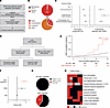 Figure 1
Ruxolitinib exposure is associated with the development of aggressive cSCC. (A) Flowchart depicting the cases included in the international pharmacovigilance study. (B) Pie chart depicting the severity of cSCC reported under ruxolitinib treatment according to the international pharmacovigilance database (VigiBase). (C) IC and PRR measuring the association between cSCC and ruxolitinib versus the indicated comparators. (D) Flowchart depicting patients diagnosed with MPNs included in the study according to their exposure to ruxolitinib and other JAK inhibitors (JAKi). (E) Cumulative incidence of cSCC for JAK inhibitor–treated patients compared with non–JAK inhibitor–treated patients. The Fine-Gray model was used for group comparison. ***P < 0.001. (F) Forest plot depicting factors independently associated with cSCC development in multivariate Fine-Gray analysis. (G) Proportion of localized cSCC (in black) and locally advanced (T3–T4) or metastatic (N+ or M+) cSCC (in red) among MPN patients treated with JAK inhibitor or not. (H) Heatmap depicting the clinical and histological aggressiveness criteria of the 6 aggressive cSCCs developed under JAK inhibitor treatment in patients with MPN. Presence or absence of aggressive criteria is represented in red or black, respectively. Absence of information is represented in gray. TNM status was defined according to AJCC8 classification.
To confirm this association, we interrogated a local cohort of 1,004 patients with MPN, including 136 patients treated with JAK inhibitors (Figure 1D and Supplemental Table 3). Fifteen patients treated with JAK inhibitors developed cSCC (11.0%), whereas only 14 patients not treated with a JAK inhibitor (1.6%) developed cSCC. Median time between JAK inhibitor treatment initiation and cSCC development was 31 months, range[1;80]. Twenty-year cSCC cumulative incidence was 13% for MPN patients treated with JAK inhibitor compared with 3% in the absence of JAK inhibitor (P < 0.001; Figure 1E). In multivariable analysis, JAK inhibitor treatment (HR = 6.52, 95%CI[2.75;15.46], P < 0.001) was associated with cSCC development, independently of age (HR = 1.04, 95%CI[1.02;1.06], P = 0.001) and male sex (HR = 3.52, 95%CI[1.39;8.93], P = 0.008), which are known cSCC risk factors (Figure 1F and Supplemental Table 4). Among the 15 patients with cSCC developed under JAK inhibitor treatment, 6 (40%) presented aggressive cSCC (locally advanced, with lymph node infiltration and/or pulmonary metastasis), while all patients with JAK inhibitor–naive cSCC had localized disease (Figure 1G). Clinical and histological aggressiveness criteria are reported in Figure 1H and Supplemental Table 5.
Ruxolitinib treatment promotes aggressive cSCC in in vitro and in vivo models. After demonstrating the epidemiological association between aggressive cSCC development and ruxolitinib, we used in vitro and in vivo models to evaluate the causal link underlying this clinical observation. In vitro, the A431 cSCC human cell line was resistant to high doses of ruxolitinib (Figure 2A). More interestingly, we observed an overgrowth of A431 cells under increasing doses of ruxolitinib, up to 12.5 μM (Figure 2B). We next used a chemically induced cSCC murine model to validate this effect in vivo and investigate whether ruxolitinib exposure results in an increased rate of premalignant-to-malignant transformation. This model resulted in the development of premalignant lesions (papillomas), of which a subset spontaneously evolved into overt cSCC (Figure 2C). At 22 weeks after first chemical agent application, mice were randomized into 2 groups (n = 9 per group) and treated with vehicle or ruxolitinib. Six weeks after treatment initiation, 11 and 5 cSCC tumors developed in the ruxolitinib and vehicle groups, respectively. cSCC incidence was higher after ruxolitinib exposure (9/9 versus 3/9) (Figure 2D). Median number of premalignant lesions was not different before (23 range[10;40] versus 22 range[13;41]) and after treatment (22 range[12;47] and 24 range[12;37]), in vehicle and ruxolitinib groups, respectively (Figure 2E). This absence of difference between both groups suggests that ruxolitinib effects on cSCC development result from an increased transformation rate of precancerous lesions, rather than de novo oncogenic lesions development. In addition to increasing the incidence of cSCC, ruxolitinib also promoted more aggressive tumors, therefore mimicking our patient cohort observations. Median tumor volume per mouse was bigger in the ruxolitinib group (vol = 337.3 mm3 range[147;720] versus vol = 114.6 mm3 range[49;800]) (Figure 2F). Moreover, 2 ruxolitinib-exposed mice developed lymph node and lung metastasis, while no metastasis was observed in the control group (Figure 2G). Ruxolitinib-promoted cSCC also presented histological aggressiveness criteria (poor differentiation, necrosis, vascular embolism, and histological metastasis) (Figure 2, G–I, and Supplemental Table 6).
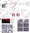 Figure 2
Ruxolitinib treatment promotes aggressive cSCC in vitro and in vivo. (A) Cell growth of A431 cSCC cell line treated with increasing doses of ruxolitinib for 3 days. Data represent mean ± SD of 8 replicates. Statistical significance was determined using 1-way ANOVA. Conditions above the red horizontal line indicate increased proliferation under ruxolitinib compared with DMSO. This experiment was performed 2 times. (B) Cell growth of A431 human cSCC cell line treated with increasing doses of ruxolitinib. Statistical significance was determined using 2-way ANOVA at day 6. Data represent mean ± SD of 8 replicates. This experiment was performed 2 times. (C) Schematic of the chemically induced murine cSCC model. DMBA was applied at weeks 0 and 3 to initiate cSCC carcinogenesis, followed by TPA twice weekly from week 4 to induce hyperplasia and premalignant lesions (papillomas). A subset of papillomas transformed into cSCC during the progression phase. At 22 weeks, mice (n = 9 per group) received vehicle or ruxolitinib (45 mg/kg bid) for 2 weeks. After 3 weeks of treatment interruption, treatment was resumed 1 week before endpoint. Created in BioRender. (D) Cutaneous carcinoma number per mouse in ruxolitinib- versus vehicle-treated mice. Statistical significance was determined using Mann-Whitney test. Data represent mean ± SEM of 9 mice. (E) Papilloma number per mouse under ruxolitinib treatment compared with control mice, before (left) and after treatment initiation (right). Statistical significance was determined using Mann-Whitney test. Data represent mean ± SEM of 9 mice. (F) Cutaneous carcinoma volume in ruxolitinib- versus vehicle-treated mice. Statistical significance was determined using Mann-Whitney test. Data represent mean ± SEM of 9 mice. (G) Heatmap depicting histological aggressiveness criteria of cSCC from ruxolitinib- versus vehicle-treated mice. Presence or absence of aggressiveness criteria is represented in red or black, respectively. (H) Representative H&E micrographs of 3 tumors per group comparing tumor differentiation degree. Scale bars: 500 μm (top), 25 μm (middle and bottom). (I) Representative H&E micrographs showing cSCC histological aggressiveness criteria in ruxolitinib-associated mice tumors. Scale bars: 250 μm (top and bottom left), 1 mm (bottom right).
Altogether, our data highlight the key causal role of ruxolitinib exposure in cutaneous oncogenesis, resulting in the promotion of aggressive secondary cSCC from precancerous lesions.
MAPK pathway activation drives ruxolitinib-mediated cutaneous carcinogenesis. We next explored the underlying mechanisms involved in ruxolitinib-mediated aggressive oncogenesis. GSEA of transcriptomic data obtained from control murine carcinomas (n = 3) compared with murine carcinomas developed under ruxolitinib treatment (n = 4) decreased JAK/STAT signaling in ruxolitinib-mediated tumors (Figure 3A). This analysis also showed that carcinoma oncogenic pathways, skin/keratinocyte proliferation pathways, and pathways involved in poor tumor differentiation were enriched in ruxolitinib tumors, in line with the increased aggressiveness of ruxolitinib-mediated cSCC (Figure 3A). Interestingly, among oncogenic signaling pathways known to be involved in cSCC proliferation, we observed an upregulation of the MAPK pathway in ruxolitinib tumors (Figure 3, A–C). Accordingly, an increased ERK phosphorylation level was detected by immunofluorescence in mice tumors developed under ruxolitinib treatment (Figure 3D). In the ruxolitinib-treated A431 cSCC cell line, ERK phosphorylation level, evaluated by Western blot, was also increased, while STAT3 and STAT5 phosphorylation was decreased (Figure 3E).
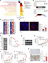 Figure 3
MAPK pathway activation drives ruxolitinib-associated cutaneous carcinogenesis. (A) GSEA of RNA-seq data identifying upregulated oncogenic pathways in cutaneous carcinomas from ruxolitinib-treated (n = 4) versus control mice (n = 3). Gene sets with FDR < 0.25 were considered enriched. NES, normalized enrichment score. (B) Representative GSEA plots showing enrichment of MAPK-related gene sets in ruxolitinib-associated tumors. (C) Heatmaps of the top 20 leading-edge genes from 2 MAPK-related gene sets comparing expression from ruxolitinib (RUXO; n = 4) and control (CTRL; n = 3) mice tumors. Blue and red indicate decreased and increased expression, respectively. (D) Representative immunofluorescence images of phosphorylated ERK (P-ERK; red) in tumors from ruxolitinib- (n = 4) and vehicle-treated mice (n = 4), with quantification of P-ERK fluorescence intensity relative to DAPI. Statistical significance was determined using Mann-Whitney test. Data represent mean ± SD of 4 mice. Scale bars: 10 μm. (E) Phosphorylation level of ERK, STAT3, and STAT5 by Western blot in A431 cSCC cell line treated with ruxolitinib (3.125 μM) compared with DMSO. (F) Growth inhibition curve and IC50 value of A431 cSCC cell line in response to increasing doses of CI-1040 after 3 days of treatment. Data represent mean ± SD of 8 replicates. This experiment was performed 2 times. (G) Phosphorylation level of ERK by Western blot in A431 cSCC cell line treated with DMSO, ruxolitinib (3.125 μM), CI-1040 (3 μM), or CI-1040 and ruxolitinib. (H) Cell growth of A431 cSCC human cell line under DMSO (in black), ruxolitinib (in red, 3.125 μM), CI-1040 (MEKi in gray, 3 μM), or both ruxolitinib and CI-1040 (in orange) treatments. Statistical significance was determined using 2-way ANOVA at day 7. Data represent mean ± SD of 8 replicates. This experiment was performed 2 times. (I and J) Cutaneous carcinoma number per mouse (I) and volume (J) in mice treated with ruxolitinib (in red, 45 mg/kg bid, n = 7), CI-1040 (MEKi in gray, 25 mg/kg qd, n = 6), or both drugs (in orange, n = 7) compared with vehicle-treated control mice (in black, n = 6). Statistical significance was determined using 2-way ANOVA at week 6 after treatment. Data represent mean ± SEM of 6 (vehicle and MEKi groups) or 7 (ruxolitinib and ruxolitinib + MEKi groups) mice. (K) Representative immunochemistry images (left) depicting ERK phosphorylation level in aggressive cSCC developed in ruxolitinib-treated MPN patients (n = 5) compared with matched spontaneously developed cSCC (n = 5), with quantification of P-ERK+ area. Statistical significance was determined using Mann-Whitney test. Data represent mean ± SD of 5 tumors. Scale bars: 100 μm.
To demonstrate the key functional role of MAPK pathway activation in ruxolitinib-mediated cutaneous oncogenesis, we next evaluated the effect of MEK inhibition. In vitro, addition of the MEK inhibitor (MEKi) CI-1040 at its IC50 (Figure 3F) reduced the ruxolitinib-induced increase in ERK phosphorylation (Figure 3G) and cell growth (Figure 3H). In vivo, the addition of CI-1040 to ruxolitinib reduced both cSCC incidence and mean tumor volume (Figure 3, I and J).
To further validate the clinical relevance of MAPK pathway hyperactivation in ruxolitinib-mediated cSCC oncogenesis in patients, we compared ERK phosphorylation levels in 5 aggressive cSCC tumors developed under ruxolitinib to 5 spontaneously developed cSCC, matched according to TNM (tumor, node, metastasis) stage and patient age. Immunochemistry quantification confirmed the increase of ERK phosphorylation level in tumors developed under ruxolitinib (Figure 3K).
Ruxolitinib-mediated cutaneous carcinogenesis results mostly from JAK2 inhibition and represents a pharmacological class effect. Determining whether ruxolitinib-mediated aggressive cSCC is specific to first-generation JAK inhibitors and whether it is related to JAK1 and/or JAK2 inhibition are key clinical objectives. Indeed, many JAK inhibitors are now available for the treatment of MPN and nonmalignant inflammatory and autoimmune disorders. Moreover, whereas ruxolitinib inhibits both JAK1 and JAK2, more selective JAK2 inhibitors, with minimal to no activity against JAK1, have recently become available for the treatment of myelofibrosis. To evaluate the pro-oncogenic effect of JAK inhibitors, we reinterrogated WHO’s pharmacovigilance database to obtain the number of cSCC cases notified over time between 2014 and 2025 under 7 different JAK inhibitors (ruxolitinib, tofacitinib, baricitinib, fedratinib, pacritinib, momelotinib, and itacitinib). The number of reported cases under ruxolitinib and tofacitinib, the oldest JAK inhibitors commercialized since 2012, increased over time (7–193 for ruxolitinib and 3–73 for tofacitinib between 2015 and 2025). However, the magnitude of increase was lower with tofacitinib. For more recently approved JAK inhibitors, such as baricitinib, fedratinib, pacritinib, and momelotinib, a lower number of cutaneous squamous cell carcinoma cases have been reported (17, 1, 1, and 0, respectively) (Supplemental Figure 1 and Supplemental Table 7).
To further assess the potential pro-oncogenic effects of different JAK inhibitors, we subsequently evaluated the effect of JAK1 or JAK2 genetic impairment, as well as the effect of 7 JAK inhibitors (ruxolitinib, fedratinib, itacitinib, momelotinib, pacritinib, tofacitinib, and baricitinib) harboring diverse JAK1/2 selectivity, on cSCC proliferation and signaling in vitro. First, to explore if ruxolitinib-mediated cutaneous carcinogenesis results from JAK1 or JAK2 inhibition, we used genetic inhibition of JAK1 or JAK2. JAK1 genetic impairment using 4 JAK1-directed shRNAs downregulated JAK1 expression without affecting JAK2 expression (Figure 4A) and decreased downstream P-STAT3 signaling (Figure 4B). Genetic inhibition of JAK1 led to an increase of MAPK pathway signaling (Figure 4B) and a mild excess of A341 cell proliferation compared with control shRNA (~1.25- to 1.5-fold) (Figure 4C). JAK2 genetic inhibition downregulated JAK2 expression without affecting JAK1 expression (Figure 4D) and decreased downstream P-STAT5 signaling (Figure 4E). More markedly than JAK1 genetic inhibition, JAK2 genetic impairment using 4 JAK2-directed shRNAs increased P-ERK expression (Figure 4E) and led to an increased cell proliferation of A431 compared with control shRNA (~2- to 5-fold) (Figure 4F).
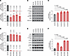 Figure 4
Ruxolitinib-associated cutaneous carcinogenesis results mostly from JAK2 inhibition. (A) RT-qPCR for JAK1 (top) and JAK2 (bottom) expression level in A431 cSCC cell line transduced with either a nontargeting control shRNA or JAK1-directed shRNAs (n = 4). Statistical significance was determined using 1-way ANOVA. Data represent mean ± SD of 4 replicates. This experiment was performed 2 times. (B) Level of JAK1, STAT3, P-STAT3, ERK, P-ERK, ACTIN, and VINCULIN by Western blot in A431 cSCC cell line transduced with either a nontargeting control shRNA or JAK1-directed shRNAs (n = 4) after 2 days of doxycycline-induced shRNA expression. (C) Cell growth of A431 cSCC cell line transduced with either a nontargeting control shRNA (shCTRL) or JAK1-directed shRNAs (n = 4) after 4 days of doxycycline-induced shRNA expression. Statistical significance was determined using 1-way ANOVA. Data represent mean ± SD of 8 replicates. This experiment was performed 2 times. (D) RT-qPCR for JAK1 (top) and JAK2 (bottom) expression level in A431 cSCC cell line transduced with either a nontargeting control shRNA or JAK2-directed shRNAs (n = 4). Statistical significance was determined using 1-way ANOVA. Data represent mean ± SD of 4 replicates. This experiment was performed 2 times. (E) Level of JAK2, STAT5, P-STAT5, ERK, P-ERK, ACTIN, and VINCULIN by Western blot in A431 cSCC cell line transduced with either a nontargeting control shRNA or JAK2-directed shRNAs (n = 4) after 2 days of doxycycline-induced shRNA expression. (F) Cell growth of A431 cSCC cell line transduced with either a nontargeting control shRNA (shCTRL) or JAK2-directed shRNAs (n = 4) after 4 days of doxycycline-induced shRNA expression. Statistical significance was determined using 1-way ANOVA. Data represent mean ± SD of 8 replicates. This experiment was performed 2 times.
For more clinical relevance, we confirmed these results using chemical inhibition. In vitro, JAK1/2 inhibitors (ruxolitinib, momelotinib, baricitinib, and tofacitinib), JAK2-specific inhibitors (fedratinib and pacritinib), and a JAK1-specific inhibitor (itacitinib) increased proliferation of A431 cells (Figure 5A). All JAK inhibitors increased P-ERK expression (Figure 5B) compared with DMSO. The JAK1 inhibitor itacitinib showed the mildest cell proliferation increase, in line with a lower increase in ERK phosphorylation (Figure 5, A and B). In vivo, using our chemically induced mouse model, we demonstrated that the number of precancerous lesions in mice treated with fedratinib, a specific JAK2 inhibitor, was not different from control mice (Figure 5C). However, both the number and volume of cSCC were increased in mice treated with fedratinib compared with control (Figure 5, D and E). Finally, in our MPN cohort, among all cSCCs associated with JAK inhibitors, 86%, 7%, and 7% were related to ruxolitinib, pacritinib, and momelotinib, respectively (Figure 5F). Altogether, these results suggest that the cutaneous pro-oncogenic effect of ruxolitinib is primarily driven by JAK2 inhibition and may be observed across all JAK2 inhibitors, including more recently approved agents.
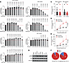 Figure 5
cSCC promotion is a JAK inhibitor–associated class effect. (A) Cell growth of A431 cSCC cell line treated with increasing doses of JAK1/2 inhibitors (ruxolitinib, momelotinib, baricitinib, and tofacitinib), JAK2-specific inhibitors (fedratinib and pacritinib), or JAK1-specific inhibitor (itacitinib) for 3 days. Data represent mean ± SD of 5 replicates. This experiment was performed 2 times. Statistical significance was determined using 1-way ANOVA. Conditions above the red horizontal line indicate increased proliferation under JAK inhibitor treatment compared with DMSO. (B) Phosphorylation level of ERK by Western blot in A431 cSCC cell line treated with JAK1/2 inhibitors (ruxolitinib, 3.125 μM; momelotinib, 0.391 μM; baricitinib, 1.562 μM; tofacitinib, 3.125 μM), JAK2-specific inhibitors (fedratinib, 0.097 μM; pacritinib, 0.391 μM), or JAK1-specific inhibitor (itacitinib, 3.125 μM). (C) Papilloma number per mouse under fedratinib (n = 15) treatment compared with control mice (n = 15), before (left) and after treatment initiation (right). Statistical significance was determined using Mann-Whitney test. Data represent mean ± SEM of 14 (vehicle group) or 13 (fedratinib group) mice. (D) Cutaneous carcinoma number per mouse in mice treated with fedratinib (n = 15) compared with vehicle-treated control mice (n = 15). Statistical significance was determined using Mann-Whitney test. Data represent mean ± SEM. (E) Cutaneous carcinoma volume in mice treated with fedratinib (n = 15) compared with vehicle-treated control mice (n = 15). Statistical significance was determined using Mann-Whitney test. Data represent mean ± SEM. (F) Pie chart reporting the proportion of all cSCCs (left) and aggressive cSCCs (right) under different JAK inhibitors in a cohort of 1,004 patients with MPN. RUXO, ruxolitinib; MOMELO, momelotinib; PACRI, pacritinib.
Ruxolitinib-induced MAPK pathway activation is mediated by the COT kinase. To better understand the mechanism underlying ruxolitinib-induced MAPK activation, we first evaluated the molecular landscape of ruxolitinib-mediated cSCC in patients since RAS pathway mutations are key drivers of cSCC oncogenesis. Next-generation sequencing (NGS) of 6 aggressive cSCCs developed under ruxolitinib treatment for MPN management revealed a high incidence of TP53 mutations (5/6), in line with the genomic landscape of non–ruxolitinib-mediated cSCC (25). Mutations in HRAS, known to be the most frequently mutated RAS isoform in cSCC, were not observed (Supplemental Figure 2, A and B). Within the rest of the RAS pathway genes, only 1 NRAS variant, c.419C>T p.P140L, of uncertain significance was detected in 1 tumor. Of note, JAK2 V617F mutation, the main driver of MPN, was detected with a low variant allele frequency (1%, 8.7%, and 2.2%) in cSCC obtained from 3 patients diagnosed with polycythemia vera. JAK2 V617F allelic burden was very high (90%, 95%, and 87%) in peripheral blood for all of them, suggesting minimal blood contamination rather than carcinoma cells harboring this mutation (Supplemental Figure 2C). Altogether, these findings argue for a nongenetic mechanism for MAPK activation in ruxolitinib-mediated cSCC.
Thus, to better understand the mechanism underlying MAPK pathway activation under ruxolitinib treatment, we first performed an unbiased proteomic analysis in mice tumors associated with ruxolitinib (n = 5) compared with control tumors (n = 4). Among the 5,743 proteins quantified, 1,100 proteins were differentially expressed between ruxolitinib and control tumors (Supplemental Table 8). GSEA of differential protein levels between ruxolitinib- and vehicle-treated tumors showed significant enrichment of cell cycle– and RTK signaling–related Gene Ontology terms in ruxolitinib-treated tumors, in line with keratinocyte proliferation induced by MAPK pathway hyperactivation in the context of ruxolitinib treatment (Supplemental Figure 2D). Cell cycle analysis of the ruxolitinib-treated A431 cell line confirmed the increased cell cycling of cSCC cells in the context of ruxolitinib treatment, with an increased proportion of cells in S and G2/M compared with DMSO-treated cells (Supplemental Figure 2E). Moreover, the addition of MEKi treatment rescued the effect of ruxolitinib on the cell cycle (Supplemental Figure 2E). Additionally, a phosphoproteomic analysis in mice tumors associated with ruxolitinib (n = 5) compared with control (n = 4) revealed that among the 9,654 phosphosites identified, 1,930 (corresponding to 801 proteins) differed between ruxolitinib and control tumors (Supplemental Table 9).
We next performed kinase enrichment analysis (KEA) (26) using differentially abundant phosphosites between ruxolitinib and control tumors. This analysis showed an enrichment of cell cycle–related kinases, such as CDK1/2, as well as multiple kinases downstream of MAP3Ks, such as ERK1/2, JNK1/2/3, and P38A/B/D (Figure 6A). This observation confirms the likely activation of the MAPK pathway in tumors treated with ruxolitinib. However, RAS and RAF activating phosphosites, known to be involved in the classical path toward MAPK pathway activation, were not enriched in the context of ruxolitinib exposure, thereby indicating a noncanonical mechanism of MAPK activation by ruxolitinib (Supplemental Table 10). Alternative kinase prediction tools (27) suggested an activation of the MAP3K COT (also named TPL2 or MAP3K8) (Figure 6B). Interestingly, COT has been shown to be an upstream activator of ERK, JNK, and P38 kinases, which is consistent with the broad activation of these kinases observed by KEA (Figure 6C). COT is an oncoprotein kinase shown to play a key role in melanoma resistance to BRAF inhibitors (28). Based on the previously described role of COT (28) and our phosphoproteomic results, we hypothesized that COT is key for alternative MAPK pathway activation, subsequently resulting in ruxolitinib-mediated aggressive cSCC. To validate this hypothesis, we first confirmed that the COT Thr290 phosphosite, known to be an activating phosphosite in the activation loop of the kinase domain of COT (29), was increased under ruxolitinib treatment of the A431 cSCC cell line, while RAF phosphorylation was decreased (Figure 6D). To explore the functional impact of COT kinase activation on ruxolitinib-mediated cSCC proliferation, we used 2 small-molecule COT inhibitors (TPL2 kinase inhibitor 1 and COT inhibitor 2) at their IC50 (Figure 7A). Addition of either COT inhibitor to ruxolitinib reduced the ruxolitinib-induced hyperproliferation phenotype (Figure 7B). Similarly, COT genetic impairment using 3 COT-directed shRNAs (Figure 7C) also dampened the ruxolitinib-induced pro-proliferative effects on cSCC cells (Figure 7D). Finally, to evaluate whether COT phosphorylation in Thr290 is critical for the ruxolitinib effect, we transduced the A431 cell line with (a) an shRNA against COT to block endogenous COT and (b) an exogenous codon-optimized WT or threonine-to-alanine (T290A) mutant form of COT. ERK phosphorylation and A431 cell proliferation increased upon ruxolitinib exposure in COT-WT–expressing cells (Figure 7, E and F). Interestingly, expression of the T290A-COT mutant impaired ruxolitinib-mediated ERK phosphorylation increase and blocked its effect on cell proliferation, suggesting that COT T290 phosphorylation is critical for ruxolitinib-mediated MAPK activation and cSCC cell proliferation. Overall, our results pinpoint a COT-mediated alternative MAPK pathway activation mechanism as being responsible for ruxolitinib-mediated aggressive cSCC development.
 Figure 6
Ruxolitinib-induced MAPK pathway activation is associated with COT kinase activation. (A) KEA in cutaneous carcinomas developed by ruxolitinib-treated mice (n = 5) compared with control mice (n = 4). Kinases are considered enriched if log2 fold change > 1 and P value < 0.05. MAPKs significantly enriched are highlighted in red. (B) Robust kinase activity inference KEA in cutaneous carcinomas developed by ruxolitinib-treated mice (n = 5) compared with control mice (n = 4). (C) Network of COT kinase substrates within the MAPK pathway. COT is highlighted in red, and its MAPK downstream signaling is in blue. Other MAPK-activating proteins are in gray. (D) Phosphorylation level of RAF, COT, and ERK by Western blot in A431 cSCC cell line treated with ruxolitinib (3.125 μM) compared with DMSO.
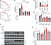 Figure 7
COT kinase phosphorylation mediates ruxolitinib-induced cutaneous carcinogenesis. (A) Growth inhibition curve and IC50 value of A431 cSCC cell line in response to increasing doses of TPL2i-1 and COTi-2 after 3 days of treatment. Data represent mean ± SD of 8 replicates. This experiment was performed 2 times. (B) Cell growth of A431 cSCC cell line after 6 days of treatment with DMSO (in black), ruxolitinib (in red, 3.125 μM), COT inhibitors (in gray, TPL2i-1 or COTi-2 at 7 μM), or ruxolitinib and COT inhibitors (in dark red). Statistical significance was determined using 1-way ANOVA. Data represent mean ± SD of 8 replicates. This experiment was performed 2 times. (C) RT-qPCR for COT expression level in A431 cSCC cell line transduced with either a nontargeting control shRNA or COT-directed shRNAs (n = 3). Statistical significance was determined using 1-way ANOVA. Data represent mean ± SD of 4 replicates. (D) Cell growth of A431 cSCC cell line transduced with either a nontargeting control shRNA (shCTRL) or COT-directed shRNAs (n = 3) after 4 days of DMSO or ruxolitinib treatment (3.125 μM). Statistical significance was determined using 1-way ANOVA. Data represent mean ± SD of 8 replicates. This experiment was performed 2 times. (E) ERK phosphorylation and COT expression levels by Western blot in A431 cSCC cell line transduced with (a) either a nontargeting control shRNA (shCTRL) or COT-directed shRNA and (b) either a codon-optimized WT COT construct or a T290A-COT mutant and treated with ruxolitinib (3.125 μM) compared with DMSO. (F) Cell growth of A431 cSCC cell line transduced with (a) either a nontargeting control shRNA (shCTRL) or COT-directed shRNA and (b) either a WT COT construct or a T290A-COT mutant after 4 days of DMSO or ruxolitinib treatment (3.125 μM). Statistical significance was determined using Mann-Whitney test. Data represent mean ± SD of 8 replicates. This experiment was performed 2 times.
Ruxolitinib-induced COT kinase activation involves a posttranslational regulation mechanism. We next set to further explore the link between JAK inhibition and COT activation. To get more mechanistic insight, we explored 2 hypotheses. Indeed, JAKs are known to impact cellular proliferation through at least 2 distinct mechanisms: transcriptional regulation through the STAT downstream factors or more directly through modulating protein phosphorylation. We thus first evaluated the effect of JAK inhibition on COT transcription in vitro in the A431 cSCC cell line. COT transcript levels were similar between ruxolitinib and control cells, as shown by RT-qPCR at 3 different time points (Figure 8A). We next hypothesized that phosphorylation changes under JAK inhibitor treatment might be involved in COT activation. In line with this hypothesis, we observed that phosphorylation of COT at T290 is an early event occurring only 15 min after ruxolitinib treatment, suggesting that ruxolitinib-mediated COT activation involves a posttranslational mechanism (Figure 8B). COT posttranslational regulation is still largely unknown. We hypothesized that, under basal conditions, JAKs phosphorylate and activate a phosphatase, which in turn dephosphorylates COT at T290, thereby reducing its activity. To test this hypothesis, we examined COT and ERK phosphorylation as well as cSCC proliferation in the presence of a phosphatase inhibitor known to target threonine and serine phosphatases. At all tested doses, threonine/serine phosphatase inhibition recapitulated the effects observed with ruxolitinib, resulting in increased COT phosphorylation at T290, enhanced ERK phosphorylation, and increased cell proliferation (Figure 8, C and D). Taken together, these results suggest that ruxolitinib might repress a COT-inactivating phosphatase, thereby promoting activation of the MAPK pathway and enhancing cell proliferation (Figure 8E).
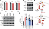 Figure 8
Ruxolitinib-induced COT kinase activation involves a posttranslational regulation mechanism. (A) RT-qPCR for COT expression level in A431 cSCC cell line treated with ruxolitinib (3.125 μM) compared with control at the indicated time points. Statistical significance was determined using Mann-Whitney test. Data represent mean ± SD of 4 replicates. (B) Phosphorylation level of COT and ERK by Western blot in A431 cSCC cell line treated with ruxolitinib (3.125 μM) compared with DMSO at the indicated time points. (C) Phosphorylation level of COT and ERK by Western blot in A431 cSCC cell line treated with ruxolitinib (3.125 μM) or increased doses of serine/threonine phosphatase inhibitor (PI) compared with DMSO. (D) Cell growth of A431 cSCC cell line after 6 days of ruxolitinib treatment (3.125 μM) and increased doses of serine/threonine PI compared with DMSO. Statistical significance was determined using 1-way ANOVA. Data represent mean ± SD of 8 replicates. This experiment was performed 2 times. (E) Schematic of the proposed model for MAPK pathway activation mechanism under ruxolitinib treatment. Activated proteins are in red, and inactive ones are represented in gray. Created in BioRender.
Ruxolitinib-mediated oncogenic effects result in an increased risk of melanoma. Melanoma is another skin cancer type exhibiting a high dependency on the MAPK pathway (30). Given ruxolitinib pro-oncogenic effects mediated by MAPK pathway activation and resulting in an increased risk of aggressive cSCC, we further explored ruxolitinib effects on melanoma. In vitro, using 2 different melanoma cell lines (B16-OVA and YUMMER1.7), we showed that ruxolitinib-induced cell proliferation compared with DMSO conditions (Figure 9A). We then confirmed these results in vivo. Indeed, in 2 different melanoma mouse models (B16-OVA and YUMMER1.7), tumor volume change from baseline was higher in the ruxolitinib-exposed mice group compared with the control group after 10 days of treatment (Figure 9B). Finally, we interrogated our MPN cohort to evaluate melanoma incidence. Importantly, melanoma incidence was higher for patients who received JAK inhibitor treatment (2.9% versus 0.35%) (Figure 9C). Altogether, these results suggest a potential role of ruxolitinib exposure in melanoma oncogenesis, and further epidemiological and mechanistic studies should explore this finding and determine clinical guidelines for patients exposed to JAK inhibitors.
 Figure 9
Ruxolitinib-mediated oncogenic effects result in an increased risk of melanoma. (A) Cell growth of B16-OVA and YUMMER1.7 melanoma cell lines treated with increasing doses of ruxolitinib for 3 days. Statistical significance was determined using 1-way ANOVA. Data represent mean ± SD of 8 replicates. This experiment was performed 2 times. Conditions above the red horizontal line indicate increased proliferation under ruxolitinib compared with DMSO. (B) Tumor volume change from baseline in 2 melanoma mouse models (B16-OVA and YUMMER1.7) treated with ruxolitinib (90 mg/kg bid; n = 6 for B16-OVA and n = 4 for YUMMER1.7) compared with vehicle-treated control mice (n = 5 for B16-OVA and n = 4 for YUMMER1.7) after 10 days of treatment. Statistical significance was determined using Mann-Whitney test. Data represent mean ± SEM. (C) Proportion of melanoma (in red) among MPN patients treated with JAK inhibitor or not.


Copyright © 2026 American Society for Clinical Investigation
ISSN: 0021-9738 (print), 1558-8238 (online)