
Advertisement
Research ArticleImmunologyInfectious diseaseInflammation
Open Access |  10.1172/JCI198863
Interferon-λ inhibits immunopathology and boosts T cell–mediated immunity in a mouse model of mucosal viral infection
Emily Feng,1,2 Alexa Mansour,1,2 Elizabeth Balint,1,2 Alyssa Mark,1,2 Sofia Tramonte,1,2 Jonathan K. Monteiro,1,2 Madison Read,1,2 Marie Joe Adaimy,1,2 Zhou Xing,1,2 Matthias Mack,3 Sergei V. Kotenko,4 Deanna M. Santer,5 and Ali A. Ashkar1,2,6
1McMaster Immunology Research Centre, Department of Medicine and
2Michael G. DeGroote Institute for Infectious Disease Research, McMaster University, Hamilton, Ontario, Canada.
3University Hospital Regensburg, Department of Nephrology, Regensburg, Germany.
4Department of Microbiology, Biochemistry, and Molecular Genetics, Rutgers New Jersey Medical School, Newark, New Jersey, USA.
5Department of Immunology, University of Manitoba, Winnipeg, Manitoba, Canada.
6Centre for Discovery in Cancer Research, McMaster University, Hamilton, Ontario, Canada.
Address correspondence to: Ali A. Ashkar, MDCL 4015, 1280 Main St. W, Hamilton, Ontario, L8S 4L8, Canada. Phone: 905.525.9140; Email: ashkara@mcmaster.ca.
Find articles by Feng, E. in: PubMed | Google Scholar
1McMaster Immunology Research Centre, Department of Medicine and
2Michael G. DeGroote Institute for Infectious Disease Research, McMaster University, Hamilton, Ontario, Canada.
3University Hospital Regensburg, Department of Nephrology, Regensburg, Germany.
4Department of Microbiology, Biochemistry, and Molecular Genetics, Rutgers New Jersey Medical School, Newark, New Jersey, USA.
5Department of Immunology, University of Manitoba, Winnipeg, Manitoba, Canada.
6Centre for Discovery in Cancer Research, McMaster University, Hamilton, Ontario, Canada.
Address correspondence to: Ali A. Ashkar, MDCL 4015, 1280 Main St. W, Hamilton, Ontario, L8S 4L8, Canada. Phone: 905.525.9140; Email: ashkara@mcmaster.ca.
Find articles by Mansour, A. in: PubMed | Google Scholar
1McMaster Immunology Research Centre, Department of Medicine and
2Michael G. DeGroote Institute for Infectious Disease Research, McMaster University, Hamilton, Ontario, Canada.
3University Hospital Regensburg, Department of Nephrology, Regensburg, Germany.
4Department of Microbiology, Biochemistry, and Molecular Genetics, Rutgers New Jersey Medical School, Newark, New Jersey, USA.
5Department of Immunology, University of Manitoba, Winnipeg, Manitoba, Canada.
6Centre for Discovery in Cancer Research, McMaster University, Hamilton, Ontario, Canada.
Address correspondence to: Ali A. Ashkar, MDCL 4015, 1280 Main St. W, Hamilton, Ontario, L8S 4L8, Canada. Phone: 905.525.9140; Email: ashkara@mcmaster.ca.
Find articles by Balint, E. in: PubMed | Google Scholar
1McMaster Immunology Research Centre, Department of Medicine and
2Michael G. DeGroote Institute for Infectious Disease Research, McMaster University, Hamilton, Ontario, Canada.
3University Hospital Regensburg, Department of Nephrology, Regensburg, Germany.
4Department of Microbiology, Biochemistry, and Molecular Genetics, Rutgers New Jersey Medical School, Newark, New Jersey, USA.
5Department of Immunology, University of Manitoba, Winnipeg, Manitoba, Canada.
6Centre for Discovery in Cancer Research, McMaster University, Hamilton, Ontario, Canada.
Address correspondence to: Ali A. Ashkar, MDCL 4015, 1280 Main St. W, Hamilton, Ontario, L8S 4L8, Canada. Phone: 905.525.9140; Email: ashkara@mcmaster.ca.
Find articles by Mark, A. in: PubMed | Google Scholar
1McMaster Immunology Research Centre, Department of Medicine and
2Michael G. DeGroote Institute for Infectious Disease Research, McMaster University, Hamilton, Ontario, Canada.
3University Hospital Regensburg, Department of Nephrology, Regensburg, Germany.
4Department of Microbiology, Biochemistry, and Molecular Genetics, Rutgers New Jersey Medical School, Newark, New Jersey, USA.
5Department of Immunology, University of Manitoba, Winnipeg, Manitoba, Canada.
6Centre for Discovery in Cancer Research, McMaster University, Hamilton, Ontario, Canada.
Address correspondence to: Ali A. Ashkar, MDCL 4015, 1280 Main St. W, Hamilton, Ontario, L8S 4L8, Canada. Phone: 905.525.9140; Email: ashkara@mcmaster.ca.
Find articles by Tramonte, S. in: PubMed | Google Scholar
1McMaster Immunology Research Centre, Department of Medicine and
2Michael G. DeGroote Institute for Infectious Disease Research, McMaster University, Hamilton, Ontario, Canada.
3University Hospital Regensburg, Department of Nephrology, Regensburg, Germany.
4Department of Microbiology, Biochemistry, and Molecular Genetics, Rutgers New Jersey Medical School, Newark, New Jersey, USA.
5Department of Immunology, University of Manitoba, Winnipeg, Manitoba, Canada.
6Centre for Discovery in Cancer Research, McMaster University, Hamilton, Ontario, Canada.
Address correspondence to: Ali A. Ashkar, MDCL 4015, 1280 Main St. W, Hamilton, Ontario, L8S 4L8, Canada. Phone: 905.525.9140; Email: ashkara@mcmaster.ca.
Find articles by Monteiro, J. in: PubMed | Google Scholar
1McMaster Immunology Research Centre, Department of Medicine and
2Michael G. DeGroote Institute for Infectious Disease Research, McMaster University, Hamilton, Ontario, Canada.
3University Hospital Regensburg, Department of Nephrology, Regensburg, Germany.
4Department of Microbiology, Biochemistry, and Molecular Genetics, Rutgers New Jersey Medical School, Newark, New Jersey, USA.
5Department of Immunology, University of Manitoba, Winnipeg, Manitoba, Canada.
6Centre for Discovery in Cancer Research, McMaster University, Hamilton, Ontario, Canada.
Address correspondence to: Ali A. Ashkar, MDCL 4015, 1280 Main St. W, Hamilton, Ontario, L8S 4L8, Canada. Phone: 905.525.9140; Email: ashkara@mcmaster.ca.
Find articles by Read, M. in: PubMed | Google Scholar
1McMaster Immunology Research Centre, Department of Medicine and
2Michael G. DeGroote Institute for Infectious Disease Research, McMaster University, Hamilton, Ontario, Canada.
3University Hospital Regensburg, Department of Nephrology, Regensburg, Germany.
4Department of Microbiology, Biochemistry, and Molecular Genetics, Rutgers New Jersey Medical School, Newark, New Jersey, USA.
5Department of Immunology, University of Manitoba, Winnipeg, Manitoba, Canada.
6Centre for Discovery in Cancer Research, McMaster University, Hamilton, Ontario, Canada.
Address correspondence to: Ali A. Ashkar, MDCL 4015, 1280 Main St. W, Hamilton, Ontario, L8S 4L8, Canada. Phone: 905.525.9140; Email: ashkara@mcmaster.ca.
Find articles by Adaimy, M. in: PubMed | Google Scholar
1McMaster Immunology Research Centre, Department of Medicine and
2Michael G. DeGroote Institute for Infectious Disease Research, McMaster University, Hamilton, Ontario, Canada.
3University Hospital Regensburg, Department of Nephrology, Regensburg, Germany.
4Department of Microbiology, Biochemistry, and Molecular Genetics, Rutgers New Jersey Medical School, Newark, New Jersey, USA.
5Department of Immunology, University of Manitoba, Winnipeg, Manitoba, Canada.
6Centre for Discovery in Cancer Research, McMaster University, Hamilton, Ontario, Canada.
Address correspondence to: Ali A. Ashkar, MDCL 4015, 1280 Main St. W, Hamilton, Ontario, L8S 4L8, Canada. Phone: 905.525.9140; Email: ashkara@mcmaster.ca.
Find articles by Xing, Z. in: PubMed | Google Scholar
1McMaster Immunology Research Centre, Department of Medicine and
2Michael G. DeGroote Institute for Infectious Disease Research, McMaster University, Hamilton, Ontario, Canada.
3University Hospital Regensburg, Department of Nephrology, Regensburg, Germany.
4Department of Microbiology, Biochemistry, and Molecular Genetics, Rutgers New Jersey Medical School, Newark, New Jersey, USA.
5Department of Immunology, University of Manitoba, Winnipeg, Manitoba, Canada.
6Centre for Discovery in Cancer Research, McMaster University, Hamilton, Ontario, Canada.
Address correspondence to: Ali A. Ashkar, MDCL 4015, 1280 Main St. W, Hamilton, Ontario, L8S 4L8, Canada. Phone: 905.525.9140; Email: ashkara@mcmaster.ca.
Find articles by
Mack, M.
in:
PubMed
|
Google Scholar
|

1McMaster Immunology Research Centre, Department of Medicine and
2Michael G. DeGroote Institute for Infectious Disease Research, McMaster University, Hamilton, Ontario, Canada.
3University Hospital Regensburg, Department of Nephrology, Regensburg, Germany.
4Department of Microbiology, Biochemistry, and Molecular Genetics, Rutgers New Jersey Medical School, Newark, New Jersey, USA.
5Department of Immunology, University of Manitoba, Winnipeg, Manitoba, Canada.
6Centre for Discovery in Cancer Research, McMaster University, Hamilton, Ontario, Canada.
Address correspondence to: Ali A. Ashkar, MDCL 4015, 1280 Main St. W, Hamilton, Ontario, L8S 4L8, Canada. Phone: 905.525.9140; Email: ashkara@mcmaster.ca.
Find articles by Kotenko, S. in: PubMed | Google Scholar
1McMaster Immunology Research Centre, Department of Medicine and
2Michael G. DeGroote Institute for Infectious Disease Research, McMaster University, Hamilton, Ontario, Canada.
3University Hospital Regensburg, Department of Nephrology, Regensburg, Germany.
4Department of Microbiology, Biochemistry, and Molecular Genetics, Rutgers New Jersey Medical School, Newark, New Jersey, USA.
5Department of Immunology, University of Manitoba, Winnipeg, Manitoba, Canada.
6Centre for Discovery in Cancer Research, McMaster University, Hamilton, Ontario, Canada.
Address correspondence to: Ali A. Ashkar, MDCL 4015, 1280 Main St. W, Hamilton, Ontario, L8S 4L8, Canada. Phone: 905.525.9140; Email: ashkara@mcmaster.ca.
Find articles by
Santer, D.
in:
PubMed
|
Google Scholar
|
1McMaster Immunology Research Centre, Department of Medicine and
2Michael G. DeGroote Institute for Infectious Disease Research, McMaster University, Hamilton, Ontario, Canada.
3University Hospital Regensburg, Department of Nephrology, Regensburg, Germany.
4Department of Microbiology, Biochemistry, and Molecular Genetics, Rutgers New Jersey Medical School, Newark, New Jersey, USA.
5Department of Immunology, University of Manitoba, Winnipeg, Manitoba, Canada.
6Centre for Discovery in Cancer Research, McMaster University, Hamilton, Ontario, Canada.
Address correspondence to: Ali A. Ashkar, MDCL 4015, 1280 Main St. W, Hamilton, Ontario, L8S 4L8, Canada. Phone: 905.525.9140; Email: ashkara@mcmaster.ca.
Find articles by Ashkar, A. in: PubMed | Google Scholar
Published October 15, 2026 - More info
J Clin Invest. 2026;136(20):e198863. https://doi.org/10.1172/JCI198863.
© 2026 Feng et al. This work is licensed under the Creative Commons Attribution 4.0 International License. To view a copy of this license, visit http://creativecommons.org/licenses/by/4.0/.
Received: August 7, 2025; Accepted: August 18, 2026

Related article:
Abstract
Authors
Pablo Penaloza-MacMaster
-
Results
IFN-λ treatment inhibits tissue pathology and viral dissemination and promotes viral clearance. IFN-λ is a powerful player in antiviral defence on mucosal surfaces, but its broader immunomodulatory roles remain underexplored. We sought to investigate whether therapeutic mucosal delivery of IFN-λ could inhibit inflammation without compromising the development of protective immune responses in the genital mucosa following intravaginal HSV-2 strain 333 infection. To investigate the functions of IFN-λ, we administered recombinant IFN-λ2 intravaginally 1 day postinfection (DPI) in Ifnar–/– mice that lack type I–IFN signaling but produce IFN-λ2/3 at levels comparable to WT mice (Figure 1A and Supplemental Figure 1A; supplemental material available online with this article; https://doi.org/10.1172/JCI198863DS1). Ifnar–/– mice serve as a model for virus-induced hyperinflammation, as they develop severe vaginal tissue pathology 3 DPI due to elevated IL-6 production (24). IFN-λ–treated mice showed reduced viral loads and increased survival compared with control-treated mice, confirming that IFN-λ can confer protection against HSV-2 infection in the absence of type I–IFN signaling (Figure 1, B and C). Strikingly, administration of IFN-λ treatment markedly reduced vaginal tissue pathology in Ifnar–/– mice 3 DPI (Figure 1, D–F). This correlated with reduced viral titers at 3 DPI and a near-complete abrogation of IL-6 production (Figure 1, G and H). We further assessed whether another subtype, IFN-λ3, would have a similar effect. IFN-λ3–treated mice similarly showed reduced viral titers and IL-6 levels (Supplemental Figure 1, B and C).
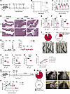 Figure 1
IFN-λ treatment inhibits tissue pathology and viral dissemination and promotes viral clearance. (A) Ifnar–/– mice were infected with 103 PFU HSV-2 333 and administered PBS or IFN-λ (n = 8, 11). Data representative of 2 pooled experiments. (B) Survival rates between PBS- and IFN-λ–treated mice. (C) Viral titers in vaginal washes. (D–J) Ifnar–/– mice were infected with 104 PFU HSV-2 strain 333 and administered PBS or IFN-λ. Data representative of 2 independent experiments. (D) Representative vaginal histology 3 DPI. Scale bars: 5 ×, 100 μm; 10 ×, 200 μm, respectively. (E) Percentage epithelial loss (n = 8, 9). (F) Pathology score. (G) Viral titers in vaginal washes 3 DPI. (H) IL-6 levels in vaginal washes (n = 10, 9). (I) IFN-γ levels in vaginal washes (n = 5, 5). mice. (J) Representative distal colon images and rates of toxic megacolon 5 DPI, pooled from 3 independent pooled experiments. (K–M) Ifnar–/– mice were infected with 103 PFU HSV-2 333 and administered PBS or IFN-λ (n = 5, 5). Data representative of 2 independent experiments. Viral titers in the (K) spine, (L) distal colon, and (M) vaginal tissue 6 DPI. (N) Primary cortical neurons from WT mice were stimulated with indicated concentrations of IFN-λ2 or IFN-β and infected with HSV-2. Infectious viral loads at the indicated time points in triplicate. (O) Ifnar–/– mice were infected with 104 PFU HSV-2 TK– and administered PBS or IFN-λ. Data shown are representative of 2 independent experiments. (P) Viral titers in vaginal washes (n = 5, 5). (Q) Rates of developing severe genital lesions. (R) Representative of genital lesion images. *P < 0.05, **P < 0.01, and ****P < 0.0001. Statistical significance determined by Kaplan-Meier survival analysis (B), repeated-measure 2-way ANOVA with Šídák’s multiple comparisons test (C, H, I, and P), ordinary 2-way ANOVA with Dunnett’s multiple comparisons test (N), and 2-tailed Student’s t test (E–G and K–M).
We further investigated the role of IFN-λ in inhibiting IL-6–mediated pathology by infecting mice deficient in IFN-λR (Ifnlr1–/–) with HSV-2. Ifnlr1–/– mice exhibited increased epithelial damage, viral titers, and IL-6, although to a lesser extent compared to Ifnar–/– mice, confirming that IFN-λ signaling suppresses IL-6 production (Supplemental Figure 2, A–E). Overall, these findings demonstrate that type III IFNs possess both antiviral and tissue-protective effects in the genital mucosa.
We have previously demonstrated that NK cell–derived IFN-γ can attenuate IL-6–driven pathology (29). As Ifnar–/– mice have deficient NK-cell–derived IFN-γ production at 2 DPI, we investigated whether IFN-λ administration could reduce tissue pathology by regulating NK cell function (27, 30). However, IFN-λ treatment did not induce IFN-γ production at 2 DPI compared with PBS-treated mice, and Ifnlr1–/– mice were not deficient in IFN-γ production compared with WT mice (Figure 1I and Supplemental Figure 2F). These results suggest that IFN-λ therapy can attenuate cytokine-driven tissue pathology independently of regulating NK cell responses.
HSV-2 genital infection causes lethal toxic megacolon through viral spread to the enteric nervous system (31). We next assessed whether IFN-λ treatment could inhibit this viral dissemination. IFN-λ treatment reduced the incidence of toxic megacolon at 5 DPI in Ifnar–/– mice (Figure 1J). This correlated with a trend toward reduced viral loads in the thoracic-lumbar spine and significantly reduced viral loads in the distal colon and vaginal tissue (Figure 1, K–M), suggesting that IFN-λ treatment inhibits not only local viral replication, but also viral spread. To model whether IFN-λ can directly induce antiviral signaling in neurons, we infected primary mouse cortical neurons derived from WT mice with HSV-2. Consistent with previous studies (32), we found that IFN-λ could not inhibit HSV-2 replication in primary mouse cortical neurons in vitro compared with IFN-β (Figure 1N). Thus, IFN-λ protects against neurodissemination independently of antiviral signaling in neurons.
To further assess the role of type III IFNs in inhibiting neurodissemination, WT and Ifnlr1–/– mice were infected with low and high doses of HSV-2. At a low inoculum of 100 PFU, Ifnlr1–/– mice were more susceptible to HSV-2 infection than WT mice (Supplemental Figure 2G). This difference in survival correlated with the ability to successfully establish infection, as no infectious virus was detected in the vaginal washes from infected WT mice, whereas virus was detected in Ifnlr1–/– mice (Supplemental Figure 2H). At a higher viral dose, Ifnlr1–/– mice had significantly higher viral titers in the vaginal washes only at 1 DPI, but not at later time points of infection (Supplemental Figure 2I). Additionally, we observed a trend toward increased viral titers in the spine, distal colon, and whole vaginal tissue in Ifnlr1–/– mice compared with WT mice (Supplemental Figure 2, J–L). These findings suggest that endogenously produced IFN-λ plays a critical role in inhibiting the establishment of viral infection and limiting early viral replication, but is less important in limiting viral spread at later time points of infection. Given the contribution of neurodissemination to disease mortality during HSV-2 infection, we sought to determine whether the tissue-protective effects of IFN-λ could be observed during a non-neurotropic mucosal infection.
Infection with thymidine kinase knockout HSV-2 (HSV-2 TK–) typically results in an asymptomatic infection in immunocompetent mice due to its inability to replicate in neuronal cells (33, 34). To assess the protective functions of IFN-λ in the absence of a neurotropic infection, we administered IFN-λ during HSV-2 TK– infection (Figure 1O). Although infectious virus was cleared in the vaginal washes by 7 DPI, Ifnar–/– mice developed severe genital lesions following HSV-2 TK– infection 3–4 weeks after infection, which was prevented by IFN-λ treatment (Figure 1, P–R). These findings suggest that Ifnar–/– mice exhibit defective viral clearance and that the protective effects of IFN-λ include not only preventing neurodissemination but also enhancing viral clearance.
The absence of type I–IFN signaling impairs local T cell immunity. CD4+ T cells are known to be the primary source of IFN-γ at later time points of HSV-2 infection, where their production of IFN-γ contributes to viral clearance, prevents infection of sensory neurons that innervate the vaginal mucosa, and promotes the recruitment of CD8+ T cells (26, 35–38). Although type I–IFN signaling is well known to be critical for NK cell–derived IFN-γ production, it is not clear whether it is also necessary for CD4+ T cell–dependent IFN-γ production. We evaluated whether Ifnar–/– mice had any deficiencies in their CD4+ T cell responses to HSV-2 infection that contributed to their greater susceptibility to HSV-2 infection compared with WT mice (Figure 2A). We observed that Ifnar–/– mice had higher viral titers starting 3 DPI and produced no IFN-γ at 5 DPI compared with WT mice (Figure 2, B and C). In contrast, Ifnlr1–/– mice produced similar levels of IFN-γ compared with WT mice (Supplemental Figure 2M). The lack of IFN-γ in Ifnar–/– mice correlated with a significant reduction in the recruitment of T cells, including CD4+, CD8+, and TCRγδ+ T cells, but not total immune cells to the vaginal tissue during HSV-2 infection compared with WT mice (Figure 2, D–H). Furthermore, we observed that the impaired recruitment of T cells correlated with reduced production of CXCL9 and CXCL10 in Ifnar–/– mice at 2 and 3 DPI (Figure 2, I and J). These results suggest that the absence of type I–IFN signaling impairs lymphocyte recruitment during genital HSV-2 infection.
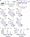 Figure 2
Absence of type I IFNs impairs adaptive immunity to HSV-2 infection. (A) WT and Ifnar–/– mice were infected with 104 PFU HSV-2 333, and the vaginal tracts were collected 5 DPI (n = 5, 5). Data representative of 2 independent experiments. (B) Viral titers in the vaginal washes. (C) IFN-γ levels in vaginal washes 5 DPI. (D) Total number of CD45+ cells in the vaginal tract. (E) Total number of T cells (CD45+/Ly6G–/CD3+). (F) Total number of CD4+ T cells (CD45+/Ly6G–/CD3+/TCRβ+/TCRγδ–/CD8–/CD4+). (G) Total number of CD8+ T cells (CD45+/Ly6G–/CD3+/TCRβ+/TCRγδ–/CD8+/CD4–). (H) Total number of γδ T cells (CD45+/Ly6G–/CD3+/TCRβ–/TCRγδ+). (I) CXCL9 levels in vaginal washes. (J) CXCL10 levels in vaginal washes. *P < 0.05. ***P < 0.001 Statistical significance determined by 2-tailed Student’s t test (C–H) and repeated measures 2-way ANOVA with Šídák’s multiple comparisons test (B, I, and J).
IFN-λ treatment boosts lymphocyte function. Although IFN-λR expression is limited in immune cell populations in mice, we hypothesized that IFN-λ treatment could protect against viral dissemination and promote viral clearance by increasing chemokine production and T cell recruitment in Ifnar–/– mice (Figure 3A). Strikingly, treatment with either IFN-λ2 or IFN-λ3 potently rescued IFN-γ production at 5 DPI (Figure 3B and Supplemental Figure 3A). Since IFN-λ treatment abrogated IL-6 production, we assessed whether this loss of IL-6 was related to the rescue in IFN-γ production. We administered an α-IL-6 antibody to Ifnar–/– mice, which depleted IL-6 levels 3 DPI (Supplemental Figure 3B). Depletion of IL-6 did not rescue IFN-γ production like IFN-λ treatment (Supplemental Figure 3C). Thus, IFN-λ treatment rescues IFN-γ production through a mechanism distinct from its ability to suppress IL-6 production.
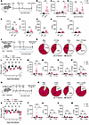 Figure 3
IFN-λ treatment boosts CD4+ T cell–mediated immunity to infection. (A–I) Ifnar–/– mice were infected with 104 PFU HSV-2 333 and administered PBS or IFN-λ (n = 9, 8). Data representative of 2 pooled experiments, except C, which is representative of 2 experiments with similar results (n = 5, 5). (B) IFN-γ levels in vaginal washes 5 DPI. (C) CXCL9 levels. (D) CXCL10 levels. (E) Total number of CD45+ cells in the vaginal tissue. (F) Total number of T cells (CD45+/Ly6G–/CD3+). (G) Total number of CD4+ T cells (CD45+/Ly6G–/CD3+/TCRβ+/TCRγδ–/CD8–/CD4+). (H) Total number of CD8 T cells (CD45+/Ly6G–/CD3+/TCRβ+/TCRγδ–/CD8+/CD4–). (I) Total number of γδ+T cells (CD45+/Ly6G–/CD3+/TCRβ–/TCRγδ+). (J–P) Ifnar–/– mice were administered normal drinking water and PBS, FTY720 and PBS, normal drinking water and IFN-λ, or FTY720 and IFN-λ (n = 4, 5, 4, 4). Data representative of 2 independent experiments, except K and M, which are representative of 2 pooled experiments (n = 8, 5, 9, 9). (K) Rates of megacolon 5 DPI. (L) Viral titers. (M) IFN-γ levels. (N) Total number of CD4+ T cells. (O) Total number of CD8+ T cells. (P) Total number of TCRγδ+ cells 5 DPI. (Q–W) Ifnar–/– mice treated with IFN-λ 1 DPI intravaginally were administered PBS, α-CD4, IFN-λ, or α-CD4+IFN-λ (n = 5, 4, 5, 5). Data representative of 2 independent experiments, except R and T, which are representative of 2 pooled experiments (n = 10, 5, 10, 10). (R) Rates of megacolon 5 DPI. (S) Viral titers. (T) IFN-γ levels. (U) Total number of CD4+ T cells. (V) Total number of CD8+ T cells. (W) Total number of TCRγδ+ cells. *P < 0.05, **P < 0.01. Statistical significance determined by 2-tailed Student’s t test (B and E–I), repeated measures 2-way ANOVA with Šídák’s multiple comparisons test (C, D, L, and S), and ordinary 2-way ANOVA with Šídák’s multiple comparisons test (M–P and T–W).
IFN-λ–treated mice had significantly higher levels of chemokines CXCL9 and CXCL10 in vaginal washes compared with PBS-treated mice, suggesting that IFN-λ signaling could increase immune cell recruitment to the genital mucosa (Figure 3, C and D). However, the total number of CD45+ leukocytes and CD3+ lymphocytes, including CD4+, CD8+, and TCRγδ+ T cells, remained unchanged in the IFN-λ–treated mice compared with control-treated mice (Figure 3, E–I). Others have shown that IFN-λ inhibits neutrophil recruitment in a mouse model of HSV-1 skin lesions by suppressing chemokine production to limit neutrophil-mediated pathology (39). We also observed a reduced number and proportion of Ly6G+ neutrophils in the vaginal tract 5 DPI, despite higher CXCL5 production (Supplemental Figure 3, E and F). Taken together, we found that, while IFN-λ potently induces IFN-γ production to limit viral spread, this is independent of increasing T cell recruitment. Thus, it is unclear whether T cells are necessary for IFN-γ production in IFN-λ–treated mice.
IFN-λ–mediated protection relies on recruited CD4+ T cells. To assess whether T cells are necessary for IFN-γ production induced by IFN-λ treatment, IFN-λ–treated mice received fingolimod (FTY720) to inhibit lymphocyte egress from secondary lymphoid tissues (Figure 3J). FTY720 treatment ablated IFN-γ production in IFN-λ–treated mice and significantly reduced the total number of CD4+, CD8+, and TCRγδ+ T cells, without altering viral titers (Figure 3, L–P). Interestingly, FTY720 treatment alone substantially reduced the incidence of toxic megacolon to rates comparable with mice treated with IFN-λ alone (Figure 3K). This finding suggests that lymphocytes recruited to the gastrointestinal tract may contribute to damage to the peripheral nervous system, similar to what has been reported during West Nile virus infection (40). The combined treatment of FTY720 and IFN-λ in Ifnar–/– mice did not confer additional protection beyond either treatment alone, suggesting that the protective functions of IFN-λ are dependent on recruited lymphocytes.
To further assess the protective mechanisms of IFN-λ, we administered an anti-CD4 depleting antibody, which effectively depleted CD4+ T cells in the draining lymph node (dLN) in both PBS- and IFN-λ–treated mice (Figure 3Q and Supplemental Figure 3G). CD4+ T cell depletion alone did not reduce the incidence of toxic megacolon or affect viral titers. However, α-CD4 treatment blunted IFN-γ production in IFN-λ–treated mice, and reversed the protective effect of IFN-λ (Figure 3, R–T). This was associated with a reduction in the number of CD4+ T cells, but not TCRγδ+ or CD8+ T cells in the vaginal tract (Figure 3, U–W). Together, these results support a requirement for CD4+ T cells, but not TCRγδ+ or CD8+ T cells, in IFN-λ–dependent IFN-γ production in the vaginal tract following HSV-2 infection.
IFN-λ does not alter T cell priming in the dLN. As there were no differences in the total number of T cells in the vaginal tissue, we assessed whether IFN-λ treatment influenced T cell priming in the dLN. Although Ifnlr1 mRNA is not expressed in murine lymphocytes, it is expressed in murine DCs (23). Previous studies have shown that IFN-λ signaling plays a key role in DC migration and priming of CD8+ T cell responses (23). We assessed whether there were differences in DC migration and activation between WT and Ifnar–/– mice, and whether IFN-λ treatment was able to influence DC activity. Ifnar–/– mice showed impaired migration of classical DCs (cDCs) to the dLN compared with WT mice; however, this was not altered by IFN-λ treatment (Supplemental Figure 6A and Figure 4A). Additionally, the total number of monocyte-derived DCs (moDCs) was unchanged among WT mice, Ifnar–/– mice, and Ifnar–/– mice treated with IFN-λ (Figure 4B). Interestingly, DCs in the dLN from WT mice had higher expression of CD86, but lower expression of CD80 compared with Ifnar–/– mice (Figure 4, C, E, and F). CD40 expression was mildly lower in Ifnar–/– mice compared with WT mice (Figure 4, C and D). Furthermore, DCs from WT mice had a distinct MHC IIhi population that was absent in Ifnar–/– mice (Figure 4, C and G). IFN-λ treatment failed to increase the expression of any of these activation markers compared with PBS-treated mice (Figure 4, C–G). Thus, while loss of type I–IFN signaling impaired DC maturation and migration to the dLN, IFN-λ was unable to rescue DC activation. While we did not observe any differences in DC migration or activation in the dLN following IFN-λ treatment, we assessed whether T cell function may still be altered by IFN-λ treatment through other mechanisms. Ifnar–/– mice had reduced numbers of T cells in the dLN compared with WT mice, which was not rescued by IFN-λ treatment (Figure 4, H–K). We further confirmed previous findings that Ifnar–/– mice do not have a defect in T cell priming, as we observed a significant increase in the proportion of IFN-γ–secreting T cells and total IFN-γ production after ex vivo HSV-2 stimulation of dLN cells from Ifnar–/– mice compared with WT mice (Figure 4, L, M, and O) (26). We also found that IFN-λ treatment did not increase the proportion of IFN-γ–secreting CD4+ or CD8+ T cells in Ifnar–/– mice (Figure 4, L and M). IL-17A+ T cells have been demonstrated to be important for optimal TH1 responses (41). However, while there was a slight increase in IL-17A–secreting CD4+ T cells in Ifnar–/– mice compared with WT mice, this was unchanged in IFN-λ–treated mice 5 DPI (Figure 4N). Finally, total IFN-γ production in the dLN did not differ between any of the groups (Figure 4O). Overall, our findings confirm that IFN-λ treatment does not improve T cell priming in the dLN or alter DC activation.
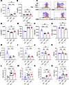 Figure 4
IFN-λ treatment does not alter T cell priming. (A–O) WT, Ifnar–/–, and Ifnar–/– administered IFN-λ 1 DPI (n = 5, 5, 4) were infected with 104 PFU HSV-2 333. Data shown in A–G are representative of 2 independent experiments; H–L are representative of 3 independent experiments with similar results; and M–O are representative of 3 independent experiments pooled (n = 10, 14, 14). The dLN was collected 5 DPI. (A) Total number of cDCs (CD45+/B220–/CD3–/CD11c+/MHC II+/CD26+/CD64–). (B) Total number of moDCs (CD45+/B220–/CD3–/CD11c+/MHC II+/CD26–/CD64+). (C) Histograms for CD40, CD80, CD86, and MHC II for total DCs. (D) CD40 gMFI. (E) CD80 gMFI. (F) CD86 gMFI. (G) MHC II gMFI. (H) Total number of CD45+ cells. (I) Total number of CD3+ T cells (CD45+/CD3+). (J) Proportion of CD4+ T cells (CD45+/CD3+/TCRβ+/TCRγδ–/CD8–/CD4+) to total CD45+ cells. (K) Proportion of CD8+ T cells (CD45+/CD3+/TCRβ+/TCRγδ–/CD8+/CD4–) to total CD45+ cells. (L) Proportion of IFN-γ+CD4+ T cells to total CD4+ cells. (M) IFN-γ+CD8+ T cells to total CD8+ cells. (N) Proportion of L. IL-17A+CD4+ T cells to total CD4+ cells. (O) IFN-γ levels in the supernatants. *P < 0.05, **P < 0.01. ***P < 0.001. Statistical significance was determined by 1-way ANOVA with Tukey’s multiple comparisons test. gMFI, geometric mean fluorescence intensity.
Type II IFN does not rescue impaired lymphocyte-dependent IFN-γ production. Type I, II, and III IFNs share many overlapping and interconnected functions that influence immune cell function. Given that Ifnar–/– mice exhibit impaired NK cell–derived IFN-γ production, we investigated whether impaired T cell production of IFN-γ was due to a loss of type I IFN signaling or NK cell–derived IFN-γ production. To model early IFN-γ production by NK cells, we administered recombinant IFN-γ 1.5 and 2 DPI to determine whether early IFN-γ production was necessary or sufficient for proper CD4+ T cell responses (Figure 5A). IFN-γ treatment did not prevent the development of megacolon in Ifnar–/– mice, and only modestly reduced viral titers at 3 DPI (Figure 5, B and C). Additionally, IFN-γ treatment did not rescue IFN-γ production by T cells at 5 DPI or increase the number of T cells in vaginal tissue (Figure 5, D–H). To support these findings, we depleted NK cells in WT mice, which reduced the total number of NK1.1+ cells in the vaginal tissue and reduced IFN-γ levels in the vaginal washes 2 DPI without altering local viral loads (Supplemental Figure 4, A–C). However, NK cell depletion did not impair T cell recruitment or T cell–dependent IFN-γ production (Supplemental Figure 4, D–G). Overall, these findings demonstrate that early IFN-γ production is neither necessary nor sufficient for an effective T cell response. Interestingly, IFN-γ treatment significantly increased the number of CD45+ cells, including T cells and DCs in the dLN, suggesting that reduced DC migration and T cell accumulation in Ifnar–/– mice may partially result from impaired NK cell function (Figure 5, I–K). Furthermore, these findings suggest that changes observed in the dLN do not necessarily reflect immune cell function at the site of infection, and that T cell function in the vaginal tissue may be influenced by the local tissue environment.
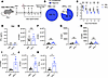 Figure 5
Innate IFN-γ does not influence local T cell responses. (A) Ifnar–/– mice were infected with 104 PFU HSV-2 333 and administered PBS 1.5 and 2 DPI intravaginally. Data shown are representative of 1 independent experiment. (B) Rates of megacolon 5 DPI (n = 4, 5). (C) Viral titers in vaginal washes (n = 4, 4). (D) IFN-γ in the vaginal washes 5 DPI (n = 4, 4). (E) Total number of CD45+ cells in the vaginal tissue 5 DPI (n = 4, 4). (F) Total number of CD3+ T cells (CD45+/Ly6G–/CD3+) in vaginal tissue. (G) Total number of CD4+ T cells (CD45+/Ly6G–/CD3+/TCRβ+/TCRγδ–/CD8–/CD4+) in the vaginal tissue. (H) Total number of CD8+ T cells (CD45+/Ly6G–/CD3+/TCRβ+/ TCRγδ–/CD8+/CD4–) in the vaginal tissue. (I) Total number of CD45+ cells in the dLN 5 DPI. (J) Total number of CD3+ T cells (CD45+/CD3+) in the dLN. (K) Total number of DCs (CD45+/B220–/CD3–/CD11c+/MHCII+) in the dLN. *P < 0.05. Statistical significance determined by 2-way ANOVA with Šídák’s multiple comparisons test (C) or 2-tailed Student’s t test (D–K).
IFN-λ–mediated induction of T cell–dependent IFN-γ production relies on CCR2+Ly6Chi cells. As there was no difference in the priming of T cell responses in the dLN following IFN-λ treatment, we assessed whether we could observe differences in T cell function in the vaginal tissue. While we did not observe differences in the proportion of IFN-γ–secreting CD4+ or CD8+ T cells in the vaginal tract 5 DPI, there was a significant increase in total IFN-γ production following ex vivo stimulation (Figure 6, A–C). These results suggest that IFN-λ treatment increased the overall IFN-γ production per T cell without increasing the frequency of IFN-γ–secreting T cells. Previous studies have shown that moDCs derived from CCR2+ monocytes are dispensable for T cell priming during HSV-2 infection, but are critical for restimulating activated T cells to induce IFN-γ production at the site of infection (26). Indeed, we find that WT mice depleted of CCR2+ monocytes have deficient IFN-γ production by NK cells 2 DPI, and T cells 5 DPI (Supplemental Figure 5A). Additionally, Ifnar–/– mice have been shown to have deficient CCR2+ monocyte recruitment due to reduced CCR2 ligand production (26, 27). Moreover, we found that Ifnar–/– mice have reduced numbers of CD11b+ DCs in the vaginal tract 5 DPI compared with WT mice (Supplemental Figure 5, B and C, and Supplemental Figure 6B). Interestingly, IFN-λ treatment significantly increased the number of CD11b+ DCs in the vaginal tract (Figure 6, D and E), but did not influence their activation, as measured by MHC II, CD40, CD80, and CD86 (Supplemental Figure 5, D–G). Additionally, while we did not observe a significant difference in the total number of monocytes at 5 DPI, there was an increase in the expression of MHC II on both Ly6ChiCCR2+ and Ly6CloCCR2– monocytes following IFN-λ treatment (Figure 6, F–J). When Ly6Chi monocytes differentiate into moDCs, they upregulate MHC II and CD11c and downregulate Ly6C and CCR2 (42, 43). These findings suggest that, in IFN-λ–treated mice, the upregulation of MHC II on Ly6Chi cells and Ly6Clo monocytes indicated an increase in differentiation into CD11b+ moDCs (Figure 6K).
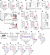 Figure 6
IFN-λ boosts CD4+ T cell function through CCR2+ inflammatory monocytes. (A–C) Ifnar–/– mice were infected with 104 PFU HSV-2 333 and administered PBS or IFN-λ 1 DPI (n = 5, 5). Data representative of 2 independent experiments. (A) Total vaginal cells were collected 5 DPI and stimulated with HSV-2 lysate for 24 hours. Proportion of IFN-γ+CD4+ T cells to total CD4+ cells. (B) Proportion of IFN-γ+CD8+ T cells to total CD8+ cells. (C) Total IFN-γ levels in supernatants. (D) Representative flow plots of CD11b+ DCs (CD45+/Ly6G–/CD3–/B220–/CD11b+/CD11c+/MHCII+) from unstimulated vaginal cells. (E) Total number of CD11b+ DCs 5 DPI (n = 5, 4). (F) Representative flow plots for Ly6ChiCCR2+ (CD45+/Ly6G–/CD3–/B220–/CD11b+/Ly6Chi/CCR2+) and Ly6CloCCR2– (CD45+/Ly6G–/CD3–/B220–/CD11b+/Ly6Clo/CCR2–) cells to assess MHC II gMFI. (G) Total number of Ly6ChiCCR2+ cells (n = 5, 4). (H) Total number of Ly6CloCCR2– cells. (I) MHC gMFI of Ly6ChiCCR2+ cells. (J) MHC gMFI of Ly6CloCCR2– cells. (K) Representative flow plots for comparing Ly6C and MHC II expression in CD11b+ cells. (L) Ifnar–/– mice infected with HSV-2 333 and administered IFN-λ were administered PBS or α-CCR2 daily (n = 4, 5). Data shown are representative of 2 independent experiments with similar results, except N, which is representative of 2 pooled experiments (n = 9, 9). (M) Viral titers. (N) IFN-γ levels in vaginal washes 5 DPI. (O) Total number of CD4+ T cells. (P) Total number of CD8+ T cells. (Q) Total number of Ly6ChiCCR2+ cells. (R) Total number of Ly6CloCCR2– cells. (S) Total number of CD11b+ DC cells. *P < 0.05, **P < 0.01 ***P < 0.0001, ****P < 0.00001. Statistical significance determined by repeated 2-way ANOVA with Šídák’s multiple comparisons test (M) or 2-tailed Student’s t test (A–C, E, G–J, and N–S). gMFI, geometric mean fluorescence intensity.
To assess whether CCR2+ monocytes and moDCs were required for IFN-λ–induced IFN-γ production, IFN-λ–treated Ifnar–/– mice were administered PBS or α-CCR2 (Figure 6L). α-CCR2 treatment did not reduce local viral titers, suggesting that recruitment of CCR2+ monocytes does not play a role in controlling local viral replication (Figure 6M). Depletion of CCR2+ cells completely abolished the induction of IFN-γ, without reducing CD4+ or CD8+ T cell recruitment (Figure 6, N–P). Additionally, depletion of CCR2+ cells reduced the total number of Ly6ChiCCR2+ monocytes, Ly6CloCCR2– monocytes, and CD11b+ DCs, suggesting that Ly6CloCCR2– monocytes and CD11b+ DCs were derived from Ly6ChiCCR2+ monocytes (Figure 6, Q–S). Overall, these findings indicate that IFN-λ relies on CCR2+ monocytes to rescue CD4+ T cell–dependent IFN-γ production.


Copyright © 2026 American Society for Clinical Investigation
ISSN: 0021-9738 (print), 1558-8238 (online)