
Advertisement
Research ArticleImmunologyOncology
Open Access |  10.1172/JCI198270
SIRT2-mediated deacetylation activates USP22 catalytic function for PD-L1 protein stabilization and tumor immune escape
Na Li,1 Qiong Gao,2 Huijun Jia,3 Guoqing Xue,3 Yuanzhang Zhou,4 Shengnan Wang,2,5 Suxian Ma,4 Bingjin Hu,4 Zhuoyue Zhao,1 Chen Su,4 Yinghong Liu,1 Wenxuan Xi,4 Zhonghao Li,4 Donna D. Zhang,6 Peng Chu,4 Zhaolin Sun,1,4 and Deyu Fang2
1Department of Biochemistry and Molecular Biology, College of Basic Medical Science, Dalian Medical University, Dalian, China.
2Department of Pathology, Northwestern University Feinberg School of Medicine, Chicago, Illinois, USA.
3Department of Immunology, College of Basic Medical Science,
4Dalian College of Pharmacy, and
5Institute of Cancer Stem Cell, Dalian Medical University, Dalian, China
6Department of Molecular Medicine, Center for Inflammation Science and Systems Medicine, The Herbert Wertheim UF Scripps Institute for Biomedical Innovation & Technology, Jupiter, Florida, USA.
Address correspondence to: Peng Chu or Zuolin Sun, Dalian Medical University, No. 9, Western Section of Lvshun South Road, Dalian, Liaoning Province, 116044, China. Phone: 8613194286901; Email: chupeng0801@163.com (PC). Phone: 8615242692543; Email: zlsun56@yeah.net (ZS). Or to: Deyu Fang, Department of Pathology, Northwestern University Feinberg School of Medicine, Chicago, Illinois 60611, USA. Phone: 312.503.3021; Email: fangd@northwestern.edu.
Find articles by Li, N. in: PubMed | Google Scholar
1Department of Biochemistry and Molecular Biology, College of Basic Medical Science, Dalian Medical University, Dalian, China.
2Department of Pathology, Northwestern University Feinberg School of Medicine, Chicago, Illinois, USA.
3Department of Immunology, College of Basic Medical Science,
4Dalian College of Pharmacy, and
5Institute of Cancer Stem Cell, Dalian Medical University, Dalian, China
6Department of Molecular Medicine, Center for Inflammation Science and Systems Medicine, The Herbert Wertheim UF Scripps Institute for Biomedical Innovation & Technology, Jupiter, Florida, USA.
Address correspondence to: Peng Chu or Zuolin Sun, Dalian Medical University, No. 9, Western Section of Lvshun South Road, Dalian, Liaoning Province, 116044, China. Phone: 8613194286901; Email: chupeng0801@163.com (PC). Phone: 8615242692543; Email: zlsun56@yeah.net (ZS). Or to: Deyu Fang, Department of Pathology, Northwestern University Feinberg School of Medicine, Chicago, Illinois 60611, USA. Phone: 312.503.3021; Email: fangd@northwestern.edu.
Find articles by Gao, Q. in: PubMed | Google Scholar
1Department of Biochemistry and Molecular Biology, College of Basic Medical Science, Dalian Medical University, Dalian, China.
2Department of Pathology, Northwestern University Feinberg School of Medicine, Chicago, Illinois, USA.
3Department of Immunology, College of Basic Medical Science,
4Dalian College of Pharmacy, and
5Institute of Cancer Stem Cell, Dalian Medical University, Dalian, China
6Department of Molecular Medicine, Center for Inflammation Science and Systems Medicine, The Herbert Wertheim UF Scripps Institute for Biomedical Innovation & Technology, Jupiter, Florida, USA.
Address correspondence to: Peng Chu or Zuolin Sun, Dalian Medical University, No. 9, Western Section of Lvshun South Road, Dalian, Liaoning Province, 116044, China. Phone: 8613194286901; Email: chupeng0801@163.com (PC). Phone: 8615242692543; Email: zlsun56@yeah.net (ZS). Or to: Deyu Fang, Department of Pathology, Northwestern University Feinberg School of Medicine, Chicago, Illinois 60611, USA. Phone: 312.503.3021; Email: fangd@northwestern.edu.
Find articles by Jia, H. in: PubMed | Google Scholar
1Department of Biochemistry and Molecular Biology, College of Basic Medical Science, Dalian Medical University, Dalian, China.
2Department of Pathology, Northwestern University Feinberg School of Medicine, Chicago, Illinois, USA.
3Department of Immunology, College of Basic Medical Science,
4Dalian College of Pharmacy, and
5Institute of Cancer Stem Cell, Dalian Medical University, Dalian, China
6Department of Molecular Medicine, Center for Inflammation Science and Systems Medicine, The Herbert Wertheim UF Scripps Institute for Biomedical Innovation & Technology, Jupiter, Florida, USA.
Address correspondence to: Peng Chu or Zuolin Sun, Dalian Medical University, No. 9, Western Section of Lvshun South Road, Dalian, Liaoning Province, 116044, China. Phone: 8613194286901; Email: chupeng0801@163.com (PC). Phone: 8615242692543; Email: zlsun56@yeah.net (ZS). Or to: Deyu Fang, Department of Pathology, Northwestern University Feinberg School of Medicine, Chicago, Illinois 60611, USA. Phone: 312.503.3021; Email: fangd@northwestern.edu.
Find articles by Xue, G. in: PubMed | Google Scholar
1Department of Biochemistry and Molecular Biology, College of Basic Medical Science, Dalian Medical University, Dalian, China.
2Department of Pathology, Northwestern University Feinberg School of Medicine, Chicago, Illinois, USA.
3Department of Immunology, College of Basic Medical Science,
4Dalian College of Pharmacy, and
5Institute of Cancer Stem Cell, Dalian Medical University, Dalian, China
6Department of Molecular Medicine, Center for Inflammation Science and Systems Medicine, The Herbert Wertheim UF Scripps Institute for Biomedical Innovation & Technology, Jupiter, Florida, USA.
Address correspondence to: Peng Chu or Zuolin Sun, Dalian Medical University, No. 9, Western Section of Lvshun South Road, Dalian, Liaoning Province, 116044, China. Phone: 8613194286901; Email: chupeng0801@163.com (PC). Phone: 8615242692543; Email: zlsun56@yeah.net (ZS). Or to: Deyu Fang, Department of Pathology, Northwestern University Feinberg School of Medicine, Chicago, Illinois 60611, USA. Phone: 312.503.3021; Email: fangd@northwestern.edu.
Find articles by Zhou, Y. in: PubMed | Google Scholar
1Department of Biochemistry and Molecular Biology, College of Basic Medical Science, Dalian Medical University, Dalian, China.
2Department of Pathology, Northwestern University Feinberg School of Medicine, Chicago, Illinois, USA.
3Department of Immunology, College of Basic Medical Science,
4Dalian College of Pharmacy, and
5Institute of Cancer Stem Cell, Dalian Medical University, Dalian, China
6Department of Molecular Medicine, Center for Inflammation Science and Systems Medicine, The Herbert Wertheim UF Scripps Institute for Biomedical Innovation & Technology, Jupiter, Florida, USA.
Address correspondence to: Peng Chu or Zuolin Sun, Dalian Medical University, No. 9, Western Section of Lvshun South Road, Dalian, Liaoning Province, 116044, China. Phone: 8613194286901; Email: chupeng0801@163.com (PC). Phone: 8615242692543; Email: zlsun56@yeah.net (ZS). Or to: Deyu Fang, Department of Pathology, Northwestern University Feinberg School of Medicine, Chicago, Illinois 60611, USA. Phone: 312.503.3021; Email: fangd@northwestern.edu.
Find articles by Wang, S. in: PubMed | Google Scholar
1Department of Biochemistry and Molecular Biology, College of Basic Medical Science, Dalian Medical University, Dalian, China.
2Department of Pathology, Northwestern University Feinberg School of Medicine, Chicago, Illinois, USA.
3Department of Immunology, College of Basic Medical Science,
4Dalian College of Pharmacy, and
5Institute of Cancer Stem Cell, Dalian Medical University, Dalian, China
6Department of Molecular Medicine, Center for Inflammation Science and Systems Medicine, The Herbert Wertheim UF Scripps Institute for Biomedical Innovation & Technology, Jupiter, Florida, USA.
Address correspondence to: Peng Chu or Zuolin Sun, Dalian Medical University, No. 9, Western Section of Lvshun South Road, Dalian, Liaoning Province, 116044, China. Phone: 8613194286901; Email: chupeng0801@163.com (PC). Phone: 8615242692543; Email: zlsun56@yeah.net (ZS). Or to: Deyu Fang, Department of Pathology, Northwestern University Feinberg School of Medicine, Chicago, Illinois 60611, USA. Phone: 312.503.3021; Email: fangd@northwestern.edu.
Find articles by Ma, S. in: PubMed | Google Scholar
1Department of Biochemistry and Molecular Biology, College of Basic Medical Science, Dalian Medical University, Dalian, China.
2Department of Pathology, Northwestern University Feinberg School of Medicine, Chicago, Illinois, USA.
3Department of Immunology, College of Basic Medical Science,
4Dalian College of Pharmacy, and
5Institute of Cancer Stem Cell, Dalian Medical University, Dalian, China
6Department of Molecular Medicine, Center for Inflammation Science and Systems Medicine, The Herbert Wertheim UF Scripps Institute for Biomedical Innovation & Technology, Jupiter, Florida, USA.
Address correspondence to: Peng Chu or Zuolin Sun, Dalian Medical University, No. 9, Western Section of Lvshun South Road, Dalian, Liaoning Province, 116044, China. Phone: 8613194286901; Email: chupeng0801@163.com (PC). Phone: 8615242692543; Email: zlsun56@yeah.net (ZS). Or to: Deyu Fang, Department of Pathology, Northwestern University Feinberg School of Medicine, Chicago, Illinois 60611, USA. Phone: 312.503.3021; Email: fangd@northwestern.edu.
Find articles by Hu, B. in: PubMed | Google Scholar
1Department of Biochemistry and Molecular Biology, College of Basic Medical Science, Dalian Medical University, Dalian, China.
2Department of Pathology, Northwestern University Feinberg School of Medicine, Chicago, Illinois, USA.
3Department of Immunology, College of Basic Medical Science,
4Dalian College of Pharmacy, and
5Institute of Cancer Stem Cell, Dalian Medical University, Dalian, China
6Department of Molecular Medicine, Center for Inflammation Science and Systems Medicine, The Herbert Wertheim UF Scripps Institute for Biomedical Innovation & Technology, Jupiter, Florida, USA.
Address correspondence to: Peng Chu or Zuolin Sun, Dalian Medical University, No. 9, Western Section of Lvshun South Road, Dalian, Liaoning Province, 116044, China. Phone: 8613194286901; Email: chupeng0801@163.com (PC). Phone: 8615242692543; Email: zlsun56@yeah.net (ZS). Or to: Deyu Fang, Department of Pathology, Northwestern University Feinberg School of Medicine, Chicago, Illinois 60611, USA. Phone: 312.503.3021; Email: fangd@northwestern.edu.
Find articles by Zhao, Z. in: PubMed | Google Scholar
1Department of Biochemistry and Molecular Biology, College of Basic Medical Science, Dalian Medical University, Dalian, China.
2Department of Pathology, Northwestern University Feinberg School of Medicine, Chicago, Illinois, USA.
3Department of Immunology, College of Basic Medical Science,
4Dalian College of Pharmacy, and
5Institute of Cancer Stem Cell, Dalian Medical University, Dalian, China
6Department of Molecular Medicine, Center for Inflammation Science and Systems Medicine, The Herbert Wertheim UF Scripps Institute for Biomedical Innovation & Technology, Jupiter, Florida, USA.
Address correspondence to: Peng Chu or Zuolin Sun, Dalian Medical University, No. 9, Western Section of Lvshun South Road, Dalian, Liaoning Province, 116044, China. Phone: 8613194286901; Email: chupeng0801@163.com (PC). Phone: 8615242692543; Email: zlsun56@yeah.net (ZS). Or to: Deyu Fang, Department of Pathology, Northwestern University Feinberg School of Medicine, Chicago, Illinois 60611, USA. Phone: 312.503.3021; Email: fangd@northwestern.edu.
Find articles by Su, C. in: PubMed | Google Scholar
1Department of Biochemistry and Molecular Biology, College of Basic Medical Science, Dalian Medical University, Dalian, China.
2Department of Pathology, Northwestern University Feinberg School of Medicine, Chicago, Illinois, USA.
3Department of Immunology, College of Basic Medical Science,
4Dalian College of Pharmacy, and
5Institute of Cancer Stem Cell, Dalian Medical University, Dalian, China
6Department of Molecular Medicine, Center for Inflammation Science and Systems Medicine, The Herbert Wertheim UF Scripps Institute for Biomedical Innovation & Technology, Jupiter, Florida, USA.
Address correspondence to: Peng Chu or Zuolin Sun, Dalian Medical University, No. 9, Western Section of Lvshun South Road, Dalian, Liaoning Province, 116044, China. Phone: 8613194286901; Email: chupeng0801@163.com (PC). Phone: 8615242692543; Email: zlsun56@yeah.net (ZS). Or to: Deyu Fang, Department of Pathology, Northwestern University Feinberg School of Medicine, Chicago, Illinois 60611, USA. Phone: 312.503.3021; Email: fangd@northwestern.edu.
Find articles by Liu, Y. in: PubMed | Google Scholar
1Department of Biochemistry and Molecular Biology, College of Basic Medical Science, Dalian Medical University, Dalian, China.
2Department of Pathology, Northwestern University Feinberg School of Medicine, Chicago, Illinois, USA.
3Department of Immunology, College of Basic Medical Science,
4Dalian College of Pharmacy, and
5Institute of Cancer Stem Cell, Dalian Medical University, Dalian, China
6Department of Molecular Medicine, Center for Inflammation Science and Systems Medicine, The Herbert Wertheim UF Scripps Institute for Biomedical Innovation & Technology, Jupiter, Florida, USA.
Address correspondence to: Peng Chu or Zuolin Sun, Dalian Medical University, No. 9, Western Section of Lvshun South Road, Dalian, Liaoning Province, 116044, China. Phone: 8613194286901; Email: chupeng0801@163.com (PC). Phone: 8615242692543; Email: zlsun56@yeah.net (ZS). Or to: Deyu Fang, Department of Pathology, Northwestern University Feinberg School of Medicine, Chicago, Illinois 60611, USA. Phone: 312.503.3021; Email: fangd@northwestern.edu.
Find articles by Xi, W. in: PubMed | Google Scholar
1Department of Biochemistry and Molecular Biology, College of Basic Medical Science, Dalian Medical University, Dalian, China.
2Department of Pathology, Northwestern University Feinberg School of Medicine, Chicago, Illinois, USA.
3Department of Immunology, College of Basic Medical Science,
4Dalian College of Pharmacy, and
5Institute of Cancer Stem Cell, Dalian Medical University, Dalian, China
6Department of Molecular Medicine, Center for Inflammation Science and Systems Medicine, The Herbert Wertheim UF Scripps Institute for Biomedical Innovation & Technology, Jupiter, Florida, USA.
Address correspondence to: Peng Chu or Zuolin Sun, Dalian Medical University, No. 9, Western Section of Lvshun South Road, Dalian, Liaoning Province, 116044, China. Phone: 8613194286901; Email: chupeng0801@163.com (PC). Phone: 8615242692543; Email: zlsun56@yeah.net (ZS). Or to: Deyu Fang, Department of Pathology, Northwestern University Feinberg School of Medicine, Chicago, Illinois 60611, USA. Phone: 312.503.3021; Email: fangd@northwestern.edu.
Find articles by Li, Z. in: PubMed | Google Scholar
1Department of Biochemistry and Molecular Biology, College of Basic Medical Science, Dalian Medical University, Dalian, China.
2Department of Pathology, Northwestern University Feinberg School of Medicine, Chicago, Illinois, USA.
3Department of Immunology, College of Basic Medical Science,
4Dalian College of Pharmacy, and
5Institute of Cancer Stem Cell, Dalian Medical University, Dalian, China
6Department of Molecular Medicine, Center for Inflammation Science and Systems Medicine, The Herbert Wertheim UF Scripps Institute for Biomedical Innovation & Technology, Jupiter, Florida, USA.
Address correspondence to: Peng Chu or Zuolin Sun, Dalian Medical University, No. 9, Western Section of Lvshun South Road, Dalian, Liaoning Province, 116044, China. Phone: 8613194286901; Email: chupeng0801@163.com (PC). Phone: 8615242692543; Email: zlsun56@yeah.net (ZS). Or to: Deyu Fang, Department of Pathology, Northwestern University Feinberg School of Medicine, Chicago, Illinois 60611, USA. Phone: 312.503.3021; Email: fangd@northwestern.edu.
Find articles by Zhang, D. in: PubMed | Google Scholar
1Department of Biochemistry and Molecular Biology, College of Basic Medical Science, Dalian Medical University, Dalian, China.
2Department of Pathology, Northwestern University Feinberg School of Medicine, Chicago, Illinois, USA.
3Department of Immunology, College of Basic Medical Science,
4Dalian College of Pharmacy, and
5Institute of Cancer Stem Cell, Dalian Medical University, Dalian, China
6Department of Molecular Medicine, Center for Inflammation Science and Systems Medicine, The Herbert Wertheim UF Scripps Institute for Biomedical Innovation & Technology, Jupiter, Florida, USA.
Address correspondence to: Peng Chu or Zuolin Sun, Dalian Medical University, No. 9, Western Section of Lvshun South Road, Dalian, Liaoning Province, 116044, China. Phone: 8613194286901; Email: chupeng0801@163.com (PC). Phone: 8615242692543; Email: zlsun56@yeah.net (ZS). Or to: Deyu Fang, Department of Pathology, Northwestern University Feinberg School of Medicine, Chicago, Illinois 60611, USA. Phone: 312.503.3021; Email: fangd@northwestern.edu.
Find articles by Chu, P. in: PubMed | Google Scholar
1Department of Biochemistry and Molecular Biology, College of Basic Medical Science, Dalian Medical University, Dalian, China.
2Department of Pathology, Northwestern University Feinberg School of Medicine, Chicago, Illinois, USA.
3Department of Immunology, College of Basic Medical Science,
4Dalian College of Pharmacy, and
5Institute of Cancer Stem Cell, Dalian Medical University, Dalian, China
6Department of Molecular Medicine, Center for Inflammation Science and Systems Medicine, The Herbert Wertheim UF Scripps Institute for Biomedical Innovation & Technology, Jupiter, Florida, USA.
Address correspondence to: Peng Chu or Zuolin Sun, Dalian Medical University, No. 9, Western Section of Lvshun South Road, Dalian, Liaoning Province, 116044, China. Phone: 8613194286901; Email: chupeng0801@163.com (PC). Phone: 8615242692543; Email: zlsun56@yeah.net (ZS). Or to: Deyu Fang, Department of Pathology, Northwestern University Feinberg School of Medicine, Chicago, Illinois 60611, USA. Phone: 312.503.3021; Email: fangd@northwestern.edu.
Find articles by Sun, Z. in: PubMed | Google Scholar
1Department of Biochemistry and Molecular Biology, College of Basic Medical Science, Dalian Medical University, Dalian, China.
2Department of Pathology, Northwestern University Feinberg School of Medicine, Chicago, Illinois, USA.
3Department of Immunology, College of Basic Medical Science,
4Dalian College of Pharmacy, and
5Institute of Cancer Stem Cell, Dalian Medical University, Dalian, China
6Department of Molecular Medicine, Center for Inflammation Science and Systems Medicine, The Herbert Wertheim UF Scripps Institute for Biomedical Innovation & Technology, Jupiter, Florida, USA.
Address correspondence to: Peng Chu or Zuolin Sun, Dalian Medical University, No. 9, Western Section of Lvshun South Road, Dalian, Liaoning Province, 116044, China. Phone: 8613194286901; Email: chupeng0801@163.com (PC). Phone: 8615242692543; Email: zlsun56@yeah.net (ZS). Or to: Deyu Fang, Department of Pathology, Northwestern University Feinberg School of Medicine, Chicago, Illinois 60611, USA. Phone: 312.503.3021; Email: fangd@northwestern.edu.
Find articles by
Fang, D.
in:
PubMed
|
Google Scholar
|

Published June 2, 2026 - More info
J Clin Invest. 2026;136(14):e198270. https://doi.org/10.1172/JCI198270.
© 2026 Li et al. This work is licensed under the Creative Commons Attribution 4.0 International License. To view a copy of this license, visit http://creativecommons.org/licenses/by/4.0/.
Received: July 22, 2025; Accepted: May 28, 2026

-
Results
Identification of SIRT2 as a positive regulator to control PD-L1 protein expression in cancer cells at the posttranscriptional level. The type III histone deacetylases have been increasingly recognized for their involvement in tumor biology, with members of this family influencing critical oncogenic processes such as cellular growth, proliferation, migration, and invasion (25–27). To investigate these roles, we employed siRNAs to individually silence each of the 7 known sirtuin family members (SIRT1–SIRT7) in T24 human bladder cancer cells. Among these, knockdown of SIRT2 led to a dramatic decrease in the expression of PD-L1, a key immune checkpoint protein known to suppress antitumor immune responses. A modest reduction in PD-L1 expression was also observed upon SIRT3 knockdown (Figure 1, A and B). To further validate the specific role of SIRT2 in PD-L1 regulation, we generated SIRT2-KO human T24 and mouse MB49 bladder cancer cells using a CRISPR/Cas9 approach. Western blotting analysis confirmed the complete SIRT2 deletion (Figure 1, C and F). Consistent with the siRNA data, complete loss of SIRT2 significantly reduced PD-L1 protein levels in both T24 and murine MB49 bladder cancer cells (Figure 1, C and F, middle panels), which was further confirmed by flow cytometry analysis (Figure 1, D, E, G, and H). Consistently, treatment of T24 human bladder cancer cells with the SIRT2-specific inhibitors thiomyristoyl (TM) and SirReal2 resulted in a dose-dependent reduction in PD-L1 expression (Figure 1, I and J).
 Figure 1
SIRT2 positively regulates PD-L1 expression in multiple cancer cell types. (A and B) T24 bladder cancer cells were transiently transfected with siRNAs targeting each of the 7 sirtuins. PD-L1 protein expression was assessed by Western blotting. Representative immunoblots are shown in A, and quantitative analysis from 3 independent experiments is presented in B. (C–E) CRISPR-mediated KO of SIRT2 in T24 cells was confirmed by Western blot (C, top panel). The effect on PD-L1 expression was examined by Western blot (C, middle panel) and flow cytometry (D, representative plots; E, quantified data from 3 independent experiments). (F–H) The impact of SIRT2 KO on PD-L1 expression was similarly assessed in MB49 murine bladder cancer cells. (I and J) T24 cells were treated with TM or SirReal2 at each indicated concentration for 8 hours, and PD-L1 expression levels were determined by Western blotting. (K–N) RT-qPCR analysis of SIRT2 (K and M) and PD-L1 (L and N) mRNA levels in T24 and MB49 cells following CRISPR-mediated SIRT2 deletion. WCL, whole-cell lysate. (O) Ubiquitination assay of PD-L1 in WT and SIRT2-KO T24 cells. (P) Western blot analysis of PD-L1 protein levels in SIRT2 KO T24 cells, with or without treatment with the proteasome inhibitor MG132. (Q) Interaction between Myc-tagged PD-L1 and Flag-tagged SIRT2. HEK293T cells were cotransfected with Myc-PD-L1 and Flag-SIRT2, followed by co-IP using an anti-Flag antibody to assess protein interaction. (R) Co-IP analysis of endogenous SIRT2 and PD-L1 interaction in T24 cells. Data are shown as mean ± SD. Statistical significance was determined using 1-way ANOVA for B and unpaired 2-tailed Student’s t test for E, H, and K–N. *P < 0.05; ***P < 0.001.
Western blot analysis revealed a more pronounced decrease in PD-L1 levels upon SIRT2 genetic deletion compared with flow cytometry, suggesting that SIRT2 may regulate intracellular PD-L1 to a greater extent than cell surface expression. To address this possibility, we performed immunofluorescence staining in WT and SIRT2-KO tumor cells, enabling simultaneous assessment of PD-L1 localization in both cytosolic and membrane compartments. These analyses demonstrated that SIRT2 deficiency leads to a more than 70% reduction of PD-L1 in both surface and cytoplasmic compartments in T24 and MB49 tumor cells (Supplemental Figure 1; supplemental material available online with this article; https://doi.org/10.1172/JCI198270DS1). Collectively, these findings indicate that SIRT2 regulates overall PD-L1 protein stability rather than selectively affecting its membrane localization.
Interestingly, real-time RT-PCR analysis did not detect any changes in PD-L1 mRNA expression levels, but confirmed a complete SIRT2 deletion, in both cancer cell lines (Figure 1, K–N). In addition, CRISPR-mediated deletion of SIRT2 led to a substantial reduction in PD-L1 protein, but not its mRNA, expression across multiple cancer cell lines, including human A375 and murine B16 melanoma cells and human endometrial cancer KLE cells (Supplemental Figure 2). These observations strongly suggest that SIRT2 acts as a common positive regulator of PD-L1 expression in cancer cells at a posttranscriptional level.
PD-L1 expression in cancer cells is known to be regulated at both transcriptional and posttranscriptional levels (28, 29). The fact that SIRT2-targeted inhibition reduced PD-L1 protein but not mRNA expression prompted us to explore whether SIRT2 influences PD-L1 expression through modulation of its ubiquitination and proteasomal degradation. Indeed, SIRT2 KO increased PD-L1 ubiquitination in T24 bladder cancer cells (Figure 1O). A similar effect was observed when T24 bladder cancer cells were treated with AGK2, a selective SIRT2 inhibitor, further confirming the role of SIRT2 in suppressing PD-L1 ubiquitination (Supplemental Figure 3). To determine whether this regulation occurs via the proteasome pathway, we treated cells with the proteasome inhibitor MG132, which restored PD-L1 protein levels in SIRT2-deficient cells to levels comparable with controls (Figure 1P). These findings collectively suggest that SIRT2 maintains PD-L1 protein stability by protecting it from ubiquitin-mediated proteasomal degradation.
We then determined whether SIRT2 exerts its effect through direct interaction with PD-L1 using co-IP experiments. However, Western blot analysis failed to detect any physical interaction between SIRT2 and PD-L1 either in transiently transfected HEK293T cells or endogenously in T24 cells (Figure 1, Q and R). These results suggest that SIRT2 likely regulates PD-L1 indirectly, potentially through modulation of upstream components of the protein degradation machinery.
SIRT2 is a de novo USP22 deacetylase. Several deubiquitinating enzymes, including USP2 (30), USP5 (31), USP7 (32), USP8 (33), USP9X (34), USP10 (35), USP19 (36), USP20 (37), USP22 (38), and CSN5 (39), deubiquitinated and stabilized PD-L1 protein in cancer cells. Interestingly, SIRT2 KO downregulated the expression of USP22, but not any others, in T24 cells (Figure 2, A and B, and Supplemental Figure 4), suggesting that SIRT2 specifically stabilizes the PD-L1 deubiquitinase USP22. Interestingly, we observed a modest but statistically significant increase in USP19 expression, suggesting a potential compensatory response to reduced USP22 levels in SIRT2-KO tumor cells. Similar trends were observed across multiple cell lines, including MB49, A375, and B16 (Figure 2C and Supplemental Figure 4). Consistently, pharmacological SIRT2 inhibition using specific inhibitors, AGK2, TM, and SirReal2, resulted in a dose-dependent reduction in USP22 protein expression (Supplemental Figure 5). In contrast, CRISPR-mediated deletion of Sirt1 or Sirt3, both known regulators of PD-L1 expression (40–42), had no detectable effect on USP22 levels in human bladder cancer cells (Supplemental Figure 6). These results indicate that SIRT2 promotes PD-L1 through upregulating the PD-L1–specific deubiquitinase USP22 expression. Supporting this speculation, USP22 overexpression fully rescued PD-L1 protein levels in SIRT2-KO cells (Figure 2, D–F). Interestingly, RT-qPCR did not detect any meaningful changes in USP22 mRNA by SIRT2 KO in T24 cells, A375 cells, and SIRT2-knockdown KLE cells (Figure 2G and Supplemental Figure 7). Collectively, these results indicate that SIRT2 upregulates USP22 expression at the posttranscriptional level.
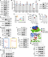 Figure 2
SIRT2 inhibits PD-L1 ubiquitination by regulating USP22. (A) Western blot analysis of deubiquitinases in SIRT2-KO T24 cells, including USP9X, USP5, USP7, USP8, USP20, USP22, and CSN5. (B) Quantification of PD-L1 expression. (C) Western blot analysis of USP22 protein levels in SIRT2-KO MB49 cells. (D–F) SIRT2-KO T24 cells were transfected with or without Myc-USP22, and PD-L1 expression was assessed by Western blotting and flow cytometry 36 hours after transfection. (G) RT-qPCR analysis of USP22 mRNA levels in WT and SIRT2-KO T24 cells. (H) Analysis of the interaction between Myc-USP22 and Flag-SIRT2 in HEK293T cells. (I) Co-IP analysis of endogenous interaction between SIRT2 and USP22 in T24 cells. (J and K) Structural modeling of SIRT2-USP22 interaction. (J) Simulated interaction diagram. (K) Dot plot representing predicted interacting amino acid residues. Blue box: N-terminal region of USP22; orange box, C-terminal region of USP22. (L) Analysis of endogenous USP22 acetylation in WT and SIRT2-KO T24 cells. (M) USP22 acetylation was detected by immunoprecipitating with anti-Myc antibodies followed by blotting with anti–pan-acetylation antibodies. (N) In vitro analysis of SIRT2 deacetylation of acetyl-USP22. Data are shown as mean ± SD. Statistical significance was determined using unpaired 2-tailed Student’s t test for B and G and 1-way ANOVA for F. **P < 0.01; ***P < 0.001.
To elucidate the molecular mechanism by which SIRT2 regulates USP22, we first examined whether these proteins physically interact. Co-IP assays revealed that SIRT2 directly interacts with USP22 in transiently transfected HEK293T cells (Figure 2H). The interaction of endogenous SIRT2 with USP22 was also confirmed at the endogenous level in T24 human bladder cancer cells (Figure 2I). Further analysis with molecular dynamics (MD) simulations and molecular mechanics/Poisson-Boltzmann surface area (MM/PBSA) binding free energy calculations demonstrated that SIRT2 binds USP22 with high affinity, with a calculated binding energy of –4,642.321 kJ/mol (Supplemental Table 1). MD simulations also identified 40 pairs of highly probable interacting amino acids between SIRT2 and USP22 (Figure 2K). Structural modeling further confirmed that these interacting residues are in close spatial proximity, suggesting a stable and specific interaction interface (Figure 2J). These results confirm the physical interaction between SIRT2 and USP22 proteins in cancer cells.
As a class III histone deacetylase, SIRT2 is known to target nonhistone proteins for deacetylation (43). Consistent with this, both CRISPR-mediated SIRT2 KO and pharmacological inhibition with AGK2 led to a more than 2-fold increase in USP22 acetylation in T24 cancer cells (Figure 2L and Supplemental Figure 8A). Conversely, overexpression of SIRT2, but not its catalytically inactive mutant, reduced USP22 acetylation (Figure 2M). These results indicate that SIRT2 directly deacetylates USP22 and that its enzymatic activity is essential for this function. To support this conclusion, in vitro deacetylation assays showed that USP22 acetylation was reduced only in the presence of both NAD+ and SIRT2, but not with NAD+ or SIRT2 alone, confirming that SIRT2 directly and specifically deacetylates USP22 in a NAD+-dependent manner (Figure 2N).
SIRT2 protects USP22 from ubiquitination-induced degradation. While competition between acetylation and ubiquitination on the same lysine (K) residues is a known mechanism of protein stability regulation (44–46), this does not appear to be the case here because SIRT2 inhibition increased both the acetylation (Figure 2M) and ubiquitination (Figure 3A) of USP22, suggesting that these modifications may not act through competition with each other. Supporting this, SIRT2 KO or pharmacological inhibition by AKG2 increased USP22 ubiquitination levels (Figure 3B and Supplemental Figure 8B).
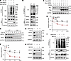 Figure 3
SIRT2 promotes USP22 ubiquitination. (A) USP22 ubiquitination was detected by IP using anti-Myc antibodies and immunoblotting with anti-HA antibodies. (B) Ubiquitination assay of USP22 in WT and SIRT2-KO T24 cells. (C and D) Stability assay of Myc-USP22 in HEK293T cells cotransfected with or without Flag-SIRT2 treated with CHX. (E and F) Stability assay of endogenous USP22 in WT and SIRT2-KO T24 cells following CHX treatment. (G–I) WT and SIRT2-KO cells were treated with NH4Cl (G) or MG132 (H and I). USP22 and SIRT2 expression and PD-L1 ubiquitination were determined. Data are shown as mean ± SD. Statistical significance was determined using unpaired 2-tailed Student’s t test for D and 1-way ANOVA for F. *P < 0.05; **P < 0.01; ***P < 0.001.
Given that SIRT2 loss enhances USP22 ubiquitination, we next assessed whether SIRT2 affects USP22 protein stability. Indeed, SIRT2 expression significantly prolonged the half-life of USP22 in HEK293T cells (Figure 3, C and D). Conversely, both CRISPR-mediated SIRT2 deletion and AGK2 treatment accelerated USP22 degradation in cancer cells (Figure 3, E and F, and Supplemental Figure 8, C and D). The lysosomal inhibitor NH4Cl failed to rescue USP22 expression in SIRT2-KO tumor cells (Figure 3G). In contrast, treatment of SIRT2-KO T24 cells with the proteasomal inhibitor MG132 largely restored both USP22 and PD-L1 protein levels (Figure 3, H and I). We also observed that the proteasomal inhibitor MG132 induced a pronounced accumulation of ubiquitinated PD-L1, as well as total PD-L1 protein levels in both WT and SIRT2-KO cells (Figure 3I). Taken together, these findings demonstrate that SIRT2 stabilizes USP22 by deacetylating it, thereby protecting USP22 from ubiquitin-mediated proteasomal degradation. Through this mechanism, SIRT2 indirectly promotes PD-L1 protein expression, positioning SIRT2 as a critical upstream regulator of tumor immune evasion.
SIRT2 specifically deacetylates USP22 at lysine residues in its deubiquitinase catalytic domain to control deubiquitination activity. To further elucidate the molecular mechanism by which SIRT2-mediated deacetylation stabilizes USP22 protein in cancer cells, we performed IP-MS analysis to identify acetylated lysine residues on USP22 that may serve as SIRT2 substrates. This analysis revealed 5 acetylated lysine residues, including K59, K88, K243, K382, and K505, that could potentially be deacetylated by SIRT2 (Supplemental Figure 9). To determine which residues are functionally relevant targets of SIRT2, we generated a panel of USP22 mutants, each with one of these lysines substituted by arginine (R). Among the 5 mutants, K505R and K382R showed a pronounced reduction in USP22 acetylation, but mutation of any of the remaining 3 lysine residues did not reduce USP22 acetylation. When both K382 and K505 were mutated, USP22 acetylation levels were largely diminished (Figure 4A), identifying 2 lysine residues as dominant acetylation sites.
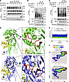 Figure 4
SIRT2-mediated deacetylation alters USP22 interaction. (A) Myc-tagged WT USP22 and its mutant by replacing each identified lysine with arginine (KR) were cotransfected into HEK293T cells with or without Flag-tagged SIRT2. USP22 acetylation was determined. (B) His-tagged ubiquitin and Flag–PD-L1 were cotransfected into HEK293T cells with or without Myc-USP22 and its K382R, K505R, or double mutant (dKR). PD-L1 ubiquitination was detected. (C) The effect of USP22 K382Q, K505Q, and dKQ mutants on PD-L1 ubiquitination was analyzed as in B. (D) Schematic diagram showing the ubiquitin-binding and acetylation sites of USP22 at K505 and K382. (Enlarged images are in Supplemental Figure 5A.) (E) Superimposition of the initial UB-USP22 complex structure with conformations obtained after 50 ns MD simulations for K505-, K382-, and double-acetylated (K505 and K382) UB-USP22 complexes. (F) Energy landscape plots of the UB-USP22 complexes with different acetylation states, generated from 50 ns MD simulations. RMSD, root mean square deviation.
Notably, both K382 and K505 reside within the catalytic domain of USP22, suggesting that acetylation at these residues may influence the enzyme’s deubiquitinating activity. We then performed functional assays to test this hypothesis. Expression of either the K382R or K505R mutant significantly enhanced USP22 deubiquitinase activity, as evidenced by their stronger suppression of PD-L1 ubiquitination (Figure 4B). In contrast, acetylation-mimicking mutants, where K382 and K505 were replaced by glutamine (Q), lost this activity, resulting in diminished ability to inhibit PD-L1 ubiquitination (Figure 4C). These results clearly indicate that acetylation of the 2 lysine residues in the enzymatic pocket of USP22 inhibits its catalytic activity.
Analysis of 50 ns MD simulations revealed that lysine acetylation at positions K505, K382, or both induces notable conformational changes in the catalytic domain of USP22, particularly within loops 253–262 and 472–478, compared with the unacetylated form. Given the critical role of C185 in catalysis, the distance between its sulfur atom and the oxygen atom of the C-terminal glycine of ubiquitin was monitored over the final 20 ns of each trajectory. The average distances were 3.2 Å (unacetylated), 4.0 Å (505K-Ac), 4.0 Å (382K-Ac), and 4.2 Å (double acetylated), indicating that the acetylation relocates the C-terminal glycine of ubiquitin away from the catalytic core of USP22. These findings suggest that lysine acetylation induces structural USP22 rearrangements that cause loop 253–262 and 472–478 movements to sterically hinder ubiquitin access to the catalytic pocket, thereby impairing USP22’s deubiquitinase activity (Figure 4, D and E, and Supplemental Figure 10, A and B). Further MM/PBSA binding free energy calculations were performed to compare nonacetylated and acetylated forms of USP22 in complex with ubiquitin. After a 50 ns simulation, nonacetylated UB-USP22 adopted a stable conformation within a single energy well, indicating strong and stable interaction. In contrast, acetylated forms, including UB-USP22(K382Ac), UB-USP22(K505Ac), and UB-USP22(K382Ac & K505Ac), displayed multiple energy wells, suggesting conformational instability and impaired ubiquitin binding (Figure 4, E and F, and Supplemental Figure 10C). Supporting this, MM/PBSA analysis showed that acetylated USP22 variants had significantly weaker binding energies with ubiquitin, with dual acetylation at K382 and K505 producing the most severe loss of binding affinity (Supplemental Table 2). These findings indicate that SIRT2 enhances USP22 catalytic activity by deacetylating K382 and K505, thereby stabilizing its conformation and ubiquitin-binding efficiency.
To further confirm whether SIRT2-mediated deacetylation enhances USP22 activity, we analyzed the effect of USP22 acetylation on its auto-deubiquitination, a mechanism critical for USP22 self-stabilization. A prior study suggested that USP22 can form homodimers (BioGRID) (47). To test this, we performed co-IP in HEK293T cells cotransfected with HA- and Myc-tagged USP22 and confirmed USP22-USP22 interaction (Figure 5A). Interestingly, mutation of the critical lysine residues K382 and K505 increased USP22 homodimerization (Figure 5B), suggesting that acetylation at these sites restrains dimer formation and that SIRT2-mediated deacetylation promotes this process. Consistently, SIRT2 overexpression enhanced USP22 self-interaction (Figure 5C), further supporting a model in which SIRT2 facilitates USP22 dimerization and functional activity.
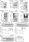 Figure 5
SIRT2-mediated deacetylation inhibits USP22 self-deubiquitination. (A) Analysis of USP22 homodimerization in HEK293 cells cotransfected with HA- and Myc-tagged USP22. (B) Analysis of USP22/KR mutant interactions as in A. (C) Interaction between HA-USP22 and Myc-USP22 with or without Flag-SIRT2 in HEK293T cells. (D) His-ubiquitin and HA-USP22 were cotransfected into HEK293T cells with Myc-USP22 or Myc-C185A-USP22. USP22 ubiquitination was immunoprecipitated with anti-HA antibodies and detected with anti-His antibody. (E and F) The effect of KR (E) or KQ (F) mutations on USP22 ubiquitination was analyzed as in D. (G–L) The effect of KR (G and H), KQ (I and J), or CA (K and L) mutations on USP22 protein stability was analyzed. Stability in transiently transfected HEK293T cells treated with CHX for the indicated time points. Western blot analysis was used to assess protein levels, and USP22 levels normalized to GAPDH were quantified. Data are shown as mean ± SD, N = 3. Statistical significance was determined using unpaired 2-tailed Student’s t test. *P < 0.05; **P < 0.01; ***P < 0.001.
Next, we determined how SIRT2 stabilizes USP22 protein in cancer cells. WT USP22, but not the catalytically inactive C185A mutant, reduced its ubiquitination, confirming that auto-deubiquitination requires catalytic activity (Figure 5D). Importantly, compared with that of WT USP22, coexpression of the K382R and K505R mutants further enhanced suppression of its auto-ubiquitination (Figure 5E), whereas the K382Q or K505Q mutant totally abolished this self-deubiquitination effect (Figure 5F). These results suggest that acetylation at K382 and K505 negatively regulates USP22 auto-deubiquitination. Consequently, coexpression of the K382R and K505R mutants significantly prolonged USP22 half-life, whereas K382Q and K505Q mutants failed to do so (Figure 5, G–J), indicating that acetylation at these sites reduces USP22 stability by promoting its degradation via ubiquitination. Indeed, SIRT2 overexpression did not stabilize the catalytically inactive C185A mutant, suggesting that SIRT2-mediated stabilization of USP22 depends on USP22’s own enzymatic activity (Figure 5, K and L).
Collectively, these findings reveal a novel mechanism by which SIRT2 stabilizes USP22 through direct deacetylation of K382 and K505, promoting auto-deubiquitination and protein stability and ultimately enhancing PD-L1 deubiquitination. This SIRT2/USP22 axis provides an important regulatory pathway for immune checkpoint control in cancer cells.
Targeted SIRT2 inhibition enhances antitumor immunity. PD-L1 expressed in tumor cells plays a key role in immune evasion by binding to PD-1 receptors on immune cells, thereby suppressing antitumor immune responses (48, 49). Our discovery that inhibition of SIRT2 reduced PD-L1 protein expression on the surface of tumor cells provided a rationale for targeting SIRT2 potentiating antitumor immunity. Consistent with this hypothesis, selective inhibition of SIRT2 in tumor cells significantly impaired syngeneic tumor growth (Figure 6, A–C). Mechanistically, SIRT2 deletion led to a pronounced decrease in PD-L1 expression in CD45+ tumor cells (Figure 6D). Importantly, tumors derived from SIRT2-KO cells showed increased infiltration of CD4+ and CD8+ T cells (Figure 6, E–G), along with a dramatic reduction in immunosuppressive FoxP3+ Tregs (Figure 6H). Notably, SIRT2 loss dramatically enhanced CD8 T cell cytotoxic function, as indicated by elevated levels of granzyme B, IFN-γ, and TNF-α (Figure 6, I–K). In contrast, the proportion of terminally exhausted/apoptotic (annexin V+) CD8+ T cells was significantly decreased in SIRT2-deficient tumors (Figure 6L).
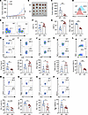 Figure 6
SIRT2 KO enhances antitumor immunity. (A–C) WT and SIRT2-KO MB49 cells were subcutaneously implanted into the right flanks of C57BL/6J mice (n = 10). Representative images of mouse tumors (B), tumor volume (A), and tumor weight (C). Scale bar: 1 cm. (D) PD-L1 expression of CD45– tumor cells (n = 10). (E) Analysis of CD4+ and CD8+ T cells in a gated CD45+ population. (F and G) Quantification of CD4+ (F) and CD8+ (G) T cells, shown as percentages of tumor-infiltrating lymphocytes in MB49 tumors. (H) Analysis of CD4+FoxP3+ Treg cells as a percentage of tumor-infiltrating CD4+ T cells. (I–L) Analysis of granzyme B+ (I), IFN-γ+ (J), and TNF-α+ (K) CD8+ T cells and expression of annexin V (L) CD8+ T cells as percentages of tumor-infiltrating CD8+ T cells in MB49 tumors (n = 10). (M–P) The expression levels of TIM-3, LAG-3, Tox, and NR4A1 on the gated intratumoral CD8+PD-1+ T cells were analyzed. Representative flow images (top panels) and data from 5 pairs of mice are shown. Data are shown as mean ± SD. Statistical significance was determined using 1-way ANOVA for A and unpaired 2-tailed Student’s t test for the remaining panels. *P < 0.05; **P < 0.01; ***P < 0.001.
Elevated PD-L1 expression promotes and sustains CD8+ T cell exhaustion, particularly under conditions of chronic antigen exposure, such as cancer or persistent viral infection (50). Consistent with this paradigm, the frequencies of PD-1+LAG3+ and PD-1+TIM-3+CD8+ T cells were significantly reduced in SIRT2-deficient tumors (Figure 6, M and N). Further analysis of TOX and NR4A1, both of which are critical for CD8+ T cell exhaustion, confirmed that SIRT2 inhibition reduced the PD-1+TOX+ and PD-1+NR4A1+ exhausted intratumoral CD8+ T cells (Figure 6, O and P). These findings suggest that the deletion of SIRT2 in tumor cells enhances antitumor immune responses primarily by relieving PD-L1–mediated suppression of CD8+ T cell function.
This immunostimulatory effect of SIRT2 inhibition was further validated in the B16 melanoma model (Supplemental Figure 11, A–C). Compared with WT B16 cells, SIRT2-KO B16 tumors exhibited significantly reduced PD-L1 expression in CD45– tumor cells (Supplemental Figure 11D). This was accompanied by increased infiltration of CD4+ and CD8+ T cells (Supplemental Figure 11, E–G) and a reduction in FoxP3+ Tregs (Supplemental Figure 11H). Enhanced cytotoxicity of CD8+ T cells was also observed, as evidenced by increased IFN-γ and granzyme B levels (Supplemental Figure 11, I and J) and a reduction in the cell surface PD-1 expression and exhausted annexin V+ phenotype (Supplemental Figure 11, K and L). Notably, SIRT2 deletion had no effect on the intrinsic proliferative capacity of MB49 cells in vitro (Supplemental Figure 12A). Furthermore, tumor growth of WT and SIRT2-KO MB49 cells was comparable in immunocompromised mice, confirming that the antitumor effects of SIRT2 loss are largely immune dependent (Supplemental Figure 12, B–D). Together, these results support that SIRT2 inhibition amplifies PD-L1–regulated antitumor immune responses across different tumor types.
SIRT2 evades CD8+ T cell antitumor immunity through USP22-mediated upregulation of PD-L1. Our studies uncover a novel mechanism by which SIRT2 promotes immune evasion from CD8+ T cell–mediated antitumor immunity, at least in part, through USP22-dependent upregulation of PD-L1. Reexpression of PD-L1 in SIRT2-deficient tumor cells largely restored tumor growth (Figure 7, A–D). We also observed slightly lower PD-L1 levels in SIRT2-KO tumor cells compared with WT cells under ectopic expression conditions, likely due to loss of SIRT2-mediated protection of PD-L1 from ubiquitination-dependent degradation (Figure 7A). Collectively, our data indicate that the protumorigenic function of SIRT2 is primarily mediated through its regulation of PD-L1 expression.
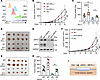 Figure 7
SIRT2 KO enhances antitumor immunity through downregulating PD-L1. (A–D) WT, SIRT2-KO, and PD-L1–reconstituted WT or SIRT2-KO MB49 cells were implanted subcutaneously into the right flanks of C57BL/6J mice (n = 5). The PD-L1 expression levels (A), tumor volume (B), tumor weight (C), and representative tumor images (D) are shown. (E–H) WT, SIRT2-KO, and USP22-reconstituted WT or SIRT2-KO MB49 cells were implanted subcutaneously into the right flanks of C57BL/6J mice (n = 5). The USP22 expression levels (E), tumor volume (F), representative tumor images (G), and tumor weight (H) are shown. (I) A proposed pathway for SIRT2 in tumor immune evasion. Scale bars: 1 cm. Data are shown as mean ± SD. Statistical significance was determined using 1-way ANOVA. *P < 0.05; **P < 0.01; ***P < 0.001.
Next, we stably overexpressed USP22 in both WT and SIRT2-deficient MB49 tumor cells to determine whether, similar to PD-L1, USP22 reconstitution could reverse the antitumor effects of SIRT2 inhibition (Figure 7E). As expected, enforced USP22 expression significantly accelerated tumor growth in WT MB49 cells, further supporting its oncogenic role. Importantly, stable reconstitution of USP22 in SIRT2-null MB49 cells largely restored tumor growth to levels comparable to WT tumors (Figure 7, F–H). Similar to PD-L1, USP22 levels were modestly reduced in SIRT2-KO tumor cells compared with WT cells under ectopic expression conditions, likely due to loss of SIRT2-mediated protection of USP22 from ubiquitination-dependent degradation (Figure 7E). These findings indicate that SIRT2 inhibition enhances tumor rejection primarily through downregulation of USP22 protein expression, thereby attenuating USP22-mediated PD-L1 upregulation and tumor immune evasion (Figure 7I). Therefore, targeted deletion or inhibition of SIRT2 enhances antitumor immunity by downregulating PD-L1, boosting T cell infiltration and function, and reversing T cell exhaustion, highlighting SIRT2 as a promising immunotherapeutic target in cancer.
SIRT2 inhibition synergizes with anti–PD-1 immune checkpoint blockade to potently suppress tumor growth. Building upon our earlier findings that CRISPR-mediated deletion of SIRT2 suppresses tumor growth and immune evasion via PD-L1 downregulation, we hypothesized that pharmacologic inhibition of SIRT2 could synergize with PD-1 immune checkpoint blockade (ICB) to enhance the efficacy of immunotherapy in bladder cancer. To test this, we treated mice bearing MB49 bladder tumors with either the SIRT2 inhibitor AGK2, anti–PD-1 antibody, or a combination of both agents. While monotherapy with either AGK2 or anti–PD-1 significantly inhibited tumor growth, their combination led to a dramatic and synergistic suppression of tumor progression. Compared with the IgG2a isotype control, mice treated with the combined AGK2 and anti–PD-1 therapy enhanced antitumor immune responses (Figure 8, A–C). Flow cytometry analysis revealed a significant increase in the infiltration of CD8+, but not CD4+, T cells in tumors from the combination group, exceeding the levels observed in either monotherapy group (Figure 8, D–F). Moreover, CD8+ T cells from the combination-treated tumors displayed enhanced effector function, as indicated by increased expression of IFN-γ and granzyme B (Figure 8, D, G, and H). These results suggest that the enhanced antitumor immunity observed with combined SIRT2 inhibition and ICB is primarily driven by reinvigoration of CD8+ cytotoxic T cell activity.
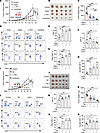 Figure 8
SIRT2 blockade combined with anti–PD-1 therapy effectively inhibited tumor growth. (A–C) MB49 cells were implanted subcutaneously into the right flanks of C57BL/6J mice (n = 5). Mice were treated with AGK2 (10 mg/kg), PD-1 antibody (50 μg per mouse), IgG2a isotype control (50 μg per mouse), or a combination via intraperitoneal injection on day 7. Tumor growth (A), tumor images (B), and tumor weight (C) are shown. (D) Representative flow cytometry plots showing CD4+, CD8+, granzyme B+, and IFN-γ+CD8+ T cells among CD45+ tumor-infiltrating cells (n = 5). (E–H) Quantification of CD8+ T cells (E), CD4+ T cells (F), IFN-γ+CD8+ T cells (G), and granzyme B+CD8+ T cells (H) within the CD45+ cell population. (I–K) MB49 cells were implanted subcutaneously into the right flanks of C57BL/6J mice (n = 5). Mice were treated with TM (20 mg/kg), PD-1 antibody (50 μg per mouse), IgG2a isotype control (50 μg per mouse), or a combination via intraperitoneal injection on day 7. Tumor growth (I), tumor images (J), and tumor weight (K) are shown. (L) Representative flow cytometry plots showing CD4+, CD8+, granzyme B+ (GB), and IFN-γ+CD8+ T cells among CD45+ tumor-infiltrating cells (n = 5). (M–O) Quantification of CD8+ T cells (M), IFN-γ+CD8+ T cells (N), and granzyme B+CD8+ T cells (O) within the CD45+ cell population. Scale bars: 1 cm. Data are shown as mean ± SD. Statistical significance was determined using 1-way ANOVA. *P < 0.05; **P < 0.01; ***P < 0.001.
AGK2 has been reported to exhibit only modest selectivity over SIRT1 and SIRT3, whereas comparative studies indicate that TM is among the most selective SIRT2 inhibitors (51, 52). However, unlike SIRT2, CRISPR-targeted deletion of SIRT1 or SIRT3 did not affect USP22 protein expression levels (Supplemental Figure 6). Consistent with our observations in SIRT2-KO tumor cells, treatment with a more specific SIRT2 inhibitor, TM, suppressed both PD-L1 and USP22 expression (Supplemental Figure 5). These results indicate that specific SIRT2 inhibition enhanced antitumor immunity. To support this, we further demonstrated that in vivo administration of TM to tumor-bearing mice significantly inhibited tumor growth (Figure 8, I–K). Immunophenotypic analysis revealed a robust increase in intratumoral CD8+ T cell infiltration, accompanied by enhanced production of IFN-γ, granzyme B, and TNF-α, indicating potentiation of antitumor effector function (Figure 8, L–O). Collectively, these findings demonstrate that SIRT2 inhibition boosts CD8+ T cell antitumor immunity, at least in part through downregulation of PD-L1 expression.
Combined SIRT2 and USP22 inhibition exhibits potent antitumor immunity. Given that USP22 and SIRT2 exert both overlapping and distinct tumor-promoting functions, there is a strong rationale for their combined inhibition to enhance antitumor immunity. Consistent with this concept, inhibition of either USP22 or SIRT2 alone significantly suppressed tumor growth, and dual targeting produced a greater effect. Specifically, cotreatment with the USP22 inhibitor S02 and the SIRT2 inhibitor AGK2 in established tumor models led to near-complete tumor suppression, supporting a cooperative, and potentially synergistic, interaction (Figure 9, A–C).
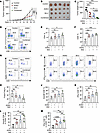 Figure 9
Combined SIR2 and USP22 inhibition in cancer treatment. (A–C) MB49 cells were implanted subcutaneously into the right flanks of C57BL/6J mice (n = 5). Mice were treated with AGK2 (10 mg/kg), USP22i-S02 (20 mg/kg), or a combination via intraperitoneal injection on day 7. Tumor growth (A), tumor images (B), and tumor weight (C) are shown. Scale bar: 1 cm. (D–F) Tumor-infiltrated CD4+ and CD8+ T cells were analyzed by flow cytometry. Representative flow cytometry plots showing CD4+ and CD8+ T cells (D) and data for 5 mice from each group (E and F) are shown. (G and H) CD4+FoxP3+ Tregs in gated CD4+ T cells were analyzed. (I–M) Granzyme B+ and IFN-γ+CD8+ T cells were determined by intracellular staining. Representative images (I) and granzyme B+ and IFN-γ+ frequency in CD8+ T cells (J and K) or CD45+ cells (L and M) are shown. Data are shown as mean ± SD. Statistical significance was determined using 1-way ANOVA. *P < 0.05; **P < 0.01; ***P < 0.001.
Further immune profiling revealed an increase in CD8+ T cell infiltration following treatment with either USP22i-S02 or AGK2, with the combination producing the most pronounced effect. In contrast, CD4+ T cell infiltration remained largely unchanged (Figure 9, D–F). Consistent with our recent findings, USP22 inhibition significantly reduced intratumoral Treg populations, an effect that was further enhanced by cotreatment with the SIRT2 inhibitor AGK2 (Figure 9, G and H). Functional analyses demonstrated a robust enhancement of CD8+ T cell effector activity by either USP22i-SO2 or AKG2 treatment, as evidenced by increased production of IFN-γ and granzyme B, and this efficacy was further enhanced by their combination (Figure 9, I–M). Collectively, these results indicate that combined treatment with S02 and AGK2 potentiates intratumoral antitumor immunity in tumor-bearing mice in a clinically relevant setting.
Positive correlation of SIRT2 with USP22 and PD-L1 expression in human cancers. Our data clearly demonstrate that SIRT2 enhances PD-L1 expression by deacetylating USP22 to promote USP22 self-deubiquitination. To evaluate the clinical relevance of these findings in human cancers, we performed IHC analysis of SIRT2, USP22, and PD-L1 protein expression, as well as CD8+ T cell infiltration in 74 bladder carcinomas, with adjacent nontumorous tissues as controls. Quantitative analysis revealed significantly elevated levels of SIRT2, USP22, and PD-L1 proteins in tumor tissues compared with adjacent nontumorous (para-cancerous) tissues (Figure 10, A and B). Consistent with our mechanistic findings, SIRT2 expression showed a strong positive correlation with both USP22 and PD-L1 levels in bladder carcinoma (Figure 10, C and D). Furthermore, USP22 and PD-L1 expression were also positively correlated in both cancer types (Figure 10E). In contrast, analysis of The Cancer Genome Atlas (TCGA) database did not show their positive correlation at mRNA levels (Supplemental Figure 13), further supporting our observations that SIRT2 regulates both USP22 and PD-L1 at the posttranscriptional level.
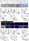 Figure 10
Positive correlation of SIRT2 with PD-L1 expression in human bladder cancer. A TMA with 70 bladder cancer sections with control adjacent normal tissue was used for the staining. (A) Representative IHC staining images of SIRT2, PD-L1, USP22, and CD8 in human bladder carcinoma and adjacent para-cancerous tissues. Scale bars: 100 μm. (B–F) Quantification of staining intensities for SIRT2, PD-L1, USP22, and CD8 in bladder carcinoma and adjacent tissues (n = 70). Correlation analysis of SIRT2 with PD-L1 (D) and USP22 (C), USP22 with PD-L1 (E), and SIRT2 with CD8 (F) protein levels in bladder carcinoma (n = 70). (G) Representative immunofluorescence staining images of SIRT2, PD-L1, USP22, and CD8 in human bladder carcinoma and adjacent para-cancerous tissues. Scale bars: 100 μm. (H) Quantification of staining intensities for SIRT2, PD-L1, USP22, and CD8 in bladder carcinoma and adjacent tissues (n = 70). (I–L) Correlation analysis of SIRT2 with PD-L1 (I) and USP22 (J), USP22 with PD-L1 (K), and SIRT2 with CD8 (L) protein levels in bladder carcinoma (n = 70). Data are shown as mean ± SD. Statistical significance was determined using an unpaired 2-tailed Student’s t test for B and H or Pearson’s correlation analysis for C–F and I–L. *P < 0.05; **P < 0.01; ***P < 0.001.
In addition, analysis of a melanoma tissue microarray (TMA) comprising 70 patient samples revealed elevated expression of SIRT2, USP22, and PD-L1, with strong positive correlations among all 3 markers (Supplemental Figure 14). These findings further support the notion that the SIRT2/USP22/PD-L1 axis represents a potentially conserved mechanism of tumor immune evasion across cancer types.
Given our data indicating that SIRT2 promotes PD-L1 expression, higher SIRT2 levels would be expected to associate with diminished CD8+ T cell–mediated antitumor immunity. However, the inverse correlation between SIRT2 expression and CD8+ T cell infiltration was modest and did not reach statistical significance by IHC (Figure 10F). Recognizing the limited sensitivity of IHC for detecting low-frequency CD8+ T cells, we further performed immunofluorescence staining. This analysis confirmed elevated expression of SIRT2, USP22, and PD-L1 in tumor tissues, with significant positive correlations among these markers (Figure 10, G–K). Consistently, the negative correlation between SIRT2 expression and CD8+ T cell infiltration remained weak and did not reach statistical significance (Figure 10L).
In summary, our study uncovers a regulatory pathway in which SIRT2-mediated deacetylation of USP22 prevents its self-ubiquitination, thereby upregulating PD-L1 expression. These findings not only deepen our understanding of PD-L1 regulation but also suggest that targeting SIRT2 in combination with PD-1/PD-L1 ICB represents a promising therapeutic strategy to enhance antitumor efficacy (Supplemental Figure 15).


Copyright © 2026 American Society for Clinical Investigation
ISSN: 0021-9738 (print), 1558-8238 (online)