
Advertisement
Clinical Research and Public HealthEndocrinologyNeuroscienceOtology
Open Access |  10.1172/JCI197932
Estrogen deficiency and risk of hearing loss in pediatric Turner syndrome
Yan Huang,1,2 Liyang Liang,3 Yanfang Ye,4 Lina Zhang,3 Li Ling,4,5 Zhe Meng,3 Wei Liu,1,2 Jia Guo,1,2 Zulin Liu,3 Zhen Zhao,1,2 Zhigang Zhang,1,2 and Yu Si1,2
1Department of Otorhinolaryngology,
2Institute of Hearing and Speech, and
3Department of Pediatrics, Sun Yat-sen Memorial Hospital, Sun Yat-sen University, Guangzhou, China.
4Clinical Research Design Division, Clinical Research Centre of Sun Yat-sen Memorial Hospital, Sun Yat-sen University, Guangzhou, China.
5Department of Medical Statistics, School of Public Health, Sun Yat-sen University, Guangzhou, China.
Address correspondence to: Yu Si or Zhigang Zhang, Department of Otorhinolaryngology, Sun Yat-sen Memorial Hospital, Sun Yat-sen University, Yan Jiang Road 107, Guangzhou, China. Phone: 86.16620081695; Email: siyu5@mail.sysu.edu.cn (YS). Phone: 86.13926108261; Email: zhangzhg@mail.sysu.edu.cn (Zhigang Zhang).
Authorship note: YH and LL contributed equally to this work.
Find articles by Huang, Y. in: PubMed | Google Scholar
1Department of Otorhinolaryngology,
2Institute of Hearing and Speech, and
3Department of Pediatrics, Sun Yat-sen Memorial Hospital, Sun Yat-sen University, Guangzhou, China.
4Clinical Research Design Division, Clinical Research Centre of Sun Yat-sen Memorial Hospital, Sun Yat-sen University, Guangzhou, China.
5Department of Medical Statistics, School of Public Health, Sun Yat-sen University, Guangzhou, China.
Address correspondence to: Yu Si or Zhigang Zhang, Department of Otorhinolaryngology, Sun Yat-sen Memorial Hospital, Sun Yat-sen University, Yan Jiang Road 107, Guangzhou, China. Phone: 86.16620081695; Email: siyu5@mail.sysu.edu.cn (YS). Phone: 86.13926108261; Email: zhangzhg@mail.sysu.edu.cn (Zhigang Zhang).
Authorship note: YH and LL contributed equally to this work.
Find articles by Liang, L. in: PubMed | Google Scholar
1Department of Otorhinolaryngology,
2Institute of Hearing and Speech, and
3Department of Pediatrics, Sun Yat-sen Memorial Hospital, Sun Yat-sen University, Guangzhou, China.
4Clinical Research Design Division, Clinical Research Centre of Sun Yat-sen Memorial Hospital, Sun Yat-sen University, Guangzhou, China.
5Department of Medical Statistics, School of Public Health, Sun Yat-sen University, Guangzhou, China.
Address correspondence to: Yu Si or Zhigang Zhang, Department of Otorhinolaryngology, Sun Yat-sen Memorial Hospital, Sun Yat-sen University, Yan Jiang Road 107, Guangzhou, China. Phone: 86.16620081695; Email: siyu5@mail.sysu.edu.cn (YS). Phone: 86.13926108261; Email: zhangzhg@mail.sysu.edu.cn (Zhigang Zhang).
Authorship note: YH and LL contributed equally to this work.
Find articles by Ye, Y. in: PubMed | Google Scholar
1Department of Otorhinolaryngology,
2Institute of Hearing and Speech, and
3Department of Pediatrics, Sun Yat-sen Memorial Hospital, Sun Yat-sen University, Guangzhou, China.
4Clinical Research Design Division, Clinical Research Centre of Sun Yat-sen Memorial Hospital, Sun Yat-sen University, Guangzhou, China.
5Department of Medical Statistics, School of Public Health, Sun Yat-sen University, Guangzhou, China.
Address correspondence to: Yu Si or Zhigang Zhang, Department of Otorhinolaryngology, Sun Yat-sen Memorial Hospital, Sun Yat-sen University, Yan Jiang Road 107, Guangzhou, China. Phone: 86.16620081695; Email: siyu5@mail.sysu.edu.cn (YS). Phone: 86.13926108261; Email: zhangzhg@mail.sysu.edu.cn (Zhigang Zhang).
Authorship note: YH and LL contributed equally to this work.
Find articles by Zhang, L. in: PubMed | Google Scholar
1Department of Otorhinolaryngology,
2Institute of Hearing and Speech, and
3Department of Pediatrics, Sun Yat-sen Memorial Hospital, Sun Yat-sen University, Guangzhou, China.
4Clinical Research Design Division, Clinical Research Centre of Sun Yat-sen Memorial Hospital, Sun Yat-sen University, Guangzhou, China.
5Department of Medical Statistics, School of Public Health, Sun Yat-sen University, Guangzhou, China.
Address correspondence to: Yu Si or Zhigang Zhang, Department of Otorhinolaryngology, Sun Yat-sen Memorial Hospital, Sun Yat-sen University, Yan Jiang Road 107, Guangzhou, China. Phone: 86.16620081695; Email: siyu5@mail.sysu.edu.cn (YS). Phone: 86.13926108261; Email: zhangzhg@mail.sysu.edu.cn (Zhigang Zhang).
Authorship note: YH and LL contributed equally to this work.
Find articles by Ling, L. in: PubMed | Google Scholar
1Department of Otorhinolaryngology,
2Institute of Hearing and Speech, and
3Department of Pediatrics, Sun Yat-sen Memorial Hospital, Sun Yat-sen University, Guangzhou, China.
4Clinical Research Design Division, Clinical Research Centre of Sun Yat-sen Memorial Hospital, Sun Yat-sen University, Guangzhou, China.
5Department of Medical Statistics, School of Public Health, Sun Yat-sen University, Guangzhou, China.
Address correspondence to: Yu Si or Zhigang Zhang, Department of Otorhinolaryngology, Sun Yat-sen Memorial Hospital, Sun Yat-sen University, Yan Jiang Road 107, Guangzhou, China. Phone: 86.16620081695; Email: siyu5@mail.sysu.edu.cn (YS). Phone: 86.13926108261; Email: zhangzhg@mail.sysu.edu.cn (Zhigang Zhang).
Authorship note: YH and LL contributed equally to this work.
Find articles by Meng, Z. in: PubMed | Google Scholar
1Department of Otorhinolaryngology,
2Institute of Hearing and Speech, and
3Department of Pediatrics, Sun Yat-sen Memorial Hospital, Sun Yat-sen University, Guangzhou, China.
4Clinical Research Design Division, Clinical Research Centre of Sun Yat-sen Memorial Hospital, Sun Yat-sen University, Guangzhou, China.
5Department of Medical Statistics, School of Public Health, Sun Yat-sen University, Guangzhou, China.
Address correspondence to: Yu Si or Zhigang Zhang, Department of Otorhinolaryngology, Sun Yat-sen Memorial Hospital, Sun Yat-sen University, Yan Jiang Road 107, Guangzhou, China. Phone: 86.16620081695; Email: siyu5@mail.sysu.edu.cn (YS). Phone: 86.13926108261; Email: zhangzhg@mail.sysu.edu.cn (Zhigang Zhang).
Authorship note: YH and LL contributed equally to this work.
Find articles by Liu, W. in: PubMed | Google Scholar
1Department of Otorhinolaryngology,
2Institute of Hearing and Speech, and
3Department of Pediatrics, Sun Yat-sen Memorial Hospital, Sun Yat-sen University, Guangzhou, China.
4Clinical Research Design Division, Clinical Research Centre of Sun Yat-sen Memorial Hospital, Sun Yat-sen University, Guangzhou, China.
5Department of Medical Statistics, School of Public Health, Sun Yat-sen University, Guangzhou, China.
Address correspondence to: Yu Si or Zhigang Zhang, Department of Otorhinolaryngology, Sun Yat-sen Memorial Hospital, Sun Yat-sen University, Yan Jiang Road 107, Guangzhou, China. Phone: 86.16620081695; Email: siyu5@mail.sysu.edu.cn (YS). Phone: 86.13926108261; Email: zhangzhg@mail.sysu.edu.cn (Zhigang Zhang).
Authorship note: YH and LL contributed equally to this work.
Find articles by Guo, J. in: PubMed | Google Scholar
1Department of Otorhinolaryngology,
2Institute of Hearing and Speech, and
3Department of Pediatrics, Sun Yat-sen Memorial Hospital, Sun Yat-sen University, Guangzhou, China.
4Clinical Research Design Division, Clinical Research Centre of Sun Yat-sen Memorial Hospital, Sun Yat-sen University, Guangzhou, China.
5Department of Medical Statistics, School of Public Health, Sun Yat-sen University, Guangzhou, China.
Address correspondence to: Yu Si or Zhigang Zhang, Department of Otorhinolaryngology, Sun Yat-sen Memorial Hospital, Sun Yat-sen University, Yan Jiang Road 107, Guangzhou, China. Phone: 86.16620081695; Email: siyu5@mail.sysu.edu.cn (YS). Phone: 86.13926108261; Email: zhangzhg@mail.sysu.edu.cn (Zhigang Zhang).
Authorship note: YH and LL contributed equally to this work.
Find articles by Liu, Z. in: PubMed | Google Scholar
1Department of Otorhinolaryngology,
2Institute of Hearing and Speech, and
3Department of Pediatrics, Sun Yat-sen Memorial Hospital, Sun Yat-sen University, Guangzhou, China.
4Clinical Research Design Division, Clinical Research Centre of Sun Yat-sen Memorial Hospital, Sun Yat-sen University, Guangzhou, China.
5Department of Medical Statistics, School of Public Health, Sun Yat-sen University, Guangzhou, China.
Address correspondence to: Yu Si or Zhigang Zhang, Department of Otorhinolaryngology, Sun Yat-sen Memorial Hospital, Sun Yat-sen University, Yan Jiang Road 107, Guangzhou, China. Phone: 86.16620081695; Email: siyu5@mail.sysu.edu.cn (YS). Phone: 86.13926108261; Email: zhangzhg@mail.sysu.edu.cn (Zhigang Zhang).
Authorship note: YH and LL contributed equally to this work.
Find articles by Zhao, Z. in: PubMed | Google Scholar
1Department of Otorhinolaryngology,
2Institute of Hearing and Speech, and
3Department of Pediatrics, Sun Yat-sen Memorial Hospital, Sun Yat-sen University, Guangzhou, China.
4Clinical Research Design Division, Clinical Research Centre of Sun Yat-sen Memorial Hospital, Sun Yat-sen University, Guangzhou, China.
5Department of Medical Statistics, School of Public Health, Sun Yat-sen University, Guangzhou, China.
Address correspondence to: Yu Si or Zhigang Zhang, Department of Otorhinolaryngology, Sun Yat-sen Memorial Hospital, Sun Yat-sen University, Yan Jiang Road 107, Guangzhou, China. Phone: 86.16620081695; Email: siyu5@mail.sysu.edu.cn (YS). Phone: 86.13926108261; Email: zhangzhg@mail.sysu.edu.cn (Zhigang Zhang).
Authorship note: YH and LL contributed equally to this work.
Find articles by Zhang, Z. in: PubMed | Google Scholar
1Department of Otorhinolaryngology,
2Institute of Hearing and Speech, and
3Department of Pediatrics, Sun Yat-sen Memorial Hospital, Sun Yat-sen University, Guangzhou, China.
4Clinical Research Design Division, Clinical Research Centre of Sun Yat-sen Memorial Hospital, Sun Yat-sen University, Guangzhou, China.
5Department of Medical Statistics, School of Public Health, Sun Yat-sen University, Guangzhou, China.
Address correspondence to: Yu Si or Zhigang Zhang, Department of Otorhinolaryngology, Sun Yat-sen Memorial Hospital, Sun Yat-sen University, Yan Jiang Road 107, Guangzhou, China. Phone: 86.16620081695; Email: siyu5@mail.sysu.edu.cn (YS). Phone: 86.13926108261; Email: zhangzhg@mail.sysu.edu.cn (Zhigang Zhang).
Authorship note: YH and LL contributed equally to this work.
Find articles by Si, Y. in: PubMed | Google Scholar
Published March 3, 2026 - More info
J Clin Invest. 2026;136(9):e197932. https://doi.org/10.1172/JCI197932.
© 2026 Huang et al. This work is licensed under the Creative Commons Attribution 4.0 International License. To view a copy of this license, visit http://creativecommons.org/licenses/by/4.0/.
Received: October 8, 2025; Accepted: February 26, 2026


-
Results
Description of the cohort.
A total of 211 potential participants with TS were initially assessed for eligibility between January, 2016 and March, 2024. After excluding 118 ineligible individuals (110 due to age >12 years and 8 with preexisting HL), 93 patients under 12 years of age were enrolled in the main prospective cohort. Following the study design, as participants reached 12 years of age, they entered the ERT-focused substudy. To date, 56 children (60.2%) from the original cohort have progressed into this follow-up substudy for longitudinal evaluation of hearing thresholds under exogenous ERT. After excluding 6 patients from the main cohort and 2 from the substudy cohort due to insufficient follow-up data (only 1 hearing test), longitudinal data from 87 participants were available for the final analysis of HL risk in the main study, while 54 participants were included in the substudy analysis of ERT (Figure 1).
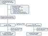 Figure 1
Flowchart of study enrollment. The ChiCTR2300068063 cohort is an ongoing cohort study, with continuous follow-up, and without a prespecified end date. The number of patients included in the cohort between January, 2016 (start of the study) and March, 2024 is presented. The “<12 Years Old” cohort (n = 87) was stratified into E2-deficient (n = 48) and E2-normal (n = 39) groups to evaluate the association between endogenous E2 status and hearing loss. The “≥12 Years Old” substudy cohort (n = 54) was used to analyze the impact of initiating exogenous E2 replacement therapy on hearing thresholds. E2, estradiol.
To ensure transparent reporting, this study adhered to the STROBE guidelines (17). The median age of children in the main cohort was 8 years (IQR 7.00–10.00). Based on CALIPER-defined thresholds (see Methods) and longitudinal profiling, 87 children with TS were stratified into an E2-deficient group (n = 48; mean E2 = 20.20 pmol/L, 95% CI 18.06–22.34) and an E2-normal group (n = 39; mean E2 = 86.11 pmol/L, 95% CI 71.39–100.82; P < 0.001, Supplemental Figure 1; supplemental material available online with this article; https://doi.org/10.1172/JCI197932DS1). The entire cohort exhibited heterogeneous karyotypes (45,X, 31.0%; mosaicism or structural variants, 69.0%) and a high prevalence of systemic comorbidities, including scoliosis (39.1%), renal dysfunction (31.0%), and thyroid disorders (24.1%). A key finding is that the E2-deficient subgroup and the E2-normal subgroup did not show statistically significant differences in demographic characteristics, karyotype, bone age, or prevalence of major systemic complications (including cardiovascular, metabolic, thyroid, and kidney diseases) (all P > 0.05). The primary divergence between the groups was confined to markers of gonadal function; the E2-deficient subgroup had significantly smaller ovarian volume (0.57 vs. 1.26 mL, P = 0.005), smaller uterine volume (1.61 vs. 4.53 mL, P = 0.029), and lower testosterone levels (0.34 vs. 0.55 nmol/L, P = 0.001), which is consistent with a phenotype of more severe primary ovarian insufficiency. This indicates that, aside from the expected gonadal function differences, the 2 groups were well balanced in terms of karyotype and major systemic comorbidities at baseline (Table 1 and Supplemental Table 1).
Association between estrogen deficiency and incident HL.
The analysis revealed a significant association between estrogen deficiency and incident HL over a median follow-up of 35 months (IQR 23–46 months). Among 87 patients with TS in the main cohort, 38 cases of HL were diagnosed. In estrogen-deficient individuals, the incidence of HL (27 cases, 56.3%; 20.6 events per 100 person-years [PY]) was significantly higher than that in estrogen-normal individuals (11 cases, 28.2%; 8.6 events per 100 PY). In the unadjusted analysis, estrogen deficiency conferred an increased risk of HL by 2.65-fold (95% CI 1.30–5.38; log-rank P = 0.005) (Figure 2). This association persisted and became more significant after stepwise adjustment of the model; after controlling for karyotype and peak growth hormone (GH) levels (Model 1), the hazard ratio (HR) remained 2.34 (95% CI 1.09–5.03; P = 0.031); further adjustment for IGF-1 concentration, height standard deviation score (SDS), and thyroid disorders (Model 2) yielded an HR of 2.59 (95% CI 1.12–5.98; P = 0.027); and in the most comprehensive model incorporating follicle-stimulating hormone (FSH) and luteinizing hormone (LH) concentrations and ovarian volume (Model 3), the risk further increased to an HR of 2.93 (95% CI 1.21–7.12; P = 0.019). The stable increase in the model HR value confirmed the independent effect of estrogen deficiency on the pathogenesis of HL, unaffected by growth, metabolism, and gonadal reserve factors (Table 2).
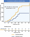 Figure 2
Kaplan-Meier curve of the cumulative incidence of HL. Kaplan-Meier analysis showing cumulative incidence of HL in 87 patients with TS, stratified by estrogen status. Cox regression yielded an unadjusted HR of 2.65 (95% CI 1.30–5.38); log-rank P < 0.01. HL, hearing loss; TS, Turner syndrome.
In order to further describe this association in the entire auditory spectrum, a single-frequency analysis of pure tone audiometry was performed. The results show that the effect of estrogen deficiency is more obvious at a lower frequency. The HR was highest at 500 Hz (HR 2.83, 95% CI 1.53–5.23; log-rank P < 0.001), followed by 1 kHz (HR 2.73, 95% CI 1.47–5.08; P = 0.001), 2 kHz (HR 2.11, 95% CI 1.10–4.03; P = 0.021), and 4 kHz (HR 2.28, 95% CI 1.14–4.56; P = 0.017). This pattern shows that the HR gradually decreases as the frequency increases. This suggests that estrogen plays a crucial role in maintaining low-frequency hearing in children with TS, which is vital for speech discrimination (Supplemental Figure 2).
Reverse causality and effect modification.
To eliminate potential confounding factors and assess the robustness of the primary findings, we performed a sensitivity analysis on the main cohort. After excluding incident HL cases that occurred within 1 to 3 years after enrollment, the association between estrogen deficiency and HL remained (HR 2.53, 95% CI 1.03–6.24; Supplemental Table 2). The longitudinal analysis suggested that baseline E2 deficiency was associated with an increased risk of new HL. Even after 3 years of follow-up, the risk was 4.70-fold higher (95% CI 0.79–27.96) (Figure 3). Notably, this association was not significantly modified by any prespecified clinical variables, including karyotype category, age strata, markers of gonadal function, or comorbidity status (all interaction P > 0.05; Figure 4), confirming the robustness of our findings across different clinical subgroups.
 Figure 3
Association between estrogen deficiency and incident HL over time. HRs with 95% CIs from multivariable Cox proportional hazards models are shown for different follow-up periods. HRs were adjusted for established risk factors (Model 3 adjustment in Table 2). No corrections for multiple testing were applied. HL, hearing loss.
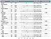 Figure 4
Potential effect modifiers in the association between estrogen deficiency and incident HL. This forest plot displays HRs and 95% CIs from multivariable Cox regression models across predefined subgroups. Stratification factors included baseline age, karyotype, and the presence of thyroid or cardiovascular disorders, as well as hormone levels (FSH, LH) and ovarian volume dichotomized by median values. No significant effect modification was observed across any variables (all interaction P values > 0.05). FSH, follicle-stimulating hormone; LH, luteinizing hormone; i(Xq)/idic(Xp), 46,X,i(Xq);46,X,idic(Xp).
Combined effects of estrogen and established risk factors.
Within the main cohort, we used multivariable Cox proportional hazards models to explore the potential synergistic effect of estrogen deficiency and known comorbidities on HL risk. Specifically, we evaluated the risk of HL associated with E2 deficiency across each comorbidity of interest (dyslipidemia, diabetes mellitus [DM], thyroid disorders, and renal dysfunction). In the comorbidity analysis, children with TS with normal E2 levels and without comorbidities were used as the reference group. All models were adjusted for established risk factors.
Estrogen deficiency may increase the risk of incident HL on top of established comorbidities (Supplemental Figure 3). The HRs for the combined presence of E2 deficiency with dyslipidemia (HR 5.75, 95% CI 1.71–19.31), thyroid disorders (HR 2.09, 95% CI 0.72–6.09), and renal dysfunction (HR 2.00, 95% CI 0.70–5.74) exceeded those of dyslipidemia (HR 4.22, 95% CI 1.03–17.21), thyroid disorders (HR 1.05, 95% CI 0.25–4.45), and renal dysfunction (HR 0.82, 95% CI 0.23–2.91) alone. Regarding DM, the combined risk of E2 deficiency and DM (HR 3.89, 95% CI 1.08–14.02) appeared to be higher than that of E2 deficiency alone (HR 2.67, 95% CI 1.15–6.17), although it did not exceed the risk associated with DM alone. In summary, estrogen deficiency may synergistically increase the risk of HL in children with TS with dyslipidemia, renal dysfunction, and possibly thyroid disease, although these observations require confirmation in larger studies due to the limited sample sizes in the present comorbidity analyses (Supplemental Figure 3).
Association between estrogen deficiency and HL types.
Analysis of 38 incident HL cases within the TS main cohort (n = 87) identified SNHL as the predominant type (68.4%, n = 26), with CHL accounting for 31.6% (n = 12). By utilizing cause-specific Cox proportional hazards models to evaluate differential associations by E2 status, we observed that incidence rates for different types of HL were consistently higher in E2-deficient patients than in those with normal E2 levels (Figure 5).
 Figure 5
Estradiol-dependent hearing loss risk stratified by audiometric subtypes. Kaplan-Meier curves showing the cumulative incidence of sensorineural hearing loss (SNHL) and conductive hearing loss (CHL) in 87 patients with TS, stratified by estrogen status. For SNHL, Cox regression yielded an unadjusted HR of 2.49 (95% CI 1.07–5.79); log-rank P = 0.028. For CHL, the unadjusted HR was 3.03 (95% CI 0.81–11.25); log-rank P = 0.083.
The E2-deficient group exhibited a significantly higher incidence rate of SNHL (13.7 events per 100 PY) compared with the E2-normal group (6.2 events per 100 PY). In the unadjusted model, E2 deficiency was associated with a 2.49-fold increased risk of developing SNHL (HR 2.49, 95% CI 1.07–5.79). Likewise, the incidence rate of CHL was higher in the E2-deficient group (6.9 events per 100 PY) than in the E2-normal group (2.3 events per 100 PY). The unadjusted HR indicated a 3.03-fold increased risk of CHL associated with E2 deficiency (HR 3.03, 95% CI 0.81–11.25). Multivariable adjustment for both CHL and SNHL outcomes did not substantially alter these associations (Figure 5 and Table 3).
Influence of abnormal DPOAEs.
In the main TS cohort, serial follow-up data for distortion product otoacoustic emissions (DPOAEs) were available for 63 patients, among whom 24 (38.1%) exhibited abnormal results. This observation aligns with the known high prevalence of hearing impairment in TS. Of these, 19 cases (48.7%; incidence: 19.8 per 100 PY) were observed in the E2-deficient group, compared with 5 cases (20.8%; incidence: 6.5 per 100 PY) in the E2-normal group. Notably, E2 deficiency independently predicted abnormal DPOAEs (HR 3.98, 95% CI 1.35–11.76; P = 0.007), with a greater impact than the incidence of HL (Figure 6). Moreover, further adjustments did not significantly alter the estimated HRs associated with E2 deficiency and abnormal DPOAEs (Supplemental Table 3).
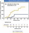 Figure 6
Kaplan-Meier curves of DPOAE abnormality incidence by E2 status. Kaplan-Meier analysis demonstrated the cumulative incidence of DPOAE abnormalities in 63 patients with TS, stratified by estrogen status. Cox regression revealed an unadjusted HR of 3.98 (95% CI 1.35–11.76; log-rank P < 0.01). DPOAE, distortion product otoacoustic emission.
To investigate the possibility of a frequency-specific association between DPOAE abnormalities and HL, we performed stratified Kaplan-Meier survival analysis at 9 frequencies (from 552 to 8838 Hz). In the low-frequency range, the cumulative incidence curve in the E2-deficient group was steeper than that in the E2-normal group and was significant (log-rank P < 0.05) at 552, 781, and 1104 Hz. Similarly, in the high-frequency range, the HR values increased, especially at 4419 Hz and 6250 Hz (both P < 0.05), which might reflect more obvious cochlear dysfunction at these frequencies. In contrast, the differences in the mid-frequency range seemed less significant, reaching statistical significance only at 2207 Hz (P = 0.022). Overall, these findings suggest a possible frequency-specific pattern, where estrogen deficiency seems to be associated with more significant DPOAE abnormalities, mainly affecting low and high frequencies, which may be related to the progressive HL pattern observed clinically (Supplemental Figure 4).
Impact of ERT on hearing preservation in the substudy.
Given the established association between estrogen deficiency and HL in the primary study population, we sought to assess whether early ERT is associated with better hearing in the peripubertal substudy. To this end, we performed a cross-sectional analysis of hearing thresholds in 54 children with TS. For patients receiving ERT (the ERT-exposed group), the regimen consisted of oral micronized 17β-estradiol, initiated at 0.25 mg/day and titrated upwards over 2–4 years to an adult maintenance dose. Concurrent ERT was associated with major bilateral hearing preservation during key developmental stages. As illustrated in Figure 7, ERT-exposed patients consistently demonstrated lower pure-tone average (PTA) thresholds compared with their ERT-unexposed counterparts across ages 15–17 years, with statistically robust differences (all P < 0.05). Considerable differences were observed for both ears at age 15 (left: 14.89 vs. 25.5 dB, P = 0.025; right: 16.22 vs. 25.88 dB, P = 0.036), age 16 (left: 15.89 vs. 27.6 dB, P = 0.012; right: 17.11 vs. 28.6 dB, P = 0.011), and age 17 (left: 19.1 vs. 28.2 dB, P = 0.012; right: 19.6 vs. 29.2 dB, P = 0.016) (Figure 7).
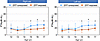 Figure 7
Age-banded hearing thresholds by ERT exposure status. Bilateral pure-tone average (PTA) thresholds, stratified by age and estrogen replacement therapy (ERT) exposure status, are shown. Hearing thresholds were consistently lower in ERT-exposed patients (red line) compared with unexposed counterparts (blue line) across key developmental stages. Statistical analysis using independent sample t tests revealed significant hearing preservation effects associated with ERT exposure at ages 15, 16, and 17 years (*P < 0.05 for all 3). Data are presented as mean ± SD; error bars indicate the dispersion of measurements around the mean. Y, years old; dB, decibel; HL, hearing level.
A total of 31 children were followed for more than 1 year (median 46 months, IQR 24–60), enabling an assessment of annual hearing decline rates. These 31 children were further divided into 20 children who began ERT between the ages of 12 and 15 years (ERT group) and 11 children who did not receive ERT during this period (Non-ERT). Baseline characteristics were generally comparable between the ERT and non-ERT subgroups. The mean age was comparable between the ERT and non-ERT groups (13.6 vs. 13.95 years). Significant differences were observed only in height SDS (ERT: –1.59 vs. Non-ERT: –2.49, P = 0.019) and ovarian volume (0.36 mL vs. 1.12 mL, P = 0.042), while all other demographic, hormonal, and physical measures showed no statistically significant differences (all P > 0.05) (Supplemental Table 4).
The longitudinal analysis of hearing trajectories demonstrated a sustained protective effect on hearing in the ERT group. The mean annual HL rate in the left ear was significantly higher in the Non-ERT group than in the ERT group (1.04 ± 1.86 vs. –1.85 ± 3.07 dB/y, P < 0.001). Similarly, the mean annual HL rate for the right ear was 1.13 ± 2.25 dB/y in the Non-ERT group, compared with –1.24 ± 2.72 dB/y in the ERT group (P = 0.001). Compared with the Non-ERT group, the annual HL rate was reduced significantly in those who initiated ERT before age 15, indicating that early intervention may alleviate long-term HL in children with TS (Figure 8).
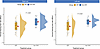 Figure 8
Effect of ERT on the annual hearing loss rate. This cloud plot illustrates the annual rate of hearing loss (dB/year) in patients receiving ERT versus untreated patients (Non-ERT). Each data point represents the annual rate of hearing loss for an individual ear. The plot integrates a half-violin plot (density distribution), a box-and-whisker plot (median [line], IQR [box bounds]), and raw data points. The mean for each group is indicated by a red diamond. An independent sample t test revealed a statistically significant difference in hearing loss progression between the 2 groups (P = 0.001). ERT, estrogen replacement therapy.
Sex hormone profiles and PTA association.
To elucidate the mechanism of action of ERT, we analyzed the levels of endogenous sex hormones in patients with TS in detail and assessed their association with hearing parameters. Longitudinal hormone level analysis in patients with TS revealed severely impaired gonadal function, characterized by persistently low E2 levels, with significantly lower E2 levels across all age groups (0–19 years) compared with the CALIPER healthy female control group (P < 0.001, Supplemental Figure 5). Notably, untreated patients with TS after age 12 typically lacked a spontaneous increase in E2 during puberty (P < 0.001 compared with CALIPER reference values). Longitudinal E2 level monitoring in 67 prepubertal patients with a median follow-up of 21.65 months showed limited intraindividual variability, with a median variation of 0.02 pmol/L (IQR 0.0–18.37 pmol/L). In most patients (79.2%), E2 levels remained stable within 0.5 standard deviations (SD) of baseline measurements (Supplemental Figures 6 and 7). Although E2 levels in adolescents with TS who started ERT after age 12 were higher than in untreated age-matched patients with TS (P = 0.008), these concentrations were still significantly lower than the physiological normal values in healthy children (P < 0.001; Supplemental Figure 8). This E2 deficiency was accompanied by systemic hypergonadotropic hypogonadism, characterized by persistently elevated FSH and LH (P < 0.001, Figure 9) and persistent testosterone/progesterone suppression across all ages (P < 0.001). Age-stratified analysis revealed different patterns of hormone-hearing associations. While circulating FSH, LH, progesterone, testosterone, and prolactin (PRL) levels showed weakly positive or no correlation with hearing thresholds across all age groups, E2 was the only hormone consistently negatively correlated (i.e., potentially protective), especially during puberty and beyond (11 to <19 years; R = –0.19 to –0.22; Supplemental Figure 9). Although these correlations did not reach statistical significance in the stratified study population, this distinct trend highlights the unique and central role of estrogen in auditory physiology relative to other reproductive hormones (Figure 9).
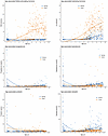 Figure 9
Comparative analysis of sex hormone levels: study cohort vs. age-matched healthy female participants (CALIPER Reference Intervals). This figure compares the longitudinal trajectories of 6 reproductive hormones — follicle-stimulating hormone (FSH), luteinizing hormone (LH), estradiol (E2), progesterone (P), prolactin (PRL), and testosterone (T) — between girls with TS and healthy controls. Individual measurements are shown as scatter points, and group trends are visualized using LOESS smoothing curves. Age-specific reference intervals for healthy girls were derived from the CALIPER database (https://caliper.research.sickkids.ca).


Copyright © 2026 American Society for Clinical Investigation
ISSN: 0021-9738 (print), 1558-8238 (online)