
Advertisement
Research ArticleCell biologyPulmonology
Open Access |  10.1172/JCI197651
Macropinocytosis inhibition attenuates profibrotic responses in lung fibroblasts and pulmonary fibrosis models
Ivan O. Rosas,1 Aaron K. McDowell-Sanchez,1 Santiago Sanchez,1 Juan D. Cala-Garcia,1 Alan R. Waich Cohen,1 Elisa Ruiz-Echartea,2 Scott A. Ochsner,2 Daniel C. Kraushaar,3 Lindsay J. Celada,1 Dandan Sun,4,5 Francesca Polverino,1 Cristian Coarfa,2 Neil J. McKenna,2 and Konstantin Tsoyi1,6
1Section of Pulmonary, Critical Care, and Sleep Medicine, Department of Medicine,
2Department of Molecular and Cellular Biology, and
3Genomic and RNA Profiling Core, Baylor College of Medicine, Houston, Texas, USA.
4Department of Neurology, Pittsburgh Institute for Neurodegenerative Disorders, University of Pittsburgh, Pittsburgh, Pennsylvania, USA.
5Veterans Affairs Pittsburgh Health Care System, Geriatric Research, Educational and Clinical Center, Pittsburgh, Pennsylvania, USA.
6Division of Pulmonary, Critical Care and Sleep Medicine, Department of Medicine, University of Maryland School of Medicine, Baltimore, Maryland, USA.
Address correspondence to: Ivan O. Rosas, Section of Pulmonary, Critical Care, and Sleep Medicine, Department of Medicine, Baylor College of Medicine, 7200 Cambridge Street, Houston, Texas, 77030, USA. Email: Ivan.Rosas@bcm.edu. Or to: Konstantin Tsoyi, Division of Pulmonary, Critical Care and Sleep Medicine, Department of Medicine, University of Maryland School of Medicine, 20 Penn Street, HSRF II, Baltimore, Maryland, 21201, USA. Email: KTsoyi@som.umaryland.edu.
Find articles by
Rosas, I.
in:
PubMed
|
Google Scholar
|

1Section of Pulmonary, Critical Care, and Sleep Medicine, Department of Medicine,
2Department of Molecular and Cellular Biology, and
3Genomic and RNA Profiling Core, Baylor College of Medicine, Houston, Texas, USA.
4Department of Neurology, Pittsburgh Institute for Neurodegenerative Disorders, University of Pittsburgh, Pittsburgh, Pennsylvania, USA.
5Veterans Affairs Pittsburgh Health Care System, Geriatric Research, Educational and Clinical Center, Pittsburgh, Pennsylvania, USA.
6Division of Pulmonary, Critical Care and Sleep Medicine, Department of Medicine, University of Maryland School of Medicine, Baltimore, Maryland, USA.
Address correspondence to: Ivan O. Rosas, Section of Pulmonary, Critical Care, and Sleep Medicine, Department of Medicine, Baylor College of Medicine, 7200 Cambridge Street, Houston, Texas, 77030, USA. Email: Ivan.Rosas@bcm.edu. Or to: Konstantin Tsoyi, Division of Pulmonary, Critical Care and Sleep Medicine, Department of Medicine, University of Maryland School of Medicine, 20 Penn Street, HSRF II, Baltimore, Maryland, 21201, USA. Email: KTsoyi@som.umaryland.edu.
Find articles by McDowell-Sanchez, A. in: PubMed | Google Scholar
1Section of Pulmonary, Critical Care, and Sleep Medicine, Department of Medicine,
2Department of Molecular and Cellular Biology, and
3Genomic and RNA Profiling Core, Baylor College of Medicine, Houston, Texas, USA.
4Department of Neurology, Pittsburgh Institute for Neurodegenerative Disorders, University of Pittsburgh, Pittsburgh, Pennsylvania, USA.
5Veterans Affairs Pittsburgh Health Care System, Geriatric Research, Educational and Clinical Center, Pittsburgh, Pennsylvania, USA.
6Division of Pulmonary, Critical Care and Sleep Medicine, Department of Medicine, University of Maryland School of Medicine, Baltimore, Maryland, USA.
Address correspondence to: Ivan O. Rosas, Section of Pulmonary, Critical Care, and Sleep Medicine, Department of Medicine, Baylor College of Medicine, 7200 Cambridge Street, Houston, Texas, 77030, USA. Email: Ivan.Rosas@bcm.edu. Or to: Konstantin Tsoyi, Division of Pulmonary, Critical Care and Sleep Medicine, Department of Medicine, University of Maryland School of Medicine, 20 Penn Street, HSRF II, Baltimore, Maryland, 21201, USA. Email: KTsoyi@som.umaryland.edu.
Find articles by Sanchez, S. in: PubMed | Google Scholar
1Section of Pulmonary, Critical Care, and Sleep Medicine, Department of Medicine,
2Department of Molecular and Cellular Biology, and
3Genomic and RNA Profiling Core, Baylor College of Medicine, Houston, Texas, USA.
4Department of Neurology, Pittsburgh Institute for Neurodegenerative Disorders, University of Pittsburgh, Pittsburgh, Pennsylvania, USA.
5Veterans Affairs Pittsburgh Health Care System, Geriatric Research, Educational and Clinical Center, Pittsburgh, Pennsylvania, USA.
6Division of Pulmonary, Critical Care and Sleep Medicine, Department of Medicine, University of Maryland School of Medicine, Baltimore, Maryland, USA.
Address correspondence to: Ivan O. Rosas, Section of Pulmonary, Critical Care, and Sleep Medicine, Department of Medicine, Baylor College of Medicine, 7200 Cambridge Street, Houston, Texas, 77030, USA. Email: Ivan.Rosas@bcm.edu. Or to: Konstantin Tsoyi, Division of Pulmonary, Critical Care and Sleep Medicine, Department of Medicine, University of Maryland School of Medicine, 20 Penn Street, HSRF II, Baltimore, Maryland, 21201, USA. Email: KTsoyi@som.umaryland.edu.
Find articles by Cala-Garcia, J. in: PubMed | Google Scholar
1Section of Pulmonary, Critical Care, and Sleep Medicine, Department of Medicine,
2Department of Molecular and Cellular Biology, and
3Genomic and RNA Profiling Core, Baylor College of Medicine, Houston, Texas, USA.
4Department of Neurology, Pittsburgh Institute for Neurodegenerative Disorders, University of Pittsburgh, Pittsburgh, Pennsylvania, USA.
5Veterans Affairs Pittsburgh Health Care System, Geriatric Research, Educational and Clinical Center, Pittsburgh, Pennsylvania, USA.
6Division of Pulmonary, Critical Care and Sleep Medicine, Department of Medicine, University of Maryland School of Medicine, Baltimore, Maryland, USA.
Address correspondence to: Ivan O. Rosas, Section of Pulmonary, Critical Care, and Sleep Medicine, Department of Medicine, Baylor College of Medicine, 7200 Cambridge Street, Houston, Texas, 77030, USA. Email: Ivan.Rosas@bcm.edu. Or to: Konstantin Tsoyi, Division of Pulmonary, Critical Care and Sleep Medicine, Department of Medicine, University of Maryland School of Medicine, 20 Penn Street, HSRF II, Baltimore, Maryland, 21201, USA. Email: KTsoyi@som.umaryland.edu.
Find articles by Waich Cohen, A. in: PubMed | Google Scholar
1Section of Pulmonary, Critical Care, and Sleep Medicine, Department of Medicine,
2Department of Molecular and Cellular Biology, and
3Genomic and RNA Profiling Core, Baylor College of Medicine, Houston, Texas, USA.
4Department of Neurology, Pittsburgh Institute for Neurodegenerative Disorders, University of Pittsburgh, Pittsburgh, Pennsylvania, USA.
5Veterans Affairs Pittsburgh Health Care System, Geriatric Research, Educational and Clinical Center, Pittsburgh, Pennsylvania, USA.
6Division of Pulmonary, Critical Care and Sleep Medicine, Department of Medicine, University of Maryland School of Medicine, Baltimore, Maryland, USA.
Address correspondence to: Ivan O. Rosas, Section of Pulmonary, Critical Care, and Sleep Medicine, Department of Medicine, Baylor College of Medicine, 7200 Cambridge Street, Houston, Texas, 77030, USA. Email: Ivan.Rosas@bcm.edu. Or to: Konstantin Tsoyi, Division of Pulmonary, Critical Care and Sleep Medicine, Department of Medicine, University of Maryland School of Medicine, 20 Penn Street, HSRF II, Baltimore, Maryland, 21201, USA. Email: KTsoyi@som.umaryland.edu.
Find articles by Ruiz-Echartea, E. in: PubMed | Google Scholar
1Section of Pulmonary, Critical Care, and Sleep Medicine, Department of Medicine,
2Department of Molecular and Cellular Biology, and
3Genomic and RNA Profiling Core, Baylor College of Medicine, Houston, Texas, USA.
4Department of Neurology, Pittsburgh Institute for Neurodegenerative Disorders, University of Pittsburgh, Pittsburgh, Pennsylvania, USA.
5Veterans Affairs Pittsburgh Health Care System, Geriatric Research, Educational and Clinical Center, Pittsburgh, Pennsylvania, USA.
6Division of Pulmonary, Critical Care and Sleep Medicine, Department of Medicine, University of Maryland School of Medicine, Baltimore, Maryland, USA.
Address correspondence to: Ivan O. Rosas, Section of Pulmonary, Critical Care, and Sleep Medicine, Department of Medicine, Baylor College of Medicine, 7200 Cambridge Street, Houston, Texas, 77030, USA. Email: Ivan.Rosas@bcm.edu. Or to: Konstantin Tsoyi, Division of Pulmonary, Critical Care and Sleep Medicine, Department of Medicine, University of Maryland School of Medicine, 20 Penn Street, HSRF II, Baltimore, Maryland, 21201, USA. Email: KTsoyi@som.umaryland.edu.
Find articles by Ochsner, S. in: PubMed | Google Scholar
1Section of Pulmonary, Critical Care, and Sleep Medicine, Department of Medicine,
2Department of Molecular and Cellular Biology, and
3Genomic and RNA Profiling Core, Baylor College of Medicine, Houston, Texas, USA.
4Department of Neurology, Pittsburgh Institute for Neurodegenerative Disorders, University of Pittsburgh, Pittsburgh, Pennsylvania, USA.
5Veterans Affairs Pittsburgh Health Care System, Geriatric Research, Educational and Clinical Center, Pittsburgh, Pennsylvania, USA.
6Division of Pulmonary, Critical Care and Sleep Medicine, Department of Medicine, University of Maryland School of Medicine, Baltimore, Maryland, USA.
Address correspondence to: Ivan O. Rosas, Section of Pulmonary, Critical Care, and Sleep Medicine, Department of Medicine, Baylor College of Medicine, 7200 Cambridge Street, Houston, Texas, 77030, USA. Email: Ivan.Rosas@bcm.edu. Or to: Konstantin Tsoyi, Division of Pulmonary, Critical Care and Sleep Medicine, Department of Medicine, University of Maryland School of Medicine, 20 Penn Street, HSRF II, Baltimore, Maryland, 21201, USA. Email: KTsoyi@som.umaryland.edu.
Find articles by Kraushaar, D. in: PubMed | Google Scholar
1Section of Pulmonary, Critical Care, and Sleep Medicine, Department of Medicine,
2Department of Molecular and Cellular Biology, and
3Genomic and RNA Profiling Core, Baylor College of Medicine, Houston, Texas, USA.
4Department of Neurology, Pittsburgh Institute for Neurodegenerative Disorders, University of Pittsburgh, Pittsburgh, Pennsylvania, USA.
5Veterans Affairs Pittsburgh Health Care System, Geriatric Research, Educational and Clinical Center, Pittsburgh, Pennsylvania, USA.
6Division of Pulmonary, Critical Care and Sleep Medicine, Department of Medicine, University of Maryland School of Medicine, Baltimore, Maryland, USA.
Address correspondence to: Ivan O. Rosas, Section of Pulmonary, Critical Care, and Sleep Medicine, Department of Medicine, Baylor College of Medicine, 7200 Cambridge Street, Houston, Texas, 77030, USA. Email: Ivan.Rosas@bcm.edu. Or to: Konstantin Tsoyi, Division of Pulmonary, Critical Care and Sleep Medicine, Department of Medicine, University of Maryland School of Medicine, 20 Penn Street, HSRF II, Baltimore, Maryland, 21201, USA. Email: KTsoyi@som.umaryland.edu.
Find articles by Celada, L. in: PubMed | Google Scholar
1Section of Pulmonary, Critical Care, and Sleep Medicine, Department of Medicine,
2Department of Molecular and Cellular Biology, and
3Genomic and RNA Profiling Core, Baylor College of Medicine, Houston, Texas, USA.
4Department of Neurology, Pittsburgh Institute for Neurodegenerative Disorders, University of Pittsburgh, Pittsburgh, Pennsylvania, USA.
5Veterans Affairs Pittsburgh Health Care System, Geriatric Research, Educational and Clinical Center, Pittsburgh, Pennsylvania, USA.
6Division of Pulmonary, Critical Care and Sleep Medicine, Department of Medicine, University of Maryland School of Medicine, Baltimore, Maryland, USA.
Address correspondence to: Ivan O. Rosas, Section of Pulmonary, Critical Care, and Sleep Medicine, Department of Medicine, Baylor College of Medicine, 7200 Cambridge Street, Houston, Texas, 77030, USA. Email: Ivan.Rosas@bcm.edu. Or to: Konstantin Tsoyi, Division of Pulmonary, Critical Care and Sleep Medicine, Department of Medicine, University of Maryland School of Medicine, 20 Penn Street, HSRF II, Baltimore, Maryland, 21201, USA. Email: KTsoyi@som.umaryland.edu.
Find articles by Sun, D. in: PubMed | Google Scholar
1Section of Pulmonary, Critical Care, and Sleep Medicine, Department of Medicine,
2Department of Molecular and Cellular Biology, and
3Genomic and RNA Profiling Core, Baylor College of Medicine, Houston, Texas, USA.
4Department of Neurology, Pittsburgh Institute for Neurodegenerative Disorders, University of Pittsburgh, Pittsburgh, Pennsylvania, USA.
5Veterans Affairs Pittsburgh Health Care System, Geriatric Research, Educational and Clinical Center, Pittsburgh, Pennsylvania, USA.
6Division of Pulmonary, Critical Care and Sleep Medicine, Department of Medicine, University of Maryland School of Medicine, Baltimore, Maryland, USA.
Address correspondence to: Ivan O. Rosas, Section of Pulmonary, Critical Care, and Sleep Medicine, Department of Medicine, Baylor College of Medicine, 7200 Cambridge Street, Houston, Texas, 77030, USA. Email: Ivan.Rosas@bcm.edu. Or to: Konstantin Tsoyi, Division of Pulmonary, Critical Care and Sleep Medicine, Department of Medicine, University of Maryland School of Medicine, 20 Penn Street, HSRF II, Baltimore, Maryland, 21201, USA. Email: KTsoyi@som.umaryland.edu.
Find articles by
Polverino, F.
in:
PubMed
|
Google Scholar
|
1Section of Pulmonary, Critical Care, and Sleep Medicine, Department of Medicine,
2Department of Molecular and Cellular Biology, and
3Genomic and RNA Profiling Core, Baylor College of Medicine, Houston, Texas, USA.
4Department of Neurology, Pittsburgh Institute for Neurodegenerative Disorders, University of Pittsburgh, Pittsburgh, Pennsylvania, USA.
5Veterans Affairs Pittsburgh Health Care System, Geriatric Research, Educational and Clinical Center, Pittsburgh, Pennsylvania, USA.
6Division of Pulmonary, Critical Care and Sleep Medicine, Department of Medicine, University of Maryland School of Medicine, Baltimore, Maryland, USA.
Address correspondence to: Ivan O. Rosas, Section of Pulmonary, Critical Care, and Sleep Medicine, Department of Medicine, Baylor College of Medicine, 7200 Cambridge Street, Houston, Texas, 77030, USA. Email: Ivan.Rosas@bcm.edu. Or to: Konstantin Tsoyi, Division of Pulmonary, Critical Care and Sleep Medicine, Department of Medicine, University of Maryland School of Medicine, 20 Penn Street, HSRF II, Baltimore, Maryland, 21201, USA. Email: KTsoyi@som.umaryland.edu.
Find articles by
Coarfa, C.
in:
PubMed
|
Google Scholar
|
1Section of Pulmonary, Critical Care, and Sleep Medicine, Department of Medicine,
2Department of Molecular and Cellular Biology, and
3Genomic and RNA Profiling Core, Baylor College of Medicine, Houston, Texas, USA.
4Department of Neurology, Pittsburgh Institute for Neurodegenerative Disorders, University of Pittsburgh, Pittsburgh, Pennsylvania, USA.
5Veterans Affairs Pittsburgh Health Care System, Geriatric Research, Educational and Clinical Center, Pittsburgh, Pennsylvania, USA.
6Division of Pulmonary, Critical Care and Sleep Medicine, Department of Medicine, University of Maryland School of Medicine, Baltimore, Maryland, USA.
Address correspondence to: Ivan O. Rosas, Section of Pulmonary, Critical Care, and Sleep Medicine, Department of Medicine, Baylor College of Medicine, 7200 Cambridge Street, Houston, Texas, 77030, USA. Email: Ivan.Rosas@bcm.edu. Or to: Konstantin Tsoyi, Division of Pulmonary, Critical Care and Sleep Medicine, Department of Medicine, University of Maryland School of Medicine, 20 Penn Street, HSRF II, Baltimore, Maryland, 21201, USA. Email: KTsoyi@som.umaryland.edu.
Find articles by McKenna, N. in: PubMed | Google Scholar
1Section of Pulmonary, Critical Care, and Sleep Medicine, Department of Medicine,
2Department of Molecular and Cellular Biology, and
3Genomic and RNA Profiling Core, Baylor College of Medicine, Houston, Texas, USA.
4Department of Neurology, Pittsburgh Institute for Neurodegenerative Disorders, University of Pittsburgh, Pittsburgh, Pennsylvania, USA.
5Veterans Affairs Pittsburgh Health Care System, Geriatric Research, Educational and Clinical Center, Pittsburgh, Pennsylvania, USA.
6Division of Pulmonary, Critical Care and Sleep Medicine, Department of Medicine, University of Maryland School of Medicine, Baltimore, Maryland, USA.
Address correspondence to: Ivan O. Rosas, Section of Pulmonary, Critical Care, and Sleep Medicine, Department of Medicine, Baylor College of Medicine, 7200 Cambridge Street, Houston, Texas, 77030, USA. Email: Ivan.Rosas@bcm.edu. Or to: Konstantin Tsoyi, Division of Pulmonary, Critical Care and Sleep Medicine, Department of Medicine, University of Maryland School of Medicine, 20 Penn Street, HSRF II, Baltimore, Maryland, 21201, USA. Email: KTsoyi@som.umaryland.edu.
Find articles by Tsoyi, K. in: PubMed | Google Scholar
Published May 1, 2026 - More info
J Clin Invest. 2026;136(12):e197651. https://doi.org/10.1172/JCI197651.
© 2026 Rosas et al. This work is licensed under the Creative Commons Attribution 4.0 International License. To view a copy of this license, visit http://creativecommons.org/licenses/by/4.0/.
Received: July 9, 2025; Accepted: April 15, 2026

-
Results
Macropinocytosis inhibition attenuates profibrotic responses in activated HLFs. Lung fibroblasts derived from IPF lung tissue possess a highcapacity for myofibroblast differentiation, ECM production, and resistance to cell death and cell invasion (12–14). We investigated whether macropinocytosis levels are elevated in IPF-derived lung fibroblasts compared with control fibroblasts. The uptake of fluorescent labeled large particles, such as 70 kDa dextran, is a widely accepted method to measure macropinocytosis in nonphagocytic cells (9, 15–17). Using fluorescence microscopy and flow cytometry techniques, IPF-derived lung fibroblasts showed increased levels of macropinocytosis compared with control cells, as measured by the uptake of 70kDa FITC-labeled dextran (Figure 1, A and B). Unlike other types of endocytosis, macropinocytosis is dependent on Na+/H+ proton exchangers (NHEs), also known as the solute carrier 9 family (SLC9s) (18–20). Thus, 5-(n-ethyl-n-isopropyl)-amiloride (EIPA), a known pan-SLC9 blocker, is considered as a first choice pharmacological inhibitor of macropinocytosis in biomedical research (16, 21–25). Consistent with prior reports, EIPA effectively inhibited macropinocytosis in IPF-derived lung fibroblasts, while neither affecting clathrin-dependent endocytosis nor expression of caveolin-1, a key protein in caveolin-mediated endocytosis (Supplemental Figure 1, A–C; supplemental material available online with this article; https://doi.org/10.1172/JCI197651DS1). We tested whether EIPA can inhibit profibrotic responses in IPF-derived lung fibroblasts. As shown in Figure 1, C and D, EIPA significantly downregulated collagen 1 (ECM protein) and α-SMA, a marker of myofibroblast transformation, protein and mRNA expression in IPF-derived lung fibroblasts. Similar results were found in TGF-β1–stimulated control lung fibroblasts, where EIPA attenuated collagen 1 production, myofibroblast transformation, and cell contractility (Figure 1, E–G). Of note, we observed a mild, but not statistically significant, decrease in fibronectin 1 (FN1) expression in response to EIPA in activated human lung fibroblasts (HLFs) (Supplemental Figure 1D). These data suggest that macropinocytosis activity is elevated in activated or IPF-derived lung fibroblasts and that inhibition of this process leads to decreased profibrotic responses in these cells.
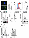 Figure 1
Macropinocytosis is elevated in IPF-derived lung fibroblasts and its inhibition regulates profibrotic responses. (A and B) Control and IPF-derived lung fibroblasts were conditioned in low-serum media for 24 hours, as described in Methods. 24 hours later, culture media was changed, and cells were incubated with FITC-dextran (70 kDa, 0.5 mg/mL) for 1 hour. Then, cells were washed 3 times with PBS. Macropinocytosis was measured by fluorescent microscopy (A) (scale bar: 25 μm) and flow cytometry (B) to measure fluorescence intensity of FITC-dextran in cells (n = 5 for A and n = 8 for B in each condition). (C and D) Cells were treated with vehicle (Veh) or EIPA (12.5 μM) for 24 hours. Then, cells were harvested and subjected to Western blot (C) and qRT-PCR (D) to measure collagen 1 and α-SMA expression, as described in Supplemental Methods and Supplemental Table 1 (n = 5 each condition). (E and F) Control HLFs were treated with EIPA (12.5 μM) with or without TGF-β1 (10 ng/mL) for 24 hours. Then, cells were harvested and subjected to Western blot (E) and qRT-PCR (F), as described in Methods (n = 4 each condition). (G) Control HLFs were mixed with collagen 1 solution, as described in Methods, and treated with EIPA (12.5 μM) with or without TGF-β1 (10 ng/mL). Gel size was measured at 0 and 24 hours after collagen gelation (n = 4 for each condition). Data are shown as the mean ± SEM. *P < 0.05, vs. unstimulated or Control + Veh; †P < 0.05, vs. IPF + Veh or TGF-β1 alone; significant comparisons by Student’s t test (A and B) or 1-way ANOVA (C–G).
Macropinocytosis regulates profibrotic responses in activated HLFs by regulating AA-mediated mTORC1 activation. Previous reports suggest that macropinocytosis tightly regulates mTORC1 activation by regulating cytosolic AA levels, which can be obtained by direct uptake of free AAs or proteins (e.g., serum albumin) from the extracellular environment (9–11, 19). In turn, mTORC1 activation is one of the central signaling pathways in promoting profibrotic responses in fibroblasts by activating multiple downstream signaling pathways and regulating cell metabolism (26–28). Accordingly, we found that EIPA inhibits the phosphorylation of p70S6K and S6, which are representative of mTORC1 activation and metabolic reprogramming, in TGF-β1–activated lung fibroblasts in low-serum (1%) culture medium without affecting cell viability (Figure 2A and Supplemental Figure 2, A and B). We also observed that EIPA effectively downregulated extracellular acidification rate (Supplemental Figure 2C). Interestingly, withdrawal of AAs, using AA-free cell culture media, completely abrogated mTORC1 activation, even in the presence of TGF-β1 (Figure 2B). The addition of AAs resulted in the upregulation of phospho-S6 in lung fibroblasts, which was further enhanced by TGF-β1 treatment (Figure 2B). EIPA significantly attenuated S6 activation under AA-supplemented conditions (Figure 2B). Furthermore, we found that macropinocytosis regulates the amount of cytosolic AAs under these conditions, suggesting that under profibrotic conditions (e.g., under TGF-β1 stimulation), extracellular AAs play a significant role in mTORC1 activation and that macropinocytosis inhibition can significantly inhibit this process (Figure 2C). Indeed, we found that free AA or albumin supplementation increased COL1A1 and ACTA2 mRNA levels compared with AA-free conditioned cells, and this effect was inhibited by EIPA treatment (Supplemental Figure 3A). Finally, mTORC1 activation by TSC1 silencing, a negative regulator of mTOR, attenuated the antifibrotic effects of EIPA (Figure 2D). Whereas, shTSC1 alone significantly upregulated the expression of COL1 and aSMA at the protein and mRNA levels in TGF-b1 stimulated lung fibroblasts (Supplemental Figure 2F and Supplemental Figure 10, C and F). These data suggest that macropinocytosis regulates the uptake of extracellular AAs, which, in turn, leads to mTORC1 activation and increased profibrotic responses in lung fibroblasts.
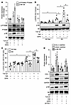 Figure 2
Macropinocytosis inhibition ameliorates mTORC1 signaling by regulating AA uptake in lung fibroblasts. (A) Control HLFs were treated with EIPA (12.5 μM) with or without TGF-β1 (10 ng/mL) for 24 hours. Then, p-P70S6K and p-S6 were measured by Western blot, as described in Methods (n = 4 each condition). (B and C) Cells were treated with EIPA (12.5 μM) with or without TGF-β1 (10 ng/mL) for 4 hours in AA-free and AA-supplemented conditions, as described in Methods. Then, cells were harvested to measure p-S6 by Western blot (B) and L-AA assay (C), as described in Methods (n = 4 or 6). (D) Cells were lentivirally transfected with scramble (Scr) or shTSC1, as described in Methods. Then, cells were treated with EIPA (12.5 μM) with or without TGF-β1 (10 ng/mL) for 24 hours, and cells were harvested, and COL1, α-SMA, and p-S6 were measured by Western blot (n = 4 each condition). Data are shown as the mean ± SEM. *P < 0.05, vs. Veh or Veh + AA free; †P < 0.05, vs. TGF-β1 + Veh or Veh + AA; §P < 0.05, vs. TGF-β1 + Veh + AA; #P < 0.05, vs. Scr + TGF-β1 + EIPA; significant comparisons by 1-way ANOVA.
MEOX1 is regulated by macropinocytosis and mTORC1 in activated HLFs. We next aimed to evaluate the effect of EIPA treatment on the TGF-β1–induced (TGFB-induced) transcriptional program in MRC5 HLFs. Consistent with a robust repressive effect of EIPA on the TGFB-induced transcriptional program, TGFB-induced genes were strongly enriched among EIPA-repressed genes (Figure 3A) (29). TGFB-induced genes repressed by EIPA treatment included several with well established roles in lung fibrosis such as ACTA2 and COL1A1. Notably, mesenchyme homeobox1 (MEOX1), recently identified as a key transcription factor in fibroblast activation and cardiac fibrosis, was among the repressed genes (30). Accordingly, we next investigated whether MEOX1 was regulated by macropinocytosis-mediated mTORC1 activation and contributed to the profibrotic phenotype of activated HLFs. We found that both EIPA and Rapalink-1, a known mTORC1 inhibitor, downregulated MEOX1 mRNA and protein expression levels (Figure 3, B and C). However, the inhibitory effect of EIPA on MEOX1 expression was ameliorated in TSC1-deficient cells, suggesting that macropinocytosis inhibition downregulates the expression of MEOX1 by regulating mTORC1 in activated lung fibroblasts (Figure 3D).
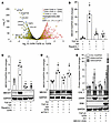 Figure 3
Macropinocytosis inhibition regulates MEOX1 expression in activated lung fibroblasts. (A) Volcano plot representing treatment of MRC5 HLFs with and without EIPA in the presence of TGFB. Overlaid in yellow is a set of TGFβ1-induced genes from a separate RNA sequencing experiment in MRC5 HLFs treated with TGF-β1 (10 ng/mL) or vehicle. MRC-5 cells were treated with EIPA (12.5 μM) with or without TGF-β1 (10 ng/mL) for 24 hours. Then, RNA was isolated and subjected to bulk RNA sequencing, as described in Methods (n = 3). (B and C) Control HLFs were treated with EIPA (12.5 μM) and Rapalink-1 (10 nM) with or without TGF-β1 (10 ng/mL) for 24 hours. Then, MEOX1 was measured at mRNA and protein levels by qRT-PCR (B) and Western blot (C), respectively (n = 4 each condition). (D) Control HLFs were lentivirally transfected with Scr or shTSC1, as described in Methods. Then, cells were treated with vehicle (Veh) or EIPA (12.5 μM) for 24 hours. Then, cells were harvested and subjected to Western blot to measure MEOX1 expression. (E) Control HLFs were lentivirally transfected with empty vector (EV) or pLenti-MEOX1-DDK, as described in Methods. Then, cells were treated with TGF-β1 (10 ng/mL) for 24 hours or not. Protein levels of COL1 and α-SMA were measured by Western blot, as described in Methods (n = 4 each condition). Data are shown as the mean ± SEM. *P < 0.05, vs. unstimulated or Scr alone; †P < 0.05, vs. TGF-β1 alone; #P < 0.05, TGF-β1+EIPA; significant comparisons by hypergeometric test (A) or 1-way ANOVA (B–E).
Silencing of regulatory-associated protein of mTOR (RAPTOR), a key component of mTORC1, but not mTORC2, inhibited MEOX1 expression in TGF-β1–activated HLFs (Supplemental Figure 3B and Supplemental Figure 10, B and E). Of note, we also found that activation of mTOR by shTSC1 significantly increased MEOX1 gene expression (Supplemental Figure 2D and Supplemental Figure 10, A and D), whereas silencing of MEOX1 significantly inhibited COL1 and α-SMA expression in activated HLFs (Supplemental Figure 3C). Finally, we found that overexpression of MEOX1 reversed the antifibrotic effect of EIPA (Figure 3E) and that EIPA significantly ameliorated the binding of MEOX1 to COL1A1 and ACTA2 genes promoters (Supplemental Figure 3D) These data suggest that macropinocytosis inhibition negatively affects profibrotic responses in fibroblasts by regulating an mTORC1/MEOX1 signaling axis.
Macropinocytosis inhibition attenuates lung fibrosis in Bleo-injured mice. Next, we examined whether genetic silencing or pharmacologic inhibition of macropinocytosis can ameliorate pulmonary fibrosis in bleomycin-injured (Bleo-injured) mice. Previously, Nhe1/Slc9a1 deficiency was shown to markedly inhibit macropinocytosis in vitro and in vivo (31). Indeed, we found that Slc9a1-deficient lung fibroblasts exhibited decreased macropinocytosis activity compared with their WT counterparts, without affecting clathrin-dependent endocytosis or caveolin1 expression (Supplemental Figure 4). We found that deficiency of Slc9a1 in fibroblasts significantly attenuated pulmonary fibrosis in mice subjected to intratracheal Bleo administration (Figure 4, A and B, and Supplemental Figure 5A). Furthermore, Slc9a1 deficiency in mouse lung fibroblasts led to a decrease in Col1a1 and α-SMA expression, mTORC1 activation, and Meox1 after TGF-β1 stimulation (Figure 4, C–E). Consistently, delayed administration of a macropinocytosis inhibitor (EIPA, 10 mg/kg) significantly reduced macropinocytosis and lung fibrosis, as determined by Trichrome and H&E staining and hydroxyproline assay, without affecting lung inflammation in Bleo-injured mice (Figure 4, F and G, Supplemental Figure 5B, and Supplemental Figure 6). Accordingly, total lung homogenates showed a significant downregulation of profibrotic gene expression and MEOX1 expression in fibroblast-specific Slc9A1 deficient and EIPA-treated mice (Supplemental Figure 7). Taken together, our data strongly suggest that macropinocytosis inhibition ameliorates pulmonary fibrosis in mice.
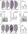 Figure 4
Macropinocytosis inhibition attenuates pulmonary fibrosis in Bleo-injured mice. (A and B) Slc9a1fl/fl (WT) or Slc9a1fl/fl Col1a2Cre-ER(T)+/0 (Slc9a1 fCKO) mice were treated with tamoxifen to induce Slc9a1 knockout in fibroblasts, as described in Methods. Then, mice were exposed to Bleo-induced pulmonary fibrosis, as described in Methods. At day 21 after Bleo exposure, lungs were harvested, stained with Masson’s trichrome staining (scale bar: 1,000 μm) (A) and subjected to hyproxyproline assay (B) (n = 5 for sham groups, n = 10–11 for Bleo groups). (C–E) Mouse lung fibroblasts from WT and Slc9a1 fCKO mice were isolated, as described in Methods. Cells were stimulated with TGF-β1 (10 ng/mL) for 24 hours. Then, cells were subjected to Western blot or qRT-PCR, as described in Methods. (F and G) WT mice were exposed to Bleo to induce pulmonary fibrosis, as described in Methods. 10 days after Bleo exposure, mice were intraperitoneally treated with EIPA (10 mg/kg) or vehicle (Veh) every other day until the end of experiment. At day 21 after Bleo exposure, lungs were harvested, stained with Masson’s trichrome staining (n = 3–4) (scale bar: 1,000 μm). (F) and subjected to hyproxyproline assay (G) (n = 5 for sham groups, n = 9 for Bleo groups). Data are shown as the mean ± SEM. *P < 0.05, vs. No Bleo (sham) or unstimulated; †P < 0.05, vs. Bleo or TGF-β1 alone; significant comparisons by 1-way ANOVA (B–E and G).
Macropinocytosis inhibition regulates the expression of profibrotic genes in different fibroblast subpopulations in IPF-derived PCLSs. Pulmonary fibroblasts represent a heterogenous cell population (32). To determine the effect of macropinocytosis inhibition on different fibroblast subpopulations in IPF lung, we treated precision cut lung slices (PCLSs) derived from individuals with IPF with EIPA and compared them with vehicle-treated PCLS from the same individuals. Single-cell RNA sequencing identified 7 cell populations of mesenchyme origin (Figure 5A and Supplemental Figure 8). Consistent with the results obtained from isolated fibroblasts and Bleo-injured mice, we observed a potent repressive effect of EIPA on the IPF-induced profibrotic gene expression in myofibroblasts (Figure 5B), known cells found to contribute to the progression of pulmonary fibrosis in mice and patients with IPF (33). Additionally, EIPA exerted a measurable effect on the other 6 mesenchymal subpopulations (Supplemental Figure 8). Gene regulatory network (GRN) analysis predicts functional footprints for transcription factors within clinical gene sets of interest (34, 35). To identify candidate transcription factors (TFs) affected by EIPA in IPF, we carried out GRN analysis on genes repressed by EIPA treatment of IPF PCLS myofibroblasts (Figure 5C) (29). Reiterating the strong antifibrotic effect of EIPA observed at the transcriptional level, the top ranked TFs from an IPF-induced PCLS myofibroblast GRN were strongly enriched among the EIPA-repressed PCLS myofibroblast GRN. Particularly strong footprints were observed for numerous TFs with established roles in pulmonary fibrosis, including NFATC1, TWIST1, and members of the SMAD family (Figure 5C), suggesting that EIPA may exert its antifibrotic effects by targeting one or more of these factors. We validated EIPA-mediated downregulation of profibrotic genes, such as periostin (POSTN), insulin growth factor binding protein-5 (IGFBP5), COL1A1, and ACTA2 by RT-qPCR in PCLS homogenates (Figure 5D). Finally, collagen 1 expression was also downregulated in EIPA-treated PCLSs compared with vehicle-treated PCLSs (Figure 5E).
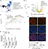 Figure 5
EIPA inhibits profibrotic responses in IPF-derived PCLSs. IPF-derived PCLSs were treated with EIPA (12.5 μM), as described in Methods. (A) UMAP plot represents different mesenchymal cell populations identified in IPF PCLSs (n of 4 for DMSO and n of 3 for EIPA-treated PCLSs). (B) Volcano plot representing treatment of IPF PCLSs with and without EIPA, as described in A. Overlaid in yellow is a set of myofibroblast IPF-induced genes from a separate PCLS experiment comparing IPF + DMSO (n of 4) and control (n of 4) PCLS samples, which were reused from McKenna et al. (29). (C) Regulatory network plot for genes repressed in EIPA-treated IPF PCLSs compared with IPF alone. TFs with the largest and most significant footprints within the gene set are distributed toward the top right of the plot. (D) After treatment, PCLSs were lysed and subjected to total RNA isolation. mRNA levels of POSTN, IGFBP5, ACTA2, and COL1A1 were measured with qRT-PCR, as described in Methods (n = 5 for each condition). (E) After treatment, PCLSs were washed with PBS and fixed for COL1 staining, as described in Methods. The COL1 fluorescent intensity was measured with ImageJ software (NIH) (n = 5 for each condition). Scale bar: 3,000 μm. Data are shown as the mean ± SEM. *P < 0.05, vs. No Bleo (sham); †P < 0.05, vs. Bleo alone; significant comparisons by hypergeometric test (B and C) and Student’s t test (D and E).
Imi inhibits fibroblast activation and pulmonary fibrosis. A previous report suggested that Imi selectively inhibits macropinocytosis over other types of endocytosis in macrophages (21). We also found that Imi strongly inhibited macropinocytosis in IPF-derived lung fibroblasts without affecting clathrin-dependent endocytosis or caveolin-1 expression (Supplemental Figure 1A). Imi effectively inhibited profibrotic responses in IPF-derived and TGF-β1–induced lung fibroblasts as well as mTORC1/MEOX1 signaling (Figure 6, A–C, and Supplemental Figure 7C). Similar to EIPA, Imi significantly ameliorated pulmonary fibrosis in Bleo-injured mice and profibrotic responses in IPF-derived PCLSs (Figure 6, D–G, and Supplemental Figure 5C). Together, these results suggest that antifibrotic effects in IPF can be achieved by targeting macropinocytosis either with EIPA or through the potential repurposing of the FDA-approved drug Imi.
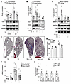 Figure 6
Imipramine ameliorates profibrotic responses in lung fibroblasts, Bleo-injured mice, and PCLSs. (A) Control and IPF-derived lung fibroblasts were treated with imipramine (Imi, 10 μM). 24 hours later cells were harvested, and COL1 and α-SMA were measured with Western blot (n = 4 each condition). (B and C) Control HLFs were treated with Imi (10 μM) with or without TGF-β1 (10 ng/mL) for 24 hours. Then, cells were harvested and subjected to Western blot to measure COL1, α-SMA, p-S6, and MEOX1 (n = 4 for each condition). (D–F) WT mice were exposed to Bleo-induced pulmonary fibrosis, as described in Methods. 10 days after Bleo exposure, mice were intraperitoneally treated with Imi (10 mg/kg) or vehicle (Veh) every other day until the end of the experiment. At day 21 after Bleo exposure, lungs were harvested, stained with Masson’s trichrome staining (n = 3–4) (D), and subjected to hyproxyproline assay (n = 5 or 10) (E) or mRNA assessment (n = 5) (F). (G) IPF-derived PCLSs were treated with Imi (10 μM), as described in Methods. Then, cells were lysed and mRNA levels of POSTN, IGFBP5, COL1A1, and ACTA2 were measured by qRT-PCR (n = 5 each condition). *P < 0.05, vs. unstimulated or Control or No Bleo (sham); †P < 0.05, vs. TGF-β1 or IPF alone or Bleo alone; significant comparisons by 1-way ANOVA (A–C, E, and F) and Student’s t test (G).


Copyright © 2026 American Society for Clinical Investigation
ISSN: 0021-9738 (print), 1558-8238 (online)