
Advertisement
Research ArticleAutoimmunityImmunology
Open Access |  10.1172/JCI196325
UTX coordinates TCF1 and STAT3 to control progenitor CD8+ T cell fate in autoimmune diabetes
Ho-Chung Chen,1 Madison F. Bang,1 Hsing-Hui Wang,2 Karl B. Shpargel,3 Lisa A. Kohn,4 David Sailer,2 Shile Zhang,5 Ethan C. McCarthy,1 Maryamsadat Seyedsadr,1 Satchel Stevens,1 Caitlyn L.H. Pham,1 Zikang Zhou,1 Xihui Yin,6 Nicole M. Wilkinson,1,7 Esther M. Peluso,5,7 Christian Bustillos,1 Jessica G. Ortega,4 Lixin Yang,3 Ashlyn A. Buzzelli,3 Reina C. Capati,8 Dennis J. Chia,8 Steven D. Mittelman,8 Christina M. Reh,8 Jason K. Whitmire,3 Melissa G. Lechner,4 Willy Hugo,4 and Maureen A. Su1,8
1Department of Microbiology, Immunology, and Molecular Genetics, UCLA David Geffen School of Medicine, Los Angeles, California, USA.
2Department of Pediatrics and
3Department of Genetics, University of North Carolina at Chapel Hill, Chapel Hill, North Carolina, USA.
4Department of Medicine and
5Department of Molecular and Medical Pharmacology, UCLA David Geffen School of Medicine, Los Angeles, California, USA.
6Department of Biology, Massachusetts Institute of Technology, Boston, Massachusetts, USA.
7UCLA/California Institute of Technology Medical Scientist Training Program and
8Department of Pediatrics, UCLA David Geffen School of Medicine, Los Angeles, California, USA.
Address correspondence to: Maureen A. Su, Department of Microbiology, Immunology, and Molecular Genetics and Pediatrics, UCLA David Geffen School of Medicine, 615 Charles E. Young Dr. South, BSRB 254, Los Angeles, California, 90095, USA. Phone: 919.597.0777; Email: masu@mednet.ucla.edu. Or to: Willy Hugo, Department of Medicine, Dermatology, UCLA David Geffen School of Medicine, 700 Tiverton Dr., Factor Building 9-240M, Los Angeles, California, 90095, USA. Phone: 310.794.6083; Email: hwilly@mednet.ucla.edu. Or to: Melissa G. Lechner, Department of Medicine, Division of Endocrinology, Diabetes, and Metabolism, UCLA David Geffen School of Medicine, 10833 Le Conte Ave., CHS 32-176, Los Angeles, California, 90095, USA. Phone: 314.651.4199; Email: mlechner@mednet.ucla.edu.
Find articles by Chen, H. in: PubMed | Google Scholar
1Department of Microbiology, Immunology, and Molecular Genetics, UCLA David Geffen School of Medicine, Los Angeles, California, USA.
2Department of Pediatrics and
3Department of Genetics, University of North Carolina at Chapel Hill, Chapel Hill, North Carolina, USA.
4Department of Medicine and
5Department of Molecular and Medical Pharmacology, UCLA David Geffen School of Medicine, Los Angeles, California, USA.
6Department of Biology, Massachusetts Institute of Technology, Boston, Massachusetts, USA.
7UCLA/California Institute of Technology Medical Scientist Training Program and
8Department of Pediatrics, UCLA David Geffen School of Medicine, Los Angeles, California, USA.
Address correspondence to: Maureen A. Su, Department of Microbiology, Immunology, and Molecular Genetics and Pediatrics, UCLA David Geffen School of Medicine, 615 Charles E. Young Dr. South, BSRB 254, Los Angeles, California, 90095, USA. Phone: 919.597.0777; Email: masu@mednet.ucla.edu. Or to: Willy Hugo, Department of Medicine, Dermatology, UCLA David Geffen School of Medicine, 700 Tiverton Dr., Factor Building 9-240M, Los Angeles, California, 90095, USA. Phone: 310.794.6083; Email: hwilly@mednet.ucla.edu. Or to: Melissa G. Lechner, Department of Medicine, Division of Endocrinology, Diabetes, and Metabolism, UCLA David Geffen School of Medicine, 10833 Le Conte Ave., CHS 32-176, Los Angeles, California, 90095, USA. Phone: 314.651.4199; Email: mlechner@mednet.ucla.edu.
Find articles by Bang, M. in: PubMed | Google Scholar
1Department of Microbiology, Immunology, and Molecular Genetics, UCLA David Geffen School of Medicine, Los Angeles, California, USA.
2Department of Pediatrics and
3Department of Genetics, University of North Carolina at Chapel Hill, Chapel Hill, North Carolina, USA.
4Department of Medicine and
5Department of Molecular and Medical Pharmacology, UCLA David Geffen School of Medicine, Los Angeles, California, USA.
6Department of Biology, Massachusetts Institute of Technology, Boston, Massachusetts, USA.
7UCLA/California Institute of Technology Medical Scientist Training Program and
8Department of Pediatrics, UCLA David Geffen School of Medicine, Los Angeles, California, USA.
Address correspondence to: Maureen A. Su, Department of Microbiology, Immunology, and Molecular Genetics and Pediatrics, UCLA David Geffen School of Medicine, 615 Charles E. Young Dr. South, BSRB 254, Los Angeles, California, 90095, USA. Phone: 919.597.0777; Email: masu@mednet.ucla.edu. Or to: Willy Hugo, Department of Medicine, Dermatology, UCLA David Geffen School of Medicine, 700 Tiverton Dr., Factor Building 9-240M, Los Angeles, California, 90095, USA. Phone: 310.794.6083; Email: hwilly@mednet.ucla.edu. Or to: Melissa G. Lechner, Department of Medicine, Division of Endocrinology, Diabetes, and Metabolism, UCLA David Geffen School of Medicine, 10833 Le Conte Ave., CHS 32-176, Los Angeles, California, 90095, USA. Phone: 314.651.4199; Email: mlechner@mednet.ucla.edu.
Find articles by Wang, H. in: PubMed | Google Scholar
1Department of Microbiology, Immunology, and Molecular Genetics, UCLA David Geffen School of Medicine, Los Angeles, California, USA.
2Department of Pediatrics and
3Department of Genetics, University of North Carolina at Chapel Hill, Chapel Hill, North Carolina, USA.
4Department of Medicine and
5Department of Molecular and Medical Pharmacology, UCLA David Geffen School of Medicine, Los Angeles, California, USA.
6Department of Biology, Massachusetts Institute of Technology, Boston, Massachusetts, USA.
7UCLA/California Institute of Technology Medical Scientist Training Program and
8Department of Pediatrics, UCLA David Geffen School of Medicine, Los Angeles, California, USA.
Address correspondence to: Maureen A. Su, Department of Microbiology, Immunology, and Molecular Genetics and Pediatrics, UCLA David Geffen School of Medicine, 615 Charles E. Young Dr. South, BSRB 254, Los Angeles, California, 90095, USA. Phone: 919.597.0777; Email: masu@mednet.ucla.edu. Or to: Willy Hugo, Department of Medicine, Dermatology, UCLA David Geffen School of Medicine, 700 Tiverton Dr., Factor Building 9-240M, Los Angeles, California, 90095, USA. Phone: 310.794.6083; Email: hwilly@mednet.ucla.edu. Or to: Melissa G. Lechner, Department of Medicine, Division of Endocrinology, Diabetes, and Metabolism, UCLA David Geffen School of Medicine, 10833 Le Conte Ave., CHS 32-176, Los Angeles, California, 90095, USA. Phone: 314.651.4199; Email: mlechner@mednet.ucla.edu.
Find articles by Shpargel, K. in: PubMed | Google Scholar
1Department of Microbiology, Immunology, and Molecular Genetics, UCLA David Geffen School of Medicine, Los Angeles, California, USA.
2Department of Pediatrics and
3Department of Genetics, University of North Carolina at Chapel Hill, Chapel Hill, North Carolina, USA.
4Department of Medicine and
5Department of Molecular and Medical Pharmacology, UCLA David Geffen School of Medicine, Los Angeles, California, USA.
6Department of Biology, Massachusetts Institute of Technology, Boston, Massachusetts, USA.
7UCLA/California Institute of Technology Medical Scientist Training Program and
8Department of Pediatrics, UCLA David Geffen School of Medicine, Los Angeles, California, USA.
Address correspondence to: Maureen A. Su, Department of Microbiology, Immunology, and Molecular Genetics and Pediatrics, UCLA David Geffen School of Medicine, 615 Charles E. Young Dr. South, BSRB 254, Los Angeles, California, 90095, USA. Phone: 919.597.0777; Email: masu@mednet.ucla.edu. Or to: Willy Hugo, Department of Medicine, Dermatology, UCLA David Geffen School of Medicine, 700 Tiverton Dr., Factor Building 9-240M, Los Angeles, California, 90095, USA. Phone: 310.794.6083; Email: hwilly@mednet.ucla.edu. Or to: Melissa G. Lechner, Department of Medicine, Division of Endocrinology, Diabetes, and Metabolism, UCLA David Geffen School of Medicine, 10833 Le Conte Ave., CHS 32-176, Los Angeles, California, 90095, USA. Phone: 314.651.4199; Email: mlechner@mednet.ucla.edu.
Find articles by Kohn, L. in: PubMed | Google Scholar
1Department of Microbiology, Immunology, and Molecular Genetics, UCLA David Geffen School of Medicine, Los Angeles, California, USA.
2Department of Pediatrics and
3Department of Genetics, University of North Carolina at Chapel Hill, Chapel Hill, North Carolina, USA.
4Department of Medicine and
5Department of Molecular and Medical Pharmacology, UCLA David Geffen School of Medicine, Los Angeles, California, USA.
6Department of Biology, Massachusetts Institute of Technology, Boston, Massachusetts, USA.
7UCLA/California Institute of Technology Medical Scientist Training Program and
8Department of Pediatrics, UCLA David Geffen School of Medicine, Los Angeles, California, USA.
Address correspondence to: Maureen A. Su, Department of Microbiology, Immunology, and Molecular Genetics and Pediatrics, UCLA David Geffen School of Medicine, 615 Charles E. Young Dr. South, BSRB 254, Los Angeles, California, 90095, USA. Phone: 919.597.0777; Email: masu@mednet.ucla.edu. Or to: Willy Hugo, Department of Medicine, Dermatology, UCLA David Geffen School of Medicine, 700 Tiverton Dr., Factor Building 9-240M, Los Angeles, California, 90095, USA. Phone: 310.794.6083; Email: hwilly@mednet.ucla.edu. Or to: Melissa G. Lechner, Department of Medicine, Division of Endocrinology, Diabetes, and Metabolism, UCLA David Geffen School of Medicine, 10833 Le Conte Ave., CHS 32-176, Los Angeles, California, 90095, USA. Phone: 314.651.4199; Email: mlechner@mednet.ucla.edu.
Find articles by Sailer, D. in: PubMed | Google Scholar
1Department of Microbiology, Immunology, and Molecular Genetics, UCLA David Geffen School of Medicine, Los Angeles, California, USA.
2Department of Pediatrics and
3Department of Genetics, University of North Carolina at Chapel Hill, Chapel Hill, North Carolina, USA.
4Department of Medicine and
5Department of Molecular and Medical Pharmacology, UCLA David Geffen School of Medicine, Los Angeles, California, USA.
6Department of Biology, Massachusetts Institute of Technology, Boston, Massachusetts, USA.
7UCLA/California Institute of Technology Medical Scientist Training Program and
8Department of Pediatrics, UCLA David Geffen School of Medicine, Los Angeles, California, USA.
Address correspondence to: Maureen A. Su, Department of Microbiology, Immunology, and Molecular Genetics and Pediatrics, UCLA David Geffen School of Medicine, 615 Charles E. Young Dr. South, BSRB 254, Los Angeles, California, 90095, USA. Phone: 919.597.0777; Email: masu@mednet.ucla.edu. Or to: Willy Hugo, Department of Medicine, Dermatology, UCLA David Geffen School of Medicine, 700 Tiverton Dr., Factor Building 9-240M, Los Angeles, California, 90095, USA. Phone: 310.794.6083; Email: hwilly@mednet.ucla.edu. Or to: Melissa G. Lechner, Department of Medicine, Division of Endocrinology, Diabetes, and Metabolism, UCLA David Geffen School of Medicine, 10833 Le Conte Ave., CHS 32-176, Los Angeles, California, 90095, USA. Phone: 314.651.4199; Email: mlechner@mednet.ucla.edu.
Find articles by
Zhang, S.
in:
PubMed
|
Google Scholar
|

1Department of Microbiology, Immunology, and Molecular Genetics, UCLA David Geffen School of Medicine, Los Angeles, California, USA.
2Department of Pediatrics and
3Department of Genetics, University of North Carolina at Chapel Hill, Chapel Hill, North Carolina, USA.
4Department of Medicine and
5Department of Molecular and Medical Pharmacology, UCLA David Geffen School of Medicine, Los Angeles, California, USA.
6Department of Biology, Massachusetts Institute of Technology, Boston, Massachusetts, USA.
7UCLA/California Institute of Technology Medical Scientist Training Program and
8Department of Pediatrics, UCLA David Geffen School of Medicine, Los Angeles, California, USA.
Address correspondence to: Maureen A. Su, Department of Microbiology, Immunology, and Molecular Genetics and Pediatrics, UCLA David Geffen School of Medicine, 615 Charles E. Young Dr. South, BSRB 254, Los Angeles, California, 90095, USA. Phone: 919.597.0777; Email: masu@mednet.ucla.edu. Or to: Willy Hugo, Department of Medicine, Dermatology, UCLA David Geffen School of Medicine, 700 Tiverton Dr., Factor Building 9-240M, Los Angeles, California, 90095, USA. Phone: 310.794.6083; Email: hwilly@mednet.ucla.edu. Or to: Melissa G. Lechner, Department of Medicine, Division of Endocrinology, Diabetes, and Metabolism, UCLA David Geffen School of Medicine, 10833 Le Conte Ave., CHS 32-176, Los Angeles, California, 90095, USA. Phone: 314.651.4199; Email: mlechner@mednet.ucla.edu.
Find articles by
McCarthy, E.
in:
PubMed
|
Google Scholar
|
1Department of Microbiology, Immunology, and Molecular Genetics, UCLA David Geffen School of Medicine, Los Angeles, California, USA.
2Department of Pediatrics and
3Department of Genetics, University of North Carolina at Chapel Hill, Chapel Hill, North Carolina, USA.
4Department of Medicine and
5Department of Molecular and Medical Pharmacology, UCLA David Geffen School of Medicine, Los Angeles, California, USA.
6Department of Biology, Massachusetts Institute of Technology, Boston, Massachusetts, USA.
7UCLA/California Institute of Technology Medical Scientist Training Program and
8Department of Pediatrics, UCLA David Geffen School of Medicine, Los Angeles, California, USA.
Address correspondence to: Maureen A. Su, Department of Microbiology, Immunology, and Molecular Genetics and Pediatrics, UCLA David Geffen School of Medicine, 615 Charles E. Young Dr. South, BSRB 254, Los Angeles, California, 90095, USA. Phone: 919.597.0777; Email: masu@mednet.ucla.edu. Or to: Willy Hugo, Department of Medicine, Dermatology, UCLA David Geffen School of Medicine, 700 Tiverton Dr., Factor Building 9-240M, Los Angeles, California, 90095, USA. Phone: 310.794.6083; Email: hwilly@mednet.ucla.edu. Or to: Melissa G. Lechner, Department of Medicine, Division of Endocrinology, Diabetes, and Metabolism, UCLA David Geffen School of Medicine, 10833 Le Conte Ave., CHS 32-176, Los Angeles, California, 90095, USA. Phone: 314.651.4199; Email: mlechner@mednet.ucla.edu.
Find articles by Seyedsadr, M. in: PubMed | Google Scholar
1Department of Microbiology, Immunology, and Molecular Genetics, UCLA David Geffen School of Medicine, Los Angeles, California, USA.
2Department of Pediatrics and
3Department of Genetics, University of North Carolina at Chapel Hill, Chapel Hill, North Carolina, USA.
4Department of Medicine and
5Department of Molecular and Medical Pharmacology, UCLA David Geffen School of Medicine, Los Angeles, California, USA.
6Department of Biology, Massachusetts Institute of Technology, Boston, Massachusetts, USA.
7UCLA/California Institute of Technology Medical Scientist Training Program and
8Department of Pediatrics, UCLA David Geffen School of Medicine, Los Angeles, California, USA.
Address correspondence to: Maureen A. Su, Department of Microbiology, Immunology, and Molecular Genetics and Pediatrics, UCLA David Geffen School of Medicine, 615 Charles E. Young Dr. South, BSRB 254, Los Angeles, California, 90095, USA. Phone: 919.597.0777; Email: masu@mednet.ucla.edu. Or to: Willy Hugo, Department of Medicine, Dermatology, UCLA David Geffen School of Medicine, 700 Tiverton Dr., Factor Building 9-240M, Los Angeles, California, 90095, USA. Phone: 310.794.6083; Email: hwilly@mednet.ucla.edu. Or to: Melissa G. Lechner, Department of Medicine, Division of Endocrinology, Diabetes, and Metabolism, UCLA David Geffen School of Medicine, 10833 Le Conte Ave., CHS 32-176, Los Angeles, California, 90095, USA. Phone: 314.651.4199; Email: mlechner@mednet.ucla.edu.
Find articles by Stevens, S. in: PubMed | Google Scholar
1Department of Microbiology, Immunology, and Molecular Genetics, UCLA David Geffen School of Medicine, Los Angeles, California, USA.
2Department of Pediatrics and
3Department of Genetics, University of North Carolina at Chapel Hill, Chapel Hill, North Carolina, USA.
4Department of Medicine and
5Department of Molecular and Medical Pharmacology, UCLA David Geffen School of Medicine, Los Angeles, California, USA.
6Department of Biology, Massachusetts Institute of Technology, Boston, Massachusetts, USA.
7UCLA/California Institute of Technology Medical Scientist Training Program and
8Department of Pediatrics, UCLA David Geffen School of Medicine, Los Angeles, California, USA.
Address correspondence to: Maureen A. Su, Department of Microbiology, Immunology, and Molecular Genetics and Pediatrics, UCLA David Geffen School of Medicine, 615 Charles E. Young Dr. South, BSRB 254, Los Angeles, California, 90095, USA. Phone: 919.597.0777; Email: masu@mednet.ucla.edu. Or to: Willy Hugo, Department of Medicine, Dermatology, UCLA David Geffen School of Medicine, 700 Tiverton Dr., Factor Building 9-240M, Los Angeles, California, 90095, USA. Phone: 310.794.6083; Email: hwilly@mednet.ucla.edu. Or to: Melissa G. Lechner, Department of Medicine, Division of Endocrinology, Diabetes, and Metabolism, UCLA David Geffen School of Medicine, 10833 Le Conte Ave., CHS 32-176, Los Angeles, California, 90095, USA. Phone: 314.651.4199; Email: mlechner@mednet.ucla.edu.
Find articles by Pham, C. in: PubMed | Google Scholar
1Department of Microbiology, Immunology, and Molecular Genetics, UCLA David Geffen School of Medicine, Los Angeles, California, USA.
2Department of Pediatrics and
3Department of Genetics, University of North Carolina at Chapel Hill, Chapel Hill, North Carolina, USA.
4Department of Medicine and
5Department of Molecular and Medical Pharmacology, UCLA David Geffen School of Medicine, Los Angeles, California, USA.
6Department of Biology, Massachusetts Institute of Technology, Boston, Massachusetts, USA.
7UCLA/California Institute of Technology Medical Scientist Training Program and
8Department of Pediatrics, UCLA David Geffen School of Medicine, Los Angeles, California, USA.
Address correspondence to: Maureen A. Su, Department of Microbiology, Immunology, and Molecular Genetics and Pediatrics, UCLA David Geffen School of Medicine, 615 Charles E. Young Dr. South, BSRB 254, Los Angeles, California, 90095, USA. Phone: 919.597.0777; Email: masu@mednet.ucla.edu. Or to: Willy Hugo, Department of Medicine, Dermatology, UCLA David Geffen School of Medicine, 700 Tiverton Dr., Factor Building 9-240M, Los Angeles, California, 90095, USA. Phone: 310.794.6083; Email: hwilly@mednet.ucla.edu. Or to: Melissa G. Lechner, Department of Medicine, Division of Endocrinology, Diabetes, and Metabolism, UCLA David Geffen School of Medicine, 10833 Le Conte Ave., CHS 32-176, Los Angeles, California, 90095, USA. Phone: 314.651.4199; Email: mlechner@mednet.ucla.edu.
Find articles by Zhou, Z. in: PubMed | Google Scholar
1Department of Microbiology, Immunology, and Molecular Genetics, UCLA David Geffen School of Medicine, Los Angeles, California, USA.
2Department of Pediatrics and
3Department of Genetics, University of North Carolina at Chapel Hill, Chapel Hill, North Carolina, USA.
4Department of Medicine and
5Department of Molecular and Medical Pharmacology, UCLA David Geffen School of Medicine, Los Angeles, California, USA.
6Department of Biology, Massachusetts Institute of Technology, Boston, Massachusetts, USA.
7UCLA/California Institute of Technology Medical Scientist Training Program and
8Department of Pediatrics, UCLA David Geffen School of Medicine, Los Angeles, California, USA.
Address correspondence to: Maureen A. Su, Department of Microbiology, Immunology, and Molecular Genetics and Pediatrics, UCLA David Geffen School of Medicine, 615 Charles E. Young Dr. South, BSRB 254, Los Angeles, California, 90095, USA. Phone: 919.597.0777; Email: masu@mednet.ucla.edu. Or to: Willy Hugo, Department of Medicine, Dermatology, UCLA David Geffen School of Medicine, 700 Tiverton Dr., Factor Building 9-240M, Los Angeles, California, 90095, USA. Phone: 310.794.6083; Email: hwilly@mednet.ucla.edu. Or to: Melissa G. Lechner, Department of Medicine, Division of Endocrinology, Diabetes, and Metabolism, UCLA David Geffen School of Medicine, 10833 Le Conte Ave., CHS 32-176, Los Angeles, California, 90095, USA. Phone: 314.651.4199; Email: mlechner@mednet.ucla.edu.
Find articles by
Yin, X.
in:
PubMed
|
Google Scholar
|
1Department of Microbiology, Immunology, and Molecular Genetics, UCLA David Geffen School of Medicine, Los Angeles, California, USA.
2Department of Pediatrics and
3Department of Genetics, University of North Carolina at Chapel Hill, Chapel Hill, North Carolina, USA.
4Department of Medicine and
5Department of Molecular and Medical Pharmacology, UCLA David Geffen School of Medicine, Los Angeles, California, USA.
6Department of Biology, Massachusetts Institute of Technology, Boston, Massachusetts, USA.
7UCLA/California Institute of Technology Medical Scientist Training Program and
8Department of Pediatrics, UCLA David Geffen School of Medicine, Los Angeles, California, USA.
Address correspondence to: Maureen A. Su, Department of Microbiology, Immunology, and Molecular Genetics and Pediatrics, UCLA David Geffen School of Medicine, 615 Charles E. Young Dr. South, BSRB 254, Los Angeles, California, 90095, USA. Phone: 919.597.0777; Email: masu@mednet.ucla.edu. Or to: Willy Hugo, Department of Medicine, Dermatology, UCLA David Geffen School of Medicine, 700 Tiverton Dr., Factor Building 9-240M, Los Angeles, California, 90095, USA. Phone: 310.794.6083; Email: hwilly@mednet.ucla.edu. Or to: Melissa G. Lechner, Department of Medicine, Division of Endocrinology, Diabetes, and Metabolism, UCLA David Geffen School of Medicine, 10833 Le Conte Ave., CHS 32-176, Los Angeles, California, 90095, USA. Phone: 314.651.4199; Email: mlechner@mednet.ucla.edu.
Find articles by Wilkinson, N. in: PubMed | Google Scholar
1Department of Microbiology, Immunology, and Molecular Genetics, UCLA David Geffen School of Medicine, Los Angeles, California, USA.
2Department of Pediatrics and
3Department of Genetics, University of North Carolina at Chapel Hill, Chapel Hill, North Carolina, USA.
4Department of Medicine and
5Department of Molecular and Medical Pharmacology, UCLA David Geffen School of Medicine, Los Angeles, California, USA.
6Department of Biology, Massachusetts Institute of Technology, Boston, Massachusetts, USA.
7UCLA/California Institute of Technology Medical Scientist Training Program and
8Department of Pediatrics, UCLA David Geffen School of Medicine, Los Angeles, California, USA.
Address correspondence to: Maureen A. Su, Department of Microbiology, Immunology, and Molecular Genetics and Pediatrics, UCLA David Geffen School of Medicine, 615 Charles E. Young Dr. South, BSRB 254, Los Angeles, California, 90095, USA. Phone: 919.597.0777; Email: masu@mednet.ucla.edu. Or to: Willy Hugo, Department of Medicine, Dermatology, UCLA David Geffen School of Medicine, 700 Tiverton Dr., Factor Building 9-240M, Los Angeles, California, 90095, USA. Phone: 310.794.6083; Email: hwilly@mednet.ucla.edu. Or to: Melissa G. Lechner, Department of Medicine, Division of Endocrinology, Diabetes, and Metabolism, UCLA David Geffen School of Medicine, 10833 Le Conte Ave., CHS 32-176, Los Angeles, California, 90095, USA. Phone: 314.651.4199; Email: mlechner@mednet.ucla.edu.
Find articles by Peluso, E. in: PubMed | Google Scholar
1Department of Microbiology, Immunology, and Molecular Genetics, UCLA David Geffen School of Medicine, Los Angeles, California, USA.
2Department of Pediatrics and
3Department of Genetics, University of North Carolina at Chapel Hill, Chapel Hill, North Carolina, USA.
4Department of Medicine and
5Department of Molecular and Medical Pharmacology, UCLA David Geffen School of Medicine, Los Angeles, California, USA.
6Department of Biology, Massachusetts Institute of Technology, Boston, Massachusetts, USA.
7UCLA/California Institute of Technology Medical Scientist Training Program and
8Department of Pediatrics, UCLA David Geffen School of Medicine, Los Angeles, California, USA.
Address correspondence to: Maureen A. Su, Department of Microbiology, Immunology, and Molecular Genetics and Pediatrics, UCLA David Geffen School of Medicine, 615 Charles E. Young Dr. South, BSRB 254, Los Angeles, California, 90095, USA. Phone: 919.597.0777; Email: masu@mednet.ucla.edu. Or to: Willy Hugo, Department of Medicine, Dermatology, UCLA David Geffen School of Medicine, 700 Tiverton Dr., Factor Building 9-240M, Los Angeles, California, 90095, USA. Phone: 310.794.6083; Email: hwilly@mednet.ucla.edu. Or to: Melissa G. Lechner, Department of Medicine, Division of Endocrinology, Diabetes, and Metabolism, UCLA David Geffen School of Medicine, 10833 Le Conte Ave., CHS 32-176, Los Angeles, California, 90095, USA. Phone: 314.651.4199; Email: mlechner@mednet.ucla.edu.
Find articles by Bustillos, C. in: PubMed | Google Scholar
1Department of Microbiology, Immunology, and Molecular Genetics, UCLA David Geffen School of Medicine, Los Angeles, California, USA.
2Department of Pediatrics and
3Department of Genetics, University of North Carolina at Chapel Hill, Chapel Hill, North Carolina, USA.
4Department of Medicine and
5Department of Molecular and Medical Pharmacology, UCLA David Geffen School of Medicine, Los Angeles, California, USA.
6Department of Biology, Massachusetts Institute of Technology, Boston, Massachusetts, USA.
7UCLA/California Institute of Technology Medical Scientist Training Program and
8Department of Pediatrics, UCLA David Geffen School of Medicine, Los Angeles, California, USA.
Address correspondence to: Maureen A. Su, Department of Microbiology, Immunology, and Molecular Genetics and Pediatrics, UCLA David Geffen School of Medicine, 615 Charles E. Young Dr. South, BSRB 254, Los Angeles, California, 90095, USA. Phone: 919.597.0777; Email: masu@mednet.ucla.edu. Or to: Willy Hugo, Department of Medicine, Dermatology, UCLA David Geffen School of Medicine, 700 Tiverton Dr., Factor Building 9-240M, Los Angeles, California, 90095, USA. Phone: 310.794.6083; Email: hwilly@mednet.ucla.edu. Or to: Melissa G. Lechner, Department of Medicine, Division of Endocrinology, Diabetes, and Metabolism, UCLA David Geffen School of Medicine, 10833 Le Conte Ave., CHS 32-176, Los Angeles, California, 90095, USA. Phone: 314.651.4199; Email: mlechner@mednet.ucla.edu.
Find articles by Ortega, J. in: PubMed | Google Scholar
1Department of Microbiology, Immunology, and Molecular Genetics, UCLA David Geffen School of Medicine, Los Angeles, California, USA.
2Department of Pediatrics and
3Department of Genetics, University of North Carolina at Chapel Hill, Chapel Hill, North Carolina, USA.
4Department of Medicine and
5Department of Molecular and Medical Pharmacology, UCLA David Geffen School of Medicine, Los Angeles, California, USA.
6Department of Biology, Massachusetts Institute of Technology, Boston, Massachusetts, USA.
7UCLA/California Institute of Technology Medical Scientist Training Program and
8Department of Pediatrics, UCLA David Geffen School of Medicine, Los Angeles, California, USA.
Address correspondence to: Maureen A. Su, Department of Microbiology, Immunology, and Molecular Genetics and Pediatrics, UCLA David Geffen School of Medicine, 615 Charles E. Young Dr. South, BSRB 254, Los Angeles, California, 90095, USA. Phone: 919.597.0777; Email: masu@mednet.ucla.edu. Or to: Willy Hugo, Department of Medicine, Dermatology, UCLA David Geffen School of Medicine, 700 Tiverton Dr., Factor Building 9-240M, Los Angeles, California, 90095, USA. Phone: 310.794.6083; Email: hwilly@mednet.ucla.edu. Or to: Melissa G. Lechner, Department of Medicine, Division of Endocrinology, Diabetes, and Metabolism, UCLA David Geffen School of Medicine, 10833 Le Conte Ave., CHS 32-176, Los Angeles, California, 90095, USA. Phone: 314.651.4199; Email: mlechner@mednet.ucla.edu.
Find articles by Yang, L. in: PubMed | Google Scholar
1Department of Microbiology, Immunology, and Molecular Genetics, UCLA David Geffen School of Medicine, Los Angeles, California, USA.
2Department of Pediatrics and
3Department of Genetics, University of North Carolina at Chapel Hill, Chapel Hill, North Carolina, USA.
4Department of Medicine and
5Department of Molecular and Medical Pharmacology, UCLA David Geffen School of Medicine, Los Angeles, California, USA.
6Department of Biology, Massachusetts Institute of Technology, Boston, Massachusetts, USA.
7UCLA/California Institute of Technology Medical Scientist Training Program and
8Department of Pediatrics, UCLA David Geffen School of Medicine, Los Angeles, California, USA.
Address correspondence to: Maureen A. Su, Department of Microbiology, Immunology, and Molecular Genetics and Pediatrics, UCLA David Geffen School of Medicine, 615 Charles E. Young Dr. South, BSRB 254, Los Angeles, California, 90095, USA. Phone: 919.597.0777; Email: masu@mednet.ucla.edu. Or to: Willy Hugo, Department of Medicine, Dermatology, UCLA David Geffen School of Medicine, 700 Tiverton Dr., Factor Building 9-240M, Los Angeles, California, 90095, USA. Phone: 310.794.6083; Email: hwilly@mednet.ucla.edu. Or to: Melissa G. Lechner, Department of Medicine, Division of Endocrinology, Diabetes, and Metabolism, UCLA David Geffen School of Medicine, 10833 Le Conte Ave., CHS 32-176, Los Angeles, California, 90095, USA. Phone: 314.651.4199; Email: mlechner@mednet.ucla.edu.
Find articles by Buzzelli, A. in: PubMed | Google Scholar
1Department of Microbiology, Immunology, and Molecular Genetics, UCLA David Geffen School of Medicine, Los Angeles, California, USA.
2Department of Pediatrics and
3Department of Genetics, University of North Carolina at Chapel Hill, Chapel Hill, North Carolina, USA.
4Department of Medicine and
5Department of Molecular and Medical Pharmacology, UCLA David Geffen School of Medicine, Los Angeles, California, USA.
6Department of Biology, Massachusetts Institute of Technology, Boston, Massachusetts, USA.
7UCLA/California Institute of Technology Medical Scientist Training Program and
8Department of Pediatrics, UCLA David Geffen School of Medicine, Los Angeles, California, USA.
Address correspondence to: Maureen A. Su, Department of Microbiology, Immunology, and Molecular Genetics and Pediatrics, UCLA David Geffen School of Medicine, 615 Charles E. Young Dr. South, BSRB 254, Los Angeles, California, 90095, USA. Phone: 919.597.0777; Email: masu@mednet.ucla.edu. Or to: Willy Hugo, Department of Medicine, Dermatology, UCLA David Geffen School of Medicine, 700 Tiverton Dr., Factor Building 9-240M, Los Angeles, California, 90095, USA. Phone: 310.794.6083; Email: hwilly@mednet.ucla.edu. Or to: Melissa G. Lechner, Department of Medicine, Division of Endocrinology, Diabetes, and Metabolism, UCLA David Geffen School of Medicine, 10833 Le Conte Ave., CHS 32-176, Los Angeles, California, 90095, USA. Phone: 314.651.4199; Email: mlechner@mednet.ucla.edu.
Find articles by Capati, R. in: PubMed | Google Scholar
1Department of Microbiology, Immunology, and Molecular Genetics, UCLA David Geffen School of Medicine, Los Angeles, California, USA.
2Department of Pediatrics and
3Department of Genetics, University of North Carolina at Chapel Hill, Chapel Hill, North Carolina, USA.
4Department of Medicine and
5Department of Molecular and Medical Pharmacology, UCLA David Geffen School of Medicine, Los Angeles, California, USA.
6Department of Biology, Massachusetts Institute of Technology, Boston, Massachusetts, USA.
7UCLA/California Institute of Technology Medical Scientist Training Program and
8Department of Pediatrics, UCLA David Geffen School of Medicine, Los Angeles, California, USA.
Address correspondence to: Maureen A. Su, Department of Microbiology, Immunology, and Molecular Genetics and Pediatrics, UCLA David Geffen School of Medicine, 615 Charles E. Young Dr. South, BSRB 254, Los Angeles, California, 90095, USA. Phone: 919.597.0777; Email: masu@mednet.ucla.edu. Or to: Willy Hugo, Department of Medicine, Dermatology, UCLA David Geffen School of Medicine, 700 Tiverton Dr., Factor Building 9-240M, Los Angeles, California, 90095, USA. Phone: 310.794.6083; Email: hwilly@mednet.ucla.edu. Or to: Melissa G. Lechner, Department of Medicine, Division of Endocrinology, Diabetes, and Metabolism, UCLA David Geffen School of Medicine, 10833 Le Conte Ave., CHS 32-176, Los Angeles, California, 90095, USA. Phone: 314.651.4199; Email: mlechner@mednet.ucla.edu.
Find articles by Chia, D. in: PubMed | Google Scholar
1Department of Microbiology, Immunology, and Molecular Genetics, UCLA David Geffen School of Medicine, Los Angeles, California, USA.
2Department of Pediatrics and
3Department of Genetics, University of North Carolina at Chapel Hill, Chapel Hill, North Carolina, USA.
4Department of Medicine and
5Department of Molecular and Medical Pharmacology, UCLA David Geffen School of Medicine, Los Angeles, California, USA.
6Department of Biology, Massachusetts Institute of Technology, Boston, Massachusetts, USA.
7UCLA/California Institute of Technology Medical Scientist Training Program and
8Department of Pediatrics, UCLA David Geffen School of Medicine, Los Angeles, California, USA.
Address correspondence to: Maureen A. Su, Department of Microbiology, Immunology, and Molecular Genetics and Pediatrics, UCLA David Geffen School of Medicine, 615 Charles E. Young Dr. South, BSRB 254, Los Angeles, California, 90095, USA. Phone: 919.597.0777; Email: masu@mednet.ucla.edu. Or to: Willy Hugo, Department of Medicine, Dermatology, UCLA David Geffen School of Medicine, 700 Tiverton Dr., Factor Building 9-240M, Los Angeles, California, 90095, USA. Phone: 310.794.6083; Email: hwilly@mednet.ucla.edu. Or to: Melissa G. Lechner, Department of Medicine, Division of Endocrinology, Diabetes, and Metabolism, UCLA David Geffen School of Medicine, 10833 Le Conte Ave., CHS 32-176, Los Angeles, California, 90095, USA. Phone: 314.651.4199; Email: mlechner@mednet.ucla.edu.
Find articles by
Mittelman, S.
in:
PubMed
|
Google Scholar
|
1Department of Microbiology, Immunology, and Molecular Genetics, UCLA David Geffen School of Medicine, Los Angeles, California, USA.
2Department of Pediatrics and
3Department of Genetics, University of North Carolina at Chapel Hill, Chapel Hill, North Carolina, USA.
4Department of Medicine and
5Department of Molecular and Medical Pharmacology, UCLA David Geffen School of Medicine, Los Angeles, California, USA.
6Department of Biology, Massachusetts Institute of Technology, Boston, Massachusetts, USA.
7UCLA/California Institute of Technology Medical Scientist Training Program and
8Department of Pediatrics, UCLA David Geffen School of Medicine, Los Angeles, California, USA.
Address correspondence to: Maureen A. Su, Department of Microbiology, Immunology, and Molecular Genetics and Pediatrics, UCLA David Geffen School of Medicine, 615 Charles E. Young Dr. South, BSRB 254, Los Angeles, California, 90095, USA. Phone: 919.597.0777; Email: masu@mednet.ucla.edu. Or to: Willy Hugo, Department of Medicine, Dermatology, UCLA David Geffen School of Medicine, 700 Tiverton Dr., Factor Building 9-240M, Los Angeles, California, 90095, USA. Phone: 310.794.6083; Email: hwilly@mednet.ucla.edu. Or to: Melissa G. Lechner, Department of Medicine, Division of Endocrinology, Diabetes, and Metabolism, UCLA David Geffen School of Medicine, 10833 Le Conte Ave., CHS 32-176, Los Angeles, California, 90095, USA. Phone: 314.651.4199; Email: mlechner@mednet.ucla.edu.
Find articles by Reh, C. in: PubMed | Google Scholar
1Department of Microbiology, Immunology, and Molecular Genetics, UCLA David Geffen School of Medicine, Los Angeles, California, USA.
2Department of Pediatrics and
3Department of Genetics, University of North Carolina at Chapel Hill, Chapel Hill, North Carolina, USA.
4Department of Medicine and
5Department of Molecular and Medical Pharmacology, UCLA David Geffen School of Medicine, Los Angeles, California, USA.
6Department of Biology, Massachusetts Institute of Technology, Boston, Massachusetts, USA.
7UCLA/California Institute of Technology Medical Scientist Training Program and
8Department of Pediatrics, UCLA David Geffen School of Medicine, Los Angeles, California, USA.
Address correspondence to: Maureen A. Su, Department of Microbiology, Immunology, and Molecular Genetics and Pediatrics, UCLA David Geffen School of Medicine, 615 Charles E. Young Dr. South, BSRB 254, Los Angeles, California, 90095, USA. Phone: 919.597.0777; Email: masu@mednet.ucla.edu. Or to: Willy Hugo, Department of Medicine, Dermatology, UCLA David Geffen School of Medicine, 700 Tiverton Dr., Factor Building 9-240M, Los Angeles, California, 90095, USA. Phone: 310.794.6083; Email: hwilly@mednet.ucla.edu. Or to: Melissa G. Lechner, Department of Medicine, Division of Endocrinology, Diabetes, and Metabolism, UCLA David Geffen School of Medicine, 10833 Le Conte Ave., CHS 32-176, Los Angeles, California, 90095, USA. Phone: 314.651.4199; Email: mlechner@mednet.ucla.edu.
Find articles by
Whitmire, J.
in:
PubMed
|
Google Scholar
|
1Department of Microbiology, Immunology, and Molecular Genetics, UCLA David Geffen School of Medicine, Los Angeles, California, USA.
2Department of Pediatrics and
3Department of Genetics, University of North Carolina at Chapel Hill, Chapel Hill, North Carolina, USA.
4Department of Medicine and
5Department of Molecular and Medical Pharmacology, UCLA David Geffen School of Medicine, Los Angeles, California, USA.
6Department of Biology, Massachusetts Institute of Technology, Boston, Massachusetts, USA.
7UCLA/California Institute of Technology Medical Scientist Training Program and
8Department of Pediatrics, UCLA David Geffen School of Medicine, Los Angeles, California, USA.
Address correspondence to: Maureen A. Su, Department of Microbiology, Immunology, and Molecular Genetics and Pediatrics, UCLA David Geffen School of Medicine, 615 Charles E. Young Dr. South, BSRB 254, Los Angeles, California, 90095, USA. Phone: 919.597.0777; Email: masu@mednet.ucla.edu. Or to: Willy Hugo, Department of Medicine, Dermatology, UCLA David Geffen School of Medicine, 700 Tiverton Dr., Factor Building 9-240M, Los Angeles, California, 90095, USA. Phone: 310.794.6083; Email: hwilly@mednet.ucla.edu. Or to: Melissa G. Lechner, Department of Medicine, Division of Endocrinology, Diabetes, and Metabolism, UCLA David Geffen School of Medicine, 10833 Le Conte Ave., CHS 32-176, Los Angeles, California, 90095, USA. Phone: 314.651.4199; Email: mlechner@mednet.ucla.edu.
Find articles by
Lechner, M.
in:
PubMed
|
Google Scholar
|
1Department of Microbiology, Immunology, and Molecular Genetics, UCLA David Geffen School of Medicine, Los Angeles, California, USA.
2Department of Pediatrics and
3Department of Genetics, University of North Carolina at Chapel Hill, Chapel Hill, North Carolina, USA.
4Department of Medicine and
5Department of Molecular and Medical Pharmacology, UCLA David Geffen School of Medicine, Los Angeles, California, USA.
6Department of Biology, Massachusetts Institute of Technology, Boston, Massachusetts, USA.
7UCLA/California Institute of Technology Medical Scientist Training Program and
8Department of Pediatrics, UCLA David Geffen School of Medicine, Los Angeles, California, USA.
Address correspondence to: Maureen A. Su, Department of Microbiology, Immunology, and Molecular Genetics and Pediatrics, UCLA David Geffen School of Medicine, 615 Charles E. Young Dr. South, BSRB 254, Los Angeles, California, 90095, USA. Phone: 919.597.0777; Email: masu@mednet.ucla.edu. Or to: Willy Hugo, Department of Medicine, Dermatology, UCLA David Geffen School of Medicine, 700 Tiverton Dr., Factor Building 9-240M, Los Angeles, California, 90095, USA. Phone: 310.794.6083; Email: hwilly@mednet.ucla.edu. Or to: Melissa G. Lechner, Department of Medicine, Division of Endocrinology, Diabetes, and Metabolism, UCLA David Geffen School of Medicine, 10833 Le Conte Ave., CHS 32-176, Los Angeles, California, 90095, USA. Phone: 314.651.4199; Email: mlechner@mednet.ucla.edu.
Find articles by
Hugo, W.
in:
PubMed
|
Google Scholar
|
1Department of Microbiology, Immunology, and Molecular Genetics, UCLA David Geffen School of Medicine, Los Angeles, California, USA.
2Department of Pediatrics and
3Department of Genetics, University of North Carolina at Chapel Hill, Chapel Hill, North Carolina, USA.
4Department of Medicine and
5Department of Molecular and Medical Pharmacology, UCLA David Geffen School of Medicine, Los Angeles, California, USA.
6Department of Biology, Massachusetts Institute of Technology, Boston, Massachusetts, USA.
7UCLA/California Institute of Technology Medical Scientist Training Program and
8Department of Pediatrics, UCLA David Geffen School of Medicine, Los Angeles, California, USA.
Address correspondence to: Maureen A. Su, Department of Microbiology, Immunology, and Molecular Genetics and Pediatrics, UCLA David Geffen School of Medicine, 615 Charles E. Young Dr. South, BSRB 254, Los Angeles, California, 90095, USA. Phone: 919.597.0777; Email: masu@mednet.ucla.edu. Or to: Willy Hugo, Department of Medicine, Dermatology, UCLA David Geffen School of Medicine, 700 Tiverton Dr., Factor Building 9-240M, Los Angeles, California, 90095, USA. Phone: 310.794.6083; Email: hwilly@mednet.ucla.edu. Or to: Melissa G. Lechner, Department of Medicine, Division of Endocrinology, Diabetes, and Metabolism, UCLA David Geffen School of Medicine, 10833 Le Conte Ave., CHS 32-176, Los Angeles, California, 90095, USA. Phone: 314.651.4199; Email: mlechner@mednet.ucla.edu.
Find articles by
Su, M.
in:
PubMed
|
Google Scholar
|
Published December 16, 2025 - More info
J Clin Invest. 2026;136(4):e196325. https://doi.org/10.1172/JCI196325.
© 2026 Chen et al. This work is licensed under the Creative Commons Attribution 4.0 International License. To view a copy of this license, visit http://creativecommons.org/licenses/by/4.0/.
Received: June 3, 2025; Accepted: December 10, 2025


-
Results
UTX in T cells of NOD mice is required for T1D and alters CD8+ T cell subset distribution. UTX has been implicated in controlling T cell differentiation in anticancer and antiviral immunity (16–18), but its role in diabetogenic T cell responses is unknown. Given genetic data linking a UTX-containing chromosomal region and T1D, we hypothesized that UTX may play a key role in T cell–mediated T1D pathogenesis. To test this, we generated NOD mice with T cell–specific UTX deletion (UTXfl/fl Lck-Cre+, herein referred to as NOD-UTXTCD mice). As expected, UTX protein was decreased in T cells isolated from NOD-UTXTCD mice, compared with UTX-sufficient littermate controls (UTXfl/fl Lck-Cre-; herein referred to as NOD-WT mice) (Supplemental Figure 1A; supplemental material available online with this article; https://doi.org/10.1172/JCI196325DS1). By 30 weeks of age, none of the NOD-UTXTCD female mice developed diabetes, in contrast to almost 80% of NOD-WT female littermate controls (Figure 1A). H&E-stained pancreas sections showed significantly reduced immune infiltration in the islets of NOD-UTXTCD mice, compared with NOD-WT controls (Figure 1, B and C). Notably, this protection from diabetes is independent of UTX’s intrinsic histone demethylase activity, since NOD mice with point mutations in UTX’s demethylase domain (NOD-UTXDemethylase-dead or NOD-UTXDMD mice) continue to develop diabetes at the same rate as NOD.WT controls (Supplemental Figure 1B). Thus, T cell–specific UTX deficiency protects against diabetes and insulitis in NOD mice in a demethylase-independent manner.
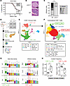 Figure 1
Resistance to T1D in NOD-UTXTCD mice is associated with altered distribution of pLN CD8+ T cells. (A) Diabetes-free incidence curves of NOD-UTXTCD vs. NOD-WT female littermates. ****P < 0.0001; log rank test. (B and C) Representative H&E-stained pancreas sections (B) and cumulative islet infiltration scores (C) from NOD-UTXTCD vs. NOD-WT female littermates. *P < 0.05; Tukey’s multiple comparisons test. Scale bar: 200 μm. (D) Scheme for 10X Genomics single-cell RNA sequencing (scRNA-seq) of CD45+ cells from pLNs of NOD-WT and NOD-UTXTCD mice. (E and F) UMAP of CD45+ cells (E) and subclustered CD8+ T cells (F). (G) Gene expression across the CD8+ T cell clusters. (H) Comparison of subset frequencies of CD8+ Tprog-like and CD8+ Tmed-like populations. CD8+ Tprog-like, *P = 0.015; CD8+ Tmed-like, **P = 0.026; 2-sided, unpaired Mann-Whitney test.
We next sought to explore the cellular mechanism by which UTX functions in T cells to promote T1D. Prior to infiltrating pancreatic islets, diabetogenic T cells are first primed in the pancreatic lymph nodes (pLNs) (3, 19). To delineate how UTX deficiency alters pLN T cell dynamics in NOD mice, we performed single-cell RNA-seq (scRNA-seq) of pLN immune cells (CD45+) from 8- and 13-week-old NOD-WT and NOD-UTXTCD mice (Figure 1D; n = 6 per genotype). These ages represent distinct stages in T1D autoimmunity development: 8 weeks represents a stage when immune cell infiltration is prominent in islets, and 13 weeks represents a stage immediately prior to overt diabetes (20). UMAP clustering of 97,400 cells showed 7 major immune cell populations, including CD8+ T cells, CD4+ T cells, B cells, and myeloid cells (Figure 1E and Supplemental Data File 1). Frequencies of these populations were similar between NOD-WT and NOD-UTXTCD mice (Supplemental Figure 1, C and D). We further evaluated CD4+ and CD8+ T cell populations to assess for primary alterations resulting from T cell–specific UTX deficiency.
Subclustering of CD4+ T cells yielded 10 populations (Supplemental Figure 2A), including T follicular helper (Tfh) and Tregs. Frequencies of key autoimmunity-associated CD4+ T cell subsets in the pLN were similar between NOD-WT and NOD-UTXTCD mice (Supplemental Figure 2B). Moreover, flow cytometric analysis of pLNs showed similar frequencies of CD4+ T cell subsets (Tfh [CXCR5+ PD1+], Th1 [T-bet+], and Treg [Foxp3+]) (Supplemental Figure 2C) and proinflammatory cytokine-producing CD4+ cells (Supplemental Figure 2D) between NOD-WT and NOD-UTXTCD mice. Thus, pLN CD4+ T cell distribution was largely unchanged by T cell–specific UTX deficiency in NOD mice.
In contrast, substantial changes were noted in the distribution of CD8+ T cell subsets between NOD-WT and NOD-UTXTCD mice. CD8+ T cells were subclustered into 8 populations (Figure 1F), which included a “CD8+ Tprog-like” population that expressed genes associated with stem-like progenitor cells (Tcf7 [which encodes TCF1], Slamf6 [which encodes Ly108], Sell, Myb, Ikzf2, Id3) (Figure 1G and Supplemental Figure 3A) and a “CD8+ Tmed-like” effector population that expressed genes associated with terminally differentiated autoimmune mediators (Nkg7, Cxcr6, Thy1) (3, 9) (Figure 1G). Strikingly, a higher frequency of CD8+ Tprog-like and a lower frequency of CD8+ Tmed-like, cells were seen in the pLNs of NOD-UTXTCD mice, compared with NOD-WT littermates (Figure 1H and Supplemental Figure 3, B and C). Thus, protection from T1D in NOD-UTXTCD mice is associated with an altered distribution of pLN CD8+ T cell subsets expressing markers of stem-like progenitors and effectors.
UTX in T cells promotes CD8+ T cell transition from progenitor to cytolytic effector state in T1D. Within the pLNs of NOD mice, stem-like progenitors (Tprog) have been reported to differentiate into terminally differentiated autoimmune mediators (Tmed), which then enter the pancreatic islets to destroy β cells (3). In line with this report, pseudotime trajectory analysis of our scRNA-seq dataset suggested a differentiation pathway that starts with the CD8+ Tprog-like population, moves through an intermediate population (CD8+ T intermediate), and ends in the CD8+ Tmed-like population (Figure 2A). Along the pseudotime axis, expression of stem-like progenitor-associated genes (Tcf7, Slamf6) was highest in CD8+ Tprog-like cells, decreased in the CD8+ T intermediate cells, and lowest in CD8+ Tmed-like cells (Figure 2, B and C). On the other hand, expression of effector-associated genes (Thy1, Cxcr6) was lowest in the CD8+ Tprog-like cells and increased in CD8+ T intermediates and CD8+ Tmed-like cells (Figure 2, B and C). Of note, T cell–specific UTX deficiency altered this differentiation trajectory. Comparison of cell densities along the pseudotime trajectory revealed relative accumulation of NOD-UTXTCD CD8+ Tprog-like cells, compared with NOD-WT cells (Figure 2D). This difference was accompanied by a relative decrease in NOD-UTXTCD CD8+ Tmed-like cells, compared with NOD-WT cells. Together, these data suggest a potential block in the conversion of CD8+ Tprog to Tmed cells in NOD-UTXTCD mice.
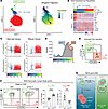 Figure 2
Impaired conversion of stem-like progenitors to effectors in NOD-UTXTCD mice. (A) UMAP plot of CD8+ Tprog-like, CD8+ T intermediate, and CD8+ Tmed-like clusters (left), with cells color-coded along Slingshot pseudotime (right). (B) Heatmap of scRNA-seq expression for key genes, plotted along Slingshot pseudotime. (C) Expression of key genes along Slingshot pseudotime trajectory, color coded by clusters shown in UMAP (A, left). (D) Comparison of cell densities of CD8+ Tprog-like, CD8+ T intermediate, and CD8+ Tmed-like cells derived from 8-week-old NOD-WT (WT) vs. NOD-UTXTCD (UTXTCD) mice, plotted along Slingshot pseudotime trajectory. P < 2.2 e-16; asymptotic 2-sample Kolmogorov-Smirnov test. (E) Gating scheme for identifying Tprog-like and Tmed-like cells by flow cytometric analysis. (F) Representative flow cytometric plot (left) and average frequencies (right) of pLN Tprog-like (Ly108+CXCR6–) and Tmed-like (Ly108–CXCR6+) cells of NOD-WT and NOD-UTXTCD female littermates (15–29 weeks of age). ****P < 0.0001; ***P < 0.001; Student’s t test. (G) Working model: UTX promotes conversion of long-lived stem-like CD8+ T cell progenitors (Tprog) to short-lived mediators (Tmed), which then enter pancreatic islets to mediate β cell destruction.
To verify this finding, we turned to flow cytometric analysis, using previously reported cell surface markers to identify Tprog-like and Tmed-like cells (4). In this approach, reciprocal expression of Ly108 (Slamf6) and CXCR6 were used to distinguish Tprog-like (Ly108+CXCR6–) and Tmed-like (Ly108–CXCR6+) populations (Figure 2E). Supporting the validity of this gating strategy, expression of the stem-like TF TCF1 was higher in Ly108+CXCR6– Tprog-like populations than that in Ly108– CXCR6+ Tmed-like populations (Supplemental Figure 4A). Furthermore, expression of the CD39 mediator marker was lower in Ly108+CXCR6– Tprog-like cells than Ly108– CXCR6+ Tmed-like populations (Supplemental Figure 4B). Using this gating strategy, a significant increase in Tprog-like cells was seen in pLNs of NOD-UTXTCD mice, compared with NOD-WT (Figure 2F). At the same time, a significant decrease in Tmed-like populations was seen in pLNs of NOD-UTXTCD mice, compared with NOD-WT mice. Taken together, our scRNA-seq and flow cytometric analyses support a model in which UTX promotes conversion of CD8+ T cell progenitors to effectors, which then enter the pancreatic islets to destroy β cells and incite diabetes (Figure 2G). In support of the importance of UTX in diabetogenic CD8+ T cells, diabetes development was delayed in immunodeficient hosts after adoptive transfer with UTX-deficient CD8+ T cells, compared with UTX-sufficient (WT) CD8+ T cells (Supplemental Figure 4C).
UTX controls progenitor to cytolytic effector differentiation in β cell–specific CD8+ T cells. CD8+ T cells recognizing the pancreatic β cell antigen islet-specific glucose-6-phosphatase catalytic subunit-related protein (IGRP) are a major pathogenic population in mice and humans with T1D (21, 22). Remarkably, Gearty et al. reported that IGRP-specific stem-like progenitors (Tprog; Ly108+CD39–) give rise to both Tprog and autoimmune mediator (Tmed; Ly108–CD39+) populations, and as few as 20 pLN Tprog cells from NOD donors can induce diabetes in immunodeficient hosts (3). Given their potent diabetes-inducing potential, we sought to define the role of UTX in controlling the differentiation of IGRP-specific Tprog cells into cytolytic Tmed cells.
In keeping with the markers used by Gearty et al., we identified pLN CD8+ Tprog cells as Ly108+CD39– cells and Tmed cells as Ly108–CD39+ cells (3) (Figure 3A). As expected, Ly108+CD39– Tprog cells expressed higher levels of the progenitor marker TCF1 (Supplemental Figure 4D) and lower levels of the mediator marker CXCR6 than Ly108–CD39+ Tmed cells (Figure 3B). We further identified IGRP-specific T cells by their expression of CD44, a marker of antigen-experienced T cells, and their binding to tetramers containing the IGRP mimotope NRP-V7 (CD44+ NRP-V7+) (23). Using this gating approach, the frequency of Tprog cells among IGRP-specific, antigen-experienced CD8+ T cells (Ly108+CD39– cells within CD8+CD44+NRP-V7+) was increased in NOD-UTXTCD pLNs compared with that in NOD-WT pLNs (Figure 3C). At the same time, the frequency of Tmed (Ly108–CD39+) cells within antigen-experienced, IGRP-specific (CD8+CD44+NRP-V7+) cells was significantly lower in NOD-UTXTCD pLNs, compared with that in NOD-WT pLNs (Figure 3C). Similar findings were seen when CXCR6, rather than CD39, was used as a mediator marker (Supplemental Figure 4E). With age, the frequency of IGRP-specific Tmed (Ly108–CD39+ cells within CD8+CD44+NRP-V7+) cells increased in NOD-WT pLNs, while IGRP-specific Tprog (Ly108+CD39– cells within CD8+CD44+NRP-V7+) cells accumulated in NOD-UTXTCD pLNs (Supplemental Figure 4F). Together, these findings demonstrate a role for UTX in promoting the conversion of β cell–specific Tprog cells to Tmed cells in the pLN.
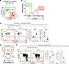 Figure 3
UTX promotes Tprog to Tmed conversion of β cell–specific CD8+ T cells. (A) Gating scheme for identifying Tprog and Tmed cells by flow cytometric analysis. (B) Representative histogram of CXCR6 expression in Tprog vs. Tmed cell populations identified by Ly108 and CD39 expression. (C) Representative flow cytometric plot (left), and average frequencies (right) of Tprog (Ly108+ CD39–) and Tmed (Ly108– CD39+) cells within antigen-experienced, IGRP-specific pLN CD8+ T cells (CD8+ CD44+ NRP-V7+) of NOD-WT and NOD-UTXTCD female littermates (15–29 weeks of age). ****P < 0.0001; ***P < 0.001; Student’s t test. (D) Scheme for adoptive transfer of pLN Tprog cells (Ly108+CD39–) from NOD-TCR 8.3 WT vs. NOD-TCR 8.3 UTXTCD mice into NOD.SCID recipients. (E) Representative flow cytometric plot (left) and average frequencies (right) of Tprog (Ly108+CD39–) or Tmed (Ly108–CD39+) among pLN antigen-experienced (CD8+CD44+) cells in hosts after adoptive transfer, as shown in D. Data from 2 replicate experiments are shown. **P < 0.01; *P < 0.05; Student’s t test.
To further delineate UTX’s functions within β cell–specific Tprog cells, we performed adoptive transfers of IGRP-specific Tprog cells into immunodeficient NOD.SCID recipients (Figure 3D). To isolate sufficient numbers of IGRP-specific Tprog cells for transfer, we exploited an NOD T cell receptor (TCR) transgenic mouse line (NOD-TCR 8.3) in which T cells express a TCR recognizing the IGRP206–214 epitope in the context of MHC class I (24). IGRP-specific CD8+ T cell progenitors (Ly108+CD39–) were sorted from 6-week-old NOD-TCR 8.3 mice with or without T cell–specific deficiency in UTX (NOD-TCR 8.3 UTXTCD or NOD-TCR 8.3 WT, respectively). Three weeks after transfer, an increased frequency of pLN Tprog cells (Ly108+CD39– among CD8+CD44+) was seen in hosts receiving UTX-deficient CD8+ Tprog cells, compared with WT cells (Figure 3E). Concomitantly, a decrease in the frequency of pLN Tmed (Ly108–CD39+ among CD8+CD44+) cells was noted in recipients of UTX-deficient Tprog cells (Figure 3E). Together, these findings support a model in which UTX functions in IGRP-specific CD8+ Tprog cells to promote their transition to Tmed cells.
We next asked whether UTX may play a parallel function in CD8+ Tprog cells within the pancreas. To address this, we performed flow cytometric analysis of pancreatic CD8+ T cell subsets in NOD-TCR 8.3 UTXTCD versus NOD-TCR 8.3 WT mice (Supplemental Figure 5A). Similar to pLN CD8+ T cells, pancreatic CD8+ T cells showed altered subset distribution with UTX deficiency. Tprog cells (Ly108+CD39–) were increased, while Tmed cells (Ly108–CD39+) were decreased, in the pancreas of NOD-TCR 8.3 UTXTCD mice compared with NOD-TCR 8.3 WT mice. These findings suggest that UTX’s role in CD8+ Tprog cells in the pancreas mirrors its role in the pLN.
UTX controls chromatin accessibility and transcriptional changes in antigen-specific pLN Tprog cells. Recent studies have demonstrated the importance of epigenetic and transcriptional regulatory programs in controlling the transition of CD8+ T cell states (25). Because UTX is an epigenetic regulator (26), we reasoned that UTX may control the Tprog to Tmed conversion by remodeling chromatin at regulatory elements of target gene loci. To examine this, we performed bulk assay for transposase-accessible chromatin sequencing (ATAC-seq) and RNA-seq in parallel on sort-purified β cell–specific CD8+ Tprog cells from pLNs of either NOD-TCR 8.3 UTXTCD or NOD-TCR 8.3 WT mice. ATAC-seq analysis demonstrated large-scale chromatin accessibility alterations in UTX-deficient CD8+ T cells. Principal component analysis (PCA) plots showed sample clustering by genotype (Figure 4A, top), highlighting the extensive effect of UTX on the chromatin landscape of CD8+ T cells. RNA-seq analysis performed in parallel also showed large-scale transcriptional changes, with PCA analysis of gene expression demonstrating genotype-specific clustering (Figure 4A, bottom). ATAC-seq revealed that 4,075 peaks decreased in accessibility and 8,807 peaks increased in accessibility in NOD-TCR 8.3 UTXTCD progenitors, compared with UTX-sufficient controls (|log2 fold change| >0.5, FDR-adjusted P < 0.05) (Supplemental Data File 2). These differentially accessible peaks corresponded to 6,859 differentially accessible genes. Furthermore, RNA-seq revealed 1,280 decreased and 1,768 increased transcripts in NOD-TCR 8.3 UTXTCD progenitors, compared with UTX-sufficient controls (|log2 fold change| >0.5, FDR-adjusted P < 0.05) (Supplemental Data File 3).
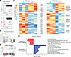 Figure 4
UTX controls chromatin accessibility at progenitor and effector gene loci in antigen-specific pLN Tprog cells. (A) PCA analysis of ATAC-seq (top) and RNA-seq (bottom) profiles of sorted Tprog (CD8+ Ly108+ CD39–) cells from pLNs of NOD-TCR 8.3 WT and NOD-TCR 8.3 UTXTCD mice (n = 3/group). (B) Heatmaps of the expression (left, RNA-seq) and chromatin accessibility (right, ATAC-seq) of selected differentially expressed and accessible genes involved in immune function. The color scale indicates the relative expression or accessibility, with red representing higher values and blue representing lower values. (C and D) Gene set enrichment analysis (GSEA) for progenitor signature (Supplemental Data File 4) (C) and effector signature (D) genes in the RNA-seq dataset of Tprog cells from NOD-TCR 8.3 WT and NOD-TCR 8.3 UTXTCD mice. (E) Pathway analysis of differentially accessible genes by ATAC-seq using Enrichr (P value by Fisher’s exact test).
Integrative analysis of ATAC- and RNA-seq identified a significant positive correlation (Pearson R = 0.73, P < 2.2 × 10⁻¹⁶) between genotype-dependent changes in chromatin accessibility and gene expression (Supplemental Figure 6A). These data suggest a direct correlation between UTX-mediated alterations in chromatin accessibility and transcription. Concurrent changes in chromatin accessibility and transcription were observed in genes with known roles in the differentiation and maintenance of stem-like progenitor CD8+ T cells. For instance, NOD-TCR 8.3 UTXTCD Tprog cells demonstrated increased chromatin accessibility, with corresponding increased gene expression, at the locus of Bcl6 (Figure 4B and Supplemental Figure 6B), a TF that prolongs stem-like progenitor persistence (27). Additionally, NOD-TCR 8.3 UTXTCD Tprog cells also showed increased chromatin accessibility and gene expression at the locus of Bach2 (Figure 4B and Supplemental Figure 6B), a transcriptional repressor that promotes the stem-like CD8+ T cell program, while suppressing terminal exhaustion programs (27). Finally, Bcl2, an anti-apoptotic factor that promotes stem-like progenitor cell survival (28), also showed increased accessibility and expression in NOD-TCR 8.3 UTXTCD Tprog cells (Figure 4B and Supplemental Figure 6B). Thus, UTX deficiency is associated with changes at loci that are important in stem-like progenitor maintenance and differentiation.
At the same time, decreased accessibility and reduced expression were seen in NOD-TCR 8.3 UTXTCD Tprog cells at genes important for CD8+ T cell effector function (Figure 4B and Supplemental Figure 6B). These genes include cytolytic molecules (e.g., Gzmb, Gzmc, Fas) important in β cell killing (29); integrins (e.g., Itga4, Itgae) (30, 31) critical in leukocyte islet infiltration; and cytokine/cytokine receptors (e.g., Ifngr1, Cxcr6) (4, 32, 33). Both Dapl1 and Nfatc2, which function together in CD8+ T cell effector function (34), were also less accessible and expressed. Moreover, genes associated with T cell activation (Fosl2, Thy1, Cd9) were also less accessible and downregulated in expression in CD8+ T cells from NOD-TCR 8.3 UTXTCD mice (35–37). Gene set enrichment analysis (GSEA) confirmed these findings, showing upregulated stem-like progenitor and downregulated effector gene signatures in NOD-TCR 8.3 UTXTCD cells (Figure 4, C and D, and Supplemental Data File 4) (4, 9).
Enrichment analysis of differentially accessible genes revealed association with pathways important in stem-like progenitor generation as well as cytolytic effector differentiation and function. For instance, Wnt signaling is critical in the generation and maintenance of stem-like memory T cells (38), and Enrichr pathway analysis of differentially accessible genes revealed upregulation of “Wnt Signaling Pathway” and “Canonical Wnt Signaling Pathway” genes in NOD-TCR 8.3 UTXTCD Tprog cells (Figure 4E). At the same time, “Lymphocyte Differentiation,” “Integrin Cell Surface Interactions,” “Positive Regulation of Lymphocyte Activation,” and other pathways associated with cytolytic effector function were downregulated in NOD-TCR 8.3 UTXTCD Tprog cells (Figure 4E). Together, these findings provide further support that UTX poises chromatin in stem-like Tprog cells for transition to cytolytic effectors.
Integrated analysis of UTX chromatin binding, accessibility, and transcription reveals STAT3 and TCF1 co-occupancy at UTX target genes. The genome-wide changes in accessibility and transcription in UTX-deficient Tprog cells may be attributed either to (a) UTX’s direct regulation of chromatin accessibility and gene expression or (b) secondary changes that result from alterations in other UTX-mediated epigenetic and transcriptional regulators. To identify gene loci directly regulated by UTX, we utilized UTX cleavage under targets and tagmentation assay (CUT&Tag) (39) to identify gene loci bound by UTX on sort-purified NOD-TCR 8.3 WT Tprog cells. Sort-purified NOD-TCR 8.3 UTXTCD Tprog cells were used as a negative control. PCA of UTX CUT&Tag data revealed sample clustering by genotype (Figure 5A). We identified 6,718 UTX-bound peaks (FDR-adjusted P < 0.05), which corresponded to 5,222 distinct genes (Supplemental Data File 5). Genomic annotation of UTX CUT&Tag peaks demonstrated a strong promoter bias, with 64.5% of UTX-bound regions localized within 1 kb of transcription start sites (Figure 5B). This promoter proximal enrichment suggests that UTX primarily engages core regulatory elements to exert its epigenetic functions in Tprog cells.
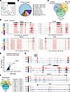 Figure 5
Integrative analysis of chromatin binding, accessibility, and transcription reveals that STAT3 and TCF1 cobind UTX-target genes. (A) PCA analysis of UTX CUT&Tag from sorted Tprog (CD8+ Ly108+ CD39–) cells from pLNs of NOD-TCR 8.3 WT and NOD-TCR 8.3 UTXTCD mice. (B) Location of UTX-bound peaks. (C) Venn diagram outlining overlapping differentially accessible (DA) genes identified by ATAC-seq and differentially expressed (DE) genes identified by RNA-seq. (D) Representative gene tracks from the UCSC Integrated Genome Browser of anti-UTX CUT&Tag, ATAC-seq, and RNA-seq of effector genes, Gzmb and Cxcr6, and progenitor genes, Bach2 and Bcl2. The y axis depicts counts per million (CPM). (E) Transcriptional factors were bound to the overlapping genes identified by UTX datasets. Overlapping genes were input into Enrichr for ChEA3 analysis. (F) Venn diagram outlining overlapping genes bound by UTX, TCF1, and STAT3 (TCF1 ChIP-seq: GSE73240, STAT3 ChIP-seq: GSE217374). (G) Gene tracks from the UCSC genome browser to demonstrate the aligned region bound by UTX, TCF1, and STAT3. The y axis depicts CPM.
We utilized this UTX CUT&Tag dataset, the ATAC-seq dataset (Figure 4B), and the RNA-seq dataset (Figure 4B) to identify 502 gene loci that are UTX-bound, differentially accessible, and differentially expressed in UTX-deficient Tprog cells (Figure 5C). These UTX-bound genes included stem-like progenitor-associated genes (i.e., Bach2, Bcl2, Bcl6) that are increased in accessibility and transcription within NOD-UTXTCD Tprog cells (Figure 5D and Supplemental Figure 7A), suggesting that UTX binds directly to stem-like progenitor-associated genes to decrease accessibility and repress transcription at these loci. Additionally, UTX-bound genes included mediator-associated genes (i.e., Gzmb, Cxcr6, Thy1) that are decreased in accessibility and transcription within NOD-UTXTCD Tprog cells (Figure 5D and Supplemental Figure 7A). These analyses demonstrate that in antigen-specific Tprog cells, UTX poises chromatin to repress transcription at key Tprog genes and to promote transcription at effector genes.
While UTX has intrinsic histone demethylase activity, our data demonstrate that UTX’s demethylase activity is dispensable in T1D pathogenesis (Supplemental Figure 1B). An alternative mechanism by which UTX exerts its regulatory effects is through interactions with TFs (40). To identify TF binding partners of UTX, we used a TF enrichment analysis tool (ChEA3 in Enrichr) to prioritize TFs potentially responsible for the observed 502 UTX-bound, differentially accessible, and differentially expressed target genes (Figure 5E) (41). Highly associated TFs included (a) TCF1, a key regulator of stem-like progenitors, (9) and (b) STAT3, a TF implicated in Tprog to effector cell conversion and a key TF in T1D pathogenesis (10–12). These findings suggest the possibility that UTX may function in concert with TCF1 and STAT3 to regulate differentiation of Tprog cells to an effector state.
We next integrated our UTX CUT&Tag dataset with publicly available chromatin immunoprecipitation datasets to determine whether gene loci may be co-occupied by UTX, TCF1, and STAT3 proteins. Cross-referencing of UTX-bound genes with TCF1-bound (GSE73240) and STAT3-bound (GSE217374) genes showed substantial overlap of 1,768 genes (Figure 5F). These overlapping genes included progenitor-associated genes (Bcl2, Bach2, Bcl6, and Lef1) and mediator-associated genes (Cxcr6, Gzmb, Ifngr1, and Thy1) that are differentially accessible and differentially expressed in NOD-UTXTCD CD8+ Tprog cells, compared with NOD-WT cells. Moreover, comparison of UTX-bound, TCF1-bound, and STAT3-bound peaks showed a high degree of overlap in multiple genes (Figure 5G and Supplemental Figure 8A). Binding by UTX, TCF1, and STAT3 was noted at key effector-associated genes (e.g., Gzmb, Cxcr6, Runx3, and Ifng) as well as progenitor-associated genes (e.g., Bach2, Ccr7, and Lef1), suggesting co-occupancy of UTX, TCF1, and STAT3 at these target gene loci. Collectively, these data suggest that UTX cooperates with TCF1 and STAT3 to influence the transition of antigen-specific Tprog cells to an effector state.
UTX promotes differentiation of human stem cell memory CD8+ T cells into a terminally differentiated effector state through binding with TCF1 and STAT3. Recent work has associated T1D in humans with increased circulating CD8+ T memory stem cells (TSCM), a long-lived, self-renewing β cell–specific population (2, 42, 43). TSCM cells play a key role in T1D pathogenesis by giving rise to terminally differentiated effector/mediator (TTE) cells that ultimately kill insulin-producing β cells. Thus, the TSCM (CCR7+CD45RA+CD95+) population in patients with T1D may have a parallel role to the Tprog population in NOD mice, while the TTE (CCR7–CD45RA+CD95+) population in patients with T1D may have a parallel role to the Tmed population in NOD mice (42, 43).
To test whether UTX controls the TSCM to TTE conversion, we utilized CRISPR/Cas9 to edit the UTX locus in CD8+ T cells isolated from PBMCs from a patient with T1D. We confirmed efficient knockdown of UTX protein expression using UTX-specific sgRNAs compared with a nontargeting control (NTC sgRNA) (Figure 6A). Following a 7-day culture with homeostatic cytokines (IL-2, IL-7, IL-15), we observed an increased frequency of TSCM cells (CCR7+CD45RA+) and a corresponding decrease in TTE cells (CCR7–CD45RA+) in UTX-deficient CD8+ T cells relative to controls (Figure 6B). This altered distribution supports a cell-intrinsic role for UTX in governing TSCM to TTE conversion in human CD8+ T cells, indicating a conserved, cross-species function for UTX.
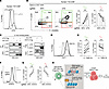 Figure 6
UTX regulates human stem cell memory CD8+ T cell differentiation into the terminally effector state and interacts with TCF1 and STAT3. (A) Representative flow cytometric plot of CD8+ T cells from patients with T1D after CRISPR-Cas9 editing with NTC (non-targeting control) sgRNA or UTX-targeting sgRNA. (B) Representative flow cytometric plots (left) and average frequency (right) of TSCM and TTE cells among CD8+CD95+ peripheral blood cells from patients with T1D. Cells were edited with either NTC sgRNA or UTX-targeting sgRNA. (C) In vitro binding assay to show the interaction of UTX-TCF1 (left) and UTX-STAT3 (right) in HEK-293T cells by cotransfection and coimmunoprecipitation. (D) Representative flow cytometric histogram plot of TCF1 expression in CD8+ T cells treated with DMSO or 5 μM TWS119 supplemented with IL-7 (10 ng/mL) and IL-21 (25 ng/mL) for 7 days. (E) Frequencies of TSCM and TTE cells within CD8+CD95+ T cells treated with DMSO or 5 μM of TWS119 for 3 days. Each dot represents a biological replicate (n = 7). **P < 0.01; paired Student’s t test. (F) Representative flow cytometric histogram plot of CD8+ T cells from patients with T1D after CRISPR/Cas9 editing with nontargeting control (NTC) sgRNA or STAT3-targeting sgRNA. (G) Frequencies of TSCM and TTE cells within CD8+CD95+ T cells after CRISPR/Cas9 editing with sgSTAT3 versus a NTC. Left: TSCM; right: TTE. Each dot represents a biological replicate (n = 3). **P < 0.01; paired Student’s t test. (H) Model of how UTX forms a complex with TCF1 and STAT3 to modulate the progenitor and effector programs in promoting T1D development.
Given that our integrative analysis suggested a high degree of cooperativity between UTX, TCF1, and STAT3 (Figure 5, E–G), we utilized a coimmunoprecipitation approach to assess the binding of UTX to TCF1 and STAT3. HEK-293T cells were cotransfected with epitope-tagged constructs encoding UTX in combination with either TCF1 or STAT3, and subsequent immunoprecipitation demonstrated interactions between UTX and both TCF1 and STAT3 (Figure 6C). In line with this finding, endogenous UTX protein coimmunoprecipitated with TCF1 and STAT3 in Jurkat cells (Supplemental Figure 9A). These data demonstrate that UTX directly binds critical TFs important in progenitor specification (9) and progenitor-to-effector differentiation (10–12).
TCF1 expression is induced by inhibitors of glycogen synthase kinase 3 β, such as TWS119 (9). Consistent with TCF1’s role in enforcing progenitor formation and maintenance, treatment of CD8+ T cells from patients with T1D with TWS119 resulted in the induction of TCF1 (Figure 6D) and an increased frequency of TSCM and a decreased frequency of TTE cells (Figure 6E). Moreover, STAT3 drives progenitor T cell conversion to effectors (11), and editing of the STAT3 locus in CD8+ T cells from patients with T1D resulted in accumulation of TSCM cells and decreased TTE cells (Figure 6, F and G). Based on these data, we propose a mechanistic model wherein UTX interacts with STAT3 to enhance effector gene transcription necessary for the progenitor-to-effector transition, while simultaneously binding TCF1 to suppress its progenitor-maintaining functions during differentiation (Figure 6H). These factors function in tandem at gene loci important in effector function and progenitor identity, an insight that enhances our understanding of the molecular mechanisms underpinning autoimmune chronicity and offers potential avenues for targeted therapeutic interventions in T1D.


Copyright © 2026 American Society for Clinical Investigation
ISSN: 0021-9738 (print), 1558-8238 (online)