
Advertisement
Research ArticleCell biologyOncology
Open Access |  10.1172/JCI195639
Therapeutic delivery of microRNAs discovered to target deregulated glioblastoma pathways inhibits tumor growth in mice
Shekhar Saha,1 Ying Zhang,1 Myron K. Gibert Jr.,1 Collin Dube,1 Farina Hanif,1,2 Elizabeth Qian Xu Mulcahy,1 Sylwia Bednarek,1 Yunan Sun,1 Pawel Marcinkiewicz,1 Xiantao Wang,3 Gijung Kwak,4,5 Ahsan Polash,3 Haolin Li,4,5,6 Kadie Hudson,1 Manikarna Dinda,7 Tapas Saha,8 Matthew McCord,9 Fadila Guessous,1,10 Nichola Cruickshanks,1 Rossymar Rivera Colon,1 Lily Dell’Olio,1 Rajitha Anbu,1 Wenjie Liu,1 Songy Choi,1 Benjamin Kefas,1,11 Pankaj Kumar,12 Alexander L. Klibanov,13 David Schiff,14 Jung Soo Suk,4,5,6,15 Justin Hanes,16 Jamie Mata,17 Markus Hafner,3 and Roger Abounader1,14,18,19
1Department of Microbiology, Immunology, and Cancer Biology, University of Virginia School of Medicine, Charlottesville, Virginia, USA.
2Department of Biochemistry, Dow International Medical College, Dow University of Health Sciences, OJHA Campus, Karachi, Pakistan.
3National Institute of Arthritis and Musculoskeletal and Skin Diseases, NIH, Bethesda, Maryland, USA.
4Department of Neurosurgery and
5Medicine Institute for Neuroscience Discovery (UM-MIND), School of Medicine, University of Maryland, Baltimore, Maryland, USA.
6Department of Chemical and Biomolecular Engineering, School of Engineering, Johns Hopkins University, Baltimore, Maryland, USA.
7Department of Biochemistry and Molecular Genetics, University of Virginia School of Medicine, Charlottesville, Virginia, USA.
8Swiss Re, Healthcare, Bengaluru, India.
9Department of Pathology, University of Virginia School of Medicine, Charlottesville, Virginia, USA.
10Laboratory of Onco-Pathology, Biology and Cancer Environment, Faculty of Medicine, Mohammed VI University of Sciences and Health, Casablanca, Morocco.
11Pharmacy, University of Virginia, Charlottesville, Virginia, USA.
12Bioinformatics Core, University of Virginia School of Medicine, Charlottesville, Virginia, USA.
13Division of Cardiovascular Medicine and
14Department of Neurology, University of Virginia, Charlottesville, Virginia, USA.
15Department of Neurosurgery, School of Medicine, Johns Hopkins University, Baltimore, Maryland, USA.
16Center for Nanomedicine at the Wilmer Eye Institute, Johns Hopkins University School of Medicine, Baltimore, Maryland, USA.
17Department of Radiology and Medical Imaging, University of Virginia School of Medicine, Charlottesville, Virginia, USA.
18Comprehensive Cancer Center and
19Center for RNA Science and Medicine, University of Virginia, Charlottesville, Virginia, USA.
Address correspondence to: Roger Abounader or Ying Zhang, Old Medical School, 21 Hospital Drive, Room 4819, Charlottesville, Virginia 22908, USA; Email: ra6u@virginia.edu (R Abounader); yz5h@virginia.edu (YZ). Or to: Markus Hafner, RNA Molecular Biology Laboratory, Building 31 – 4C32, 31 Center Drive, Bethesda, Maryland 20892-0004, USA. Email: markus.hafner@nih.gov. Or to: Jamie Mata, Department of Radiology and Medical Imaging, 480 Ray C Hunt Drive, Snyder Building, Charlottesville, Virginia 22903, USA. Email: jfm4q@virginia.edu.
Authorship note: SS, YZ, MKG, and CD contributed equally to this work.
Find articles by
Saha, S.
in:
PubMed
|
Google Scholar
|

1Department of Microbiology, Immunology, and Cancer Biology, University of Virginia School of Medicine, Charlottesville, Virginia, USA.
2Department of Biochemistry, Dow International Medical College, Dow University of Health Sciences, OJHA Campus, Karachi, Pakistan.
3National Institute of Arthritis and Musculoskeletal and Skin Diseases, NIH, Bethesda, Maryland, USA.
4Department of Neurosurgery and
5Medicine Institute for Neuroscience Discovery (UM-MIND), School of Medicine, University of Maryland, Baltimore, Maryland, USA.
6Department of Chemical and Biomolecular Engineering, School of Engineering, Johns Hopkins University, Baltimore, Maryland, USA.
7Department of Biochemistry and Molecular Genetics, University of Virginia School of Medicine, Charlottesville, Virginia, USA.
8Swiss Re, Healthcare, Bengaluru, India.
9Department of Pathology, University of Virginia School of Medicine, Charlottesville, Virginia, USA.
10Laboratory of Onco-Pathology, Biology and Cancer Environment, Faculty of Medicine, Mohammed VI University of Sciences and Health, Casablanca, Morocco.
11Pharmacy, University of Virginia, Charlottesville, Virginia, USA.
12Bioinformatics Core, University of Virginia School of Medicine, Charlottesville, Virginia, USA.
13Division of Cardiovascular Medicine and
14Department of Neurology, University of Virginia, Charlottesville, Virginia, USA.
15Department of Neurosurgery, School of Medicine, Johns Hopkins University, Baltimore, Maryland, USA.
16Center for Nanomedicine at the Wilmer Eye Institute, Johns Hopkins University School of Medicine, Baltimore, Maryland, USA.
17Department of Radiology and Medical Imaging, University of Virginia School of Medicine, Charlottesville, Virginia, USA.
18Comprehensive Cancer Center and
19Center for RNA Science and Medicine, University of Virginia, Charlottesville, Virginia, USA.
Address correspondence to: Roger Abounader or Ying Zhang, Old Medical School, 21 Hospital Drive, Room 4819, Charlottesville, Virginia 22908, USA; Email: ra6u@virginia.edu (R Abounader); yz5h@virginia.edu (YZ). Or to: Markus Hafner, RNA Molecular Biology Laboratory, Building 31 – 4C32, 31 Center Drive, Bethesda, Maryland 20892-0004, USA. Email: markus.hafner@nih.gov. Or to: Jamie Mata, Department of Radiology and Medical Imaging, 480 Ray C Hunt Drive, Snyder Building, Charlottesville, Virginia 22903, USA. Email: jfm4q@virginia.edu.
Authorship note: SS, YZ, MKG, and CD contributed equally to this work.
Find articles by Zhang, Y. in: PubMed | Google Scholar
1Department of Microbiology, Immunology, and Cancer Biology, University of Virginia School of Medicine, Charlottesville, Virginia, USA.
2Department of Biochemistry, Dow International Medical College, Dow University of Health Sciences, OJHA Campus, Karachi, Pakistan.
3National Institute of Arthritis and Musculoskeletal and Skin Diseases, NIH, Bethesda, Maryland, USA.
4Department of Neurosurgery and
5Medicine Institute for Neuroscience Discovery (UM-MIND), School of Medicine, University of Maryland, Baltimore, Maryland, USA.
6Department of Chemical and Biomolecular Engineering, School of Engineering, Johns Hopkins University, Baltimore, Maryland, USA.
7Department of Biochemistry and Molecular Genetics, University of Virginia School of Medicine, Charlottesville, Virginia, USA.
8Swiss Re, Healthcare, Bengaluru, India.
9Department of Pathology, University of Virginia School of Medicine, Charlottesville, Virginia, USA.
10Laboratory of Onco-Pathology, Biology and Cancer Environment, Faculty of Medicine, Mohammed VI University of Sciences and Health, Casablanca, Morocco.
11Pharmacy, University of Virginia, Charlottesville, Virginia, USA.
12Bioinformatics Core, University of Virginia School of Medicine, Charlottesville, Virginia, USA.
13Division of Cardiovascular Medicine and
14Department of Neurology, University of Virginia, Charlottesville, Virginia, USA.
15Department of Neurosurgery, School of Medicine, Johns Hopkins University, Baltimore, Maryland, USA.
16Center for Nanomedicine at the Wilmer Eye Institute, Johns Hopkins University School of Medicine, Baltimore, Maryland, USA.
17Department of Radiology and Medical Imaging, University of Virginia School of Medicine, Charlottesville, Virginia, USA.
18Comprehensive Cancer Center and
19Center for RNA Science and Medicine, University of Virginia, Charlottesville, Virginia, USA.
Address correspondence to: Roger Abounader or Ying Zhang, Old Medical School, 21 Hospital Drive, Room 4819, Charlottesville, Virginia 22908, USA; Email: ra6u@virginia.edu (R Abounader); yz5h@virginia.edu (YZ). Or to: Markus Hafner, RNA Molecular Biology Laboratory, Building 31 – 4C32, 31 Center Drive, Bethesda, Maryland 20892-0004, USA. Email: markus.hafner@nih.gov. Or to: Jamie Mata, Department of Radiology and Medical Imaging, 480 Ray C Hunt Drive, Snyder Building, Charlottesville, Virginia 22903, USA. Email: jfm4q@virginia.edu.
Authorship note: SS, YZ, MKG, and CD contributed equally to this work.
Find articles by Gibert, M. in: PubMed | Google Scholar
1Department of Microbiology, Immunology, and Cancer Biology, University of Virginia School of Medicine, Charlottesville, Virginia, USA.
2Department of Biochemistry, Dow International Medical College, Dow University of Health Sciences, OJHA Campus, Karachi, Pakistan.
3National Institute of Arthritis and Musculoskeletal and Skin Diseases, NIH, Bethesda, Maryland, USA.
4Department of Neurosurgery and
5Medicine Institute for Neuroscience Discovery (UM-MIND), School of Medicine, University of Maryland, Baltimore, Maryland, USA.
6Department of Chemical and Biomolecular Engineering, School of Engineering, Johns Hopkins University, Baltimore, Maryland, USA.
7Department of Biochemistry and Molecular Genetics, University of Virginia School of Medicine, Charlottesville, Virginia, USA.
8Swiss Re, Healthcare, Bengaluru, India.
9Department of Pathology, University of Virginia School of Medicine, Charlottesville, Virginia, USA.
10Laboratory of Onco-Pathology, Biology and Cancer Environment, Faculty of Medicine, Mohammed VI University of Sciences and Health, Casablanca, Morocco.
11Pharmacy, University of Virginia, Charlottesville, Virginia, USA.
12Bioinformatics Core, University of Virginia School of Medicine, Charlottesville, Virginia, USA.
13Division of Cardiovascular Medicine and
14Department of Neurology, University of Virginia, Charlottesville, Virginia, USA.
15Department of Neurosurgery, School of Medicine, Johns Hopkins University, Baltimore, Maryland, USA.
16Center for Nanomedicine at the Wilmer Eye Institute, Johns Hopkins University School of Medicine, Baltimore, Maryland, USA.
17Department of Radiology and Medical Imaging, University of Virginia School of Medicine, Charlottesville, Virginia, USA.
18Comprehensive Cancer Center and
19Center for RNA Science and Medicine, University of Virginia, Charlottesville, Virginia, USA.
Address correspondence to: Roger Abounader or Ying Zhang, Old Medical School, 21 Hospital Drive, Room 4819, Charlottesville, Virginia 22908, USA; Email: ra6u@virginia.edu (R Abounader); yz5h@virginia.edu (YZ). Or to: Markus Hafner, RNA Molecular Biology Laboratory, Building 31 – 4C32, 31 Center Drive, Bethesda, Maryland 20892-0004, USA. Email: markus.hafner@nih.gov. Or to: Jamie Mata, Department of Radiology and Medical Imaging, 480 Ray C Hunt Drive, Snyder Building, Charlottesville, Virginia 22903, USA. Email: jfm4q@virginia.edu.
Authorship note: SS, YZ, MKG, and CD contributed equally to this work.
Find articles by Dube, C. in: PubMed | Google Scholar
1Department of Microbiology, Immunology, and Cancer Biology, University of Virginia School of Medicine, Charlottesville, Virginia, USA.
2Department of Biochemistry, Dow International Medical College, Dow University of Health Sciences, OJHA Campus, Karachi, Pakistan.
3National Institute of Arthritis and Musculoskeletal and Skin Diseases, NIH, Bethesda, Maryland, USA.
4Department of Neurosurgery and
5Medicine Institute for Neuroscience Discovery (UM-MIND), School of Medicine, University of Maryland, Baltimore, Maryland, USA.
6Department of Chemical and Biomolecular Engineering, School of Engineering, Johns Hopkins University, Baltimore, Maryland, USA.
7Department of Biochemistry and Molecular Genetics, University of Virginia School of Medicine, Charlottesville, Virginia, USA.
8Swiss Re, Healthcare, Bengaluru, India.
9Department of Pathology, University of Virginia School of Medicine, Charlottesville, Virginia, USA.
10Laboratory of Onco-Pathology, Biology and Cancer Environment, Faculty of Medicine, Mohammed VI University of Sciences and Health, Casablanca, Morocco.
11Pharmacy, University of Virginia, Charlottesville, Virginia, USA.
12Bioinformatics Core, University of Virginia School of Medicine, Charlottesville, Virginia, USA.
13Division of Cardiovascular Medicine and
14Department of Neurology, University of Virginia, Charlottesville, Virginia, USA.
15Department of Neurosurgery, School of Medicine, Johns Hopkins University, Baltimore, Maryland, USA.
16Center for Nanomedicine at the Wilmer Eye Institute, Johns Hopkins University School of Medicine, Baltimore, Maryland, USA.
17Department of Radiology and Medical Imaging, University of Virginia School of Medicine, Charlottesville, Virginia, USA.
18Comprehensive Cancer Center and
19Center for RNA Science and Medicine, University of Virginia, Charlottesville, Virginia, USA.
Address correspondence to: Roger Abounader or Ying Zhang, Old Medical School, 21 Hospital Drive, Room 4819, Charlottesville, Virginia 22908, USA; Email: ra6u@virginia.edu (R Abounader); yz5h@virginia.edu (YZ). Or to: Markus Hafner, RNA Molecular Biology Laboratory, Building 31 – 4C32, 31 Center Drive, Bethesda, Maryland 20892-0004, USA. Email: markus.hafner@nih.gov. Or to: Jamie Mata, Department of Radiology and Medical Imaging, 480 Ray C Hunt Drive, Snyder Building, Charlottesville, Virginia 22903, USA. Email: jfm4q@virginia.edu.
Authorship note: SS, YZ, MKG, and CD contributed equally to this work.
Find articles by Hanif, F. in: PubMed | Google Scholar
1Department of Microbiology, Immunology, and Cancer Biology, University of Virginia School of Medicine, Charlottesville, Virginia, USA.
2Department of Biochemistry, Dow International Medical College, Dow University of Health Sciences, OJHA Campus, Karachi, Pakistan.
3National Institute of Arthritis and Musculoskeletal and Skin Diseases, NIH, Bethesda, Maryland, USA.
4Department of Neurosurgery and
5Medicine Institute for Neuroscience Discovery (UM-MIND), School of Medicine, University of Maryland, Baltimore, Maryland, USA.
6Department of Chemical and Biomolecular Engineering, School of Engineering, Johns Hopkins University, Baltimore, Maryland, USA.
7Department of Biochemistry and Molecular Genetics, University of Virginia School of Medicine, Charlottesville, Virginia, USA.
8Swiss Re, Healthcare, Bengaluru, India.
9Department of Pathology, University of Virginia School of Medicine, Charlottesville, Virginia, USA.
10Laboratory of Onco-Pathology, Biology and Cancer Environment, Faculty of Medicine, Mohammed VI University of Sciences and Health, Casablanca, Morocco.
11Pharmacy, University of Virginia, Charlottesville, Virginia, USA.
12Bioinformatics Core, University of Virginia School of Medicine, Charlottesville, Virginia, USA.
13Division of Cardiovascular Medicine and
14Department of Neurology, University of Virginia, Charlottesville, Virginia, USA.
15Department of Neurosurgery, School of Medicine, Johns Hopkins University, Baltimore, Maryland, USA.
16Center for Nanomedicine at the Wilmer Eye Institute, Johns Hopkins University School of Medicine, Baltimore, Maryland, USA.
17Department of Radiology and Medical Imaging, University of Virginia School of Medicine, Charlottesville, Virginia, USA.
18Comprehensive Cancer Center and
19Center for RNA Science and Medicine, University of Virginia, Charlottesville, Virginia, USA.
Address correspondence to: Roger Abounader or Ying Zhang, Old Medical School, 21 Hospital Drive, Room 4819, Charlottesville, Virginia 22908, USA; Email: ra6u@virginia.edu (R Abounader); yz5h@virginia.edu (YZ). Or to: Markus Hafner, RNA Molecular Biology Laboratory, Building 31 – 4C32, 31 Center Drive, Bethesda, Maryland 20892-0004, USA. Email: markus.hafner@nih.gov. Or to: Jamie Mata, Department of Radiology and Medical Imaging, 480 Ray C Hunt Drive, Snyder Building, Charlottesville, Virginia 22903, USA. Email: jfm4q@virginia.edu.
Authorship note: SS, YZ, MKG, and CD contributed equally to this work.
Find articles by Mulcahy, E. in: PubMed | Google Scholar
1Department of Microbiology, Immunology, and Cancer Biology, University of Virginia School of Medicine, Charlottesville, Virginia, USA.
2Department of Biochemistry, Dow International Medical College, Dow University of Health Sciences, OJHA Campus, Karachi, Pakistan.
3National Institute of Arthritis and Musculoskeletal and Skin Diseases, NIH, Bethesda, Maryland, USA.
4Department of Neurosurgery and
5Medicine Institute for Neuroscience Discovery (UM-MIND), School of Medicine, University of Maryland, Baltimore, Maryland, USA.
6Department of Chemical and Biomolecular Engineering, School of Engineering, Johns Hopkins University, Baltimore, Maryland, USA.
7Department of Biochemistry and Molecular Genetics, University of Virginia School of Medicine, Charlottesville, Virginia, USA.
8Swiss Re, Healthcare, Bengaluru, India.
9Department of Pathology, University of Virginia School of Medicine, Charlottesville, Virginia, USA.
10Laboratory of Onco-Pathology, Biology and Cancer Environment, Faculty of Medicine, Mohammed VI University of Sciences and Health, Casablanca, Morocco.
11Pharmacy, University of Virginia, Charlottesville, Virginia, USA.
12Bioinformatics Core, University of Virginia School of Medicine, Charlottesville, Virginia, USA.
13Division of Cardiovascular Medicine and
14Department of Neurology, University of Virginia, Charlottesville, Virginia, USA.
15Department of Neurosurgery, School of Medicine, Johns Hopkins University, Baltimore, Maryland, USA.
16Center for Nanomedicine at the Wilmer Eye Institute, Johns Hopkins University School of Medicine, Baltimore, Maryland, USA.
17Department of Radiology and Medical Imaging, University of Virginia School of Medicine, Charlottesville, Virginia, USA.
18Comprehensive Cancer Center and
19Center for RNA Science and Medicine, University of Virginia, Charlottesville, Virginia, USA.
Address correspondence to: Roger Abounader or Ying Zhang, Old Medical School, 21 Hospital Drive, Room 4819, Charlottesville, Virginia 22908, USA; Email: ra6u@virginia.edu (R Abounader); yz5h@virginia.edu (YZ). Or to: Markus Hafner, RNA Molecular Biology Laboratory, Building 31 – 4C32, 31 Center Drive, Bethesda, Maryland 20892-0004, USA. Email: markus.hafner@nih.gov. Or to: Jamie Mata, Department of Radiology and Medical Imaging, 480 Ray C Hunt Drive, Snyder Building, Charlottesville, Virginia 22903, USA. Email: jfm4q@virginia.edu.
Authorship note: SS, YZ, MKG, and CD contributed equally to this work.
Find articles by Bednarek, S. in: PubMed | Google Scholar
1Department of Microbiology, Immunology, and Cancer Biology, University of Virginia School of Medicine, Charlottesville, Virginia, USA.
2Department of Biochemistry, Dow International Medical College, Dow University of Health Sciences, OJHA Campus, Karachi, Pakistan.
3National Institute of Arthritis and Musculoskeletal and Skin Diseases, NIH, Bethesda, Maryland, USA.
4Department of Neurosurgery and
5Medicine Institute for Neuroscience Discovery (UM-MIND), School of Medicine, University of Maryland, Baltimore, Maryland, USA.
6Department of Chemical and Biomolecular Engineering, School of Engineering, Johns Hopkins University, Baltimore, Maryland, USA.
7Department of Biochemistry and Molecular Genetics, University of Virginia School of Medicine, Charlottesville, Virginia, USA.
8Swiss Re, Healthcare, Bengaluru, India.
9Department of Pathology, University of Virginia School of Medicine, Charlottesville, Virginia, USA.
10Laboratory of Onco-Pathology, Biology and Cancer Environment, Faculty of Medicine, Mohammed VI University of Sciences and Health, Casablanca, Morocco.
11Pharmacy, University of Virginia, Charlottesville, Virginia, USA.
12Bioinformatics Core, University of Virginia School of Medicine, Charlottesville, Virginia, USA.
13Division of Cardiovascular Medicine and
14Department of Neurology, University of Virginia, Charlottesville, Virginia, USA.
15Department of Neurosurgery, School of Medicine, Johns Hopkins University, Baltimore, Maryland, USA.
16Center for Nanomedicine at the Wilmer Eye Institute, Johns Hopkins University School of Medicine, Baltimore, Maryland, USA.
17Department of Radiology and Medical Imaging, University of Virginia School of Medicine, Charlottesville, Virginia, USA.
18Comprehensive Cancer Center and
19Center for RNA Science and Medicine, University of Virginia, Charlottesville, Virginia, USA.
Address correspondence to: Roger Abounader or Ying Zhang, Old Medical School, 21 Hospital Drive, Room 4819, Charlottesville, Virginia 22908, USA; Email: ra6u@virginia.edu (R Abounader); yz5h@virginia.edu (YZ). Or to: Markus Hafner, RNA Molecular Biology Laboratory, Building 31 – 4C32, 31 Center Drive, Bethesda, Maryland 20892-0004, USA. Email: markus.hafner@nih.gov. Or to: Jamie Mata, Department of Radiology and Medical Imaging, 480 Ray C Hunt Drive, Snyder Building, Charlottesville, Virginia 22903, USA. Email: jfm4q@virginia.edu.
Authorship note: SS, YZ, MKG, and CD contributed equally to this work.
Find articles by Sun, Y. in: PubMed | Google Scholar
1Department of Microbiology, Immunology, and Cancer Biology, University of Virginia School of Medicine, Charlottesville, Virginia, USA.
2Department of Biochemistry, Dow International Medical College, Dow University of Health Sciences, OJHA Campus, Karachi, Pakistan.
3National Institute of Arthritis and Musculoskeletal and Skin Diseases, NIH, Bethesda, Maryland, USA.
4Department of Neurosurgery and
5Medicine Institute for Neuroscience Discovery (UM-MIND), School of Medicine, University of Maryland, Baltimore, Maryland, USA.
6Department of Chemical and Biomolecular Engineering, School of Engineering, Johns Hopkins University, Baltimore, Maryland, USA.
7Department of Biochemistry and Molecular Genetics, University of Virginia School of Medicine, Charlottesville, Virginia, USA.
8Swiss Re, Healthcare, Bengaluru, India.
9Department of Pathology, University of Virginia School of Medicine, Charlottesville, Virginia, USA.
10Laboratory of Onco-Pathology, Biology and Cancer Environment, Faculty of Medicine, Mohammed VI University of Sciences and Health, Casablanca, Morocco.
11Pharmacy, University of Virginia, Charlottesville, Virginia, USA.
12Bioinformatics Core, University of Virginia School of Medicine, Charlottesville, Virginia, USA.
13Division of Cardiovascular Medicine and
14Department of Neurology, University of Virginia, Charlottesville, Virginia, USA.
15Department of Neurosurgery, School of Medicine, Johns Hopkins University, Baltimore, Maryland, USA.
16Center for Nanomedicine at the Wilmer Eye Institute, Johns Hopkins University School of Medicine, Baltimore, Maryland, USA.
17Department of Radiology and Medical Imaging, University of Virginia School of Medicine, Charlottesville, Virginia, USA.
18Comprehensive Cancer Center and
19Center for RNA Science and Medicine, University of Virginia, Charlottesville, Virginia, USA.
Address correspondence to: Roger Abounader or Ying Zhang, Old Medical School, 21 Hospital Drive, Room 4819, Charlottesville, Virginia 22908, USA; Email: ra6u@virginia.edu (R Abounader); yz5h@virginia.edu (YZ). Or to: Markus Hafner, RNA Molecular Biology Laboratory, Building 31 – 4C32, 31 Center Drive, Bethesda, Maryland 20892-0004, USA. Email: markus.hafner@nih.gov. Or to: Jamie Mata, Department of Radiology and Medical Imaging, 480 Ray C Hunt Drive, Snyder Building, Charlottesville, Virginia 22903, USA. Email: jfm4q@virginia.edu.
Authorship note: SS, YZ, MKG, and CD contributed equally to this work.
Find articles by Marcinkiewicz, P. in: PubMed | Google Scholar
1Department of Microbiology, Immunology, and Cancer Biology, University of Virginia School of Medicine, Charlottesville, Virginia, USA.
2Department of Biochemistry, Dow International Medical College, Dow University of Health Sciences, OJHA Campus, Karachi, Pakistan.
3National Institute of Arthritis and Musculoskeletal and Skin Diseases, NIH, Bethesda, Maryland, USA.
4Department of Neurosurgery and
5Medicine Institute for Neuroscience Discovery (UM-MIND), School of Medicine, University of Maryland, Baltimore, Maryland, USA.
6Department of Chemical and Biomolecular Engineering, School of Engineering, Johns Hopkins University, Baltimore, Maryland, USA.
7Department of Biochemistry and Molecular Genetics, University of Virginia School of Medicine, Charlottesville, Virginia, USA.
8Swiss Re, Healthcare, Bengaluru, India.
9Department of Pathology, University of Virginia School of Medicine, Charlottesville, Virginia, USA.
10Laboratory of Onco-Pathology, Biology and Cancer Environment, Faculty of Medicine, Mohammed VI University of Sciences and Health, Casablanca, Morocco.
11Pharmacy, University of Virginia, Charlottesville, Virginia, USA.
12Bioinformatics Core, University of Virginia School of Medicine, Charlottesville, Virginia, USA.
13Division of Cardiovascular Medicine and
14Department of Neurology, University of Virginia, Charlottesville, Virginia, USA.
15Department of Neurosurgery, School of Medicine, Johns Hopkins University, Baltimore, Maryland, USA.
16Center for Nanomedicine at the Wilmer Eye Institute, Johns Hopkins University School of Medicine, Baltimore, Maryland, USA.
17Department of Radiology and Medical Imaging, University of Virginia School of Medicine, Charlottesville, Virginia, USA.
18Comprehensive Cancer Center and
19Center for RNA Science and Medicine, University of Virginia, Charlottesville, Virginia, USA.
Address correspondence to: Roger Abounader or Ying Zhang, Old Medical School, 21 Hospital Drive, Room 4819, Charlottesville, Virginia 22908, USA; Email: ra6u@virginia.edu (R Abounader); yz5h@virginia.edu (YZ). Or to: Markus Hafner, RNA Molecular Biology Laboratory, Building 31 – 4C32, 31 Center Drive, Bethesda, Maryland 20892-0004, USA. Email: markus.hafner@nih.gov. Or to: Jamie Mata, Department of Radiology and Medical Imaging, 480 Ray C Hunt Drive, Snyder Building, Charlottesville, Virginia 22903, USA. Email: jfm4q@virginia.edu.
Authorship note: SS, YZ, MKG, and CD contributed equally to this work.
Find articles by Wang, X. in: PubMed | Google Scholar
1Department of Microbiology, Immunology, and Cancer Biology, University of Virginia School of Medicine, Charlottesville, Virginia, USA.
2Department of Biochemistry, Dow International Medical College, Dow University of Health Sciences, OJHA Campus, Karachi, Pakistan.
3National Institute of Arthritis and Musculoskeletal and Skin Diseases, NIH, Bethesda, Maryland, USA.
4Department of Neurosurgery and
5Medicine Institute for Neuroscience Discovery (UM-MIND), School of Medicine, University of Maryland, Baltimore, Maryland, USA.
6Department of Chemical and Biomolecular Engineering, School of Engineering, Johns Hopkins University, Baltimore, Maryland, USA.
7Department of Biochemistry and Molecular Genetics, University of Virginia School of Medicine, Charlottesville, Virginia, USA.
8Swiss Re, Healthcare, Bengaluru, India.
9Department of Pathology, University of Virginia School of Medicine, Charlottesville, Virginia, USA.
10Laboratory of Onco-Pathology, Biology and Cancer Environment, Faculty of Medicine, Mohammed VI University of Sciences and Health, Casablanca, Morocco.
11Pharmacy, University of Virginia, Charlottesville, Virginia, USA.
12Bioinformatics Core, University of Virginia School of Medicine, Charlottesville, Virginia, USA.
13Division of Cardiovascular Medicine and
14Department of Neurology, University of Virginia, Charlottesville, Virginia, USA.
15Department of Neurosurgery, School of Medicine, Johns Hopkins University, Baltimore, Maryland, USA.
16Center for Nanomedicine at the Wilmer Eye Institute, Johns Hopkins University School of Medicine, Baltimore, Maryland, USA.
17Department of Radiology and Medical Imaging, University of Virginia School of Medicine, Charlottesville, Virginia, USA.
18Comprehensive Cancer Center and
19Center for RNA Science and Medicine, University of Virginia, Charlottesville, Virginia, USA.
Address correspondence to: Roger Abounader or Ying Zhang, Old Medical School, 21 Hospital Drive, Room 4819, Charlottesville, Virginia 22908, USA; Email: ra6u@virginia.edu (R Abounader); yz5h@virginia.edu (YZ). Or to: Markus Hafner, RNA Molecular Biology Laboratory, Building 31 – 4C32, 31 Center Drive, Bethesda, Maryland 20892-0004, USA. Email: markus.hafner@nih.gov. Or to: Jamie Mata, Department of Radiology and Medical Imaging, 480 Ray C Hunt Drive, Snyder Building, Charlottesville, Virginia 22903, USA. Email: jfm4q@virginia.edu.
Authorship note: SS, YZ, MKG, and CD contributed equally to this work.
Find articles by Kwak, G. in: PubMed | Google Scholar
1Department of Microbiology, Immunology, and Cancer Biology, University of Virginia School of Medicine, Charlottesville, Virginia, USA.
2Department of Biochemistry, Dow International Medical College, Dow University of Health Sciences, OJHA Campus, Karachi, Pakistan.
3National Institute of Arthritis and Musculoskeletal and Skin Diseases, NIH, Bethesda, Maryland, USA.
4Department of Neurosurgery and
5Medicine Institute for Neuroscience Discovery (UM-MIND), School of Medicine, University of Maryland, Baltimore, Maryland, USA.
6Department of Chemical and Biomolecular Engineering, School of Engineering, Johns Hopkins University, Baltimore, Maryland, USA.
7Department of Biochemistry and Molecular Genetics, University of Virginia School of Medicine, Charlottesville, Virginia, USA.
8Swiss Re, Healthcare, Bengaluru, India.
9Department of Pathology, University of Virginia School of Medicine, Charlottesville, Virginia, USA.
10Laboratory of Onco-Pathology, Biology and Cancer Environment, Faculty of Medicine, Mohammed VI University of Sciences and Health, Casablanca, Morocco.
11Pharmacy, University of Virginia, Charlottesville, Virginia, USA.
12Bioinformatics Core, University of Virginia School of Medicine, Charlottesville, Virginia, USA.
13Division of Cardiovascular Medicine and
14Department of Neurology, University of Virginia, Charlottesville, Virginia, USA.
15Department of Neurosurgery, School of Medicine, Johns Hopkins University, Baltimore, Maryland, USA.
16Center for Nanomedicine at the Wilmer Eye Institute, Johns Hopkins University School of Medicine, Baltimore, Maryland, USA.
17Department of Radiology and Medical Imaging, University of Virginia School of Medicine, Charlottesville, Virginia, USA.
18Comprehensive Cancer Center and
19Center for RNA Science and Medicine, University of Virginia, Charlottesville, Virginia, USA.
Address correspondence to: Roger Abounader or Ying Zhang, Old Medical School, 21 Hospital Drive, Room 4819, Charlottesville, Virginia 22908, USA; Email: ra6u@virginia.edu (R Abounader); yz5h@virginia.edu (YZ). Or to: Markus Hafner, RNA Molecular Biology Laboratory, Building 31 – 4C32, 31 Center Drive, Bethesda, Maryland 20892-0004, USA. Email: markus.hafner@nih.gov. Or to: Jamie Mata, Department of Radiology and Medical Imaging, 480 Ray C Hunt Drive, Snyder Building, Charlottesville, Virginia 22903, USA. Email: jfm4q@virginia.edu.
Authorship note: SS, YZ, MKG, and CD contributed equally to this work.
Find articles by Polash, A. in: PubMed | Google Scholar
1Department of Microbiology, Immunology, and Cancer Biology, University of Virginia School of Medicine, Charlottesville, Virginia, USA.
2Department of Biochemistry, Dow International Medical College, Dow University of Health Sciences, OJHA Campus, Karachi, Pakistan.
3National Institute of Arthritis and Musculoskeletal and Skin Diseases, NIH, Bethesda, Maryland, USA.
4Department of Neurosurgery and
5Medicine Institute for Neuroscience Discovery (UM-MIND), School of Medicine, University of Maryland, Baltimore, Maryland, USA.
6Department of Chemical and Biomolecular Engineering, School of Engineering, Johns Hopkins University, Baltimore, Maryland, USA.
7Department of Biochemistry and Molecular Genetics, University of Virginia School of Medicine, Charlottesville, Virginia, USA.
8Swiss Re, Healthcare, Bengaluru, India.
9Department of Pathology, University of Virginia School of Medicine, Charlottesville, Virginia, USA.
10Laboratory of Onco-Pathology, Biology and Cancer Environment, Faculty of Medicine, Mohammed VI University of Sciences and Health, Casablanca, Morocco.
11Pharmacy, University of Virginia, Charlottesville, Virginia, USA.
12Bioinformatics Core, University of Virginia School of Medicine, Charlottesville, Virginia, USA.
13Division of Cardiovascular Medicine and
14Department of Neurology, University of Virginia, Charlottesville, Virginia, USA.
15Department of Neurosurgery, School of Medicine, Johns Hopkins University, Baltimore, Maryland, USA.
16Center for Nanomedicine at the Wilmer Eye Institute, Johns Hopkins University School of Medicine, Baltimore, Maryland, USA.
17Department of Radiology and Medical Imaging, University of Virginia School of Medicine, Charlottesville, Virginia, USA.
18Comprehensive Cancer Center and
19Center for RNA Science and Medicine, University of Virginia, Charlottesville, Virginia, USA.
Address correspondence to: Roger Abounader or Ying Zhang, Old Medical School, 21 Hospital Drive, Room 4819, Charlottesville, Virginia 22908, USA; Email: ra6u@virginia.edu (R Abounader); yz5h@virginia.edu (YZ). Or to: Markus Hafner, RNA Molecular Biology Laboratory, Building 31 – 4C32, 31 Center Drive, Bethesda, Maryland 20892-0004, USA. Email: markus.hafner@nih.gov. Or to: Jamie Mata, Department of Radiology and Medical Imaging, 480 Ray C Hunt Drive, Snyder Building, Charlottesville, Virginia 22903, USA. Email: jfm4q@virginia.edu.
Authorship note: SS, YZ, MKG, and CD contributed equally to this work.
Find articles by Li, H. in: PubMed | Google Scholar
1Department of Microbiology, Immunology, and Cancer Biology, University of Virginia School of Medicine, Charlottesville, Virginia, USA.
2Department of Biochemistry, Dow International Medical College, Dow University of Health Sciences, OJHA Campus, Karachi, Pakistan.
3National Institute of Arthritis and Musculoskeletal and Skin Diseases, NIH, Bethesda, Maryland, USA.
4Department of Neurosurgery and
5Medicine Institute for Neuroscience Discovery (UM-MIND), School of Medicine, University of Maryland, Baltimore, Maryland, USA.
6Department of Chemical and Biomolecular Engineering, School of Engineering, Johns Hopkins University, Baltimore, Maryland, USA.
7Department of Biochemistry and Molecular Genetics, University of Virginia School of Medicine, Charlottesville, Virginia, USA.
8Swiss Re, Healthcare, Bengaluru, India.
9Department of Pathology, University of Virginia School of Medicine, Charlottesville, Virginia, USA.
10Laboratory of Onco-Pathology, Biology and Cancer Environment, Faculty of Medicine, Mohammed VI University of Sciences and Health, Casablanca, Morocco.
11Pharmacy, University of Virginia, Charlottesville, Virginia, USA.
12Bioinformatics Core, University of Virginia School of Medicine, Charlottesville, Virginia, USA.
13Division of Cardiovascular Medicine and
14Department of Neurology, University of Virginia, Charlottesville, Virginia, USA.
15Department of Neurosurgery, School of Medicine, Johns Hopkins University, Baltimore, Maryland, USA.
16Center for Nanomedicine at the Wilmer Eye Institute, Johns Hopkins University School of Medicine, Baltimore, Maryland, USA.
17Department of Radiology and Medical Imaging, University of Virginia School of Medicine, Charlottesville, Virginia, USA.
18Comprehensive Cancer Center and
19Center for RNA Science and Medicine, University of Virginia, Charlottesville, Virginia, USA.
Address correspondence to: Roger Abounader or Ying Zhang, Old Medical School, 21 Hospital Drive, Room 4819, Charlottesville, Virginia 22908, USA; Email: ra6u@virginia.edu (R Abounader); yz5h@virginia.edu (YZ). Or to: Markus Hafner, RNA Molecular Biology Laboratory, Building 31 – 4C32, 31 Center Drive, Bethesda, Maryland 20892-0004, USA. Email: markus.hafner@nih.gov. Or to: Jamie Mata, Department of Radiology and Medical Imaging, 480 Ray C Hunt Drive, Snyder Building, Charlottesville, Virginia 22903, USA. Email: jfm4q@virginia.edu.
Authorship note: SS, YZ, MKG, and CD contributed equally to this work.
Find articles by Hudson, K. in: PubMed | Google Scholar
1Department of Microbiology, Immunology, and Cancer Biology, University of Virginia School of Medicine, Charlottesville, Virginia, USA.
2Department of Biochemistry, Dow International Medical College, Dow University of Health Sciences, OJHA Campus, Karachi, Pakistan.
3National Institute of Arthritis and Musculoskeletal and Skin Diseases, NIH, Bethesda, Maryland, USA.
4Department of Neurosurgery and
5Medicine Institute for Neuroscience Discovery (UM-MIND), School of Medicine, University of Maryland, Baltimore, Maryland, USA.
6Department of Chemical and Biomolecular Engineering, School of Engineering, Johns Hopkins University, Baltimore, Maryland, USA.
7Department of Biochemistry and Molecular Genetics, University of Virginia School of Medicine, Charlottesville, Virginia, USA.
8Swiss Re, Healthcare, Bengaluru, India.
9Department of Pathology, University of Virginia School of Medicine, Charlottesville, Virginia, USA.
10Laboratory of Onco-Pathology, Biology and Cancer Environment, Faculty of Medicine, Mohammed VI University of Sciences and Health, Casablanca, Morocco.
11Pharmacy, University of Virginia, Charlottesville, Virginia, USA.
12Bioinformatics Core, University of Virginia School of Medicine, Charlottesville, Virginia, USA.
13Division of Cardiovascular Medicine and
14Department of Neurology, University of Virginia, Charlottesville, Virginia, USA.
15Department of Neurosurgery, School of Medicine, Johns Hopkins University, Baltimore, Maryland, USA.
16Center for Nanomedicine at the Wilmer Eye Institute, Johns Hopkins University School of Medicine, Baltimore, Maryland, USA.
17Department of Radiology and Medical Imaging, University of Virginia School of Medicine, Charlottesville, Virginia, USA.
18Comprehensive Cancer Center and
19Center for RNA Science and Medicine, University of Virginia, Charlottesville, Virginia, USA.
Address correspondence to: Roger Abounader or Ying Zhang, Old Medical School, 21 Hospital Drive, Room 4819, Charlottesville, Virginia 22908, USA; Email: ra6u@virginia.edu (R Abounader); yz5h@virginia.edu (YZ). Or to: Markus Hafner, RNA Molecular Biology Laboratory, Building 31 – 4C32, 31 Center Drive, Bethesda, Maryland 20892-0004, USA. Email: markus.hafner@nih.gov. Or to: Jamie Mata, Department of Radiology and Medical Imaging, 480 Ray C Hunt Drive, Snyder Building, Charlottesville, Virginia 22903, USA. Email: jfm4q@virginia.edu.
Authorship note: SS, YZ, MKG, and CD contributed equally to this work.
Find articles by Dinda, M. in: PubMed | Google Scholar
1Department of Microbiology, Immunology, and Cancer Biology, University of Virginia School of Medicine, Charlottesville, Virginia, USA.
2Department of Biochemistry, Dow International Medical College, Dow University of Health Sciences, OJHA Campus, Karachi, Pakistan.
3National Institute of Arthritis and Musculoskeletal and Skin Diseases, NIH, Bethesda, Maryland, USA.
4Department of Neurosurgery and
5Medicine Institute for Neuroscience Discovery (UM-MIND), School of Medicine, University of Maryland, Baltimore, Maryland, USA.
6Department of Chemical and Biomolecular Engineering, School of Engineering, Johns Hopkins University, Baltimore, Maryland, USA.
7Department of Biochemistry and Molecular Genetics, University of Virginia School of Medicine, Charlottesville, Virginia, USA.
8Swiss Re, Healthcare, Bengaluru, India.
9Department of Pathology, University of Virginia School of Medicine, Charlottesville, Virginia, USA.
10Laboratory of Onco-Pathology, Biology and Cancer Environment, Faculty of Medicine, Mohammed VI University of Sciences and Health, Casablanca, Morocco.
11Pharmacy, University of Virginia, Charlottesville, Virginia, USA.
12Bioinformatics Core, University of Virginia School of Medicine, Charlottesville, Virginia, USA.
13Division of Cardiovascular Medicine and
14Department of Neurology, University of Virginia, Charlottesville, Virginia, USA.
15Department of Neurosurgery, School of Medicine, Johns Hopkins University, Baltimore, Maryland, USA.
16Center for Nanomedicine at the Wilmer Eye Institute, Johns Hopkins University School of Medicine, Baltimore, Maryland, USA.
17Department of Radiology and Medical Imaging, University of Virginia School of Medicine, Charlottesville, Virginia, USA.
18Comprehensive Cancer Center and
19Center for RNA Science and Medicine, University of Virginia, Charlottesville, Virginia, USA.
Address correspondence to: Roger Abounader or Ying Zhang, Old Medical School, 21 Hospital Drive, Room 4819, Charlottesville, Virginia 22908, USA; Email: ra6u@virginia.edu (R Abounader); yz5h@virginia.edu (YZ). Or to: Markus Hafner, RNA Molecular Biology Laboratory, Building 31 – 4C32, 31 Center Drive, Bethesda, Maryland 20892-0004, USA. Email: markus.hafner@nih.gov. Or to: Jamie Mata, Department of Radiology and Medical Imaging, 480 Ray C Hunt Drive, Snyder Building, Charlottesville, Virginia 22903, USA. Email: jfm4q@virginia.edu.
Authorship note: SS, YZ, MKG, and CD contributed equally to this work.
Find articles by Saha, T. in: PubMed | Google Scholar
1Department of Microbiology, Immunology, and Cancer Biology, University of Virginia School of Medicine, Charlottesville, Virginia, USA.
2Department of Biochemistry, Dow International Medical College, Dow University of Health Sciences, OJHA Campus, Karachi, Pakistan.
3National Institute of Arthritis and Musculoskeletal and Skin Diseases, NIH, Bethesda, Maryland, USA.
4Department of Neurosurgery and
5Medicine Institute for Neuroscience Discovery (UM-MIND), School of Medicine, University of Maryland, Baltimore, Maryland, USA.
6Department of Chemical and Biomolecular Engineering, School of Engineering, Johns Hopkins University, Baltimore, Maryland, USA.
7Department of Biochemistry and Molecular Genetics, University of Virginia School of Medicine, Charlottesville, Virginia, USA.
8Swiss Re, Healthcare, Bengaluru, India.
9Department of Pathology, University of Virginia School of Medicine, Charlottesville, Virginia, USA.
10Laboratory of Onco-Pathology, Biology and Cancer Environment, Faculty of Medicine, Mohammed VI University of Sciences and Health, Casablanca, Morocco.
11Pharmacy, University of Virginia, Charlottesville, Virginia, USA.
12Bioinformatics Core, University of Virginia School of Medicine, Charlottesville, Virginia, USA.
13Division of Cardiovascular Medicine and
14Department of Neurology, University of Virginia, Charlottesville, Virginia, USA.
15Department of Neurosurgery, School of Medicine, Johns Hopkins University, Baltimore, Maryland, USA.
16Center for Nanomedicine at the Wilmer Eye Institute, Johns Hopkins University School of Medicine, Baltimore, Maryland, USA.
17Department of Radiology and Medical Imaging, University of Virginia School of Medicine, Charlottesville, Virginia, USA.
18Comprehensive Cancer Center and
19Center for RNA Science and Medicine, University of Virginia, Charlottesville, Virginia, USA.
Address correspondence to: Roger Abounader or Ying Zhang, Old Medical School, 21 Hospital Drive, Room 4819, Charlottesville, Virginia 22908, USA; Email: ra6u@virginia.edu (R Abounader); yz5h@virginia.edu (YZ). Or to: Markus Hafner, RNA Molecular Biology Laboratory, Building 31 – 4C32, 31 Center Drive, Bethesda, Maryland 20892-0004, USA. Email: markus.hafner@nih.gov. Or to: Jamie Mata, Department of Radiology and Medical Imaging, 480 Ray C Hunt Drive, Snyder Building, Charlottesville, Virginia 22903, USA. Email: jfm4q@virginia.edu.
Authorship note: SS, YZ, MKG, and CD contributed equally to this work.
Find articles by McCord, M. in: PubMed | Google Scholar
1Department of Microbiology, Immunology, and Cancer Biology, University of Virginia School of Medicine, Charlottesville, Virginia, USA.
2Department of Biochemistry, Dow International Medical College, Dow University of Health Sciences, OJHA Campus, Karachi, Pakistan.
3National Institute of Arthritis and Musculoskeletal and Skin Diseases, NIH, Bethesda, Maryland, USA.
4Department of Neurosurgery and
5Medicine Institute for Neuroscience Discovery (UM-MIND), School of Medicine, University of Maryland, Baltimore, Maryland, USA.
6Department of Chemical and Biomolecular Engineering, School of Engineering, Johns Hopkins University, Baltimore, Maryland, USA.
7Department of Biochemistry and Molecular Genetics, University of Virginia School of Medicine, Charlottesville, Virginia, USA.
8Swiss Re, Healthcare, Bengaluru, India.
9Department of Pathology, University of Virginia School of Medicine, Charlottesville, Virginia, USA.
10Laboratory of Onco-Pathology, Biology and Cancer Environment, Faculty of Medicine, Mohammed VI University of Sciences and Health, Casablanca, Morocco.
11Pharmacy, University of Virginia, Charlottesville, Virginia, USA.
12Bioinformatics Core, University of Virginia School of Medicine, Charlottesville, Virginia, USA.
13Division of Cardiovascular Medicine and
14Department of Neurology, University of Virginia, Charlottesville, Virginia, USA.
15Department of Neurosurgery, School of Medicine, Johns Hopkins University, Baltimore, Maryland, USA.
16Center for Nanomedicine at the Wilmer Eye Institute, Johns Hopkins University School of Medicine, Baltimore, Maryland, USA.
17Department of Radiology and Medical Imaging, University of Virginia School of Medicine, Charlottesville, Virginia, USA.
18Comprehensive Cancer Center and
19Center for RNA Science and Medicine, University of Virginia, Charlottesville, Virginia, USA.
Address correspondence to: Roger Abounader or Ying Zhang, Old Medical School, 21 Hospital Drive, Room 4819, Charlottesville, Virginia 22908, USA; Email: ra6u@virginia.edu (R Abounader); yz5h@virginia.edu (YZ). Or to: Markus Hafner, RNA Molecular Biology Laboratory, Building 31 – 4C32, 31 Center Drive, Bethesda, Maryland 20892-0004, USA. Email: markus.hafner@nih.gov. Or to: Jamie Mata, Department of Radiology and Medical Imaging, 480 Ray C Hunt Drive, Snyder Building, Charlottesville, Virginia 22903, USA. Email: jfm4q@virginia.edu.
Authorship note: SS, YZ, MKG, and CD contributed equally to this work.
Find articles by Guessous, F. in: PubMed | Google Scholar
1Department of Microbiology, Immunology, and Cancer Biology, University of Virginia School of Medicine, Charlottesville, Virginia, USA.
2Department of Biochemistry, Dow International Medical College, Dow University of Health Sciences, OJHA Campus, Karachi, Pakistan.
3National Institute of Arthritis and Musculoskeletal and Skin Diseases, NIH, Bethesda, Maryland, USA.
4Department of Neurosurgery and
5Medicine Institute for Neuroscience Discovery (UM-MIND), School of Medicine, University of Maryland, Baltimore, Maryland, USA.
6Department of Chemical and Biomolecular Engineering, School of Engineering, Johns Hopkins University, Baltimore, Maryland, USA.
7Department of Biochemistry and Molecular Genetics, University of Virginia School of Medicine, Charlottesville, Virginia, USA.
8Swiss Re, Healthcare, Bengaluru, India.
9Department of Pathology, University of Virginia School of Medicine, Charlottesville, Virginia, USA.
10Laboratory of Onco-Pathology, Biology and Cancer Environment, Faculty of Medicine, Mohammed VI University of Sciences and Health, Casablanca, Morocco.
11Pharmacy, University of Virginia, Charlottesville, Virginia, USA.
12Bioinformatics Core, University of Virginia School of Medicine, Charlottesville, Virginia, USA.
13Division of Cardiovascular Medicine and
14Department of Neurology, University of Virginia, Charlottesville, Virginia, USA.
15Department of Neurosurgery, School of Medicine, Johns Hopkins University, Baltimore, Maryland, USA.
16Center for Nanomedicine at the Wilmer Eye Institute, Johns Hopkins University School of Medicine, Baltimore, Maryland, USA.
17Department of Radiology and Medical Imaging, University of Virginia School of Medicine, Charlottesville, Virginia, USA.
18Comprehensive Cancer Center and
19Center for RNA Science and Medicine, University of Virginia, Charlottesville, Virginia, USA.
Address correspondence to: Roger Abounader or Ying Zhang, Old Medical School, 21 Hospital Drive, Room 4819, Charlottesville, Virginia 22908, USA; Email: ra6u@virginia.edu (R Abounader); yz5h@virginia.edu (YZ). Or to: Markus Hafner, RNA Molecular Biology Laboratory, Building 31 – 4C32, 31 Center Drive, Bethesda, Maryland 20892-0004, USA. Email: markus.hafner@nih.gov. Or to: Jamie Mata, Department of Radiology and Medical Imaging, 480 Ray C Hunt Drive, Snyder Building, Charlottesville, Virginia 22903, USA. Email: jfm4q@virginia.edu.
Authorship note: SS, YZ, MKG, and CD contributed equally to this work.
Find articles by Cruickshanks, N. in: PubMed | Google Scholar
1Department of Microbiology, Immunology, and Cancer Biology, University of Virginia School of Medicine, Charlottesville, Virginia, USA.
2Department of Biochemistry, Dow International Medical College, Dow University of Health Sciences, OJHA Campus, Karachi, Pakistan.
3National Institute of Arthritis and Musculoskeletal and Skin Diseases, NIH, Bethesda, Maryland, USA.
4Department of Neurosurgery and
5Medicine Institute for Neuroscience Discovery (UM-MIND), School of Medicine, University of Maryland, Baltimore, Maryland, USA.
6Department of Chemical and Biomolecular Engineering, School of Engineering, Johns Hopkins University, Baltimore, Maryland, USA.
7Department of Biochemistry and Molecular Genetics, University of Virginia School of Medicine, Charlottesville, Virginia, USA.
8Swiss Re, Healthcare, Bengaluru, India.
9Department of Pathology, University of Virginia School of Medicine, Charlottesville, Virginia, USA.
10Laboratory of Onco-Pathology, Biology and Cancer Environment, Faculty of Medicine, Mohammed VI University of Sciences and Health, Casablanca, Morocco.
11Pharmacy, University of Virginia, Charlottesville, Virginia, USA.
12Bioinformatics Core, University of Virginia School of Medicine, Charlottesville, Virginia, USA.
13Division of Cardiovascular Medicine and
14Department of Neurology, University of Virginia, Charlottesville, Virginia, USA.
15Department of Neurosurgery, School of Medicine, Johns Hopkins University, Baltimore, Maryland, USA.
16Center for Nanomedicine at the Wilmer Eye Institute, Johns Hopkins University School of Medicine, Baltimore, Maryland, USA.
17Department of Radiology and Medical Imaging, University of Virginia School of Medicine, Charlottesville, Virginia, USA.
18Comprehensive Cancer Center and
19Center for RNA Science and Medicine, University of Virginia, Charlottesville, Virginia, USA.
Address correspondence to: Roger Abounader or Ying Zhang, Old Medical School, 21 Hospital Drive, Room 4819, Charlottesville, Virginia 22908, USA; Email: ra6u@virginia.edu (R Abounader); yz5h@virginia.edu (YZ). Or to: Markus Hafner, RNA Molecular Biology Laboratory, Building 31 – 4C32, 31 Center Drive, Bethesda, Maryland 20892-0004, USA. Email: markus.hafner@nih.gov. Or to: Jamie Mata, Department of Radiology and Medical Imaging, 480 Ray C Hunt Drive, Snyder Building, Charlottesville, Virginia 22903, USA. Email: jfm4q@virginia.edu.
Authorship note: SS, YZ, MKG, and CD contributed equally to this work.
Find articles by Rivera Colon, R. in: PubMed | Google Scholar
1Department of Microbiology, Immunology, and Cancer Biology, University of Virginia School of Medicine, Charlottesville, Virginia, USA.
2Department of Biochemistry, Dow International Medical College, Dow University of Health Sciences, OJHA Campus, Karachi, Pakistan.
3National Institute of Arthritis and Musculoskeletal and Skin Diseases, NIH, Bethesda, Maryland, USA.
4Department of Neurosurgery and
5Medicine Institute for Neuroscience Discovery (UM-MIND), School of Medicine, University of Maryland, Baltimore, Maryland, USA.
6Department of Chemical and Biomolecular Engineering, School of Engineering, Johns Hopkins University, Baltimore, Maryland, USA.
7Department of Biochemistry and Molecular Genetics, University of Virginia School of Medicine, Charlottesville, Virginia, USA.
8Swiss Re, Healthcare, Bengaluru, India.
9Department of Pathology, University of Virginia School of Medicine, Charlottesville, Virginia, USA.
10Laboratory of Onco-Pathology, Biology and Cancer Environment, Faculty of Medicine, Mohammed VI University of Sciences and Health, Casablanca, Morocco.
11Pharmacy, University of Virginia, Charlottesville, Virginia, USA.
12Bioinformatics Core, University of Virginia School of Medicine, Charlottesville, Virginia, USA.
13Division of Cardiovascular Medicine and
14Department of Neurology, University of Virginia, Charlottesville, Virginia, USA.
15Department of Neurosurgery, School of Medicine, Johns Hopkins University, Baltimore, Maryland, USA.
16Center for Nanomedicine at the Wilmer Eye Institute, Johns Hopkins University School of Medicine, Baltimore, Maryland, USA.
17Department of Radiology and Medical Imaging, University of Virginia School of Medicine, Charlottesville, Virginia, USA.
18Comprehensive Cancer Center and
19Center for RNA Science and Medicine, University of Virginia, Charlottesville, Virginia, USA.
Address correspondence to: Roger Abounader or Ying Zhang, Old Medical School, 21 Hospital Drive, Room 4819, Charlottesville, Virginia 22908, USA; Email: ra6u@virginia.edu (R Abounader); yz5h@virginia.edu (YZ). Or to: Markus Hafner, RNA Molecular Biology Laboratory, Building 31 – 4C32, 31 Center Drive, Bethesda, Maryland 20892-0004, USA. Email: markus.hafner@nih.gov. Or to: Jamie Mata, Department of Radiology and Medical Imaging, 480 Ray C Hunt Drive, Snyder Building, Charlottesville, Virginia 22903, USA. Email: jfm4q@virginia.edu.
Authorship note: SS, YZ, MKG, and CD contributed equally to this work.
Find articles by Dell’Olio, L. in: PubMed | Google Scholar
1Department of Microbiology, Immunology, and Cancer Biology, University of Virginia School of Medicine, Charlottesville, Virginia, USA.
2Department of Biochemistry, Dow International Medical College, Dow University of Health Sciences, OJHA Campus, Karachi, Pakistan.
3National Institute of Arthritis and Musculoskeletal and Skin Diseases, NIH, Bethesda, Maryland, USA.
4Department of Neurosurgery and
5Medicine Institute for Neuroscience Discovery (UM-MIND), School of Medicine, University of Maryland, Baltimore, Maryland, USA.
6Department of Chemical and Biomolecular Engineering, School of Engineering, Johns Hopkins University, Baltimore, Maryland, USA.
7Department of Biochemistry and Molecular Genetics, University of Virginia School of Medicine, Charlottesville, Virginia, USA.
8Swiss Re, Healthcare, Bengaluru, India.
9Department of Pathology, University of Virginia School of Medicine, Charlottesville, Virginia, USA.
10Laboratory of Onco-Pathology, Biology and Cancer Environment, Faculty of Medicine, Mohammed VI University of Sciences and Health, Casablanca, Morocco.
11Pharmacy, University of Virginia, Charlottesville, Virginia, USA.
12Bioinformatics Core, University of Virginia School of Medicine, Charlottesville, Virginia, USA.
13Division of Cardiovascular Medicine and
14Department of Neurology, University of Virginia, Charlottesville, Virginia, USA.
15Department of Neurosurgery, School of Medicine, Johns Hopkins University, Baltimore, Maryland, USA.
16Center for Nanomedicine at the Wilmer Eye Institute, Johns Hopkins University School of Medicine, Baltimore, Maryland, USA.
17Department of Radiology and Medical Imaging, University of Virginia School of Medicine, Charlottesville, Virginia, USA.
18Comprehensive Cancer Center and
19Center for RNA Science and Medicine, University of Virginia, Charlottesville, Virginia, USA.
Address correspondence to: Roger Abounader or Ying Zhang, Old Medical School, 21 Hospital Drive, Room 4819, Charlottesville, Virginia 22908, USA; Email: ra6u@virginia.edu (R Abounader); yz5h@virginia.edu (YZ). Or to: Markus Hafner, RNA Molecular Biology Laboratory, Building 31 – 4C32, 31 Center Drive, Bethesda, Maryland 20892-0004, USA. Email: markus.hafner@nih.gov. Or to: Jamie Mata, Department of Radiology and Medical Imaging, 480 Ray C Hunt Drive, Snyder Building, Charlottesville, Virginia 22903, USA. Email: jfm4q@virginia.edu.
Authorship note: SS, YZ, MKG, and CD contributed equally to this work.
Find articles by Anbu, R. in: PubMed | Google Scholar
1Department of Microbiology, Immunology, and Cancer Biology, University of Virginia School of Medicine, Charlottesville, Virginia, USA.
2Department of Biochemistry, Dow International Medical College, Dow University of Health Sciences, OJHA Campus, Karachi, Pakistan.
3National Institute of Arthritis and Musculoskeletal and Skin Diseases, NIH, Bethesda, Maryland, USA.
4Department of Neurosurgery and
5Medicine Institute for Neuroscience Discovery (UM-MIND), School of Medicine, University of Maryland, Baltimore, Maryland, USA.
6Department of Chemical and Biomolecular Engineering, School of Engineering, Johns Hopkins University, Baltimore, Maryland, USA.
7Department of Biochemistry and Molecular Genetics, University of Virginia School of Medicine, Charlottesville, Virginia, USA.
8Swiss Re, Healthcare, Bengaluru, India.
9Department of Pathology, University of Virginia School of Medicine, Charlottesville, Virginia, USA.
10Laboratory of Onco-Pathology, Biology and Cancer Environment, Faculty of Medicine, Mohammed VI University of Sciences and Health, Casablanca, Morocco.
11Pharmacy, University of Virginia, Charlottesville, Virginia, USA.
12Bioinformatics Core, University of Virginia School of Medicine, Charlottesville, Virginia, USA.
13Division of Cardiovascular Medicine and
14Department of Neurology, University of Virginia, Charlottesville, Virginia, USA.
15Department of Neurosurgery, School of Medicine, Johns Hopkins University, Baltimore, Maryland, USA.
16Center for Nanomedicine at the Wilmer Eye Institute, Johns Hopkins University School of Medicine, Baltimore, Maryland, USA.
17Department of Radiology and Medical Imaging, University of Virginia School of Medicine, Charlottesville, Virginia, USA.
18Comprehensive Cancer Center and
19Center for RNA Science and Medicine, University of Virginia, Charlottesville, Virginia, USA.
Address correspondence to: Roger Abounader or Ying Zhang, Old Medical School, 21 Hospital Drive, Room 4819, Charlottesville, Virginia 22908, USA; Email: ra6u@virginia.edu (R Abounader); yz5h@virginia.edu (YZ). Or to: Markus Hafner, RNA Molecular Biology Laboratory, Building 31 – 4C32, 31 Center Drive, Bethesda, Maryland 20892-0004, USA. Email: markus.hafner@nih.gov. Or to: Jamie Mata, Department of Radiology and Medical Imaging, 480 Ray C Hunt Drive, Snyder Building, Charlottesville, Virginia 22903, USA. Email: jfm4q@virginia.edu.
Authorship note: SS, YZ, MKG, and CD contributed equally to this work.
Find articles by Liu, W. in: PubMed | Google Scholar
1Department of Microbiology, Immunology, and Cancer Biology, University of Virginia School of Medicine, Charlottesville, Virginia, USA.
2Department of Biochemistry, Dow International Medical College, Dow University of Health Sciences, OJHA Campus, Karachi, Pakistan.
3National Institute of Arthritis and Musculoskeletal and Skin Diseases, NIH, Bethesda, Maryland, USA.
4Department of Neurosurgery and
5Medicine Institute for Neuroscience Discovery (UM-MIND), School of Medicine, University of Maryland, Baltimore, Maryland, USA.
6Department of Chemical and Biomolecular Engineering, School of Engineering, Johns Hopkins University, Baltimore, Maryland, USA.
7Department of Biochemistry and Molecular Genetics, University of Virginia School of Medicine, Charlottesville, Virginia, USA.
8Swiss Re, Healthcare, Bengaluru, India.
9Department of Pathology, University of Virginia School of Medicine, Charlottesville, Virginia, USA.
10Laboratory of Onco-Pathology, Biology and Cancer Environment, Faculty of Medicine, Mohammed VI University of Sciences and Health, Casablanca, Morocco.
11Pharmacy, University of Virginia, Charlottesville, Virginia, USA.
12Bioinformatics Core, University of Virginia School of Medicine, Charlottesville, Virginia, USA.
13Division of Cardiovascular Medicine and
14Department of Neurology, University of Virginia, Charlottesville, Virginia, USA.
15Department of Neurosurgery, School of Medicine, Johns Hopkins University, Baltimore, Maryland, USA.
16Center for Nanomedicine at the Wilmer Eye Institute, Johns Hopkins University School of Medicine, Baltimore, Maryland, USA.
17Department of Radiology and Medical Imaging, University of Virginia School of Medicine, Charlottesville, Virginia, USA.
18Comprehensive Cancer Center and
19Center for RNA Science and Medicine, University of Virginia, Charlottesville, Virginia, USA.
Address correspondence to: Roger Abounader or Ying Zhang, Old Medical School, 21 Hospital Drive, Room 4819, Charlottesville, Virginia 22908, USA; Email: ra6u@virginia.edu (R Abounader); yz5h@virginia.edu (YZ). Or to: Markus Hafner, RNA Molecular Biology Laboratory, Building 31 – 4C32, 31 Center Drive, Bethesda, Maryland 20892-0004, USA. Email: markus.hafner@nih.gov. Or to: Jamie Mata, Department of Radiology and Medical Imaging, 480 Ray C Hunt Drive, Snyder Building, Charlottesville, Virginia 22903, USA. Email: jfm4q@virginia.edu.
Authorship note: SS, YZ, MKG, and CD contributed equally to this work.
Find articles by Choi, S. in: PubMed | Google Scholar
1Department of Microbiology, Immunology, and Cancer Biology, University of Virginia School of Medicine, Charlottesville, Virginia, USA.
2Department of Biochemistry, Dow International Medical College, Dow University of Health Sciences, OJHA Campus, Karachi, Pakistan.
3National Institute of Arthritis and Musculoskeletal and Skin Diseases, NIH, Bethesda, Maryland, USA.
4Department of Neurosurgery and
5Medicine Institute for Neuroscience Discovery (UM-MIND), School of Medicine, University of Maryland, Baltimore, Maryland, USA.
6Department of Chemical and Biomolecular Engineering, School of Engineering, Johns Hopkins University, Baltimore, Maryland, USA.
7Department of Biochemistry and Molecular Genetics, University of Virginia School of Medicine, Charlottesville, Virginia, USA.
8Swiss Re, Healthcare, Bengaluru, India.
9Department of Pathology, University of Virginia School of Medicine, Charlottesville, Virginia, USA.
10Laboratory of Onco-Pathology, Biology and Cancer Environment, Faculty of Medicine, Mohammed VI University of Sciences and Health, Casablanca, Morocco.
11Pharmacy, University of Virginia, Charlottesville, Virginia, USA.
12Bioinformatics Core, University of Virginia School of Medicine, Charlottesville, Virginia, USA.
13Division of Cardiovascular Medicine and
14Department of Neurology, University of Virginia, Charlottesville, Virginia, USA.
15Department of Neurosurgery, School of Medicine, Johns Hopkins University, Baltimore, Maryland, USA.
16Center for Nanomedicine at the Wilmer Eye Institute, Johns Hopkins University School of Medicine, Baltimore, Maryland, USA.
17Department of Radiology and Medical Imaging, University of Virginia School of Medicine, Charlottesville, Virginia, USA.
18Comprehensive Cancer Center and
19Center for RNA Science and Medicine, University of Virginia, Charlottesville, Virginia, USA.
Address correspondence to: Roger Abounader or Ying Zhang, Old Medical School, 21 Hospital Drive, Room 4819, Charlottesville, Virginia 22908, USA; Email: ra6u@virginia.edu (R Abounader); yz5h@virginia.edu (YZ). Or to: Markus Hafner, RNA Molecular Biology Laboratory, Building 31 – 4C32, 31 Center Drive, Bethesda, Maryland 20892-0004, USA. Email: markus.hafner@nih.gov. Or to: Jamie Mata, Department of Radiology and Medical Imaging, 480 Ray C Hunt Drive, Snyder Building, Charlottesville, Virginia 22903, USA. Email: jfm4q@virginia.edu.
Authorship note: SS, YZ, MKG, and CD contributed equally to this work.
Find articles by Kefas, B. in: PubMed | Google Scholar
1Department of Microbiology, Immunology, and Cancer Biology, University of Virginia School of Medicine, Charlottesville, Virginia, USA.
2Department of Biochemistry, Dow International Medical College, Dow University of Health Sciences, OJHA Campus, Karachi, Pakistan.
3National Institute of Arthritis and Musculoskeletal and Skin Diseases, NIH, Bethesda, Maryland, USA.
4Department of Neurosurgery and
5Medicine Institute for Neuroscience Discovery (UM-MIND), School of Medicine, University of Maryland, Baltimore, Maryland, USA.
6Department of Chemical and Biomolecular Engineering, School of Engineering, Johns Hopkins University, Baltimore, Maryland, USA.
7Department of Biochemistry and Molecular Genetics, University of Virginia School of Medicine, Charlottesville, Virginia, USA.
8Swiss Re, Healthcare, Bengaluru, India.
9Department of Pathology, University of Virginia School of Medicine, Charlottesville, Virginia, USA.
10Laboratory of Onco-Pathology, Biology and Cancer Environment, Faculty of Medicine, Mohammed VI University of Sciences and Health, Casablanca, Morocco.
11Pharmacy, University of Virginia, Charlottesville, Virginia, USA.
12Bioinformatics Core, University of Virginia School of Medicine, Charlottesville, Virginia, USA.
13Division of Cardiovascular Medicine and
14Department of Neurology, University of Virginia, Charlottesville, Virginia, USA.
15Department of Neurosurgery, School of Medicine, Johns Hopkins University, Baltimore, Maryland, USA.
16Center for Nanomedicine at the Wilmer Eye Institute, Johns Hopkins University School of Medicine, Baltimore, Maryland, USA.
17Department of Radiology and Medical Imaging, University of Virginia School of Medicine, Charlottesville, Virginia, USA.
18Comprehensive Cancer Center and
19Center for RNA Science and Medicine, University of Virginia, Charlottesville, Virginia, USA.
Address correspondence to: Roger Abounader or Ying Zhang, Old Medical School, 21 Hospital Drive, Room 4819, Charlottesville, Virginia 22908, USA; Email: ra6u@virginia.edu (R Abounader); yz5h@virginia.edu (YZ). Or to: Markus Hafner, RNA Molecular Biology Laboratory, Building 31 – 4C32, 31 Center Drive, Bethesda, Maryland 20892-0004, USA. Email: markus.hafner@nih.gov. Or to: Jamie Mata, Department of Radiology and Medical Imaging, 480 Ray C Hunt Drive, Snyder Building, Charlottesville, Virginia 22903, USA. Email: jfm4q@virginia.edu.
Authorship note: SS, YZ, MKG, and CD contributed equally to this work.
Find articles by Kumar, P. in: PubMed | Google Scholar
1Department of Microbiology, Immunology, and Cancer Biology, University of Virginia School of Medicine, Charlottesville, Virginia, USA.
2Department of Biochemistry, Dow International Medical College, Dow University of Health Sciences, OJHA Campus, Karachi, Pakistan.
3National Institute of Arthritis and Musculoskeletal and Skin Diseases, NIH, Bethesda, Maryland, USA.
4Department of Neurosurgery and
5Medicine Institute for Neuroscience Discovery (UM-MIND), School of Medicine, University of Maryland, Baltimore, Maryland, USA.
6Department of Chemical and Biomolecular Engineering, School of Engineering, Johns Hopkins University, Baltimore, Maryland, USA.
7Department of Biochemistry and Molecular Genetics, University of Virginia School of Medicine, Charlottesville, Virginia, USA.
8Swiss Re, Healthcare, Bengaluru, India.
9Department of Pathology, University of Virginia School of Medicine, Charlottesville, Virginia, USA.
10Laboratory of Onco-Pathology, Biology and Cancer Environment, Faculty of Medicine, Mohammed VI University of Sciences and Health, Casablanca, Morocco.
11Pharmacy, University of Virginia, Charlottesville, Virginia, USA.
12Bioinformatics Core, University of Virginia School of Medicine, Charlottesville, Virginia, USA.
13Division of Cardiovascular Medicine and
14Department of Neurology, University of Virginia, Charlottesville, Virginia, USA.
15Department of Neurosurgery, School of Medicine, Johns Hopkins University, Baltimore, Maryland, USA.
16Center for Nanomedicine at the Wilmer Eye Institute, Johns Hopkins University School of Medicine, Baltimore, Maryland, USA.
17Department of Radiology and Medical Imaging, University of Virginia School of Medicine, Charlottesville, Virginia, USA.
18Comprehensive Cancer Center and
19Center for RNA Science and Medicine, University of Virginia, Charlottesville, Virginia, USA.
Address correspondence to: Roger Abounader or Ying Zhang, Old Medical School, 21 Hospital Drive, Room 4819, Charlottesville, Virginia 22908, USA; Email: ra6u@virginia.edu (R Abounader); yz5h@virginia.edu (YZ). Or to: Markus Hafner, RNA Molecular Biology Laboratory, Building 31 – 4C32, 31 Center Drive, Bethesda, Maryland 20892-0004, USA. Email: markus.hafner@nih.gov. Or to: Jamie Mata, Department of Radiology and Medical Imaging, 480 Ray C Hunt Drive, Snyder Building, Charlottesville, Virginia 22903, USA. Email: jfm4q@virginia.edu.
Authorship note: SS, YZ, MKG, and CD contributed equally to this work.
Find articles by Klibanov, A. in: PubMed | Google Scholar
1Department of Microbiology, Immunology, and Cancer Biology, University of Virginia School of Medicine, Charlottesville, Virginia, USA.
2Department of Biochemistry, Dow International Medical College, Dow University of Health Sciences, OJHA Campus, Karachi, Pakistan.
3National Institute of Arthritis and Musculoskeletal and Skin Diseases, NIH, Bethesda, Maryland, USA.
4Department of Neurosurgery and
5Medicine Institute for Neuroscience Discovery (UM-MIND), School of Medicine, University of Maryland, Baltimore, Maryland, USA.
6Department of Chemical and Biomolecular Engineering, School of Engineering, Johns Hopkins University, Baltimore, Maryland, USA.
7Department of Biochemistry and Molecular Genetics, University of Virginia School of Medicine, Charlottesville, Virginia, USA.
8Swiss Re, Healthcare, Bengaluru, India.
9Department of Pathology, University of Virginia School of Medicine, Charlottesville, Virginia, USA.
10Laboratory of Onco-Pathology, Biology and Cancer Environment, Faculty of Medicine, Mohammed VI University of Sciences and Health, Casablanca, Morocco.
11Pharmacy, University of Virginia, Charlottesville, Virginia, USA.
12Bioinformatics Core, University of Virginia School of Medicine, Charlottesville, Virginia, USA.
13Division of Cardiovascular Medicine and
14Department of Neurology, University of Virginia, Charlottesville, Virginia, USA.
15Department of Neurosurgery, School of Medicine, Johns Hopkins University, Baltimore, Maryland, USA.
16Center for Nanomedicine at the Wilmer Eye Institute, Johns Hopkins University School of Medicine, Baltimore, Maryland, USA.
17Department of Radiology and Medical Imaging, University of Virginia School of Medicine, Charlottesville, Virginia, USA.
18Comprehensive Cancer Center and
19Center for RNA Science and Medicine, University of Virginia, Charlottesville, Virginia, USA.
Address correspondence to: Roger Abounader or Ying Zhang, Old Medical School, 21 Hospital Drive, Room 4819, Charlottesville, Virginia 22908, USA; Email: ra6u@virginia.edu (R Abounader); yz5h@virginia.edu (YZ). Or to: Markus Hafner, RNA Molecular Biology Laboratory, Building 31 – 4C32, 31 Center Drive, Bethesda, Maryland 20892-0004, USA. Email: markus.hafner@nih.gov. Or to: Jamie Mata, Department of Radiology and Medical Imaging, 480 Ray C Hunt Drive, Snyder Building, Charlottesville, Virginia 22903, USA. Email: jfm4q@virginia.edu.
Authorship note: SS, YZ, MKG, and CD contributed equally to this work.
Find articles by Schiff, D. in: PubMed | Google Scholar
1Department of Microbiology, Immunology, and Cancer Biology, University of Virginia School of Medicine, Charlottesville, Virginia, USA.
2Department of Biochemistry, Dow International Medical College, Dow University of Health Sciences, OJHA Campus, Karachi, Pakistan.
3National Institute of Arthritis and Musculoskeletal and Skin Diseases, NIH, Bethesda, Maryland, USA.
4Department of Neurosurgery and
5Medicine Institute for Neuroscience Discovery (UM-MIND), School of Medicine, University of Maryland, Baltimore, Maryland, USA.
6Department of Chemical and Biomolecular Engineering, School of Engineering, Johns Hopkins University, Baltimore, Maryland, USA.
7Department of Biochemistry and Molecular Genetics, University of Virginia School of Medicine, Charlottesville, Virginia, USA.
8Swiss Re, Healthcare, Bengaluru, India.
9Department of Pathology, University of Virginia School of Medicine, Charlottesville, Virginia, USA.
10Laboratory of Onco-Pathology, Biology and Cancer Environment, Faculty of Medicine, Mohammed VI University of Sciences and Health, Casablanca, Morocco.
11Pharmacy, University of Virginia, Charlottesville, Virginia, USA.
12Bioinformatics Core, University of Virginia School of Medicine, Charlottesville, Virginia, USA.
13Division of Cardiovascular Medicine and
14Department of Neurology, University of Virginia, Charlottesville, Virginia, USA.
15Department of Neurosurgery, School of Medicine, Johns Hopkins University, Baltimore, Maryland, USA.
16Center for Nanomedicine at the Wilmer Eye Institute, Johns Hopkins University School of Medicine, Baltimore, Maryland, USA.
17Department of Radiology and Medical Imaging, University of Virginia School of Medicine, Charlottesville, Virginia, USA.
18Comprehensive Cancer Center and
19Center for RNA Science and Medicine, University of Virginia, Charlottesville, Virginia, USA.
Address correspondence to: Roger Abounader or Ying Zhang, Old Medical School, 21 Hospital Drive, Room 4819, Charlottesville, Virginia 22908, USA; Email: ra6u@virginia.edu (R Abounader); yz5h@virginia.edu (YZ). Or to: Markus Hafner, RNA Molecular Biology Laboratory, Building 31 – 4C32, 31 Center Drive, Bethesda, Maryland 20892-0004, USA. Email: markus.hafner@nih.gov. Or to: Jamie Mata, Department of Radiology and Medical Imaging, 480 Ray C Hunt Drive, Snyder Building, Charlottesville, Virginia 22903, USA. Email: jfm4q@virginia.edu.
Authorship note: SS, YZ, MKG, and CD contributed equally to this work.
Find articles by Suk, J. in: PubMed | Google Scholar
1Department of Microbiology, Immunology, and Cancer Biology, University of Virginia School of Medicine, Charlottesville, Virginia, USA.
2Department of Biochemistry, Dow International Medical College, Dow University of Health Sciences, OJHA Campus, Karachi, Pakistan.
3National Institute of Arthritis and Musculoskeletal and Skin Diseases, NIH, Bethesda, Maryland, USA.
4Department of Neurosurgery and
5Medicine Institute for Neuroscience Discovery (UM-MIND), School of Medicine, University of Maryland, Baltimore, Maryland, USA.
6Department of Chemical and Biomolecular Engineering, School of Engineering, Johns Hopkins University, Baltimore, Maryland, USA.
7Department of Biochemistry and Molecular Genetics, University of Virginia School of Medicine, Charlottesville, Virginia, USA.
8Swiss Re, Healthcare, Bengaluru, India.
9Department of Pathology, University of Virginia School of Medicine, Charlottesville, Virginia, USA.
10Laboratory of Onco-Pathology, Biology and Cancer Environment, Faculty of Medicine, Mohammed VI University of Sciences and Health, Casablanca, Morocco.
11Pharmacy, University of Virginia, Charlottesville, Virginia, USA.
12Bioinformatics Core, University of Virginia School of Medicine, Charlottesville, Virginia, USA.
13Division of Cardiovascular Medicine and
14Department of Neurology, University of Virginia, Charlottesville, Virginia, USA.
15Department of Neurosurgery, School of Medicine, Johns Hopkins University, Baltimore, Maryland, USA.
16Center for Nanomedicine at the Wilmer Eye Institute, Johns Hopkins University School of Medicine, Baltimore, Maryland, USA.
17Department of Radiology and Medical Imaging, University of Virginia School of Medicine, Charlottesville, Virginia, USA.
18Comprehensive Cancer Center and
19Center for RNA Science and Medicine, University of Virginia, Charlottesville, Virginia, USA.
Address correspondence to: Roger Abounader or Ying Zhang, Old Medical School, 21 Hospital Drive, Room 4819, Charlottesville, Virginia 22908, USA; Email: ra6u@virginia.edu (R Abounader); yz5h@virginia.edu (YZ). Or to: Markus Hafner, RNA Molecular Biology Laboratory, Building 31 – 4C32, 31 Center Drive, Bethesda, Maryland 20892-0004, USA. Email: markus.hafner@nih.gov. Or to: Jamie Mata, Department of Radiology and Medical Imaging, 480 Ray C Hunt Drive, Snyder Building, Charlottesville, Virginia 22903, USA. Email: jfm4q@virginia.edu.
Authorship note: SS, YZ, MKG, and CD contributed equally to this work.
Find articles by Hanes, J. in: PubMed | Google Scholar
1Department of Microbiology, Immunology, and Cancer Biology, University of Virginia School of Medicine, Charlottesville, Virginia, USA.
2Department of Biochemistry, Dow International Medical College, Dow University of Health Sciences, OJHA Campus, Karachi, Pakistan.
3National Institute of Arthritis and Musculoskeletal and Skin Diseases, NIH, Bethesda, Maryland, USA.
4Department of Neurosurgery and
5Medicine Institute for Neuroscience Discovery (UM-MIND), School of Medicine, University of Maryland, Baltimore, Maryland, USA.
6Department of Chemical and Biomolecular Engineering, School of Engineering, Johns Hopkins University, Baltimore, Maryland, USA.
7Department of Biochemistry and Molecular Genetics, University of Virginia School of Medicine, Charlottesville, Virginia, USA.
8Swiss Re, Healthcare, Bengaluru, India.
9Department of Pathology, University of Virginia School of Medicine, Charlottesville, Virginia, USA.
10Laboratory of Onco-Pathology, Biology and Cancer Environment, Faculty of Medicine, Mohammed VI University of Sciences and Health, Casablanca, Morocco.
11Pharmacy, University of Virginia, Charlottesville, Virginia, USA.
12Bioinformatics Core, University of Virginia School of Medicine, Charlottesville, Virginia, USA.
13Division of Cardiovascular Medicine and
14Department of Neurology, University of Virginia, Charlottesville, Virginia, USA.
15Department of Neurosurgery, School of Medicine, Johns Hopkins University, Baltimore, Maryland, USA.
16Center for Nanomedicine at the Wilmer Eye Institute, Johns Hopkins University School of Medicine, Baltimore, Maryland, USA.
17Department of Radiology and Medical Imaging, University of Virginia School of Medicine, Charlottesville, Virginia, USA.
18Comprehensive Cancer Center and
19Center for RNA Science and Medicine, University of Virginia, Charlottesville, Virginia, USA.
Address correspondence to: Roger Abounader or Ying Zhang, Old Medical School, 21 Hospital Drive, Room 4819, Charlottesville, Virginia 22908, USA; Email: ra6u@virginia.edu (R Abounader); yz5h@virginia.edu (YZ). Or to: Markus Hafner, RNA Molecular Biology Laboratory, Building 31 – 4C32, 31 Center Drive, Bethesda, Maryland 20892-0004, USA. Email: markus.hafner@nih.gov. Or to: Jamie Mata, Department of Radiology and Medical Imaging, 480 Ray C Hunt Drive, Snyder Building, Charlottesville, Virginia 22903, USA. Email: jfm4q@virginia.edu.
Authorship note: SS, YZ, MKG, and CD contributed equally to this work.
Find articles by Mata, J. in: PubMed | Google Scholar
1Department of Microbiology, Immunology, and Cancer Biology, University of Virginia School of Medicine, Charlottesville, Virginia, USA.
2Department of Biochemistry, Dow International Medical College, Dow University of Health Sciences, OJHA Campus, Karachi, Pakistan.
3National Institute of Arthritis and Musculoskeletal and Skin Diseases, NIH, Bethesda, Maryland, USA.
4Department of Neurosurgery and
5Medicine Institute for Neuroscience Discovery (UM-MIND), School of Medicine, University of Maryland, Baltimore, Maryland, USA.
6Department of Chemical and Biomolecular Engineering, School of Engineering, Johns Hopkins University, Baltimore, Maryland, USA.
7Department of Biochemistry and Molecular Genetics, University of Virginia School of Medicine, Charlottesville, Virginia, USA.
8Swiss Re, Healthcare, Bengaluru, India.
9Department of Pathology, University of Virginia School of Medicine, Charlottesville, Virginia, USA.
10Laboratory of Onco-Pathology, Biology and Cancer Environment, Faculty of Medicine, Mohammed VI University of Sciences and Health, Casablanca, Morocco.
11Pharmacy, University of Virginia, Charlottesville, Virginia, USA.
12Bioinformatics Core, University of Virginia School of Medicine, Charlottesville, Virginia, USA.
13Division of Cardiovascular Medicine and
14Department of Neurology, University of Virginia, Charlottesville, Virginia, USA.
15Department of Neurosurgery, School of Medicine, Johns Hopkins University, Baltimore, Maryland, USA.
16Center for Nanomedicine at the Wilmer Eye Institute, Johns Hopkins University School of Medicine, Baltimore, Maryland, USA.
17Department of Radiology and Medical Imaging, University of Virginia School of Medicine, Charlottesville, Virginia, USA.
18Comprehensive Cancer Center and
19Center for RNA Science and Medicine, University of Virginia, Charlottesville, Virginia, USA.
Address correspondence to: Roger Abounader or Ying Zhang, Old Medical School, 21 Hospital Drive, Room 4819, Charlottesville, Virginia 22908, USA; Email: ra6u@virginia.edu (R Abounader); yz5h@virginia.edu (YZ). Or to: Markus Hafner, RNA Molecular Biology Laboratory, Building 31 – 4C32, 31 Center Drive, Bethesda, Maryland 20892-0004, USA. Email: markus.hafner@nih.gov. Or to: Jamie Mata, Department of Radiology and Medical Imaging, 480 Ray C Hunt Drive, Snyder Building, Charlottesville, Virginia 22903, USA. Email: jfm4q@virginia.edu.
Authorship note: SS, YZ, MKG, and CD contributed equally to this work.
Find articles by Hafner, M. in: PubMed | Google Scholar
1Department of Microbiology, Immunology, and Cancer Biology, University of Virginia School of Medicine, Charlottesville, Virginia, USA.
2Department of Biochemistry, Dow International Medical College, Dow University of Health Sciences, OJHA Campus, Karachi, Pakistan.
3National Institute of Arthritis and Musculoskeletal and Skin Diseases, NIH, Bethesda, Maryland, USA.
4Department of Neurosurgery and
5Medicine Institute for Neuroscience Discovery (UM-MIND), School of Medicine, University of Maryland, Baltimore, Maryland, USA.
6Department of Chemical and Biomolecular Engineering, School of Engineering, Johns Hopkins University, Baltimore, Maryland, USA.
7Department of Biochemistry and Molecular Genetics, University of Virginia School of Medicine, Charlottesville, Virginia, USA.
8Swiss Re, Healthcare, Bengaluru, India.
9Department of Pathology, University of Virginia School of Medicine, Charlottesville, Virginia, USA.
10Laboratory of Onco-Pathology, Biology and Cancer Environment, Faculty of Medicine, Mohammed VI University of Sciences and Health, Casablanca, Morocco.
11Pharmacy, University of Virginia, Charlottesville, Virginia, USA.
12Bioinformatics Core, University of Virginia School of Medicine, Charlottesville, Virginia, USA.
13Division of Cardiovascular Medicine and
14Department of Neurology, University of Virginia, Charlottesville, Virginia, USA.
15Department of Neurosurgery, School of Medicine, Johns Hopkins University, Baltimore, Maryland, USA.
16Center for Nanomedicine at the Wilmer Eye Institute, Johns Hopkins University School of Medicine, Baltimore, Maryland, USA.
17Department of Radiology and Medical Imaging, University of Virginia School of Medicine, Charlottesville, Virginia, USA.
18Comprehensive Cancer Center and
19Center for RNA Science and Medicine, University of Virginia, Charlottesville, Virginia, USA.
Address correspondence to: Roger Abounader or Ying Zhang, Old Medical School, 21 Hospital Drive, Room 4819, Charlottesville, Virginia 22908, USA; Email: ra6u@virginia.edu (R Abounader); yz5h@virginia.edu (YZ). Or to: Markus Hafner, RNA Molecular Biology Laboratory, Building 31 – 4C32, 31 Center Drive, Bethesda, Maryland 20892-0004, USA. Email: markus.hafner@nih.gov. Or to: Jamie Mata, Department of Radiology and Medical Imaging, 480 Ray C Hunt Drive, Snyder Building, Charlottesville, Virginia 22903, USA. Email: jfm4q@virginia.edu.
Authorship note: SS, YZ, MKG, and CD contributed equally to this work.
Find articles by Abounader, R. in: PubMed | Google Scholar
Published June 30, 2026 - More info
J Clin Invest. 2026;136(16):e195639. https://doi.org/10.1172/JCI195639.
© 2026 Saha et al. This work is licensed under the Creative Commons Attribution 4.0 International License. To view a copy of this license, visit http://creativecommons.org/licenses/by/4.0/.
Received: May 20, 2025; Accepted: June 24, 2026


-
Results
Identification of genome-wide miRNA targets in glioblastoma via PAR-CLIP. Several online miRNA target prediction tools are available, but they can yield false positives and do not always capture all relevant targets. To identify regulatory miRNAs in glioblastoma, we therefore first used PAR-CLIP to experimentally identify genome-wide targets for all miRNAs in glioblastoma cells (Figure 1A). PAR-CLIP was conducted in 2 biological replicates and independent experiments in U87 cells overexpressing FLAG-tagged AGO1, AGO2, or AGO3. The cells were cultured in the presence of 4SU, and the cross-linked RNA fragments bound by AGO1, AGO2, AGO3 were immunoprecipitated with anti-FLAG antibodies. Cross-linked RNAs were end-labeled using 32P, and immunoprecipitates were fractionated by SDS-PAGE. Ribonucleoproteins (RNPs) were visualized by phosphorimaging; the RNP corresponding to AGO was isolated; and cross-linked RNA fragments were recovered, reverse-transcribed, and deep-sequenced. Sequence reads were mapped to the human genome and grouped into clusters using PARalyzer to identify those enriched in T-to-C mutations, which are induced by cross-linking of 4SU-containing RNA (Figure 1, B and C, and Supplemental Figure 1, A–E; supplemental material available online with this article; https://doi.org/10.1172/JCI195639DS1). Resulting clusters of sequence reads were then analyzed to identify sites compatible with canonical seed pairing of miRNAs. We observed similar binding patterns for all 3 human AGO proteins (Figure 1, D and E). A total of 19,483 clusters were obtained from the 3 AGO PAR-CLIP libraries, with 6,412 for AGO1, 7,007 for AGO2, and 6,064 for AGO3. A total of 4,068 clusters were common across all 3 AGO samples (Figure 1D and Supplemental Table 1). Among them, 279 mapped to 5′-UTRs, 4,635 to CDSs, 4,750 to 3′-UTRs, and 513 to intronic regions (Figure 1E and Supplemental Figure 1, G–I). These clusters correspond to 4,583 transcripts, representing approximately 23% of all protein-coding genes. A comprehensive list of mRNA targets for AGO1, AGO2, AGO3, along with their associated 611 expressed miRNAs, is provided in Supplemental Table 1. These findings describe all mRNA targets of all miRNAs expressed in glioblastoma cells. They offer valuable insights into the genome-wide interactions between miRNAs and their targets in glioblastoma cells, illuminating the complex regulatory networks through which miRNAs modulate multiple targets.
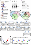 Figure 1
Identification and prioritization of regulatory miRNAs in glioblastoma. (A) Schematic overview of PAR-CLIP. 4SU, 4-thiouridine; CIP, calf intestinal phosphatase; PNK, polynucleotide kinase. (B) The phosphorimage of SDS-PAGE gel of RNA-argonaute (Ago) complexes labeled with 5′-32P that were immunoprecipitated with a FLAG-tag antibody. The complex is expected to appear near 100 kDa. (C) Agarose gel separation of PCR products from AGO1, AGO2, and AGO3 PAR-CLIP cDNA libraries. (D) Venn diagram illustrating the overlap in miRNA clusters identified with AGO1, AGO2, and AGO3. (E) Venn diagram showing the distribution of identified targets in 3′-UTRs from AGO1, AGO2, and AGO3 PAR-CLIP. (F) Diagram depicting the algorithm used for determining regulatory miRNAs in glioblastoma. ONC, oncogene; TSG, tumor suppressor gene. (G) Volcano plot denoting the statistically significant miRNAs based on Cox proportional hazard. Blue denotes downregulated miRNAs and red upregulated miRNAs. (H) Classification of miRNAs as oncogenic and tumor suppressive based on their targets derived from AGO1, AGO2, and AGO3 PAR-CLIP.
Ranking of regulatory miRNAs based on the importance of their targets in human TCGA tumors. Having identified all targets of all miRNAs in glioblastoma cells, we next sought to determine the relevance of these targets, and consequently of their targeting miRNAs, to glioblastoma malignancy. This process is described in the Methods section (Figure 1F). Briefly, we analyzed TCGA data for the PAR-CLIP–identified miRNA targets in human tumors. We identified significantly deregulated targets (FDR ≤ 0.05) with a greater than 2-fold change in human tumors using TCGA database. The targets were then scored based on the magnitude and frequency of deregulation, as well as their correlation with patient survival, using a Cox coefficient threshold of 0.2 (Figure 1G and Supplemental Tables 2 and 3). Each target gene was assigned a composite score calculated from its absolute expression level in glioma samples and its fold-change relative to healthy brain tissue. These 2 metrics were independently converted into percentile ranks (ranging from 0 to 1), and their sum represented the gene’s composite score, with a theoretical range from 0 (lowest expression and change) to 2 (highest in both categories). For each miRNA, 2 aggregate scores were then computed: a TSG score, representing the average composite score of its oncogenic targets, and an ONC score, reflecting the average composite score of its tumor-suppressive targets. To enable comparison across the entire miRNA dataset, these scores were further normalized by converting them into percentile ranks across all miRNAs. To determine the functional role of each miRNA, we calculated the difference between its TSG and ONC percentile ranks. We added the scores for each target and then added all scores for all targets of each miRNA. We subsequently averaged the target scores for both tumor suppressor and oncogenic miRNAs across the AGO1, AGO2, and AGO3 PAR-CLIP datasets, then consolidated the comprehensive set of targets for each miRNA (Supplemental Tables 4 and 5). The final score for each miRNA represents its regulatory and therapeutic potential because it is a reflection of both the number of its targets and the importance of these targets in glioblastoma biology (Supplemental Tables 6 and 7).
Using this computational workflow, we identified several miRNAs that targeted many highly relevant genes that are deregulated in glioblastoma (Figure 1H, Supplemental Figure 1F, and Supplemental Tables 1–5). We designated these miRNAs as regulators because they can simultaneously target multiple significantly deregulated and biologically relevant genes and inhibit their expressions. This approach identifies miRNAs that are likely to exert strong therapeutic effects when delivered (tumor-suppressive miRNAs targeting numerous oncogenes) or inhibited (oncogenic miRNAs that target numerous tumor suppressors). The comprehensive list of miRNAs, their targets, and their scores can be found in Supplemental Tables 1–7.
Regulatory miRNAs miR-340 and miR-382 bind multiple targets and decrease their expression. To prioritize regulatory miRNAs for functional validation, we incorporated evolutionary conservation as an additional criterion and selected the top 5 regulatory miRNAs from both the tumor suppressor (Supplemental Table 4) and oncogenic (Supplemental Table 5) lists for further analysis. Among these, we focused on 2 tumor-suppressive miRNAs, miR-340 and miR-382, because these miRNAs had high scores based on the algorithm described above. Our analysis determined that the tumor-suppressive regulatory miRNAs, miR-340 and miR-382, have 114 and 89 targets (Supplemental Table 4), respectively. From these, we selected a subset for validation based on their known deregulation in glioblastoma and other cancers and their widespread expression in glioblastoma samples and patient-derived glioblastoma stem cells. We checked the expression of these selective targets, CD44, TOP2A, MDM2, RHOC, HMGA2, PLAU, and NUSAP1, in TCGA glioma patient samples and found that they are highly overexpressed in tumor samples compared with Genotype-Tissue Expression (GTEx) normal brain samples (Supplemental Figure 2, A–G). We performed extensive target validation using immunoblot and 3′-UTR reporter analyses. We transfected precursor miR-340 and miR-382 into glioblastoma cell lines A172, U87, and U251, as well as patient-derived glioblastoma stem cell lines GSC-34 and GSC-28. Cell lysates were then subjected to immunoblot. Overexpression of these miRNAs in various glioblastoma cells, and stem cells, led to a marked reduction in protein levels across all tested cell lines (Figure 2, A–L, and Supplemental Figure 3, A–I). To determine if miR-340 and miR-382 directly target CD44, TOP2A, RHOC, MDM2, HMGA2, EGFR, PDGFRA, NUSAP1, and PLAU by binding to their 3′-UTRs, we amplified the 3′-UTR sequences containing the miR-340 and miR-382 binding sites from genomic DNA and subcloned them into the psiCheck2 luciferase reporter plasmid under the control of the T7 promoter. These 3′-UTR luciferase reporter constructs were then transiently transfected into U87 cells along with either a scrambled control miRNA mimic or mimics of miR-340 or miR-382. Overexpression of miR-340 resulted in a significant decrease in luciferase signals for CD44, RHOC, HMGA2, MDM2, EGFR, and PDGFRA (P < 0.05). Similarly, miR-382 overexpression led to a significant reduction in 3′-UTR luciferase signals for PLAU, CD44, NUSAP1, and MDM2 (P < 0.05), compared with scrambled control transfections (Figure 2, M and N). The predicted miRNA response elements in these targets were identified based on 6-mer seed complementarity from PAR-CLIP analysis. Importantly, mutation of these seed-matching regions within the respective 3′-UTRs abolished this repression, confirming the specificity of miR-340 and miR-382 targeting. These findings provide evidence that miR-340 and miR-382 directly bind to their respective 3′-UTRs and significantly repress the expression of numerous deregulated genes in glioblastoma.
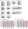 Figure 2
Validation of miR-340 and miR-382 targets in glioblastoma cells and patient-derived stem cells. (A–L) Glioblastoma cell lines A172, U87, U251, and patient-derived stem cell lines GSC-28 and GSC-34 were transfected with a scrambled negative control, miR-340, or miR-382. Immunoblots were probed with antibodies against TOP2A, RHOC, CD44, HMGA2, MDM2, CD44, NUSAP1, PLAU, and HMGA2. GAPDH served as the internal loading control. The data show that miR-340 and miR-382 downregulated protein expression compared with negative controls. (M and N) 3′-UTR luciferase activity assays in U87 cells were performed by cotransfecting cells with miR-340 (M) or miR-382 (N) and a psiCheck2 luciferase reporter plasmid containing the wild-type 3′-UTRs of target genes CD44, EGFR, HMGA2, MDM2, PDGFRA, and RHOC (M) or CD44, MDM2, NUSAP1, and PLAU (N) along with corresponding mutant 3′-UTR constructs in which the predicted miRNA binding sites were disrupted. The data show that miR-340 and miR-382 decreased luciferase activity for all respective wild-type targets compared with controls, while this repression was abolished or significantly reduced in the mutant constructs. The pink color on the immunoblots represents a higher intensity signal. Data represent mean ± SEM from 3 independent experiments. Statistical significance was determined using a 2-tailed Student’s t test, with *P < 0.05.
miR-17 is an oncogenic regulatory miRNA in glioblastoma. We identified miR-17 as one of the top oncogenic regulatory miRNAs that targets several deregulated TSGs in glioblastoma. We determined the expression of the selective targets in TCGA glioma samples and found they are highly downregulated in tumor samples compared with GTEx healthy brain samples (Supplemental Figure 4, A–D). We validated some of these targets using immunoblot analysis and 3′-UTR reporter assays in several glioma cell lines and patient-derived GSC lines. Since miR-17 is upregulated in glioblastoma and acts as an oncogene, we inhibited miR-17 by transfecting a miR-17 inhibitor into glioma cell lines (A172, U87, U251) and patient-derived GSC lines (GSC-28 and GSC-34). We selected specific targets — ZBTB4, ANKRD11, EHD3, and EPHA4 — based on their widespread published roles in glioblastoma. Inhibiting miR-17 resulted in an increased expression of all these targets in both glioblastoma cells and stem cells, indicating that miR-17 suppresses their expression (Figure 3, A–D, and Supplemental Figure 5, A–D). To confirm that miR-17 directly binds to the 3′-UTRs of ANKRD11, EHD3, and EPHA4, we conducted a luciferase reporter assay. The 3′-UTRs of these genes, which contain the miR-17 binding sites, were subcloned into a psiCheck2 reporter plasmid. In addition, mutant constructs harboring point mutations in the predicted miR-17 seed-binding sites were generated. Glioblastoma cells were transfected with either a scrambled anti-miR or an miR-17 inhibitor, along with the luciferase reporter plasmid containing the wild-type or mutant miR-17 target binding sites. Transfection with the miR-17 inhibitor led to a significant derepression (increase) in luciferase signals for ANKRD11, EHD3, and EPHA4 (P < 0.05), compared with the scrambled control (Figure 3E), whereas mutation of the binding sites abolished this effect. These data show that miR-17 directly binds to and regulates several deregulated mRNAs in glioblastoma.
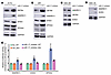 Figure 3
Validation of miR-17 targets in glioblastoma cells and patient-derived stem cells. (A–D) Glioma cell lines A172, U251, GSC-28, and GSC-34 were transfected with a miRNA inhibitor targeting miR-17. Immunoblots were performed with antibodies against ANKRD11, EHD3, and EPHA4. GAPDH served as the internal control for loading. The data show that the miR-17 inhibitor increased protein expression compared with negative controls. (E) U87 cells were cotransfected with miR-17 or miR-17 inhibitor along with psiCheck2 luciferase reporter plasmids containing the wild-type 3′-UTRs of targets ANKRD11, EHD3, EPHA4, and ZBTB4, as well as corresponding mutant 3′-UTR constructs in which the predicted miR-17 binding sites were disrupted. The data show that the miR-17 inhibitor increased luciferase activity for all respective wild-type 3′-UTR targets compared with negative controls, while this effect was abolished or significantly reduced in the mutant constructs. Forty-eight hours after transfection, cells were lysed and luciferase signals were measured. Data represent mean ± SEM from 3 independent experiments. *P < 0.05 by 2-tailed Student’s t test.
Further, we investigated whether the targets of the regulatory miRNAs, miR-340, miR-382, and miR-17, are associated with patient survival. To evaluate survival outcomes, we utilized 2 independent cohorts of primary glioma patient samples from TCGA and the Chinese Glioma Genome Atlas (CGGA) for survival analysis. Kaplan-Meier survival curves were generated and analyzed using the online tools GEPIA and CGGA, where patients were stratified into high– and low–gene expression groups based on target expression levels. For the tumor suppressor targets of miR-340 and miR-382, high expression was correlated with poorer patient survival, while low expression was associated with more favorable outcomes in both TCGA and CGGA cohorts (Supplemental Figure 6, A–G, and Supplemental Figure 7, A–G). Conversely, for the oncogenic miR-17, high expression of its targets was linked to better survival, while low expression was associated with poorer patient prognosis (Supplemental Figure 8, A and B, and Supplemental Figure 9, A and B). Additionally, we performed survival analysis using CGGA data from recurrent glioma patient samples, which revealed similar survival trends for both tumor suppressor and oncogenic miRNA targets (Supplemental Figure 10, A–F, and Supplemental Figure 11). These findings underscore the importance of regulatory miRNAs and their target genes in determining glioma patient outcomes.
Regulatory miRNAs miR-340 and miR-382 regulate multiple pathways in glioblastoma. After identifying and validating regulatory miRNAs and their targets, we investigated their functions. We first performed pathway analyses with the targets of miR-340 and miR-382. We performed GO term, KEGG, and Hallmark pathway analysis. The most significant pathways associated with miR-340 were the mitotic cell cycle phase transition, stem cell differentiation, miRNAs in cancer, PI3K-Akt signaling, proteoglycan in cancer, the hallmarks of apoptosis, G2/M checkpoint, and epithelial to mesenchymal transition (Supplemental Figure 12, A–C). The pathways enriched for miR-382 targets were response to hypoxia, signaling pathways associated with miRNA metabolic process, proteoglycan in cancer, miRNAs in cancer, G2/M checkpoint, and epithelial to mesenchymal transition (Supplemental Figure 12, D–F). These data suggest that these miRNAs regulate different cancer-related pathways, and inhibiting them could attenuate glioma growth by inhibiting numerous oncogenic pathways.
Regulatory miRNAs miR-340 and miR-382 inhibit cell proliferation, invasion, and neurosphere formation in several glioblastoma cell lines. We then performed a thorough assessment of the functional and experimental therapeutic impacts of miR-340 and miR-382. We first quantified their endogenous expression levels in glioblastoma cell lines, GSCs, and banked human tumors including cell lines (A172, T98G, LN18, U251, U87, SNB19), and stem cell lines (GSC-28, GSC-20, GSC-34, GSC-627, GSC-267), as well as patient glioblastoma samples, using quantitative PCR. Normal human astrocytes and normal human cortex were used as a control. We observed a significant downregulation of miR-340 and miR-382 expression in cell lines compared with normal human astrocytes, mirroring the pattern observed in patient glioblastoma samples compared with normal brain samples (Supplemental Figure 13, A–D). Notably, miR-382 showed upregulation in only a single tumor tissue sample, while all other samples remained consistently downregulated. For miR-340, a majority of the cell lines showed downregulation, although a few cell lines exhibited relatively unchanged expression. These variations are consistent with the well-established intratumoral heterogeneity of glioblastoma. This consistent downregulation hinted at a potential tumor suppressor role for these miRNAs, which was consistent with our analyses (Supplemental Figure 13, A–D). Subsequently, we conducted cell proliferation and invasion assays to determine the biological significance of miR-340 and miR-382 in vitro across different glioblastoma cell lines. Glioblastoma cells were transfected with either scrambled control miRNA or precursor miR-340 or precursor miR-382, and cell proliferation was assessed through trypan blue staining and live-cell counting. Overexpression of precursor miR-340 or miR-382 led to a significant reduction in cell proliferation compared with the scrambled negative control (Figure 4, A–D). To investigate the influence of these miRNAs on glioma cell invasion, we performed Transwell invasion assays using glioblastoma cell lines. The Transwell invasion chambers were precoated with collagen IV, one of the abundant ECM components in the brain. Glioblastoma cell lines were transfected with scrambled control miRNA, or miR-340 or miR-382 precursors, before being plated in the Transwell chambers. These cells were allowed to invade through the collagen IV layer. Transient overexpression of either miR-340 or miR-382 led to a substantial reduction in glioblastoma cell invasion compared with scrambled negative control miRNA-transfected cells (Figure 4, E–J).
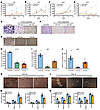 Figure 4
miR-340 and miR-382 inhibit cell proliferation, invasion, and neurosphere formation in glioblastoma. (A–D) Glioblastoma cell lines U87 and U251 were transfected with a scrambled control, miR-340 precursor, or miR-382 precursor. Cell counts were performed at various time points, and miR-340 and miR-382 showed decreased cell growth compared with negative controls. (E–J) A172, U87, and U251 cells were transfected with mimic miR-340 and miR-382, and invasion assays were executed. Invaded images from 5–10 random fields were captured and analyzed using ImageJ software (NIH) for quantification. miR-340 and miR-382 decreased invasion compared with negative controls. (E–G, K, and L) Original magnification, ×10. (K–N) Six-well plates were precoated with poly-ornithine, and glioblastoma stem cell lines GSC-28 and GSC-34 were plated and transfected with scrambled control miRNA, miR-340, or miR-382. Neurosphere images were taken from 5 distinct fields 72 hours posttransfection and categorized into large, medium, and small using ImageJ software, with quantifications presented (M and N). miR-340 and miR-382 decreased neurosphere formation compared with negative controls. LS, large sphere; MS, medium sphere; SS, small sphere. Data represent mean ± SEM from 3 independent experiments. *P < 0.05 by 2-tailed Student’s t test.
Glioblastoma contains self-renewing stem cells contributing to tumor initiation and resistance to therapy (24). To elucidate the effect of the regulatory miRNAs on the self-renewal property of GSCs, we conducted neurosphere assays using 2 patient-derived glioblastoma stem cell lines, GSC-34 and GSC-28. We transfected precursor miR-340 or precursor miR-382 or scrambled controls into the glioblastoma stem cells and counted the number of neurospheres 7 days after miRNA transfection. Neurospheres were categorized based on their size under the microscope (10× original magnification) into large, medium, and small groups. Overexpression of miR-340 and miR-382 resulted in a reduction in the total number of neurospheres in both GSCs. These findings suggest that these miRNAs impair neurosphere formation, potentially reflecting effects on self-renewal capacity (Figure 4, K–N).
Inhibiting oncogenic miR-17 in glioma cells decreases cell proliferation, invasion, and neurosphere formation in glioblastoma cell lines and GSCs. miR-17 is one of the top oncogenic regulatory miRNAs identified by our integrated approach. Similar to the tumor-suppressive regulatory miRNAs, we first checked the endogenous expression of miR-17 in multiple glioma and stem cell lines by RT-qPCR (Supplemental Figure 13, E and F). We observed an increased expression pattern in all glioma and stem cell lines, consistent with our algorithm that identified miR-17 as an oncogenic miRNA. Therefore, we performed a series of functional assays with miR-17 in a spectrum of glioblastoma cell lines, including patient-derived glioblastoma stem cells. For the cell proliferation assay, we transfected the inhibitor against miR-17 into multiple glioma cell lines, then counted the cells on different days. We observed a decrease in cell proliferation, supporting the oncogenic nature of miR-17 in glioma cells (Figure 5, A–C). Transwell cell invasion assays were carried out with miR-17 inhibitor in glioblastoma cell lines. The invasion assays were carried out with a collagen IV–coated Transwell chamber. Inhibition of miR-17 in glioblastoma cell lines led to a decrease in cell invasion through collagen-coated chambers, which were counted by taking images from 5 random microscopic fields and quantified with ImageJ software (NIH) (Figure 5, D–I). The neurosphere formation assay was performed with GSC lines GSC-28 and GSC-34. The transfection of GSC lines with an inhibitor of miR-17 decreases the overall neurosphere formation. We categorized the neurosphere size into 3 different groups, large, medium, and small, based on the size calculator with ImageJ software. The overall neurosphere number in different categories decreased significantly after miR-17 inhibitor treatment compared with the scrambled control treatment (Figure 5, J–M).
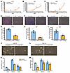 Figure 5
Effects of miR-17 inhibition on cell proliferation, invasion, and neurosphere formation in glioblastoma. (A–C) A172, U87, and U251 were transfected with either scrambled control miRNA or a miR-17 inhibitor. Cells were counted 48 hours posttransfection using trypan blue exclusion at various intervals to assess viability. (D–I) A172, U87, and U251 cells were transfected with control or miR-17 inhibitor. Invasion assay was carried out, the invaded cells were stained with crystal violet, and images were captured and analyzed using ImageJ software for quantification. (D, F, J, and L) Original magnification, ×10. (J–M) Glioma stem cell lines GSC-28 and GSC-34, plated on poly-ornithine–coated, 6-well plates, were transfected with either a scrambled control miRNA or an miR-17 inhibitor. Neurospheres were imaged 72 hours posttransfection in neurobasal complete growth medium. Images from 5 microscopic fields were taken, and neurosphere sizes were categorized into large, medium, and small for quantification using ImageJ software. Data are presented as mean ± SEM from 3 independent experiments. *P < 0.05 by 2-tailed Student’s t test.
Inhibition of oncogenic miR-17 and overexpression of tumor-suppressive miR-340 and miR-382 reduce in vivo glioblastoma growth. The impact of miR-340 and miR-382 overexpression on xenografted tumor growth in mice was investigated. We transfected 3 × 10⁵ U87 cells with a scrambled negative control miRNA (n = 7), precursor miR-340 (n = 7), precursor miR-382 (n = 7), or miR-17 inhibitor (n = 7) (Figure 6A). The transfected cells were then stereotactically implanted into the striata of 6-week-old immunodeficient mice. Over 3 weeks, the mice were closely monitored for tumor growth and survival, and MRI images were obtained to visualize the tumors. After 3 weeks, the scrambled control U87 group exhibited significant tumor growth. In contrast, the groups treated with miR-340 and miR-382 displayed a significant reduction in tumor volume (Figure 6, B and C). Specifically, the scrambled control group reached a tumor volume of 7.58 ± 2.26 mm3, while the mice bearing miR-340– or miR-382–transfected tumors showed tumor volumes of 4.59 ± 1.44 mm3 and 2.86 ± 0.9 mm3, respectively (Figure 6C). The mice implanted with miR-17 inhibitor–transfected cells showed a substantial decrease in tumor volume compared with scrambled control (Figure 6, D and E). The scrambled control mouse group showed tumor volumes of 5.3 ± 1.94 mm3, whereas miR-17 inhibitor group exhibited tumor volumes of 0.68 ± 0.29 mm3. Collectively, these in vivo findings support the use of miR-340, miR-382, the miR-17 inhibitor, or a combination of them as new therapeutics for glioblastoma.
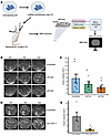 Figure 6
miR-340, miR-382, and miR-17 regulate in vivo tumor growth. (A) Schematic overview of the experimental design for tumor implantation and timeline for MRI to assess tumor volume. Each group comprised 7 mice for surgery. (B–E) U87 cells were transfected with scrambled negative miRNA, miR-340, miR-382, or an inhibitor of miR-17. At 48 hours posttransfection, cells were implanted intracranially into the striata of 5- to 6-week-old immunodeficient mice. Mice were monitored 3–4 weeks, and MRI was performed to evaluate tumor generation. Representative MRIs and quantification showing that miR-340, miR-382, and miR-17 inhibitor showed reduction in tumor volume compared with scrambled controls. *P < 0.05 by 2-tailed Student’s t test.
Therapeutic delivery of PEG-PBAE BPNs carrying plasmids encoding miR-340 and miR-382 to mice bearing glioblastoma tumors by MRIgFUS inhibits tumor growth. For the therapeutic delivery of regulatory miRNA, we developed and employed an approach consisting of MRIgFUS and MBs (FUS-MB) to facilitate the delivery of BPNs carrying miR-340 and miR-382 encoding lentiviral plasmids. This innovative approach (abbreviated FUS-MB-BPN) circumvents the hurdles of miRNA therapeutics, particularly the blood-brain barrier and tumor penetration and transfection, and delivers cargo loads to tumor cells. The schematic plan for FUS-MB-BPN is depicted in Figure 7, A and B. We generated glioblastoma xenografts in immunodeficient mice, accomplished by intracranial implantation of 3 × 10⁵ U87 cells into the striata of mice brains. Once the tumors had formed (7–10 days postinjection), we verified tumor formation by MRI and the safe and effective opening of the blood-brain barrier. This step was achieved through a meticulously orchestrated combination of microbubbles and sonication, resulting in a conspicuous increase in MRI signal intensity surrounding the tumors. This increased signal intensity is attributed to the leakage of the MRI contrast agent, gadobenate dimeglumine, into the brain parenchyma after blood-brain barrier opening (Figure 7C). To induce robust miRNA expression in brain tumors, BPNs were formulated with lentiviral plasmids encoding miR-340 or miR-382. We also prepared the BPNs with plasmids encoding scrambled miRNA sequences as a control group. We extensively characterized these BPNs because the ζ-potential and size of the particles are important parameters for effective blood-brain barrier penetration. The hydrodynamic diameter and ζ-potential of BPNs carrying plasmids encoding scrambled miRNA, miR-340, and miR-382 were measured as 70.2 ± 4.7 nm, 2.8 ± 0.8 mV; 71.2 ± 1.2 nm 2.3 ± 0.6 mV; and 70.7 ± 1.0 nm, 1.8 ± 0.7 mV, respectively (Supplemental Table 8). After tumor implantation, we divided the brain tumor–bearing mice into 2 cohorts receiving plasmids encoding scrambled miRNA or tumor-suppressive miR-340 and miR-382. Each group consisted of 7 mice. The MRIgFUS-MB-BPN procedure was then executed. The BPNs carrying plasmids encoding scrambled miRNA, miR-340, or miR-382 were intravenously injected alongside MBs, and FUS was applied to the tumor regions to transiently open the blood-brain barrier and facilitate the delivery of the plasmid cargoes into the tumor. One week after the FUS-MB-BPN procedure, MRI images were taken to visualize and measure the tumor volumes. Delivery of miR-340 or miR-382 by FUS-MB-BPN procedure into the tumors significantly reduced tumor burden compared with the control group (n = 7 mice per group, P < 0.05) (Figure 7, D and E). This represents to our best knowledge the first successful FUS-MB-BPN-procedure–based experimental therapeutic delivery of a miRNA to inhibit tumor growth. We also closely monitored the post–FUS-treated mice for survival, which revealed that delivery of miR-340 or miR-382 into brain tumors significantly extended the survival of these mice compared with those treated with the scrambled control miRNA, with miR-382 providing greater survival benefit (Figure 7F). To determine whether the therapeutic efficacy of FUS-MB-BPN–mediated miRNA delivery was restricted to U87 xenografts, we next extended our analysis to additional models. First, intracranial xenografts were established using patient-derived GSCs (GSC-34) and treated with the same FUS-MB-BPN protocol. Delivery of the tumor-suppressive miRNAs miR-340 or miR-382 significantly reduced tumor burden and improved survival relative to the scrambled control group (Figure 7, G–I). To further evaluate treatment kinetics, longitudinal MRI was performed on days 4 and 7 post-FUS, revealing a progressive decline in tumor volume over time and thereby indicating sustained therapeutic efficacy (Supplemental Figure 14, A–D). Importantly, delivery of BPNs carrying miR-340 or miR-382 in the absence of FUS did not produce a substantial reduction in tumor burden (FUS group: scrambled, miR-340, and miR-382: 64.81 ± 10.38, 15.65 ± 3.19, and 22.31 ± 9.99 mm3, respectively; without FUS: 62.67 ± 11.97, 77.89 ± 14.67, and 72.09 ± 3.60 mm3), highlighting the necessity of FUS-mediated blood-brain barrier opening for effective tumor delivery (Figure 7, G–I, and Supplemental Figure 15, A–H). Collectively, these findings demonstrate that FUS-MB-BPN–mediated delivery is broadly applicable across multiple glioblastoma models and effective for therapeutic miRNA delivery. To assess potential toxicity from FUS-MB, we harvested various organs including the liver, kidney, brain, heart, spleen, lung, lymph nodes, and pancreas from treated mice. We then performed H&E staining on these organs, which were analyzed by a neuropathologist. The data showed no apparent damage to the liver, kidney, brain, or heart in treated mice compared to the nontreated controls (Supplemental Figure 16, A and B). These data demonstrate that FUS-MB-BPN is a safe and effective strategy for delivering miRNAs into brain tumors.
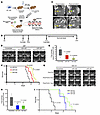 Figure 7
Inhibition of in vivo glioma growth through MRI-guided focused ultrasound, microbubble, and brain-penetrating nanoparticle delivery of miR-340 or miR-382. (A) A schematic representation of FUS-MB-BPN–mediated miRNA delivery into mice. (B) Overall experimental plan and timeline for FUS-MB-BPN. (C) Tumors in mice were sonicated before and after FUS-MB-BPN, and images were captured to validate blood-brain barrier opening. MBs were employed to facilitate blood-brain barrier opening. Arrow indicates the tumor location and show dispersion of the contrast, demonstrating successful opening of the blood-brain barrier. MRI images were acquired using a small-animal MRI system with a slice thickness of 0.7 mm. The acquisition parameters included a repetition time (TR) of 600 ms and an echo time (TE) of 11 ms. (D and E) MBs were injected through mice’s tail veins, and MRIgFUS was conducted. Upon confirmation of blood-brain barrier opening, BPN conjugated with scrambled, miR-340, or miR-382 were injected through mice’s tail veins. The mice were imaged using MRI, and tumor volume shows significant reduction upon delivery of miR-340 or miR-382 compared with scrambled controls at day 15. Box plots show the interquartile range, median (line), and minimum and maximum (whiskers). (F) Kaplan-Meier survival curve showing miR-340 or miR-382 significantly prolonged survival compared with scrambled control mice. (G–I) For patient-derived glioma stem cell (GSC-34) intracranial xenografts, the same FUS-MB-BPN protocol was applied using miR-340 and miR-382. MRI analysis at day 4 and day 7 post-FUS showed a reduction in tumor volume over time, and Kaplan-Meier survival analysis demonstrated improved survival in miR-340– and miR-382–treated groups compared with scrambled controls. n = 7 mice per group. *P < 0.05 by 2-tailed Student’s t test for miR-340–treated vs. scrambled control.
In vivo target repression and durability following MRIgFUS-mediated delivery of miR-340 and miR-382 to GSC-34 xenografts. To determine whether the therapeutic effects observed following FUS-MB-BPN–mediated delivery of miR-340 and miR-382 were associated with effective and sustained repression of their molecular targets in vivo, we performed studies using GSC-34 glioma xenografts. Intracranial tumors were generated by implantation of GSC-34 cells into nude mice. After confirmation of tumor establishment by MRI, mice underwent MRIgFUS in combination with MBs to transiently open the blood-brain barrier, followed by intravenous administration of BPNs carrying plasmids encoding miR-340, miR-382, or scrambled control miRNA. Tumor tissues were harvested at 4 and 7 days after FUS treatment for molecular analyses. Quantitative PCR analysis revealed significant downregulation of validated target transcripts for both miR-340 and miR-382 at 4 days posttreatment compared with scrambled controls (Figure 8, A–K). While several targets showed robust repression at this early time point, a subset demonstrated more modest changes at day 4. Notably, by day 7, repression was consistently observed across all examined targets, indicating progressive and sustained transcript-level suppression following FUS-mediated miRNA delivery (Figure 8, A–K). To determine whether transcript repression translated into functional protein downregulation, we analyzed protein expression in tumor lysates derived from the same GSC-34 samples. Western blot analysis at day 7 demonstrated marked reduction of validated on-target proteins in tumors treated with miR-340 or miR-382 relative to scrambled controls (Figure 8, L and M). In contrast, expression levels of selected off-target proteins remained unchanged, supporting the specificity of miRNA-mediated repression in vivo (Figure 8N). Collectively, these data demonstrate that FUS-MB-BPN delivery of miR-340 and miR-382 into GSC-34 brain tumors induces specific and durable suppression of molecular targets at both the mRNA and protein levels. The progressive repression observed between days 4 and 7 further supports sustained biological activity of the delivered miRNAs in vivo and provides molecular evidence underlying the observed antitumor effects.
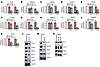 Figure 8
In vivo target repression and durability following MRIgFUS-mediated delivery of miR-340 and miR-382 in GSC-34 glioma xenografts. (A–K) Quantitative PCR analysis of validated target transcripts demonstrates significant downregulation in tumors treated with miR-340 or miR-382 compared with scrambled controls at day 4, with more uniform and sustained repression observed at day 7. While some targets exhibited modest suppression at day 4, consistent transcript-level repression across all examined targets was evident by day 7, indicating progressive and durable in vivo silencing following FUS-mediated delivery. (L and M) Western blot analysis of tumor lysates at day 7 confirms reduced protein expression of validated on-target genes in miR-340– and miR-382–treated tumors relative to controls. (N) Expression of selected nontarget proteins remains unchanged, supporting specificity of miRNA-mediated repression. Statistical significance was determined using a 2-tailed Student’s t test. *P < 0.05.


Copyright © 2026 American Society for Clinical Investigation
ISSN: 0021-9738 (print), 1558-8238 (online)