
Advertisement
Research ArticleCardiologyMetabolism
Open Access |  10.1172/JCI193499
The liver regulates ectopic calcification in Abcc6-deficient models of pseudoxanthoma elasticum
Yijie Wang,1,2,3,4,5,6 Baiming Sun,1,2,3,4,5,6 Feiyang Ma,3 Bo Tao,1,2,3,4,5,6 Yiqian Gu,3,7 Zhiqiang Zhou,1,8,9 Jason Kim,1 Linlin Zhang,1,2,3,4,5,6 Zhihao Liu,1,2,3,4,5,6 Johanna ten Hoeve,6,10,11,12 Linsey Stiles,12,13 Lucia Fernandez del Rio,13 Calvin Pan,1,8,9 Orian Shirihai,12,13 Shili Xu,11,12,14 Thomas G. Graeber,6,10,11 Tamer Sallam,1,5 Matteo Pellegrini,3,4,15 Aldons J. Lusis,1,10,11 and Arjun Deb1,2,3,4,5,6
1Division of Cardiology, Department of Medicine, David Geffen School of Medicine,
2UCLA Cardiovascular Theme, David Geffen School of Medicine,
3Department of Molecular, Cell and Developmental Biology,
4Eli and Edythe Broad Center of Regenerative Medicine and Stem Cell Research,
5Molecular Biology Institute,
6California Nanosystems Institute,
7Bioinformatics Interdepartmental Program,
8Department of Microbiology, Immunology and Molecular Genetics,
9Department of Human Genetics, David Geffen School of Medicine,
10UCLA Metabolomics Center,
11Crump Institute of Molecular Imaging,
12Department of Molecular and Medical Pharmacology,
13Department of Medicine, Endocrinology, David Geffen School of Medicine,
14Jonsson Comprehensive Cancer Center, David Geffen School of Medicine, and
15Eli and Edythe Broad Center of Regenerative Medicine and Stem Cell Research, UCLA, Los Angeles, California, USA.
Address correspondence to: Arjun Deb, McDonald Research Laboratories 3641A, UCLA, Los Angeles, California, 90095, USA. Phone: 310.983.1017; Email: adeb@mednet.ucla.edu.
Find articles by
Wang, Y.
in:
PubMed
|
Google Scholar
|

1Division of Cardiology, Department of Medicine, David Geffen School of Medicine,
2UCLA Cardiovascular Theme, David Geffen School of Medicine,
3Department of Molecular, Cell and Developmental Biology,
4Eli and Edythe Broad Center of Regenerative Medicine and Stem Cell Research,
5Molecular Biology Institute,
6California Nanosystems Institute,
7Bioinformatics Interdepartmental Program,
8Department of Microbiology, Immunology and Molecular Genetics,
9Department of Human Genetics, David Geffen School of Medicine,
10UCLA Metabolomics Center,
11Crump Institute of Molecular Imaging,
12Department of Molecular and Medical Pharmacology,
13Department of Medicine, Endocrinology, David Geffen School of Medicine,
14Jonsson Comprehensive Cancer Center, David Geffen School of Medicine, and
15Eli and Edythe Broad Center of Regenerative Medicine and Stem Cell Research, UCLA, Los Angeles, California, USA.
Address correspondence to: Arjun Deb, McDonald Research Laboratories 3641A, UCLA, Los Angeles, California, 90095, USA. Phone: 310.983.1017; Email: adeb@mednet.ucla.edu.
Find articles by Sun, B. in: PubMed | Google Scholar
1Division of Cardiology, Department of Medicine, David Geffen School of Medicine,
2UCLA Cardiovascular Theme, David Geffen School of Medicine,
3Department of Molecular, Cell and Developmental Biology,
4Eli and Edythe Broad Center of Regenerative Medicine and Stem Cell Research,
5Molecular Biology Institute,
6California Nanosystems Institute,
7Bioinformatics Interdepartmental Program,
8Department of Microbiology, Immunology and Molecular Genetics,
9Department of Human Genetics, David Geffen School of Medicine,
10UCLA Metabolomics Center,
11Crump Institute of Molecular Imaging,
12Department of Molecular and Medical Pharmacology,
13Department of Medicine, Endocrinology, David Geffen School of Medicine,
14Jonsson Comprehensive Cancer Center, David Geffen School of Medicine, and
15Eli and Edythe Broad Center of Regenerative Medicine and Stem Cell Research, UCLA, Los Angeles, California, USA.
Address correspondence to: Arjun Deb, McDonald Research Laboratories 3641A, UCLA, Los Angeles, California, 90095, USA. Phone: 310.983.1017; Email: adeb@mednet.ucla.edu.
Find articles by
Ma, F.
in:
PubMed
|
Google Scholar
|
1Division of Cardiology, Department of Medicine, David Geffen School of Medicine,
2UCLA Cardiovascular Theme, David Geffen School of Medicine,
3Department of Molecular, Cell and Developmental Biology,
4Eli and Edythe Broad Center of Regenerative Medicine and Stem Cell Research,
5Molecular Biology Institute,
6California Nanosystems Institute,
7Bioinformatics Interdepartmental Program,
8Department of Microbiology, Immunology and Molecular Genetics,
9Department of Human Genetics, David Geffen School of Medicine,
10UCLA Metabolomics Center,
11Crump Institute of Molecular Imaging,
12Department of Molecular and Medical Pharmacology,
13Department of Medicine, Endocrinology, David Geffen School of Medicine,
14Jonsson Comprehensive Cancer Center, David Geffen School of Medicine, and
15Eli and Edythe Broad Center of Regenerative Medicine and Stem Cell Research, UCLA, Los Angeles, California, USA.
Address correspondence to: Arjun Deb, McDonald Research Laboratories 3641A, UCLA, Los Angeles, California, 90095, USA. Phone: 310.983.1017; Email: adeb@mednet.ucla.edu.
Find articles by Tao, B. in: PubMed | Google Scholar
1Division of Cardiology, Department of Medicine, David Geffen School of Medicine,
2UCLA Cardiovascular Theme, David Geffen School of Medicine,
3Department of Molecular, Cell and Developmental Biology,
4Eli and Edythe Broad Center of Regenerative Medicine and Stem Cell Research,
5Molecular Biology Institute,
6California Nanosystems Institute,
7Bioinformatics Interdepartmental Program,
8Department of Microbiology, Immunology and Molecular Genetics,
9Department of Human Genetics, David Geffen School of Medicine,
10UCLA Metabolomics Center,
11Crump Institute of Molecular Imaging,
12Department of Molecular and Medical Pharmacology,
13Department of Medicine, Endocrinology, David Geffen School of Medicine,
14Jonsson Comprehensive Cancer Center, David Geffen School of Medicine, and
15Eli and Edythe Broad Center of Regenerative Medicine and Stem Cell Research, UCLA, Los Angeles, California, USA.
Address correspondence to: Arjun Deb, McDonald Research Laboratories 3641A, UCLA, Los Angeles, California, 90095, USA. Phone: 310.983.1017; Email: adeb@mednet.ucla.edu.
Find articles by Gu, Y. in: PubMed | Google Scholar
1Division of Cardiology, Department of Medicine, David Geffen School of Medicine,
2UCLA Cardiovascular Theme, David Geffen School of Medicine,
3Department of Molecular, Cell and Developmental Biology,
4Eli and Edythe Broad Center of Regenerative Medicine and Stem Cell Research,
5Molecular Biology Institute,
6California Nanosystems Institute,
7Bioinformatics Interdepartmental Program,
8Department of Microbiology, Immunology and Molecular Genetics,
9Department of Human Genetics, David Geffen School of Medicine,
10UCLA Metabolomics Center,
11Crump Institute of Molecular Imaging,
12Department of Molecular and Medical Pharmacology,
13Department of Medicine, Endocrinology, David Geffen School of Medicine,
14Jonsson Comprehensive Cancer Center, David Geffen School of Medicine, and
15Eli and Edythe Broad Center of Regenerative Medicine and Stem Cell Research, UCLA, Los Angeles, California, USA.
Address correspondence to: Arjun Deb, McDonald Research Laboratories 3641A, UCLA, Los Angeles, California, 90095, USA. Phone: 310.983.1017; Email: adeb@mednet.ucla.edu.
Find articles by Zhou, Z. in: PubMed | Google Scholar
1Division of Cardiology, Department of Medicine, David Geffen School of Medicine,
2UCLA Cardiovascular Theme, David Geffen School of Medicine,
3Department of Molecular, Cell and Developmental Biology,
4Eli and Edythe Broad Center of Regenerative Medicine and Stem Cell Research,
5Molecular Biology Institute,
6California Nanosystems Institute,
7Bioinformatics Interdepartmental Program,
8Department of Microbiology, Immunology and Molecular Genetics,
9Department of Human Genetics, David Geffen School of Medicine,
10UCLA Metabolomics Center,
11Crump Institute of Molecular Imaging,
12Department of Molecular and Medical Pharmacology,
13Department of Medicine, Endocrinology, David Geffen School of Medicine,
14Jonsson Comprehensive Cancer Center, David Geffen School of Medicine, and
15Eli and Edythe Broad Center of Regenerative Medicine and Stem Cell Research, UCLA, Los Angeles, California, USA.
Address correspondence to: Arjun Deb, McDonald Research Laboratories 3641A, UCLA, Los Angeles, California, 90095, USA. Phone: 310.983.1017; Email: adeb@mednet.ucla.edu.
Find articles by Kim, J. in: PubMed | Google Scholar
1Division of Cardiology, Department of Medicine, David Geffen School of Medicine,
2UCLA Cardiovascular Theme, David Geffen School of Medicine,
3Department of Molecular, Cell and Developmental Biology,
4Eli and Edythe Broad Center of Regenerative Medicine and Stem Cell Research,
5Molecular Biology Institute,
6California Nanosystems Institute,
7Bioinformatics Interdepartmental Program,
8Department of Microbiology, Immunology and Molecular Genetics,
9Department of Human Genetics, David Geffen School of Medicine,
10UCLA Metabolomics Center,
11Crump Institute of Molecular Imaging,
12Department of Molecular and Medical Pharmacology,
13Department of Medicine, Endocrinology, David Geffen School of Medicine,
14Jonsson Comprehensive Cancer Center, David Geffen School of Medicine, and
15Eli and Edythe Broad Center of Regenerative Medicine and Stem Cell Research, UCLA, Los Angeles, California, USA.
Address correspondence to: Arjun Deb, McDonald Research Laboratories 3641A, UCLA, Los Angeles, California, 90095, USA. Phone: 310.983.1017; Email: adeb@mednet.ucla.edu.
Find articles by Zhang, L. in: PubMed | Google Scholar
1Division of Cardiology, Department of Medicine, David Geffen School of Medicine,
2UCLA Cardiovascular Theme, David Geffen School of Medicine,
3Department of Molecular, Cell and Developmental Biology,
4Eli and Edythe Broad Center of Regenerative Medicine and Stem Cell Research,
5Molecular Biology Institute,
6California Nanosystems Institute,
7Bioinformatics Interdepartmental Program,
8Department of Microbiology, Immunology and Molecular Genetics,
9Department of Human Genetics, David Geffen School of Medicine,
10UCLA Metabolomics Center,
11Crump Institute of Molecular Imaging,
12Department of Molecular and Medical Pharmacology,
13Department of Medicine, Endocrinology, David Geffen School of Medicine,
14Jonsson Comprehensive Cancer Center, David Geffen School of Medicine, and
15Eli and Edythe Broad Center of Regenerative Medicine and Stem Cell Research, UCLA, Los Angeles, California, USA.
Address correspondence to: Arjun Deb, McDonald Research Laboratories 3641A, UCLA, Los Angeles, California, 90095, USA. Phone: 310.983.1017; Email: adeb@mednet.ucla.edu.
Find articles by Liu, Z. in: PubMed | Google Scholar
1Division of Cardiology, Department of Medicine, David Geffen School of Medicine,
2UCLA Cardiovascular Theme, David Geffen School of Medicine,
3Department of Molecular, Cell and Developmental Biology,
4Eli and Edythe Broad Center of Regenerative Medicine and Stem Cell Research,
5Molecular Biology Institute,
6California Nanosystems Institute,
7Bioinformatics Interdepartmental Program,
8Department of Microbiology, Immunology and Molecular Genetics,
9Department of Human Genetics, David Geffen School of Medicine,
10UCLA Metabolomics Center,
11Crump Institute of Molecular Imaging,
12Department of Molecular and Medical Pharmacology,
13Department of Medicine, Endocrinology, David Geffen School of Medicine,
14Jonsson Comprehensive Cancer Center, David Geffen School of Medicine, and
15Eli and Edythe Broad Center of Regenerative Medicine and Stem Cell Research, UCLA, Los Angeles, California, USA.
Address correspondence to: Arjun Deb, McDonald Research Laboratories 3641A, UCLA, Los Angeles, California, 90095, USA. Phone: 310.983.1017; Email: adeb@mednet.ucla.edu.
Find articles by ten Hoeve, J. in: PubMed | Google Scholar
1Division of Cardiology, Department of Medicine, David Geffen School of Medicine,
2UCLA Cardiovascular Theme, David Geffen School of Medicine,
3Department of Molecular, Cell and Developmental Biology,
4Eli and Edythe Broad Center of Regenerative Medicine and Stem Cell Research,
5Molecular Biology Institute,
6California Nanosystems Institute,
7Bioinformatics Interdepartmental Program,
8Department of Microbiology, Immunology and Molecular Genetics,
9Department of Human Genetics, David Geffen School of Medicine,
10UCLA Metabolomics Center,
11Crump Institute of Molecular Imaging,
12Department of Molecular and Medical Pharmacology,
13Department of Medicine, Endocrinology, David Geffen School of Medicine,
14Jonsson Comprehensive Cancer Center, David Geffen School of Medicine, and
15Eli and Edythe Broad Center of Regenerative Medicine and Stem Cell Research, UCLA, Los Angeles, California, USA.
Address correspondence to: Arjun Deb, McDonald Research Laboratories 3641A, UCLA, Los Angeles, California, 90095, USA. Phone: 310.983.1017; Email: adeb@mednet.ucla.edu.
Find articles by Stiles, L. in: PubMed | Google Scholar
1Division of Cardiology, Department of Medicine, David Geffen School of Medicine,
2UCLA Cardiovascular Theme, David Geffen School of Medicine,
3Department of Molecular, Cell and Developmental Biology,
4Eli and Edythe Broad Center of Regenerative Medicine and Stem Cell Research,
5Molecular Biology Institute,
6California Nanosystems Institute,
7Bioinformatics Interdepartmental Program,
8Department of Microbiology, Immunology and Molecular Genetics,
9Department of Human Genetics, David Geffen School of Medicine,
10UCLA Metabolomics Center,
11Crump Institute of Molecular Imaging,
12Department of Molecular and Medical Pharmacology,
13Department of Medicine, Endocrinology, David Geffen School of Medicine,
14Jonsson Comprehensive Cancer Center, David Geffen School of Medicine, and
15Eli and Edythe Broad Center of Regenerative Medicine and Stem Cell Research, UCLA, Los Angeles, California, USA.
Address correspondence to: Arjun Deb, McDonald Research Laboratories 3641A, UCLA, Los Angeles, California, 90095, USA. Phone: 310.983.1017; Email: adeb@mednet.ucla.edu.
Find articles by Fernandez del Rio, L. in: PubMed | Google Scholar
1Division of Cardiology, Department of Medicine, David Geffen School of Medicine,
2UCLA Cardiovascular Theme, David Geffen School of Medicine,
3Department of Molecular, Cell and Developmental Biology,
4Eli and Edythe Broad Center of Regenerative Medicine and Stem Cell Research,
5Molecular Biology Institute,
6California Nanosystems Institute,
7Bioinformatics Interdepartmental Program,
8Department of Microbiology, Immunology and Molecular Genetics,
9Department of Human Genetics, David Geffen School of Medicine,
10UCLA Metabolomics Center,
11Crump Institute of Molecular Imaging,
12Department of Molecular and Medical Pharmacology,
13Department of Medicine, Endocrinology, David Geffen School of Medicine,
14Jonsson Comprehensive Cancer Center, David Geffen School of Medicine, and
15Eli and Edythe Broad Center of Regenerative Medicine and Stem Cell Research, UCLA, Los Angeles, California, USA.
Address correspondence to: Arjun Deb, McDonald Research Laboratories 3641A, UCLA, Los Angeles, California, 90095, USA. Phone: 310.983.1017; Email: adeb@mednet.ucla.edu.
Find articles by Pan, C. in: PubMed | Google Scholar
1Division of Cardiology, Department of Medicine, David Geffen School of Medicine,
2UCLA Cardiovascular Theme, David Geffen School of Medicine,
3Department of Molecular, Cell and Developmental Biology,
4Eli and Edythe Broad Center of Regenerative Medicine and Stem Cell Research,
5Molecular Biology Institute,
6California Nanosystems Institute,
7Bioinformatics Interdepartmental Program,
8Department of Microbiology, Immunology and Molecular Genetics,
9Department of Human Genetics, David Geffen School of Medicine,
10UCLA Metabolomics Center,
11Crump Institute of Molecular Imaging,
12Department of Molecular and Medical Pharmacology,
13Department of Medicine, Endocrinology, David Geffen School of Medicine,
14Jonsson Comprehensive Cancer Center, David Geffen School of Medicine, and
15Eli and Edythe Broad Center of Regenerative Medicine and Stem Cell Research, UCLA, Los Angeles, California, USA.
Address correspondence to: Arjun Deb, McDonald Research Laboratories 3641A, UCLA, Los Angeles, California, 90095, USA. Phone: 310.983.1017; Email: adeb@mednet.ucla.edu.
Find articles by
Shirihai, O.
in:
PubMed
|
Google Scholar
|
1Division of Cardiology, Department of Medicine, David Geffen School of Medicine,
2UCLA Cardiovascular Theme, David Geffen School of Medicine,
3Department of Molecular, Cell and Developmental Biology,
4Eli and Edythe Broad Center of Regenerative Medicine and Stem Cell Research,
5Molecular Biology Institute,
6California Nanosystems Institute,
7Bioinformatics Interdepartmental Program,
8Department of Microbiology, Immunology and Molecular Genetics,
9Department of Human Genetics, David Geffen School of Medicine,
10UCLA Metabolomics Center,
11Crump Institute of Molecular Imaging,
12Department of Molecular and Medical Pharmacology,
13Department of Medicine, Endocrinology, David Geffen School of Medicine,
14Jonsson Comprehensive Cancer Center, David Geffen School of Medicine, and
15Eli and Edythe Broad Center of Regenerative Medicine and Stem Cell Research, UCLA, Los Angeles, California, USA.
Address correspondence to: Arjun Deb, McDonald Research Laboratories 3641A, UCLA, Los Angeles, California, 90095, USA. Phone: 310.983.1017; Email: adeb@mednet.ucla.edu.
Find articles by Xu, S. in: PubMed | Google Scholar
1Division of Cardiology, Department of Medicine, David Geffen School of Medicine,
2UCLA Cardiovascular Theme, David Geffen School of Medicine,
3Department of Molecular, Cell and Developmental Biology,
4Eli and Edythe Broad Center of Regenerative Medicine and Stem Cell Research,
5Molecular Biology Institute,
6California Nanosystems Institute,
7Bioinformatics Interdepartmental Program,
8Department of Microbiology, Immunology and Molecular Genetics,
9Department of Human Genetics, David Geffen School of Medicine,
10UCLA Metabolomics Center,
11Crump Institute of Molecular Imaging,
12Department of Molecular and Medical Pharmacology,
13Department of Medicine, Endocrinology, David Geffen School of Medicine,
14Jonsson Comprehensive Cancer Center, David Geffen School of Medicine, and
15Eli and Edythe Broad Center of Regenerative Medicine and Stem Cell Research, UCLA, Los Angeles, California, USA.
Address correspondence to: Arjun Deb, McDonald Research Laboratories 3641A, UCLA, Los Angeles, California, 90095, USA. Phone: 310.983.1017; Email: adeb@mednet.ucla.edu.
Find articles by
Graeber, T.
in:
PubMed
|
Google Scholar
|
1Division of Cardiology, Department of Medicine, David Geffen School of Medicine,
2UCLA Cardiovascular Theme, David Geffen School of Medicine,
3Department of Molecular, Cell and Developmental Biology,
4Eli and Edythe Broad Center of Regenerative Medicine and Stem Cell Research,
5Molecular Biology Institute,
6California Nanosystems Institute,
7Bioinformatics Interdepartmental Program,
8Department of Microbiology, Immunology and Molecular Genetics,
9Department of Human Genetics, David Geffen School of Medicine,
10UCLA Metabolomics Center,
11Crump Institute of Molecular Imaging,
12Department of Molecular and Medical Pharmacology,
13Department of Medicine, Endocrinology, David Geffen School of Medicine,
14Jonsson Comprehensive Cancer Center, David Geffen School of Medicine, and
15Eli and Edythe Broad Center of Regenerative Medicine and Stem Cell Research, UCLA, Los Angeles, California, USA.
Address correspondence to: Arjun Deb, McDonald Research Laboratories 3641A, UCLA, Los Angeles, California, 90095, USA. Phone: 310.983.1017; Email: adeb@mednet.ucla.edu.
Find articles by
Sallam, T.
in:
PubMed
|
Google Scholar
|
1Division of Cardiology, Department of Medicine, David Geffen School of Medicine,
2UCLA Cardiovascular Theme, David Geffen School of Medicine,
3Department of Molecular, Cell and Developmental Biology,
4Eli and Edythe Broad Center of Regenerative Medicine and Stem Cell Research,
5Molecular Biology Institute,
6California Nanosystems Institute,
7Bioinformatics Interdepartmental Program,
8Department of Microbiology, Immunology and Molecular Genetics,
9Department of Human Genetics, David Geffen School of Medicine,
10UCLA Metabolomics Center,
11Crump Institute of Molecular Imaging,
12Department of Molecular and Medical Pharmacology,
13Department of Medicine, Endocrinology, David Geffen School of Medicine,
14Jonsson Comprehensive Cancer Center, David Geffen School of Medicine, and
15Eli and Edythe Broad Center of Regenerative Medicine and Stem Cell Research, UCLA, Los Angeles, California, USA.
Address correspondence to: Arjun Deb, McDonald Research Laboratories 3641A, UCLA, Los Angeles, California, 90095, USA. Phone: 310.983.1017; Email: adeb@mednet.ucla.edu.
Find articles by
Pellegrini, M.
in:
PubMed
|
Google Scholar
|
1Division of Cardiology, Department of Medicine, David Geffen School of Medicine,
2UCLA Cardiovascular Theme, David Geffen School of Medicine,
3Department of Molecular, Cell and Developmental Biology,
4Eli and Edythe Broad Center of Regenerative Medicine and Stem Cell Research,
5Molecular Biology Institute,
6California Nanosystems Institute,
7Bioinformatics Interdepartmental Program,
8Department of Microbiology, Immunology and Molecular Genetics,
9Department of Human Genetics, David Geffen School of Medicine,
10UCLA Metabolomics Center,
11Crump Institute of Molecular Imaging,
12Department of Molecular and Medical Pharmacology,
13Department of Medicine, Endocrinology, David Geffen School of Medicine,
14Jonsson Comprehensive Cancer Center, David Geffen School of Medicine, and
15Eli and Edythe Broad Center of Regenerative Medicine and Stem Cell Research, UCLA, Los Angeles, California, USA.
Address correspondence to: Arjun Deb, McDonald Research Laboratories 3641A, UCLA, Los Angeles, California, 90095, USA. Phone: 310.983.1017; Email: adeb@mednet.ucla.edu.
Find articles by
Lusis, A.
in:
PubMed
|
Google Scholar
|
1Division of Cardiology, Department of Medicine, David Geffen School of Medicine,
2UCLA Cardiovascular Theme, David Geffen School of Medicine,
3Department of Molecular, Cell and Developmental Biology,
4Eli and Edythe Broad Center of Regenerative Medicine and Stem Cell Research,
5Molecular Biology Institute,
6California Nanosystems Institute,
7Bioinformatics Interdepartmental Program,
8Department of Microbiology, Immunology and Molecular Genetics,
9Department of Human Genetics, David Geffen School of Medicine,
10UCLA Metabolomics Center,
11Crump Institute of Molecular Imaging,
12Department of Molecular and Medical Pharmacology,
13Department of Medicine, Endocrinology, David Geffen School of Medicine,
14Jonsson Comprehensive Cancer Center, David Geffen School of Medicine, and
15Eli and Edythe Broad Center of Regenerative Medicine and Stem Cell Research, UCLA, Los Angeles, California, USA.
Address correspondence to: Arjun Deb, McDonald Research Laboratories 3641A, UCLA, Los Angeles, California, 90095, USA. Phone: 310.983.1017; Email: adeb@mednet.ucla.edu.
Find articles by
Deb, A.
in:
PubMed
|
Google Scholar
|
Published March 10, 2026 - More info
J Clin Invest. 2026;136(9):e193499. https://doi.org/10.1172/JCI193499.
© 2026 Wang et al. This work is licensed under the Creative Commons Attribution 4.0 International License. To view a copy of this license, visit http://creativecommons.org/licenses/by/4.0/.
Received: March 28, 2025; Accepted: February 17, 2026

-
Results
Animals globally deficient in Abcc6 develop rapid and robust cardiac calcification after heart injury. To investigate the underlying mechanisms of ectopic calcification in PXE, we created a model of robust cardiac calcification that occurs rapidly in Abcc6-deficient animals (11). We subjected animals that were globally deficient in Abcc6 (Abcc6 KO) to a phosphate-rich, magnesium-deficient diet for 3 days prior to and 3 days following cardiac cryo-injury (Figure 1A). Following cryo-injury to the left ventricle, gross inspection revealed extensive calcification in the injured cardiac region of Abcc6-KO animals 3 days after injury, whereas the WT hearts exhibited only pale tissue at the injury site without any evidence of mineralization (Figure 1B). Micro-CT imaging with 3D rendering of the heart and thoracic cage confirmed the presence of cardiac calcification in Abcc6-KO animals but not in WT controls (Figure 1, C–E). Von-Kossa staining of tissue sections confirmed the presence of calcium deposits in the injured left ventricle of Abcc6-KO animals (Figure 1, F and G). Immunofluorescence staining with dyes that bind to hydroxyapatite confirmed the mineral deposits to be hydroxyapatite (Figure 1, H and I). Biochemical measurement of calcium and phosphate in tissue homogenates of injured heart confirmed increased calcium and phosphate deposition in Abcc6-KO animals (0.8894 ± 0.6156 vs. 18.49 ± 5.619 μg/mg tissue of Ca2+ and 19.43 ± 12.75 vs. 228.7 ± 84.27 μM/mg tissue of phosphate in WT and Abcc6-KO animals, respectively, P < 0.001, mean ± SD) (Figure 1, J and K). The serum calcium, phosphorus, and magnesium levels were not significantly different between WT and the Abcc6-KO animals (Supplemental Figure 1; supplemental material available online with this article; https://doi.org/10.1172/JCI193499DS1). TTC staining of the harvested heart 3 days following injury demonstrated similar degrees of dead and viable myocardium in WT and Abcc6-KO hearts (Supplemental Figure 2, A and B), and the number of accumulated fibroblasts in the injured region was not different between the WT and the Abcc6-KO animals (Supplemental Figure 2, C and D), suggesting that differences in myocyte viability did not affect the calcific response. The calcification persisted beyond the acute phase of injury and remained visible at 4 months after cardiac cryo-injury (Figure 1, L–P).
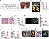 Figure 1
Genetic deletion of Abcc6 leads to persistent myocardial calcification after heart injury. (A) Schematic of the experimental design with cryo-induced injury of WT or Abcc6-KO animals and harvesting 3 days after injury. A high-phosphate, low-magnesium diet was administered starting 3 days before surgery. (B) Cryo-injured WT and Abcc6-KO animals demonstrating cardiac calcification (black arrow) on gross inspection and calcification in the Abcc6-KO heart, visualized with (C) CT scan and (D) following 3D rendering (red circles and insets indicate the calcific lesions). (E) Quantitative analysis of calcium content in scar tissue as measured by CT imaging (n = 6 in each group, mean ± SD, **P < 0.01, by 2-tailed, unpaired t test, WT vs. Abcc6-KO group). (F and G) Histological staining of cryo-injured myocardium in WT and Abcc6-KO animals with (F) Von-Kossa staining (scale bars: 100 μm) and (G) corresponding quantitative analysis showing calcium deposits (black arrows) (n = 6 in each group, mean ± SD, ***P < 0.001, by 2-tailed, unpaired t test, WT vs. Abcc6-KO group). (H) Immunostaining for hydroxyapatite (white arrows, scale bars: 10 μm) and (I) corresponding quantitative analysis. (n = 6 in each group, mean ± SD, ***P < 0.001 WT versus Abcc6-KO group, P value was calculated by 2-tailed, unpaired t test). (J and K) Biochemical measurements of (J) myocardial calcium and (K) phosphate deposits. All staining and measurements were performed on day 3 following injury (n = 6 in each group, mean ± SD, ***P < 0.001, by 2-tailed, unpaired t test, WT vs. Abcc6-KO group). (L–P) Persistence of cardiac calcification with (L) a gross image of the heart 4 months after cryo-injury (black arrowhead indicates the calcific lesion), (M) CT scan (white arrowhead), and (N) 3D rendering showing the calcification region. Red circles (L) and insets (N) indicate the calcific lesions. (O and P) Histological analysis of cryo-injured myocardium 4 months after surgery. (O) Von-Kossa staining (scale bar: 100 μm) and (P) immunostaining for hydroxyapatite (black and white arrowheads; scale bar: 50 μm). (n = 6 per group; WT vs. Abcc6-KO).
The immune system probably does not contribute to calcification in Abcc6-deficient animals. To gain insight into the transcriptional changes that are associated with such rapid calcification of the injured heart, we performed bulk RNA-seq of injured calcific tissue of the Abcc6-KO animal versus the injured noncalcific tissue of the WT animal at day 3 of cryo-injury. Gene ontogeny analysis demonstrated that the principal pathways differentially upregulated in calcific tissue of Abcc6-KO hearts related to extracellular matrix (ECM) and inflammation (Supplemental Figure 3A). Genes downregulated in calcific tissue were related to cardiac muscle contraction and myofibril assembly (Supplemental Figure 3B). Inflammation has been strongly related to calcification of cardiovascular tissues (20, 21).
Given the profound differences in inflammation and ECM gene expression, we performed single-cell transcriptomics to obtain greater insight into cell-specific transcriptomics including differences in the inflammatory infiltrate that could create a more permissible environment for calcification. The cell population was clustered into typical cellular identities on the basis of expression of canonical genes in specific cell types (Supplemental Figure 4), and we could discern multiple populations of cells including cardiac muscle cells, endothelial cells, fibroblasts, macrophages, and other immune cell subsets (Supplemental Figure 3C). We found that cell populations were equally distributed between the Abcc6-KO and WT genotypes with the exception of macrophages, which showed distinct subsets between the WT and the Abcc6-KO injured hearts (Supplemental Figure 3D). The macrophage population could be separated into distinct subclusters (Supplemental Figure 3E) according to gene expression profile, and clusters 1, 3, and 4 predominated more in the tissue of Abcc6-KO hearts (Supplemental Figure 3F), and the macrophage population enriched in the calcific tissue of Abcc6-KO animals represented both monocyte-derived macrophages and tissue repair macrophages (M2) (Supplemental Figure 3G). However, the fraction of macrophages recruited to the injured hearts of WT and Abcc6-KO mice was similar, as shown by both flow cytometry (Supplemental Figure 5, A and B) and immunofluorescence staining (Supplemental Figure 5, C and D)
As the accumulation of different subsets of macrophages could be secondary to the mineralization itself rather than being causal, we next investigated, using bone marrow transplantation (BMT) experiments, whether the inflammatory cells played a critical causal role in mediating calcification in Abcc6-deficient states. For this purpose, we transplanted bone marrow from CD45.1 WT animals into irradiated CD45.2 Abcc6-KO mice (Figure 2A). We used an additional set of controls, in which Abcc6-KO bone marrow was transplanted into irradiated Abcc6-deficient animals. Eight weeks after transplantation, we performed chimerism analysis, and then 1 week later subjected the animals to cardiac cryo-injury to determine the effects on cardiac calcification (Figure 2A). Analysis of peripheral blood cells 8 weeks after BMT demonstrated that 96.5 ± 1.258 (mean ± SD) cells in the Abcc6-KO animals were of WT origin, while Abcc6-deficient animals that received Abcc6-deficient bone marrow cells did not exhibit detectable CD45.1+ cells (Figure 2B). We then subjected the Abcc6-KO animals that had been transplanted with WT bone marrow to cardiac cryo-injury. Despite successful chimerism, the Abcc6-KO animals transplanted with WT bone marrow cells developed myocardial calcification, as did those transplanted with Abcc6-KO bone marrow (Figure 2C). CT imaging demonstrated the presence of cardiac calcification in Abcc6-KO recipient animals transplanted with either WT or Abcc6-KO bone marrow (Figure 2, D–F). Consistent with these imaging findings, Von-Kossa staining of injured cardiac tissue (Figure 2, G and H), and immunofluorescence labeling of hydroxyapatite (Figure 2, I and J) confirmed that both groups developed comparable mineral deposits. Biochemical analysis of the calcified tissue showed similar degrees of calcium and phosphate deposition in both groups (Figure 2, K and L).
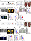 Figure 2
Bone marrow–derived cells do not affect ectopic cardiac calcification. (A) Schematic of BMT. CD45.2 Abcc6-KO recipients were irradiated and transplanted with bone marrow from CD45.1 WT or CD45.2 Abcc6-KO donors, followed by chimerism assessment at 8 weeks and cryo-injury at 9 weeks. (B) Analysis of chimerism in peripheral blood of CD45.2 recipients (n = 6 per group). (C) Gross images of hearts 3 days after cryo-injury in Abcc6-KO recipients; red dotted lines indicate injury; arrowheads indicate calcific lesions. (D) CT scan and (E) 3D rendering highlighting calcific lesions. (F) Quantitative analysis of calcification in scar tissue from CT data (n = 6 per group; mean ± SD; 2-tailed, unpaired Student’s t test) (G and H) Histological assessment with (G) Von-Kossa staining and (H) quantification. Scale bars: 100 μm (n = 6 per group; mean ± SD; 2-tailed, unpaired Student’s t test). (I) Immunostaining for hydroxyapatite and (J) quantification. Scale bars: 50 μm. (n = 6 per group; mean ± SD; 2-tailed, unpaired Student’s t test). (K and L) Biochemical measurements of myocardial (K) calcium and (L) phosphate (n = 6 per group; mean ± SD; 2-tailed, unpaired Student’s t test). (M) Schematic of BMT in CD45.1 WT recipients transplanted with CD45.2 Abcc6-KO marrow, followed by chimerism assessment and cryo-injury. (N) Chimerism analysis of peripheral blood from CD45.1 recipients (n = 6 per group). (O) Gross images of hearts 3 days after cryo-injury showing an absence of calcification, consistent with observations from (P) CT scans and (Q) 3D rendering. (R) Quantitative analysis of calcium content in scar tissue. (S) Von-Kossa staining and (T) quantification. Scale bars: 100 μm. (U) Immunostaining for hydroxyapatite and (V) quantification. Scale bars: 50 μm. (W and X) Biochemical measurements of myocardial (W) calcium and (X) phosphate (n = 6 per group; mean ± SD; 2-tailed, unpaired Student’s t test).
We next performed another series of BMT experiments to determine whether the presence of Abcc6-KO bone marrow cells would be sufficient to cause calcification. For this purpose, we irradiated and transplanted CD45.1 WT animals with donor Abcc6-KO bone marrow (CD45.2) (Figure 2M). Following successful engraftment, we subjected the recipient WT animals to cardiac cryo-injury (Figure 2M). We observed successful chimerism of donor Abcc6-KO cells in WT animals (Figure 2N). Analysis of hearts following cardiac cryo-injury demonstrated the absence of calcification in WT animals transplanted with Abcc6-KO bone marrow as well as in those transplanted with WT bone marrow (Figure 2O). This absence of calcification was further confirmed by micro-CT imaging (Figure 2, P–R), Von-Kossa staining (Figure 2, S and T), and hydroxyapatite immunofluorescence staining (Figure 2, U and V). Biochemical analysis demonstrated the absence of mineralization (Figure 2, W and X). Taken together, these experiments demonstrate that bone marrow–derived cells did not likely to contribute to the calcific phenotype in Abcc6-deficient states.
To further investigate the role of macrophages, we performed experiments to either block or deplete macrophage activity to confirm the phenotype seen in the BMT experiments. We subjected Abcc6-KO animals to a low-magnesium, high-phosphate diet as described above and subjected the animals to cardiac cryo-injury. We administered a monoclonal antibody (mAb) against the CSF1R or IgG control at 3 days and 1 day prior to injury and administered another dose 1 day after injury (Supplemental Figure 6A). Previous studies have demonstrated that injection of the CSF1R antibody depleted macrophages (22, 23), and flow cytometry of the injured heart harvested at 3 days after cryo-injury showed a significant reduction of macrophages in the injured region in the CSF1R mAb–injected animals (Supplemental Figure 6B). However, macrophage inhibition via administration of the CSF1R mAb did not have an effect on calcification. On gross inspection, we observed similar degrees of calcification between the IgG- and CSF1R mAb–injected groups (Supplemental Figure 6C). CT scanning revealed that CSF1R mAb treatment in Abcc6-KO mice did not result in any difference in cardiac tissue mineralization (Supplemental Figure 6, D–F). Histopathological staining and immunofluorescence detection of hydroxyapatite confirmed the inability of CSF1R mAb to affect calcification compared with IgG-injected controls (Supplemental Figure 6, G–J). Biochemical analysis of the calcified tissue revealed a similar amount of calcium and phosphate deposition in the IgG- and CSF1R mAb–treated groups (Supplemental Figure 6, K and L). Next, we performed a circulating monocyte/macrophage depletion experiment. For this purpose, we crossed transgenic mice expressing the diphtheria toxin receptor (DTR) under the CD11b promoter (CD11b-DTR), which enables selective depletion of CD11b cells, with the Abcc6-KO animals to create CD11b-DTR Abcc6-KO animals. The CD11b-DTR animals express the diphtheria toxin (DT) receptor under the control of the human CD11b promoter; thus, administration of DT would lead to ablation of CD11b circulating monocyte/macrophage lineages (24, 25). Macrophage depletion was achieved by 2 i.p. injections of DT (15 ng/g body weight) to ablate CD11b-expressing cells at 3 days and 1 day before injury (Supplemental Figure 6M). Using flow cytometry, we observed decreased macrophages in the hearts of animals that received DT (Supplemental Figure 6N), but on gross inspection, no difference in calcification was noted between the animals that had macrophages ablated and the control animals (Supplemental Figure 6O). CT scanning, histological staining, and immunofluorescence labeling of hydroxyapatite confirmed a similar degree of calcium deposition between the CD11b-ablated animals and the controls (Supplemental Figure 6, P–V). Consistent with these findings, biochemical analyses showed similar levels of calcium and phosphate in the injured cardiac tissue from animals of both groups (Supplemental Figure 6, W and X). Collectively, these experiments using BMT and macrophage depletion techniques strongly indicated that immune cells did not directly affect the degree of calcification in Abcc6-KO animals after cardiac cryo-injury.
Deficiency of Abcc6 in the liver but not in the heart regulates ectopic calcification in the heart. Given that bone marrow–derived cells do not contribute to ectopic calcification, we next investigated how the expression of Abcc6 in the heart or other tissues affects the calcific phenotype. To investigate this, we first analyzed Abcc6 expression across various organs using the Genotype-Tissue Expression (GTEx) database. Our analysis revealed that Abcc6 was most highly expressed in the liver, whereas cardiac tissue had minimal expression (Supplemental Figure 7).
In Abcc6-KO injury, we observed calcific deposits to be initially formed in regions of cell death (Supplemental Figure 8). To determine whether Abcc6 causes calcification in a myocyte-autonomous manner, we first deleted Abcc6 conditionally (conditional knockout [CKO]) in cardiac muscle cells. For this purpose, we crossed Myh6-Cre mice with animals that had Abcc6 alleles floxed to create Myh6-Cre Abcc6-CKO animals. We subjected the Myh6-Cre Abcc6-CKO animals to cardiac cryo-injury as described earlier (Figure 3A). However, we found that cardiac-specific deletion of Abcc6 did not lead to cardiac calcification (Figure 3B). This finding was corroborated by CT imaging (Figure 3, C–E), Von-Kossa staining (Figure 3, F and G), immunofluorescence labeling of hydroxyapatite (Figure 3, H and I), and biochemical quantification of calcium and phosphate (Figure 3, J and K).
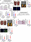 Figure 3
Liver- but not cardiac muscle–specific deletion of Abcc6 leads to ectopic calcification following heart injury. (A) Experimental design of cryo-injury in Myh6-Cre: Abcc6-CKO animals on a high-phosphate, low-magnesium diet. (B) Gross inspection of the cryo-injured hearts from Abcc6fl/fl and Myh6-Cre Abcc6-CKO mice 3 days after injury. (C) CT scan, (D) corresponding 3D rendering, and (E) quantitative analysis of calcium contents in scar tissue demonstrating a lack of detectable calcification (n = 6 per group; mean ± SD; 2-tailed, unpaired t test). (F and G) Histological staining of cryo-injured myocardium in WT and Abcc6-KO animals with (F) Von-Kossa staining and (G) corresponding quantitative analysis showing calcium deposits (n = 6 per group; mean ± SD; 2-tailed, unpaired t test). (H) immunostaining for hydroxyapatite and (I) corresponding quantitative analysis (n = 6 per group; mean ± SD; 2-tailed, unpaired t test). (J and K) Biochemical measurements of (J) myocardial calcium (K) and phosphate deposits in the injured region (n = 6 per group; mean ± SD; 2-tailed, unpaired t test). (L) Experimental design of inducing cryo-injury in animals with liver-specific deletion of Abcc6. (M) Gross inspection of cryo-injured hearts from Abcc6fl/fl and Alb-Cre Abcc6-CKO animals 3 days after injury. Red dashed circles indicate the injury region with visible calcification in animals with liver-specific Abcc6 deletion. Black arrowhead indicates the calcification. (N) CT scan, (O) 3D rendering, and (P) corresponding quantitative analysis of calcium content in scar tissue showing calcification in the injured hearts of liver-specific Abcc6-deleted animals (n = 6 per group; mean ± SD; ***P < 0.001, by 2-tailed, unpaired t test). (Q and R) Myocardial calcification region with (Q) Von-Kossa staining and (R) the corresponding quantitative analysis (n = 6 per group; mean ± SD; ***P < 0.001, by 2-tailed, unpaired t test). (S) Immunostaining for hydroxyapatite in injured hearts of Abcc6fl/fl and Alb-Cre: Abcc6-CKO mice and (T) the corresponding quantitative analysis (n = 6 per group; mean ± SD; **P < 0.01, by 2-tailed, unpaired t test). (U and V) Biochemical measurements of (U) myocardial calcium and (V) phosphate deposits in the region of injured myocardium (n = 6 per group; mean ± SD; ***P < 0.001, by 2-tailed, unpaired t test). Scale bars: 100 μm (F and Q) and 50 μm (H and S).
As the liver is known to express Abcc6, we next investigated whether liver-specific expression regulates cardiac calcification. For this purpose, we crossed animals expressing the liver-specific albumin Cre driver with Abcc6fl/fl animals to create progeny liver-specific Abcc6-CKO animals. The liver-specific Abcc6-CKO animals were maintained on a similar low-magnesium, high-phosphate diet and subjected to cryo-injury (Figure 3L). We observed extensive myocardial calcification in the liver-specific Abcc6-CKO animals (Figure 3M). Micro-CT imaging revealed calcified lesions in these animals (Figure 3, N–P). Histology with Von-Kossa staining and immunofluorescence staining confirmed the calcium deposits in injured cardiac tissue of liver-specific Abcc6-CKO animals (Figure 3, Q–T), and this was further corroborated by biochemical measurement of calcium and phosphate (Figure 3, U and V). These observations confirm that Abcc6 acts in a hepatic cell–autonomous manner to affect ectopic cardiac calcification in Abcc6-deficient states and that tissue-specific deletion of Abcc6 does not determine calcification in that tissue.
Global Abcc6 deficiency leads to deficiencies in metabolites regulating nucleotide metabolism. Abcc6 is thought to be a metabolite transporter, although the exact identity of the metabolite(s) it transports remains unknown. To determine the metabolic abnormalities that could predispose to the development of cardiac calcification in Abcc6-deficient states, we first subjected animals with global Abcc6 deficiency to cardiac cryo-injury and harvested the hearts of WT and Abcc6-KO animals on day 3 of cryo-injury for metabolomics analysis (Figure 4A). The predominant metabolic response was the depletion of metabolites affecting nucleotide metabolism (Figure 4B). Purine and pyrimidine nucleotides such as ATP, ADP, AMP, and GMP were downregulated in Abcc6-KO heart tissue (Figure 4B). Metabolites involved in cardiac energetics, particularly the NAD+ metabolite, were downregulated as well after cardiac injury. A Kyoto Encyclopedia of Genes and Genomes (KEGG) pathway analysis demonstrated metabolites related to purine, arginine, and pyrimidine metabolism to be significantly downregulated (Figure 4C). Taken together, these data indicate that aberrations in nucleotide metabolism were present in calcific tissues that underwent calcification in Abcc6-KO animals. As we have shown previously, the deficiency of the gene in the liver regulates cardiac calcification, we also harvested livers from Abcc6-KO mice (Figure 4A) and observed that similar metabolic abnormalities of nucleotide metabolism with decreased purine and pyrimidine metabolites were present in the liver as well (Figure 4, D and E).
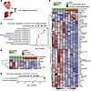 Figure 4
Loss of Abcc6 alters metabolites in the injured myocardium. (A) Schematic of injured cardiac tissue and liver tissue collected from WT and Abcc6-KO animals for untargeted metabolomics 3 days following injury. (B) Untargeted metabolomics by liquid chromatography/mass spectrometry (LC/MS) of injured heart tissue from WT and Abcc6-KO animals at post–cryo-injury day 3, showing significantly altered metabolites in Abcc6-KO hearts versus WT hearts (n = 5 per group; *P < 0.05, **P < 0.01, ***P < 0.001, determined by one-way ANOVA. Significance is noted in Supporting Data Values). (C) KEGG analysis showing the top metabolic pathways significantly downregulated in injured Abcc6-KO hearts compared with WT. (D) Untargeted metabolomics by LC/MS of liver tissue from WT and Abcc6-KO animals at day 3 after cryo-injury of the heart, showing significantly altered metabolites (P < 0.05 (n=5 per group; *P < 0.05, **P < 0.01, ***P < 0.001 determined by one-way ANOVA) in Abcc6-KO livers versus WT livers. Significance is noted in the Supporting Data Values file. (E) KEGG analysis showing the top metabolic pathways significantly downregulated in Abcc6-KO liver tissue compared with WT after cardiac injury.
Liver-specific Abcc6 deficiency mirrors the metabolic abnormalities seen in animals globally deficient in Abcc6. Our experiments demonstrate that liver-specific deletion of Abcc6 led to ectopic cardiovascular calcification and recapitulated the phenotype observed in global Abcc6-KO animals. We next subjected liver-specific Abcc6-KO animals to cryo-injury and harvested the injured cardiac tissue for metabolomics analysis (Figure 5A), which demonstrated a significant reduction of nucleotides, including purines and pyrimidines. Purine, pyrimidine, and nicotinamide nucleotides were markedly downregulated in cardiac tissues of liver-specific Abcc6-CKO animals (Figure 5B). To identify the metabolites that were commonly dysregulated in injured heart tissue from global and liver-specific deletion in Abcc6 animals, we compared the differentially up-/downregulated metabolites in each instance (Figure 5C). We observed that 60% of the metabolites were commonly dysregulated in injured heart tissues of animals that exhibited liver-specific Abcc6 deletion and global Abcc6 deletion (Figure 5C). KEGG pathway analysis demonstrated that nucleotide metabolism, including NAD and purine metabolism, was commonly downregulated in the hearts of liver-specific Abcc6-CKO and global-KO animals, demonstrating that liver-specific deletion of Abcc6 affected similar metabolic pathways in the injured heart tissue as global Abcc6 deficiency (Figure 5D). Taken together, these experiments suggest that the liver drives metabolic abnormalities in the injured heart tissue that create a permissive environment for ectopic calcification.
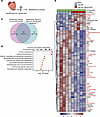 Figure 5
Liver-specific deletion of Abcc6 leads to metabolic abnormalities in the injured heart similar to metabolic pathways affected in the injured hearts of Abcc6-KO animals. (A) Schematic of scar tissue collection from Abcc6fl/fl and Alb-Cre: Abcc6-CKO animals for metabolomic analysis. (B) Untargeted metabolomics by LC/MS of injured tissue from Abcc6fl/fl and Alb-Cre Abcc6-CKO animals at day 3 after cryo-injury, highlighting significantly altered metabolites in Alb-Cre: Abcc6-CKO hearts (n=5 per group; *P < 0.05, **P < 0.01, ***P < 0.001 determined by one-way ANOVA). Metabolites labeled in red overlap with those altered in injured hearts of global Abcc6-KO versus WT animals. (C) Venn diagram depicting metabolites differentially present between injured hearts of Abcc6-KO versus WT (left) and Alb-Cre Abcc6-CKO versus Abcc6fl/fl (right) mice. (D) KEGG analysis of metabolic pathways of the commonly altered metabolites that were significantly downregulated in injured tissue of Abcc6-KO versus WT and Alb-Cre Abcc6-CKO versus Abcc6fl/fl mice.
The liver regulates cardiac mitochondrial function in an Abcc6-dependent manner. Given the metabolic abnormalities noted in the injured heart in animals with global Abcc6 deficiency, as well as in animals with liver-specific deletion of Abcc6, we next investigated the functional consequences of such metabolic abnormalities. Mitochondrial abnormalities have been noted in patients with PXE (26), and in animal models, calcification has been thought to originate around mitochondrial structures (17). To determine whether the mitochondria exhibited functional abnormalities in the injured heart in animals deficient in Abcc6, we subjected Abcc6-KO animals to cardiac cryo-injury and isolated mitochondria from their hearts at post-injury day 3 and measured oxygen consumption rates (OCRs) with the Seahorse analyzer. We focused on state 3 respiration, which in isolated mitochondria represents the mitochondria′s capacity to generate ATP when provided with specific substrates and ADP (27). We provided the isolated mitochondria substrates for complex I (pyruvate and malate), complex II (succinate with rotenone to inhibit complex I), and fatty acid oxidation (palmitoyl-carnitine) (Figure 6A). We observed that after adding the complex I electron transport chain substrates pyruvate and malate (28), there was no difference in OCRs between mitochondria obtained from uninjured hearts of WT and animals globally deficient in Abcc6 (Figure 6B). However, mitochondria from injured hearts of Abcc6-KO animals exhibited a marked reduction in OCRs compared with mitochondria isolated from injured hearts of WT animals (Figure 6B). When complex II substrate was provided, no differences in OCRs were observed in the isolated mitochondria from injured heart tissue of WT and Abcc6-KO animals (Figure 6C). Similarly, mitochondria supplied with palmitoyl-carnitine, the substrate for fatty acid oxidation, showed decreased OCRs in isolated Abcc6-KO animals versus WT animals (Figure 6D). State 3 respiration was decreased in mitochondria from Abcc6-KO versus WT hearts when mitochondria were treated with pyruvate and malate or palmitoyl-carnitine but not succinate and rotenone (Figure 6, E–G). Taken together, the data suggest that differences in state 3 respiration were primarily due to abnormalities in complex I.
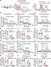 Figure 6
Mitochondrial oxygen consumption of injured heart tissue is reduced in global and liver-specific Abcc6-KO animals. (A) Schematic of Seahorse assays to measure mitochondrial oxygen consumption with the addition of complex-specific substrates and inhibitors. (B–D) Representative trace of OCRs in isolated mitochondria from uninjured and injured heart tissue of WT and Abcc6-KO animals treated with (B) complex I substrates (pyruvate and malate), (C) complex II substrate (succinate) and complex I inhibitor (rotenone), and (D) fatty acid oxidation substrates (palmitoyl-carnitine). In all cases, the OCR was normalized to the amount of mitochondrial protein loaded. (E–G) State 3 respiration quantification of isolated cardiac mitochondria from uninjured and injured cardiac tissue of WT and Abcc6-KO animals. Mitochondria were supplemented with (E) pyruvate and malate or (F) succinate and rotenone, or (G) palmitoyl-carnitine (n = 6 per group; mean ± SD; *P < 0.05, by 2-way ANOVA with Tukey’s multiple-comparison test). (H–J) Representative respirometry trace in isolated mitochondria from Abcc6fl/fl and Alb-Cre: Abcc6-CKO uninjured and injured cardiac tissue 3 days after surgery. Mitochondria were supplemented with (H) pyruvate and malate, (I) succinate and rotenone, or (J) palmitoyl-carnitine. (K–M) State 3 respiration of isolated cardiac mitochondria from uninjured and injured cardiac tissue of Abcc6fl/fl and Alb-Cre Abcc6-CKO animals. Mitochondria were treated with (K) substrates of pyruvate and malate, (L) succinate and rotenone, or (M) palmitoyl-carnitine (n = 6 per group; mean ± SD; *P < 0.05 and **P < 0.01, by 2-way ANOVA with Tukey’s multiple-comparison test).
As liver-specific deletion of Abcc6 was sufficient to induce cardiac calcification, we next examined whether liver-specific deletion of Abcc6 leads to functional abnormalities of cardiac mitochondria. For this purpose, we isolated mitochondria from injured and uninjured hearts of animals with liver-specific Abcc6-CKO and respective Cre (–) Abcc6fl/fl controls. Measurement of the OCR demonstrated similar abnormalities in mitochondrial respiration rates, as noted in the animals with global Abcc6 deficiency. There were no significant differences in OCRs in mitochondria isolated from uninjured hearts between the 2 groups, but mitochondria from injured hearts of liver-specific Abcc6-CKO animals showed a profound reduction in respiration with pyruvate and malate as the substrate (Figure 6H) but not with succinate and rotenone (Figure 6I).We observed a similar reduction in respiration with palmitoyl-carnitine (Figure 6J). Quantification of state 3 OCRs with the addition of complex I substrates confirmed the profound reduction of cellular respiration in mitochondria isolated from injured heart tissue from liver-specific Abcc6-CKO animals compared with Cre (–) controls (Figure 6K). State 3 OCRs with complex II substrate and complex I inhibitor did not show any significant abnormalities (Figure 6L). A similar reduction in the state 3 respiratory rate was also seen in mitochondria from injured hearts of animals with liver-specific Abcc6 deletion, upon addition of palmitoyl-carnitine (Figure 6M). We also performed Western blotting and observed no change in the amount of oxidative phosphorylation proteins in isolated mitochondria from injured heart tissues of Abcc6-KO animals and their respective controls (Supplemental Figure 9). We did not observe impaired function of complex I in the mitochondrial electron transport chain, dependent on Abcc6 in Myh6-Cre Abcc6-CKO animals, suggesting again that Abcc6 acted in a nonmyocyte-autonomous manner (Supplemental Figure 10). These data, taken together, demonstrate that hepatic expression of Abcc6, likely through an altered systemic metabolic milieu, altered mitochondrial function in the heart following injury and suggest an altered systemic metabolic milieu as the basis of functional mitochondrial defects in PXE.
The mineralization inhibitor clodronate prevents the development of cardiac calcification in animals deficient in Abcc6. Ectopic cardiac calcification is thought to parallel osteogenesis, with expression of the osteogenic transcriptional program in mesenchymal cells in the region undergoing ectopic calcification. In contrast, dystrophic calcification occurs after cell death or in scar tissue and represents mineralization of the matrix from hydroxyapatite mineral deposition and growth without an induction of an osteogenic transcriptional program. We first examined bulk sequencing gene expression data from injured heart tissue and examined expression levels of a panel of genes that are known to be associated with induction of an osteogenic transcription program, but we found no evidence of differential expression of osteogenic genes between animals with global Abcc6 deficiency and WT controls (Figure 7A). Dystrophic calcification is initiated with cell death, and we performed immunostaining of the injured region and observed hydroxyapatite crystals within cardiomyocytes (Figure 7B). As dystrophic calcification is thought to be secondary to mineral crystallization and growth, we administered the first-generation bisphosphonate clodronate (1.8 mg/30 g of BW via i.p. injection) to determine the effects on cardiac calcification. Abcc6-KO animals were subjected to cardiac cryo-injury, and clodronate liposomes were administered 3 days and 1 day prior to cryo-injury (Figure 7C). On gross inspection, we observed complete rescue of calcification in the hearts of Abcc6-KO animals that had been administered clodronate liposomes compared with vehicle liposome–injected animals (Figure 7D). Micro-CT imaging of the heart and thorax showed an absence of calcium mineral deposits in the Abcc6-KO animals administered clodronate liposomes (Figure 7, E–G). Von-Kossa staining and immunofluorescence staining for hydroxyapatite confirmed the complete rescue of calcification in Abcc6-KO animals (Figure 7, H–K). Biochemical analysis demonstrated a significant reduction in calcium (Figure 7L) and phosphate (Figure 7M) deposition in injured hearts of animals injected with clodronate. Consistent with the known effects of liposomal clodronate on macrophage depletion (29), we observed significant depletion of both macrophages and neutrophils in the injured hearts of animals receiving clodronate liposomes versus vehicle (Supplemental Figure 11). To confirm the effects of bisphosphonate in reducing calcification in Abcc6-KO animals, we injected the bisphosphonate etidronate into Abcc6-KO animals subjected to cardiac cryo-injury. Etidronate is not known to deplete macrophages or neutrophils. Animals received 4 doses with each dose administered i.p. daily starting from the day prior to injury (Supplemental Figure 12A). On gross inspection, we observed complete rescue of cardiac calcification in animals that received etidronate (Supplemental Figure 12B), and CT scanning confirmed the absence of calcification in etidronate-injected animals (Supplemental Figure 12, C–E). Histological staining with Von-Kossa and immunofluorescence labeling of hydroxyapatite and biochemical measurements of calcium and phosphate confirmed the absence of calcification in injured hearts of animals that received etidronate (Supplemental Figure 12, F–K). Taken together, these data suggest that tissues undergo dystrophic calcification in Abcc6-deficient states and that mineralization inhibitors can attenuate or rescue the development of post-injury ectopic calcification.
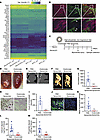 Figure 7
Clodronate inhibits cardiac calcification in animals deficient in Abcc6. (A) Heatmap showing the expression levels of a set of osteogenic genes in WT and Abcc6-KO injured heart tissue on day 3 after surgery demonstrating no significant differential expression (n = 3 in each group, P > 0.05). (B) Immunostaining for hydroxyapatite (green) and myocytes (red) in injured hearts from Abcc6-KO mice (inset shows high magnification). The arrow indicates the calcific area within the myocyte. Scale bars: 100 μm (inset: original magnification, ×60). (C) Experimental design for the administration of clodronate liposomes to Abcc6-KO animals to determine the effects on cardiac calcification. (D) Gross images 3 days after cryo-injury of hearts from Abcc6-KO mice that received PBS liposomes or clodronate liposomes (red dotted lines indicate the area of injury; arrowhead indicates the calcific lesion). Note that the animals that received clodronate liposomes showed no mineralization. (E) CT scan, (F) 3D rendering, and (G) quantitative analysis of calcium content in heart scar tissue (n = 6 in each group; mean ± SD; ***P < 0.001, by 2-tailed, unpaired Student’s t test comparing PBS liposome and clodronate liposome groups). (H and I) Histological staining of cryo-injured myocardium with (H) Von-Kossa staining and (I) corresponding quantitative analysis showing calcium deposits. Scale bars: 100 μm (n = 6 in each group; mean ± SD; ***P < 0.001, by 2-tailed, unpaired Student’s t test). (J) Immunostaining for hydroxyapatite and (K) corresponding quantitative analysis. Scale bars: 50 μm (n = 6 in each group; mean ± SD; **P < 0.01, by 2-tailed, unpaired Student’s t test). (L and M) Biochemical measurements of (L) myocardial calcium and (M) phosphate deposits in the injured region (n = 6 in each group; mean ± SD; **P < 0.01, by 2-tailed, unpaired Student’s t test, comparing PBS liposome and clodronate liposome groups).


Copyright © 2026 American Society for Clinical Investigation
ISSN: 0021-9738 (print), 1558-8238 (online)