
Advertisement
Research ArticleImmunologyOncology
Open Access |  10.1172/JCI192885
Bispecific antibodies and CAR T cells targeting a TP53 mutation–associated neoantigen show discordant affinity requirements
Sarah R. DiNapoli,1,2,3 Katharine M. Wright,2,4,5 Brian J. Mog,1,2,3,6 Alexander H. Pearlman,1,2,3 Tushar D. Nichakawade,1,2,3,7,8 Nikita Marcou,1,2,3 Emily Han-Chung Hsiue,1,2,3 Michael S. Hwang,1,2,3 Jacqueline Douglass,1,2,3,9 Qiang Liu,1,2,3 Evangeline Watson,1,2,3 Marco Dal Molin,1,2,3,10 Joshua D. Cohen,1,2,3,6 Maria Popoli,1,2,3 Suman Paul,1,9 Maximilian F. Konig,1,2,3,11 Nicolas Wyhs,1,9 P. Aitana Azurmendi,2,4,5 Stephanie Glavaris,1,2,3 Jiaxin Ge,1,2,3 Tolulope O. Awosika,1,2,3 Jin Liu,1,2,3 Kathleen L. Gabrielson,9,12,13 Sandra B. Gabelli,4,5,9,14 Drew M. Pardoll,4,9 Chetan Bettegowda,1,3,9,15 Nickolas Papadopoulos,1,3,9,13 Kenneth W. Kinzler,1,3,4,9 Shibin Zhou,1,3,4,9 and Bert Vogelstein1,2,3,4,9,13
1Ludwig Center, Sidney Kimmel Comprehensive Cancer Center, Johns Hopkins University School of Medicine, Baltimore, Maryland, USA.
2Howard Hughes Medical Institute, Chevy Chase, Maryland, USA.
3Lustgarten Pancreatic Cancer Research Laboratory and
4Bloomberg~Kimmel Institute for Cancer Immunotherapy, Sidney Kimmel Comprehensive Cancer Center, Johns Hopkins University School of Medicine, Baltimore, Maryland, USA.
5Department of Biophysics and Biophysical Chemistry, Johns Hopkins University School of Medicine, Baltimore, Maryland, USA.
6Department of Biomedical Engineering,
7Department of Chemical and Biomolecular Engineering, and
8Institute for NanoBioTechnology, Johns Hopkins University, Baltimore, Maryland, USA.
9Department of Oncology,
10Department of Surgery,
11Division of Rheumatology, Department of Medicine,
12Department of Molecular and Comparative Pathobiology,
13Department of Pathology,
14Department of Medicine, and
15Department of Neurosurgery, Johns Hopkins University School of Medicine, Baltimore, Maryland, USA.
Address correspondence to: Bert Vogelstein, Ludwig Center, Sidney Kimmel Comprehensive Cancer Center, Johns Hopkins University School of Medicine, 1650 Orleans St., CRB 1, Room 589, Baltimore, Maryland 21231, USA. Phone: 410.955.8878; Email: vogelbe@jhmi.edu.
Find articles by
DiNapoli, S.
in:
PubMed
|
Google Scholar
|

1Ludwig Center, Sidney Kimmel Comprehensive Cancer Center, Johns Hopkins University School of Medicine, Baltimore, Maryland, USA.
2Howard Hughes Medical Institute, Chevy Chase, Maryland, USA.
3Lustgarten Pancreatic Cancer Research Laboratory and
4Bloomberg~Kimmel Institute for Cancer Immunotherapy, Sidney Kimmel Comprehensive Cancer Center, Johns Hopkins University School of Medicine, Baltimore, Maryland, USA.
5Department of Biophysics and Biophysical Chemistry, Johns Hopkins University School of Medicine, Baltimore, Maryland, USA.
6Department of Biomedical Engineering,
7Department of Chemical and Biomolecular Engineering, and
8Institute for NanoBioTechnology, Johns Hopkins University, Baltimore, Maryland, USA.
9Department of Oncology,
10Department of Surgery,
11Division of Rheumatology, Department of Medicine,
12Department of Molecular and Comparative Pathobiology,
13Department of Pathology,
14Department of Medicine, and
15Department of Neurosurgery, Johns Hopkins University School of Medicine, Baltimore, Maryland, USA.
Address correspondence to: Bert Vogelstein, Ludwig Center, Sidney Kimmel Comprehensive Cancer Center, Johns Hopkins University School of Medicine, 1650 Orleans St., CRB 1, Room 589, Baltimore, Maryland 21231, USA. Phone: 410.955.8878; Email: vogelbe@jhmi.edu.
Find articles by Wright, K. in: PubMed | Google Scholar
1Ludwig Center, Sidney Kimmel Comprehensive Cancer Center, Johns Hopkins University School of Medicine, Baltimore, Maryland, USA.
2Howard Hughes Medical Institute, Chevy Chase, Maryland, USA.
3Lustgarten Pancreatic Cancer Research Laboratory and
4Bloomberg~Kimmel Institute for Cancer Immunotherapy, Sidney Kimmel Comprehensive Cancer Center, Johns Hopkins University School of Medicine, Baltimore, Maryland, USA.
5Department of Biophysics and Biophysical Chemistry, Johns Hopkins University School of Medicine, Baltimore, Maryland, USA.
6Department of Biomedical Engineering,
7Department of Chemical and Biomolecular Engineering, and
8Institute for NanoBioTechnology, Johns Hopkins University, Baltimore, Maryland, USA.
9Department of Oncology,
10Department of Surgery,
11Division of Rheumatology, Department of Medicine,
12Department of Molecular and Comparative Pathobiology,
13Department of Pathology,
14Department of Medicine, and
15Department of Neurosurgery, Johns Hopkins University School of Medicine, Baltimore, Maryland, USA.
Address correspondence to: Bert Vogelstein, Ludwig Center, Sidney Kimmel Comprehensive Cancer Center, Johns Hopkins University School of Medicine, 1650 Orleans St., CRB 1, Room 589, Baltimore, Maryland 21231, USA. Phone: 410.955.8878; Email: vogelbe@jhmi.edu.
Find articles by Mog, B. in: PubMed | Google Scholar
1Ludwig Center, Sidney Kimmel Comprehensive Cancer Center, Johns Hopkins University School of Medicine, Baltimore, Maryland, USA.
2Howard Hughes Medical Institute, Chevy Chase, Maryland, USA.
3Lustgarten Pancreatic Cancer Research Laboratory and
4Bloomberg~Kimmel Institute for Cancer Immunotherapy, Sidney Kimmel Comprehensive Cancer Center, Johns Hopkins University School of Medicine, Baltimore, Maryland, USA.
5Department of Biophysics and Biophysical Chemistry, Johns Hopkins University School of Medicine, Baltimore, Maryland, USA.
6Department of Biomedical Engineering,
7Department of Chemical and Biomolecular Engineering, and
8Institute for NanoBioTechnology, Johns Hopkins University, Baltimore, Maryland, USA.
9Department of Oncology,
10Department of Surgery,
11Division of Rheumatology, Department of Medicine,
12Department of Molecular and Comparative Pathobiology,
13Department of Pathology,
14Department of Medicine, and
15Department of Neurosurgery, Johns Hopkins University School of Medicine, Baltimore, Maryland, USA.
Address correspondence to: Bert Vogelstein, Ludwig Center, Sidney Kimmel Comprehensive Cancer Center, Johns Hopkins University School of Medicine, 1650 Orleans St., CRB 1, Room 589, Baltimore, Maryland 21231, USA. Phone: 410.955.8878; Email: vogelbe@jhmi.edu.
Find articles by Pearlman, A. in: PubMed | Google Scholar
1Ludwig Center, Sidney Kimmel Comprehensive Cancer Center, Johns Hopkins University School of Medicine, Baltimore, Maryland, USA.
2Howard Hughes Medical Institute, Chevy Chase, Maryland, USA.
3Lustgarten Pancreatic Cancer Research Laboratory and
4Bloomberg~Kimmel Institute for Cancer Immunotherapy, Sidney Kimmel Comprehensive Cancer Center, Johns Hopkins University School of Medicine, Baltimore, Maryland, USA.
5Department of Biophysics and Biophysical Chemistry, Johns Hopkins University School of Medicine, Baltimore, Maryland, USA.
6Department of Biomedical Engineering,
7Department of Chemical and Biomolecular Engineering, and
8Institute for NanoBioTechnology, Johns Hopkins University, Baltimore, Maryland, USA.
9Department of Oncology,
10Department of Surgery,
11Division of Rheumatology, Department of Medicine,
12Department of Molecular and Comparative Pathobiology,
13Department of Pathology,
14Department of Medicine, and
15Department of Neurosurgery, Johns Hopkins University School of Medicine, Baltimore, Maryland, USA.
Address correspondence to: Bert Vogelstein, Ludwig Center, Sidney Kimmel Comprehensive Cancer Center, Johns Hopkins University School of Medicine, 1650 Orleans St., CRB 1, Room 589, Baltimore, Maryland 21231, USA. Phone: 410.955.8878; Email: vogelbe@jhmi.edu.
Find articles by Nichakawade, T. in: PubMed | Google Scholar
1Ludwig Center, Sidney Kimmel Comprehensive Cancer Center, Johns Hopkins University School of Medicine, Baltimore, Maryland, USA.
2Howard Hughes Medical Institute, Chevy Chase, Maryland, USA.
3Lustgarten Pancreatic Cancer Research Laboratory and
4Bloomberg~Kimmel Institute for Cancer Immunotherapy, Sidney Kimmel Comprehensive Cancer Center, Johns Hopkins University School of Medicine, Baltimore, Maryland, USA.
5Department of Biophysics and Biophysical Chemistry, Johns Hopkins University School of Medicine, Baltimore, Maryland, USA.
6Department of Biomedical Engineering,
7Department of Chemical and Biomolecular Engineering, and
8Institute for NanoBioTechnology, Johns Hopkins University, Baltimore, Maryland, USA.
9Department of Oncology,
10Department of Surgery,
11Division of Rheumatology, Department of Medicine,
12Department of Molecular and Comparative Pathobiology,
13Department of Pathology,
14Department of Medicine, and
15Department of Neurosurgery, Johns Hopkins University School of Medicine, Baltimore, Maryland, USA.
Address correspondence to: Bert Vogelstein, Ludwig Center, Sidney Kimmel Comprehensive Cancer Center, Johns Hopkins University School of Medicine, 1650 Orleans St., CRB 1, Room 589, Baltimore, Maryland 21231, USA. Phone: 410.955.8878; Email: vogelbe@jhmi.edu.
Find articles by Marcou, N. in: PubMed | Google Scholar
1Ludwig Center, Sidney Kimmel Comprehensive Cancer Center, Johns Hopkins University School of Medicine, Baltimore, Maryland, USA.
2Howard Hughes Medical Institute, Chevy Chase, Maryland, USA.
3Lustgarten Pancreatic Cancer Research Laboratory and
4Bloomberg~Kimmel Institute for Cancer Immunotherapy, Sidney Kimmel Comprehensive Cancer Center, Johns Hopkins University School of Medicine, Baltimore, Maryland, USA.
5Department of Biophysics and Biophysical Chemistry, Johns Hopkins University School of Medicine, Baltimore, Maryland, USA.
6Department of Biomedical Engineering,
7Department of Chemical and Biomolecular Engineering, and
8Institute for NanoBioTechnology, Johns Hopkins University, Baltimore, Maryland, USA.
9Department of Oncology,
10Department of Surgery,
11Division of Rheumatology, Department of Medicine,
12Department of Molecular and Comparative Pathobiology,
13Department of Pathology,
14Department of Medicine, and
15Department of Neurosurgery, Johns Hopkins University School of Medicine, Baltimore, Maryland, USA.
Address correspondence to: Bert Vogelstein, Ludwig Center, Sidney Kimmel Comprehensive Cancer Center, Johns Hopkins University School of Medicine, 1650 Orleans St., CRB 1, Room 589, Baltimore, Maryland 21231, USA. Phone: 410.955.8878; Email: vogelbe@jhmi.edu.
Find articles by
Hsiue, E.
in:
PubMed
|
Google Scholar
|
1Ludwig Center, Sidney Kimmel Comprehensive Cancer Center, Johns Hopkins University School of Medicine, Baltimore, Maryland, USA.
2Howard Hughes Medical Institute, Chevy Chase, Maryland, USA.
3Lustgarten Pancreatic Cancer Research Laboratory and
4Bloomberg~Kimmel Institute for Cancer Immunotherapy, Sidney Kimmel Comprehensive Cancer Center, Johns Hopkins University School of Medicine, Baltimore, Maryland, USA.
5Department of Biophysics and Biophysical Chemistry, Johns Hopkins University School of Medicine, Baltimore, Maryland, USA.
6Department of Biomedical Engineering,
7Department of Chemical and Biomolecular Engineering, and
8Institute for NanoBioTechnology, Johns Hopkins University, Baltimore, Maryland, USA.
9Department of Oncology,
10Department of Surgery,
11Division of Rheumatology, Department of Medicine,
12Department of Molecular and Comparative Pathobiology,
13Department of Pathology,
14Department of Medicine, and
15Department of Neurosurgery, Johns Hopkins University School of Medicine, Baltimore, Maryland, USA.
Address correspondence to: Bert Vogelstein, Ludwig Center, Sidney Kimmel Comprehensive Cancer Center, Johns Hopkins University School of Medicine, 1650 Orleans St., CRB 1, Room 589, Baltimore, Maryland 21231, USA. Phone: 410.955.8878; Email: vogelbe@jhmi.edu.
Find articles by Hwang, M. in: PubMed | Google Scholar
1Ludwig Center, Sidney Kimmel Comprehensive Cancer Center, Johns Hopkins University School of Medicine, Baltimore, Maryland, USA.
2Howard Hughes Medical Institute, Chevy Chase, Maryland, USA.
3Lustgarten Pancreatic Cancer Research Laboratory and
4Bloomberg~Kimmel Institute for Cancer Immunotherapy, Sidney Kimmel Comprehensive Cancer Center, Johns Hopkins University School of Medicine, Baltimore, Maryland, USA.
5Department of Biophysics and Biophysical Chemistry, Johns Hopkins University School of Medicine, Baltimore, Maryland, USA.
6Department of Biomedical Engineering,
7Department of Chemical and Biomolecular Engineering, and
8Institute for NanoBioTechnology, Johns Hopkins University, Baltimore, Maryland, USA.
9Department of Oncology,
10Department of Surgery,
11Division of Rheumatology, Department of Medicine,
12Department of Molecular and Comparative Pathobiology,
13Department of Pathology,
14Department of Medicine, and
15Department of Neurosurgery, Johns Hopkins University School of Medicine, Baltimore, Maryland, USA.
Address correspondence to: Bert Vogelstein, Ludwig Center, Sidney Kimmel Comprehensive Cancer Center, Johns Hopkins University School of Medicine, 1650 Orleans St., CRB 1, Room 589, Baltimore, Maryland 21231, USA. Phone: 410.955.8878; Email: vogelbe@jhmi.edu.
Find articles by Douglass, J. in: PubMed | Google Scholar
1Ludwig Center, Sidney Kimmel Comprehensive Cancer Center, Johns Hopkins University School of Medicine, Baltimore, Maryland, USA.
2Howard Hughes Medical Institute, Chevy Chase, Maryland, USA.
3Lustgarten Pancreatic Cancer Research Laboratory and
4Bloomberg~Kimmel Institute for Cancer Immunotherapy, Sidney Kimmel Comprehensive Cancer Center, Johns Hopkins University School of Medicine, Baltimore, Maryland, USA.
5Department of Biophysics and Biophysical Chemistry, Johns Hopkins University School of Medicine, Baltimore, Maryland, USA.
6Department of Biomedical Engineering,
7Department of Chemical and Biomolecular Engineering, and
8Institute for NanoBioTechnology, Johns Hopkins University, Baltimore, Maryland, USA.
9Department of Oncology,
10Department of Surgery,
11Division of Rheumatology, Department of Medicine,
12Department of Molecular and Comparative Pathobiology,
13Department of Pathology,
14Department of Medicine, and
15Department of Neurosurgery, Johns Hopkins University School of Medicine, Baltimore, Maryland, USA.
Address correspondence to: Bert Vogelstein, Ludwig Center, Sidney Kimmel Comprehensive Cancer Center, Johns Hopkins University School of Medicine, 1650 Orleans St., CRB 1, Room 589, Baltimore, Maryland 21231, USA. Phone: 410.955.8878; Email: vogelbe@jhmi.edu.
Find articles by Liu, Q. in: PubMed | Google Scholar
1Ludwig Center, Sidney Kimmel Comprehensive Cancer Center, Johns Hopkins University School of Medicine, Baltimore, Maryland, USA.
2Howard Hughes Medical Institute, Chevy Chase, Maryland, USA.
3Lustgarten Pancreatic Cancer Research Laboratory and
4Bloomberg~Kimmel Institute for Cancer Immunotherapy, Sidney Kimmel Comprehensive Cancer Center, Johns Hopkins University School of Medicine, Baltimore, Maryland, USA.
5Department of Biophysics and Biophysical Chemistry, Johns Hopkins University School of Medicine, Baltimore, Maryland, USA.
6Department of Biomedical Engineering,
7Department of Chemical and Biomolecular Engineering, and
8Institute for NanoBioTechnology, Johns Hopkins University, Baltimore, Maryland, USA.
9Department of Oncology,
10Department of Surgery,
11Division of Rheumatology, Department of Medicine,
12Department of Molecular and Comparative Pathobiology,
13Department of Pathology,
14Department of Medicine, and
15Department of Neurosurgery, Johns Hopkins University School of Medicine, Baltimore, Maryland, USA.
Address correspondence to: Bert Vogelstein, Ludwig Center, Sidney Kimmel Comprehensive Cancer Center, Johns Hopkins University School of Medicine, 1650 Orleans St., CRB 1, Room 589, Baltimore, Maryland 21231, USA. Phone: 410.955.8878; Email: vogelbe@jhmi.edu.
Find articles by Watson, E. in: PubMed | Google Scholar
1Ludwig Center, Sidney Kimmel Comprehensive Cancer Center, Johns Hopkins University School of Medicine, Baltimore, Maryland, USA.
2Howard Hughes Medical Institute, Chevy Chase, Maryland, USA.
3Lustgarten Pancreatic Cancer Research Laboratory and
4Bloomberg~Kimmel Institute for Cancer Immunotherapy, Sidney Kimmel Comprehensive Cancer Center, Johns Hopkins University School of Medicine, Baltimore, Maryland, USA.
5Department of Biophysics and Biophysical Chemistry, Johns Hopkins University School of Medicine, Baltimore, Maryland, USA.
6Department of Biomedical Engineering,
7Department of Chemical and Biomolecular Engineering, and
8Institute for NanoBioTechnology, Johns Hopkins University, Baltimore, Maryland, USA.
9Department of Oncology,
10Department of Surgery,
11Division of Rheumatology, Department of Medicine,
12Department of Molecular and Comparative Pathobiology,
13Department of Pathology,
14Department of Medicine, and
15Department of Neurosurgery, Johns Hopkins University School of Medicine, Baltimore, Maryland, USA.
Address correspondence to: Bert Vogelstein, Ludwig Center, Sidney Kimmel Comprehensive Cancer Center, Johns Hopkins University School of Medicine, 1650 Orleans St., CRB 1, Room 589, Baltimore, Maryland 21231, USA. Phone: 410.955.8878; Email: vogelbe@jhmi.edu.
Find articles by Dal Molin, M. in: PubMed | Google Scholar
1Ludwig Center, Sidney Kimmel Comprehensive Cancer Center, Johns Hopkins University School of Medicine, Baltimore, Maryland, USA.
2Howard Hughes Medical Institute, Chevy Chase, Maryland, USA.
3Lustgarten Pancreatic Cancer Research Laboratory and
4Bloomberg~Kimmel Institute for Cancer Immunotherapy, Sidney Kimmel Comprehensive Cancer Center, Johns Hopkins University School of Medicine, Baltimore, Maryland, USA.
5Department of Biophysics and Biophysical Chemistry, Johns Hopkins University School of Medicine, Baltimore, Maryland, USA.
6Department of Biomedical Engineering,
7Department of Chemical and Biomolecular Engineering, and
8Institute for NanoBioTechnology, Johns Hopkins University, Baltimore, Maryland, USA.
9Department of Oncology,
10Department of Surgery,
11Division of Rheumatology, Department of Medicine,
12Department of Molecular and Comparative Pathobiology,
13Department of Pathology,
14Department of Medicine, and
15Department of Neurosurgery, Johns Hopkins University School of Medicine, Baltimore, Maryland, USA.
Address correspondence to: Bert Vogelstein, Ludwig Center, Sidney Kimmel Comprehensive Cancer Center, Johns Hopkins University School of Medicine, 1650 Orleans St., CRB 1, Room 589, Baltimore, Maryland 21231, USA. Phone: 410.955.8878; Email: vogelbe@jhmi.edu.
Find articles by Cohen, J. in: PubMed | Google Scholar
1Ludwig Center, Sidney Kimmel Comprehensive Cancer Center, Johns Hopkins University School of Medicine, Baltimore, Maryland, USA.
2Howard Hughes Medical Institute, Chevy Chase, Maryland, USA.
3Lustgarten Pancreatic Cancer Research Laboratory and
4Bloomberg~Kimmel Institute for Cancer Immunotherapy, Sidney Kimmel Comprehensive Cancer Center, Johns Hopkins University School of Medicine, Baltimore, Maryland, USA.
5Department of Biophysics and Biophysical Chemistry, Johns Hopkins University School of Medicine, Baltimore, Maryland, USA.
6Department of Biomedical Engineering,
7Department of Chemical and Biomolecular Engineering, and
8Institute for NanoBioTechnology, Johns Hopkins University, Baltimore, Maryland, USA.
9Department of Oncology,
10Department of Surgery,
11Division of Rheumatology, Department of Medicine,
12Department of Molecular and Comparative Pathobiology,
13Department of Pathology,
14Department of Medicine, and
15Department of Neurosurgery, Johns Hopkins University School of Medicine, Baltimore, Maryland, USA.
Address correspondence to: Bert Vogelstein, Ludwig Center, Sidney Kimmel Comprehensive Cancer Center, Johns Hopkins University School of Medicine, 1650 Orleans St., CRB 1, Room 589, Baltimore, Maryland 21231, USA. Phone: 410.955.8878; Email: vogelbe@jhmi.edu.
Find articles by Popoli, M. in: PubMed | Google Scholar
1Ludwig Center, Sidney Kimmel Comprehensive Cancer Center, Johns Hopkins University School of Medicine, Baltimore, Maryland, USA.
2Howard Hughes Medical Institute, Chevy Chase, Maryland, USA.
3Lustgarten Pancreatic Cancer Research Laboratory and
4Bloomberg~Kimmel Institute for Cancer Immunotherapy, Sidney Kimmel Comprehensive Cancer Center, Johns Hopkins University School of Medicine, Baltimore, Maryland, USA.
5Department of Biophysics and Biophysical Chemistry, Johns Hopkins University School of Medicine, Baltimore, Maryland, USA.
6Department of Biomedical Engineering,
7Department of Chemical and Biomolecular Engineering, and
8Institute for NanoBioTechnology, Johns Hopkins University, Baltimore, Maryland, USA.
9Department of Oncology,
10Department of Surgery,
11Division of Rheumatology, Department of Medicine,
12Department of Molecular and Comparative Pathobiology,
13Department of Pathology,
14Department of Medicine, and
15Department of Neurosurgery, Johns Hopkins University School of Medicine, Baltimore, Maryland, USA.
Address correspondence to: Bert Vogelstein, Ludwig Center, Sidney Kimmel Comprehensive Cancer Center, Johns Hopkins University School of Medicine, 1650 Orleans St., CRB 1, Room 589, Baltimore, Maryland 21231, USA. Phone: 410.955.8878; Email: vogelbe@jhmi.edu.
Find articles by Paul, S. in: PubMed | Google Scholar
1Ludwig Center, Sidney Kimmel Comprehensive Cancer Center, Johns Hopkins University School of Medicine, Baltimore, Maryland, USA.
2Howard Hughes Medical Institute, Chevy Chase, Maryland, USA.
3Lustgarten Pancreatic Cancer Research Laboratory and
4Bloomberg~Kimmel Institute for Cancer Immunotherapy, Sidney Kimmel Comprehensive Cancer Center, Johns Hopkins University School of Medicine, Baltimore, Maryland, USA.
5Department of Biophysics and Biophysical Chemistry, Johns Hopkins University School of Medicine, Baltimore, Maryland, USA.
6Department of Biomedical Engineering,
7Department of Chemical and Biomolecular Engineering, and
8Institute for NanoBioTechnology, Johns Hopkins University, Baltimore, Maryland, USA.
9Department of Oncology,
10Department of Surgery,
11Division of Rheumatology, Department of Medicine,
12Department of Molecular and Comparative Pathobiology,
13Department of Pathology,
14Department of Medicine, and
15Department of Neurosurgery, Johns Hopkins University School of Medicine, Baltimore, Maryland, USA.
Address correspondence to: Bert Vogelstein, Ludwig Center, Sidney Kimmel Comprehensive Cancer Center, Johns Hopkins University School of Medicine, 1650 Orleans St., CRB 1, Room 589, Baltimore, Maryland 21231, USA. Phone: 410.955.8878; Email: vogelbe@jhmi.edu.
Find articles by
Konig, M.
in:
PubMed
|
Google Scholar
|
1Ludwig Center, Sidney Kimmel Comprehensive Cancer Center, Johns Hopkins University School of Medicine, Baltimore, Maryland, USA.
2Howard Hughes Medical Institute, Chevy Chase, Maryland, USA.
3Lustgarten Pancreatic Cancer Research Laboratory and
4Bloomberg~Kimmel Institute for Cancer Immunotherapy, Sidney Kimmel Comprehensive Cancer Center, Johns Hopkins University School of Medicine, Baltimore, Maryland, USA.
5Department of Biophysics and Biophysical Chemistry, Johns Hopkins University School of Medicine, Baltimore, Maryland, USA.
6Department of Biomedical Engineering,
7Department of Chemical and Biomolecular Engineering, and
8Institute for NanoBioTechnology, Johns Hopkins University, Baltimore, Maryland, USA.
9Department of Oncology,
10Department of Surgery,
11Division of Rheumatology, Department of Medicine,
12Department of Molecular and Comparative Pathobiology,
13Department of Pathology,
14Department of Medicine, and
15Department of Neurosurgery, Johns Hopkins University School of Medicine, Baltimore, Maryland, USA.
Address correspondence to: Bert Vogelstein, Ludwig Center, Sidney Kimmel Comprehensive Cancer Center, Johns Hopkins University School of Medicine, 1650 Orleans St., CRB 1, Room 589, Baltimore, Maryland 21231, USA. Phone: 410.955.8878; Email: vogelbe@jhmi.edu.
Find articles by Wyhs, N. in: PubMed | Google Scholar
1Ludwig Center, Sidney Kimmel Comprehensive Cancer Center, Johns Hopkins University School of Medicine, Baltimore, Maryland, USA.
2Howard Hughes Medical Institute, Chevy Chase, Maryland, USA.
3Lustgarten Pancreatic Cancer Research Laboratory and
4Bloomberg~Kimmel Institute for Cancer Immunotherapy, Sidney Kimmel Comprehensive Cancer Center, Johns Hopkins University School of Medicine, Baltimore, Maryland, USA.
5Department of Biophysics and Biophysical Chemistry, Johns Hopkins University School of Medicine, Baltimore, Maryland, USA.
6Department of Biomedical Engineering,
7Department of Chemical and Biomolecular Engineering, and
8Institute for NanoBioTechnology, Johns Hopkins University, Baltimore, Maryland, USA.
9Department of Oncology,
10Department of Surgery,
11Division of Rheumatology, Department of Medicine,
12Department of Molecular and Comparative Pathobiology,
13Department of Pathology,
14Department of Medicine, and
15Department of Neurosurgery, Johns Hopkins University School of Medicine, Baltimore, Maryland, USA.
Address correspondence to: Bert Vogelstein, Ludwig Center, Sidney Kimmel Comprehensive Cancer Center, Johns Hopkins University School of Medicine, 1650 Orleans St., CRB 1, Room 589, Baltimore, Maryland 21231, USA. Phone: 410.955.8878; Email: vogelbe@jhmi.edu.
Find articles by Azurmendi, P. in: PubMed | Google Scholar
1Ludwig Center, Sidney Kimmel Comprehensive Cancer Center, Johns Hopkins University School of Medicine, Baltimore, Maryland, USA.
2Howard Hughes Medical Institute, Chevy Chase, Maryland, USA.
3Lustgarten Pancreatic Cancer Research Laboratory and
4Bloomberg~Kimmel Institute for Cancer Immunotherapy, Sidney Kimmel Comprehensive Cancer Center, Johns Hopkins University School of Medicine, Baltimore, Maryland, USA.
5Department of Biophysics and Biophysical Chemistry, Johns Hopkins University School of Medicine, Baltimore, Maryland, USA.
6Department of Biomedical Engineering,
7Department of Chemical and Biomolecular Engineering, and
8Institute for NanoBioTechnology, Johns Hopkins University, Baltimore, Maryland, USA.
9Department of Oncology,
10Department of Surgery,
11Division of Rheumatology, Department of Medicine,
12Department of Molecular and Comparative Pathobiology,
13Department of Pathology,
14Department of Medicine, and
15Department of Neurosurgery, Johns Hopkins University School of Medicine, Baltimore, Maryland, USA.
Address correspondence to: Bert Vogelstein, Ludwig Center, Sidney Kimmel Comprehensive Cancer Center, Johns Hopkins University School of Medicine, 1650 Orleans St., CRB 1, Room 589, Baltimore, Maryland 21231, USA. Phone: 410.955.8878; Email: vogelbe@jhmi.edu.
Find articles by
Glavaris, S.
in:
PubMed
|
Google Scholar
|
1Ludwig Center, Sidney Kimmel Comprehensive Cancer Center, Johns Hopkins University School of Medicine, Baltimore, Maryland, USA.
2Howard Hughes Medical Institute, Chevy Chase, Maryland, USA.
3Lustgarten Pancreatic Cancer Research Laboratory and
4Bloomberg~Kimmel Institute for Cancer Immunotherapy, Sidney Kimmel Comprehensive Cancer Center, Johns Hopkins University School of Medicine, Baltimore, Maryland, USA.
5Department of Biophysics and Biophysical Chemistry, Johns Hopkins University School of Medicine, Baltimore, Maryland, USA.
6Department of Biomedical Engineering,
7Department of Chemical and Biomolecular Engineering, and
8Institute for NanoBioTechnology, Johns Hopkins University, Baltimore, Maryland, USA.
9Department of Oncology,
10Department of Surgery,
11Division of Rheumatology, Department of Medicine,
12Department of Molecular and Comparative Pathobiology,
13Department of Pathology,
14Department of Medicine, and
15Department of Neurosurgery, Johns Hopkins University School of Medicine, Baltimore, Maryland, USA.
Address correspondence to: Bert Vogelstein, Ludwig Center, Sidney Kimmel Comprehensive Cancer Center, Johns Hopkins University School of Medicine, 1650 Orleans St., CRB 1, Room 589, Baltimore, Maryland 21231, USA. Phone: 410.955.8878; Email: vogelbe@jhmi.edu.
Find articles by Ge, J. in: PubMed | Google Scholar
1Ludwig Center, Sidney Kimmel Comprehensive Cancer Center, Johns Hopkins University School of Medicine, Baltimore, Maryland, USA.
2Howard Hughes Medical Institute, Chevy Chase, Maryland, USA.
3Lustgarten Pancreatic Cancer Research Laboratory and
4Bloomberg~Kimmel Institute for Cancer Immunotherapy, Sidney Kimmel Comprehensive Cancer Center, Johns Hopkins University School of Medicine, Baltimore, Maryland, USA.
5Department of Biophysics and Biophysical Chemistry, Johns Hopkins University School of Medicine, Baltimore, Maryland, USA.
6Department of Biomedical Engineering,
7Department of Chemical and Biomolecular Engineering, and
8Institute for NanoBioTechnology, Johns Hopkins University, Baltimore, Maryland, USA.
9Department of Oncology,
10Department of Surgery,
11Division of Rheumatology, Department of Medicine,
12Department of Molecular and Comparative Pathobiology,
13Department of Pathology,
14Department of Medicine, and
15Department of Neurosurgery, Johns Hopkins University School of Medicine, Baltimore, Maryland, USA.
Address correspondence to: Bert Vogelstein, Ludwig Center, Sidney Kimmel Comprehensive Cancer Center, Johns Hopkins University School of Medicine, 1650 Orleans St., CRB 1, Room 589, Baltimore, Maryland 21231, USA. Phone: 410.955.8878; Email: vogelbe@jhmi.edu.
Find articles by Awosika, T. in: PubMed | Google Scholar
1Ludwig Center, Sidney Kimmel Comprehensive Cancer Center, Johns Hopkins University School of Medicine, Baltimore, Maryland, USA.
2Howard Hughes Medical Institute, Chevy Chase, Maryland, USA.
3Lustgarten Pancreatic Cancer Research Laboratory and
4Bloomberg~Kimmel Institute for Cancer Immunotherapy, Sidney Kimmel Comprehensive Cancer Center, Johns Hopkins University School of Medicine, Baltimore, Maryland, USA.
5Department of Biophysics and Biophysical Chemistry, Johns Hopkins University School of Medicine, Baltimore, Maryland, USA.
6Department of Biomedical Engineering,
7Department of Chemical and Biomolecular Engineering, and
8Institute for NanoBioTechnology, Johns Hopkins University, Baltimore, Maryland, USA.
9Department of Oncology,
10Department of Surgery,
11Division of Rheumatology, Department of Medicine,
12Department of Molecular and Comparative Pathobiology,
13Department of Pathology,
14Department of Medicine, and
15Department of Neurosurgery, Johns Hopkins University School of Medicine, Baltimore, Maryland, USA.
Address correspondence to: Bert Vogelstein, Ludwig Center, Sidney Kimmel Comprehensive Cancer Center, Johns Hopkins University School of Medicine, 1650 Orleans St., CRB 1, Room 589, Baltimore, Maryland 21231, USA. Phone: 410.955.8878; Email: vogelbe@jhmi.edu.
Find articles by Liu, J. in: PubMed | Google Scholar
1Ludwig Center, Sidney Kimmel Comprehensive Cancer Center, Johns Hopkins University School of Medicine, Baltimore, Maryland, USA.
2Howard Hughes Medical Institute, Chevy Chase, Maryland, USA.
3Lustgarten Pancreatic Cancer Research Laboratory and
4Bloomberg~Kimmel Institute for Cancer Immunotherapy, Sidney Kimmel Comprehensive Cancer Center, Johns Hopkins University School of Medicine, Baltimore, Maryland, USA.
5Department of Biophysics and Biophysical Chemistry, Johns Hopkins University School of Medicine, Baltimore, Maryland, USA.
6Department of Biomedical Engineering,
7Department of Chemical and Biomolecular Engineering, and
8Institute for NanoBioTechnology, Johns Hopkins University, Baltimore, Maryland, USA.
9Department of Oncology,
10Department of Surgery,
11Division of Rheumatology, Department of Medicine,
12Department of Molecular and Comparative Pathobiology,
13Department of Pathology,
14Department of Medicine, and
15Department of Neurosurgery, Johns Hopkins University School of Medicine, Baltimore, Maryland, USA.
Address correspondence to: Bert Vogelstein, Ludwig Center, Sidney Kimmel Comprehensive Cancer Center, Johns Hopkins University School of Medicine, 1650 Orleans St., CRB 1, Room 589, Baltimore, Maryland 21231, USA. Phone: 410.955.8878; Email: vogelbe@jhmi.edu.
Find articles by Gabrielson, K. in: PubMed | Google Scholar
1Ludwig Center, Sidney Kimmel Comprehensive Cancer Center, Johns Hopkins University School of Medicine, Baltimore, Maryland, USA.
2Howard Hughes Medical Institute, Chevy Chase, Maryland, USA.
3Lustgarten Pancreatic Cancer Research Laboratory and
4Bloomberg~Kimmel Institute for Cancer Immunotherapy, Sidney Kimmel Comprehensive Cancer Center, Johns Hopkins University School of Medicine, Baltimore, Maryland, USA.
5Department of Biophysics and Biophysical Chemistry, Johns Hopkins University School of Medicine, Baltimore, Maryland, USA.
6Department of Biomedical Engineering,
7Department of Chemical and Biomolecular Engineering, and
8Institute for NanoBioTechnology, Johns Hopkins University, Baltimore, Maryland, USA.
9Department of Oncology,
10Department of Surgery,
11Division of Rheumatology, Department of Medicine,
12Department of Molecular and Comparative Pathobiology,
13Department of Pathology,
14Department of Medicine, and
15Department of Neurosurgery, Johns Hopkins University School of Medicine, Baltimore, Maryland, USA.
Address correspondence to: Bert Vogelstein, Ludwig Center, Sidney Kimmel Comprehensive Cancer Center, Johns Hopkins University School of Medicine, 1650 Orleans St., CRB 1, Room 589, Baltimore, Maryland 21231, USA. Phone: 410.955.8878; Email: vogelbe@jhmi.edu.
Find articles by Gabelli, S. in: PubMed | Google Scholar
1Ludwig Center, Sidney Kimmel Comprehensive Cancer Center, Johns Hopkins University School of Medicine, Baltimore, Maryland, USA.
2Howard Hughes Medical Institute, Chevy Chase, Maryland, USA.
3Lustgarten Pancreatic Cancer Research Laboratory and
4Bloomberg~Kimmel Institute for Cancer Immunotherapy, Sidney Kimmel Comprehensive Cancer Center, Johns Hopkins University School of Medicine, Baltimore, Maryland, USA.
5Department of Biophysics and Biophysical Chemistry, Johns Hopkins University School of Medicine, Baltimore, Maryland, USA.
6Department of Biomedical Engineering,
7Department of Chemical and Biomolecular Engineering, and
8Institute for NanoBioTechnology, Johns Hopkins University, Baltimore, Maryland, USA.
9Department of Oncology,
10Department of Surgery,
11Division of Rheumatology, Department of Medicine,
12Department of Molecular and Comparative Pathobiology,
13Department of Pathology,
14Department of Medicine, and
15Department of Neurosurgery, Johns Hopkins University School of Medicine, Baltimore, Maryland, USA.
Address correspondence to: Bert Vogelstein, Ludwig Center, Sidney Kimmel Comprehensive Cancer Center, Johns Hopkins University School of Medicine, 1650 Orleans St., CRB 1, Room 589, Baltimore, Maryland 21231, USA. Phone: 410.955.8878; Email: vogelbe@jhmi.edu.
Find articles by Pardoll, D. in: PubMed | Google Scholar
1Ludwig Center, Sidney Kimmel Comprehensive Cancer Center, Johns Hopkins University School of Medicine, Baltimore, Maryland, USA.
2Howard Hughes Medical Institute, Chevy Chase, Maryland, USA.
3Lustgarten Pancreatic Cancer Research Laboratory and
4Bloomberg~Kimmel Institute for Cancer Immunotherapy, Sidney Kimmel Comprehensive Cancer Center, Johns Hopkins University School of Medicine, Baltimore, Maryland, USA.
5Department of Biophysics and Biophysical Chemistry, Johns Hopkins University School of Medicine, Baltimore, Maryland, USA.
6Department of Biomedical Engineering,
7Department of Chemical and Biomolecular Engineering, and
8Institute for NanoBioTechnology, Johns Hopkins University, Baltimore, Maryland, USA.
9Department of Oncology,
10Department of Surgery,
11Division of Rheumatology, Department of Medicine,
12Department of Molecular and Comparative Pathobiology,
13Department of Pathology,
14Department of Medicine, and
15Department of Neurosurgery, Johns Hopkins University School of Medicine, Baltimore, Maryland, USA.
Address correspondence to: Bert Vogelstein, Ludwig Center, Sidney Kimmel Comprehensive Cancer Center, Johns Hopkins University School of Medicine, 1650 Orleans St., CRB 1, Room 589, Baltimore, Maryland 21231, USA. Phone: 410.955.8878; Email: vogelbe@jhmi.edu.
Find articles by
Bettegowda, C.
in:
PubMed
|
Google Scholar
|
1Ludwig Center, Sidney Kimmel Comprehensive Cancer Center, Johns Hopkins University School of Medicine, Baltimore, Maryland, USA.
2Howard Hughes Medical Institute, Chevy Chase, Maryland, USA.
3Lustgarten Pancreatic Cancer Research Laboratory and
4Bloomberg~Kimmel Institute for Cancer Immunotherapy, Sidney Kimmel Comprehensive Cancer Center, Johns Hopkins University School of Medicine, Baltimore, Maryland, USA.
5Department of Biophysics and Biophysical Chemistry, Johns Hopkins University School of Medicine, Baltimore, Maryland, USA.
6Department of Biomedical Engineering,
7Department of Chemical and Biomolecular Engineering, and
8Institute for NanoBioTechnology, Johns Hopkins University, Baltimore, Maryland, USA.
9Department of Oncology,
10Department of Surgery,
11Division of Rheumatology, Department of Medicine,
12Department of Molecular and Comparative Pathobiology,
13Department of Pathology,
14Department of Medicine, and
15Department of Neurosurgery, Johns Hopkins University School of Medicine, Baltimore, Maryland, USA.
Address correspondence to: Bert Vogelstein, Ludwig Center, Sidney Kimmel Comprehensive Cancer Center, Johns Hopkins University School of Medicine, 1650 Orleans St., CRB 1, Room 589, Baltimore, Maryland 21231, USA. Phone: 410.955.8878; Email: vogelbe@jhmi.edu.
Find articles by Papadopoulos, N. in: PubMed | Google Scholar
1Ludwig Center, Sidney Kimmel Comprehensive Cancer Center, Johns Hopkins University School of Medicine, Baltimore, Maryland, USA.
2Howard Hughes Medical Institute, Chevy Chase, Maryland, USA.
3Lustgarten Pancreatic Cancer Research Laboratory and
4Bloomberg~Kimmel Institute for Cancer Immunotherapy, Sidney Kimmel Comprehensive Cancer Center, Johns Hopkins University School of Medicine, Baltimore, Maryland, USA.
5Department of Biophysics and Biophysical Chemistry, Johns Hopkins University School of Medicine, Baltimore, Maryland, USA.
6Department of Biomedical Engineering,
7Department of Chemical and Biomolecular Engineering, and
8Institute for NanoBioTechnology, Johns Hopkins University, Baltimore, Maryland, USA.
9Department of Oncology,
10Department of Surgery,
11Division of Rheumatology, Department of Medicine,
12Department of Molecular and Comparative Pathobiology,
13Department of Pathology,
14Department of Medicine, and
15Department of Neurosurgery, Johns Hopkins University School of Medicine, Baltimore, Maryland, USA.
Address correspondence to: Bert Vogelstein, Ludwig Center, Sidney Kimmel Comprehensive Cancer Center, Johns Hopkins University School of Medicine, 1650 Orleans St., CRB 1, Room 589, Baltimore, Maryland 21231, USA. Phone: 410.955.8878; Email: vogelbe@jhmi.edu.
Find articles by Kinzler, K. in: PubMed | Google Scholar
1Ludwig Center, Sidney Kimmel Comprehensive Cancer Center, Johns Hopkins University School of Medicine, Baltimore, Maryland, USA.
2Howard Hughes Medical Institute, Chevy Chase, Maryland, USA.
3Lustgarten Pancreatic Cancer Research Laboratory and
4Bloomberg~Kimmel Institute for Cancer Immunotherapy, Sidney Kimmel Comprehensive Cancer Center, Johns Hopkins University School of Medicine, Baltimore, Maryland, USA.
5Department of Biophysics and Biophysical Chemistry, Johns Hopkins University School of Medicine, Baltimore, Maryland, USA.
6Department of Biomedical Engineering,
7Department of Chemical and Biomolecular Engineering, and
8Institute for NanoBioTechnology, Johns Hopkins University, Baltimore, Maryland, USA.
9Department of Oncology,
10Department of Surgery,
11Division of Rheumatology, Department of Medicine,
12Department of Molecular and Comparative Pathobiology,
13Department of Pathology,
14Department of Medicine, and
15Department of Neurosurgery, Johns Hopkins University School of Medicine, Baltimore, Maryland, USA.
Address correspondence to: Bert Vogelstein, Ludwig Center, Sidney Kimmel Comprehensive Cancer Center, Johns Hopkins University School of Medicine, 1650 Orleans St., CRB 1, Room 589, Baltimore, Maryland 21231, USA. Phone: 410.955.8878; Email: vogelbe@jhmi.edu.
Find articles by
Zhou, S.
in:
PubMed
|
Google Scholar
|
1Ludwig Center, Sidney Kimmel Comprehensive Cancer Center, Johns Hopkins University School of Medicine, Baltimore, Maryland, USA.
2Howard Hughes Medical Institute, Chevy Chase, Maryland, USA.
3Lustgarten Pancreatic Cancer Research Laboratory and
4Bloomberg~Kimmel Institute for Cancer Immunotherapy, Sidney Kimmel Comprehensive Cancer Center, Johns Hopkins University School of Medicine, Baltimore, Maryland, USA.
5Department of Biophysics and Biophysical Chemistry, Johns Hopkins University School of Medicine, Baltimore, Maryland, USA.
6Department of Biomedical Engineering,
7Department of Chemical and Biomolecular Engineering, and
8Institute for NanoBioTechnology, Johns Hopkins University, Baltimore, Maryland, USA.
9Department of Oncology,
10Department of Surgery,
11Division of Rheumatology, Department of Medicine,
12Department of Molecular and Comparative Pathobiology,
13Department of Pathology,
14Department of Medicine, and
15Department of Neurosurgery, Johns Hopkins University School of Medicine, Baltimore, Maryland, USA.
Address correspondence to: Bert Vogelstein, Ludwig Center, Sidney Kimmel Comprehensive Cancer Center, Johns Hopkins University School of Medicine, 1650 Orleans St., CRB 1, Room 589, Baltimore, Maryland 21231, USA. Phone: 410.955.8878; Email: vogelbe@jhmi.edu.
Find articles by Vogelstein, B. in: PubMed | Google Scholar
Published January 16, 2026 - More info
J Clin Invest. 2026;136(2):e192885. https://doi.org/10.1172/JCI192885.
© 2026 DiNapoli et al. This work is licensed under the Creative Commons Attribution 4.0 International License. To view a copy of this license, visit http://creativecommons.org/licenses/by/4.0/.
Received: February 27, 2025; Accepted: November 19, 2025


-
Results
Selecting H2 variants with strong binding to the p53 R175H MANA. To discover variants of the H2 scFv with improved binding affinity, we used biopanning with 4 rounds of negative and positive selection to screen a phage display library of 1,159 single–amino acid variants covering each site in the 6 complementarity determining regions (CDRs) of the H2 scFv (Supplemental Table 1 and Supplemental Methods; supplemental material available online with this article; https://doi.org/10.1172/JCI192885DS1).
We previously observed that after biopanning of a single–amino acid variant library for a KRAS-targeting MANAbody, >95% of randomly selected phage clones retained specificity for their target MANA (34). We therefore attempted to prioritize a subset of 117 randomly selected H2 variant clones after biopanning by using the previously reported crystal structure of the H2 Fab in complex with R175H/A2 (5) and identified 19 monoclonal phage clones to test for specific binding to the R175H/A2 monomer by ELISA (Supplemental Methods). Six of 19 (32%) had increased off-target binding to the WT p53 HMTEVVRRC peptide presented on HLA-A*02:01 (R175WT/A2), and 8/19 (42%) had little or no binding to the R175H/A2 monomer (Supplemental Figure 1 and Supplemental Methods). While the remaining 5/19 (26%) variants had comparable specificity to the original H2 clone by ELISA, the low yield of promising variants by structural inference led us to explore alternative methods to differentiate the H2 variants after biopanning.
We adopted a thiocyanate elution method described over 30 years ago to screen for variants with higher relative binding affinity without using structural data (35, 36). After 4 rounds of enrichment by biopanning, we applied the H2 variant phage display library to plates coated with the R175H/A2 monomer. Unbound phage were washed from the plates, and strong binders were then selected by thiocyanate elution, where plates were incubated overnight in buffer with or without 0.5 M ammonium thiocyanate (Supplemental Figure 2A). Bound phage remaining after overnight incubation were then identified by massively parallel sequencing (Supplemental Figure 2, B and C, and Supplemental Methods). Sequencing results showed that variants were enriched both through panning (Supplemental Figure 2, D and E) and with thiocyanate elution (Supplemental Figure 2, C, F, and G). Sequenced variants were ranked by their read fraction and enrichment scores (comparing variant frequency with and without thiocyanate) to select 8 scFvs for further functional testing (see Methods).
H2 variant bispecific antibodies redirect T cells against the p53 R175H MANA. Eight H2 variants were expressed as bispecific antibodies consisting of the p53 MANA-targeting scFv and an anti-CD3ε scFv (UCHT1) in single-chain diabody (scDb) format (4, 5). Each scDb was then tested in cocultures of primary human T cells and TAP-deficient T2A3 target cells (TAP: transporter associated with antigen processing) pulsed with the p53 R175H or WT p53 peptide. Notably, all variants selected from the thiocyanate screening stimulated substantially more IFN-γ release (Figure 1A) and cytotoxicity at low antigen density (peptide concentration) compared with the original H2 scDb (particularly evident at 10–9 M peptide in Figure 1B and Supplemental Table 2). The bispecific antibodies were then tested against a multiple myeloma cell line, KMS26, which has an endogenous TP53R175H mutation and expresses approximately 2.4 copies of the R175H/A2 MANA per cell (5). Coculture with KMS26 target cells or isogenic cells in which the TP53 gene had been inactivated via CRISPR showed that all variants selected by thiocyanate elution had increased IFN-γ release and T cell–mediated cytotoxicity compared with the original H2 scDb, particularly at low scDb concentrations (Figure 1, C and D). However, 5 of the H2 variants elicited off-target cytotoxicity against the isogenic control cells (R24DCDRL1, S26PCDRL1, V29DCDRL1, V29ECDRL1, and Y57LCDRL2) and were not considered further.
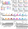 Figure 1
Functional testing of variants selected by thiocyanate enrichment screen. (A and B) Variants were expressed as scDbs. 5 × 10⁴ primary human T cells were cocultured overnight with 2.5 × 10⁴ T2A3 target cells pulsed with (A) 10 nM or (B) varying concentrations of the p53 R175H or R175WT 9mer peptide in the presence of 1 nM scDb. (A) IFN-γ production and (B) percent cytotoxicity compared with no scDb control by CellTiterGlo assay (n = 3 biological replicates, single experiment). (C and D) 5 × 10⁴ T cells were cocultured with 2.5 × 10⁴ luciferase+ KMS26 target cells (p53 R175H or p53 KO) and scDb for 18 hours. (C) IFN-γ production. (D) Percent cytotoxicity measured by SteadyGlo assay (n = 3 biological replicates, single experiment). All data are shown as the mean ± SD. (E and F) Top variants were combined into double-mutant scDbs. (E) scDb was applied to plates coated with CD3ε/δ heterodimer, or R175H/A2 or R175WT/A2 monomer. Relative binding was calculated by normalizing mean monomer binding to mean CD3 binding (n = 3 technical replicates, single experiment). (F) 3 × 10⁴ T cells were cocultured with 1.5 × 10⁴ KMS26 target cells in the presence of scDb for 19 hours. Percent cytotoxicity by SteadyGlo assay (n = 3 biological replicates per condition, single experiment).
Variants with similar or improved potency compared with the original H2 were combined in double mutant scDbs to determine whether any pair of changes further increased bispecific antibody performance. All single and double mutants tested had increased binding to the R175H/A2 monomer compared with the original H2 scDb by ELISA, suggesting increased binding affinity (Figure 1E). Double mutants also yielded increased T cell activation in coculture with KMS26 target cells compared with H2 scDb (Figure 1F). However, 12 of the 17 variant combinations led to an increase in off-target cytotoxicity against the isogenic control cells not expressing p53 R175H (Figure 1F).
Selected H2 variants have higher binding affinity. To determine how H2 variants differed in binding affinity, 3 scDbs with improved cytotoxicity and cytokine production were selected for affinity characterization by surface plasmon resonance (SPR): F53SCDRL2, Y57ICDRH2, and F53SCDRL2 Y57ICDRH2. The original H2 scDb had an affinity (KD) of 29.5 nM (Figure 2A and Table 1). The F53S, Y57I, and F53S Y57I variants each had higher affinities than the original H2 scDb (12.9, 6.8, and 3.3 nM, respectively), confirming that the thiocyanate elution method selected for strong binders. No variant had appreciable binding to the p53 WT/HLA-A2 monomer by SPR (Supplemental Figure 3A). All H2 bispecific antibodies had similar stability by differential scanning fluorimetry and solubility (Supplemental Figure 3, B–E, and Supplemental Methods). For convenience, these H2 scDb variants will be referred to by their affinity: H212.9, H26.8, and H23.3. The original H2 scDb will henceforth be referred to as H229.5.
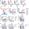 Figure 2
Higher-affinity bispecific antibodies have improved activity in vitro. (A) Single-cycle binding kinetics for each H2 variant scDb binding to the p53 R175H/HLA-A*02:01 monomer measured by SPR. Dashed line indicates curve fit. (B) Coculture design testing scDb cytotoxicity against isogenic p53R175H and p53–/– cell lines or p53R175WT cell lines. (C and D) 5 × 10⁴ T cells were cocultured with 2.5 × 10⁴ KMS26 target cells in the presence of 0–3 nM scDb for 20 hours. (C) Percent cytotoxicity compared with no scDb was measured by SteadyGlo. (D) IFN-γ production (n = 3, representative of 3 independent experiments, curve fitting and EC50s determined by 5-parameter log fit). (E) 1 × 10⁴ GFP+ TYK-nu target cells were incubated with 2 × 10⁴ T cells and 0.01 nM scDb and counted by live-cell imaging (n = 3, representative of 2 independent experiments). (F) Mean GFP+ target cells at 120 hours (ordinary 1-way ANOVA with Tukey’s multiple comparisons). (G and H) 5 × 10⁴ T cells were cocultured with 2.5 × 10⁴ target cells and 0.01 nM scDb for 20 hours. Percent cytotoxicity compared with no scDb was measured by SteadyGlo (n = 3, representative of 3 independent experiments, 1-way ANOVA with Holm-Šidák multiple comparisons). All data are shown as the mean ± SD.
Higher-affinity H2 variants elicit stronger T cell responses against cancer cells in vitro. We next compared the H2 scDb variants’ activity against various patient-derived cancer cell lines expressing endogenous levels of HLA-A2 and p53R175H by coculture with primary human T cells (Figure 2B). All 3 higher-affinity variants had improved cytotoxicity (lower EC50s) against KMS26 cells compared with the original H229.5 scDb (Table 2 and Figure 2, C and D). Indeed, the highest-affinity H23.3 scDb had a 7 times lower EC50 than H229.5 (4.2 vs. 31 pM). In addition, while the original H2 scDb had limited activity against TYK-nu, an ovarian cancer cell line that carries an endogenous TP53R175H mutation and an HLA-A*02:01 allele (1.5 copies of R175H/A2 per cell) (5), the higher-affinity variants all mediated cytotoxicity against TYK-nu without off-target activity against its isogenic control cell line (CRISPR-mediated KO of the TP53 gene) (Figure 2, E and F). H212.9, H26.8, and H23.3 all elicited significantly improved cytotoxicity against endogenous antigen levels at very low scDb concentrations (0.01 nM) without cross-reactivity in isogenic TP53-KO control cell lines (Figure 2G). We also tested scDb activity against NALM6 cells edited to express p53R175H via CRISPR-mediated knockin (NALM6R175H, 1.3 copies of R175H/A2 MANA per cell) (37) compared with the isogenic control parental NALM6 (p53R175WT) (Figure 2H). IFN-γ release similarly increased as affinity increased for each of the H2 variants (Supplemental Figure 4). Taken together, increasing H2 MANAbody affinity yielded increased T cell activation against cells expressing endogenous levels of the p53 MANA without an increase in off-target activity.
Increasing bispecific antibody affinity improved tumor control in vivo. We chose the H26.8 variant for in vivo testing given its potency in the absence of any detectable off-target toxicity at low antibody concentrations (10 pM; Figure 2, G and H). We previously reported that the original H229.5 MANAbody can limit KMS26 growth in a NOD.Cg-PrkdcscidIl2rgtm1Wjl/SzJ (NSG) mouse hematologic xenograft model (5). We used a considerably more challenging variation of this model in the current study. Specifically, we employed a delayed treatment model in which mice were treated 7 days after tumor inoculation with 3.5 × 10⁵ KMS26 cells i.v. (Figure 3A). Mice were inoculated with 107 primary human T cells i.v. and treated with a low dose of bispecific antibody (0.075 mg/kg/d vs. previously reported 0.15–0.3 mg/kg/d) (5) delivered via a 14-day continuous release pump i.p. Measurement of tumor burden by bioluminescence imaging showed that mice treated with H26.8 had significantly lower tumor burden at the end of treatment compared with mice treated with H229.5 (day 14, P = 0.0317, n = 5 per group; Figure 3, B and C, and Supplemental Figure 5A). Moreover, tumors were undetectable in more mice at day 21 in the H26.8 variant group (3/5 mice) compared with those in the H229.5 group (0/5 mice) (Supplemental Figure 5A).
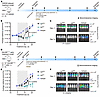 Figure 3
Higher-affinity H26.8 has improved tumor control in vivo. (A) Delayed treatment model design. NSG mice were inoculated with 3.5 × 10⁵ KMS26 cells i.v. 7 days prior to treatment. On treatment day 0, mice received 107 human T cells i.v. and surgically implanted micro-osmotic continuous release pumps i.p. scDb was dosed at 0.075 mg/kg/d for 14 days. Tumor burden was measured by bioluminescence imaging. (B) Tumor burden and (C) bioluminescent images at days –1 and 14 (average radiance, means compared at day 14 by Mann-Whitney test, n = 5 per group; data are shown as mean ± SD). (D) Early treatment model design. NSG mice were inoculated with 5 × 10⁵ NALM6R175H cells and 107 human T cells i.v. 2 days prior to treatment start. On treatment day 0, mice received surgically implanted continuous release pumps i.p. scDb was dosed at 0.075 mg/kg/d for 14 days. (E) Tumor burden and (F) bioluminescent images of mice at days –1 and 14 (average radiance, means compared at day 14 by Mann-Whitney test, n = 5 per group, representative of 2 experiments; data are shown as mean ± SD). Pump treatment period indicated by gray boxes.
We additionally tested H26.8 in an aggressive tumor model employing NALM6R175H cells, which divide rapidly in mice. Mice were treated with a low scDb dose (0.075 mg/kg/d) via continuous release pump 2 days after receiving 5 × 10⁵ NALM6R175H cells and 1 × 10⁷ human T cells i.v. (Figure 3D). In this model, the original H229.5 scDb did not control tumor growth, while mice treated with the higher-affinity H26.8 scDb induced tumor regression that lasted the duration of infusion (14 days). Tumor burdens on day 14 were significantly lower in the mice treated with H26.8 versus the original H229.5 (day 14, P = 0.0079, n = 5 per group; Figure 3, E and F, and Supplemental Figure 5B). Treatment with bispecific antibodies was well tolerated with stable body weights in all animals over the duration of treatment (Supplemental Figure 5C).
Increasing H2 scFv affinity does not improve CAR function. Low antigen density presents a challenge for CAR T cell therapies and is a major limitation for MANA-targeting CARs (31). While some groups have shown that increased scFv affinity increases CAR activation at low antigen density (38, 39), others have shown that higher-affinity CAR scFvs have reduced T cell proliferation and activation (40–42). In most cases, comparisons of different affinities involved unrelated scFvs, complicating conclusions about the reasons for the differences in performance. Because the H2 variants all recognize the identical, precisely defined epitope and differ by only 1 or 2 amino acids, they provide an ideal opportunity to assess the influence of affinity on CAR T cell performance.
To test the higher-affinity H2 variants in CAR T cells, we first generated second-generation CD28-CD3ζ CAR T cells (2G CAR T cells) (43) by CRISPR knockin at the CD3G locus in primary human T cells (Supplemental Figure 6, A and B). We chose this method of creating CAR T cells, rather than lentivirus transduction, to ensure similar expression of each construct. Edited CAR T cell frequency was analyzed by flow cytometry with no significant differences in CD4+ or CD8+ T cell frequencies or expression levels across CAR types (Supplemental Figure 6, C–F).
The 2G CAR T cells were then cocultured with TAP-deficient T2 target cells pulsed with exogenous p53 R175H 9mer peptide to test for T cell activation at different antigen densities. At 24 hours, all higher-affinity H2 2G CAR T cells had lower IFN-γ production compared with the original H2 2G CAR (Supplemental Figure 7, A and B), which was precisely the opposite of what was observed with the corresponding bispecific antibodies (Figure 2, C and D). This decreased cytokine production with increasing scFv affinity was observed at high peptide pulsing conditions (1–10 μM), while lower peptide concentrations elicited minimal 2G CAR T cell activation for all 2G CARs (Supplemental Figure 7, A and B).
CD3γ TCR CARs improve efficacy at low antigen densities. Second-generation CARs are known to have limited activity when antigen levels drop below 100–1,000 copies per cell (44), making targeting MANAs with traditional CARs challenging. The H2 2G CARs constructed with the original H2 MANAbody had limited activity at low p53 R175H/A2 antigen density, consistent with previous work (37). One strategy to increase CAR sensitivity is to append the scFv to the TCR complex, which may increase activity by better activating canonical immunological synapse signaling (37, 45–47). We next appended the H2 scFv to the CD3γ subunit in the TCR complex to generate a CD3γ-TRuC T cell (TRuC: T cell receptor fusion construct) (45) (Supplemental Figure 6, A, B, and G–J). We observed increased T cell activation with the CD3γ-TRuC compared with the 2G CAR (Supplemental Figure 7, C and D, compared with A and B). However, a similar trend of decreasing sensitivity with increasing scFv affinities was observed for the CD3γ-TRuC T cells (Supplemental Figure 7, C and D).
STAR T cells mimic native TCR structure and have increased tetramer binding with higher affinity. We and others have shown that antigen sensitivity can be improved using TCR-based CAR T cells called STARs (synthetic TCR and antigen receptor T cells) (37, 46, 47). STARs consist of an engineered receptor where the CAR scFv is divided into variable heavy and light chains appended to the TCRα and TCRβ constant domains. To determine whether we could improve CAR T cell MANA targeting with higher-affinity STARs, we generated H2 STAR T cells with CRISPR editing at the TRAC locus to append the H2 scFv variable light chain and variable heavy chain to TCRα and TCRβ constant domains, respectively, using a modified murine TRAC and TRBC to maximize TCR assembly (Figure 4, A and B) (37, 48). We also evaluated a standard TCR (AV6/BV11) derived from a patient harboring a TP53R175H mutant cancer (49). The only difference between this TCR and the STAR was that the variable chains of AV6/BV11 TCR were substituted for the variable chains of the H2 scFv. Both were appended to the constant regions of the modified murine TCR (Figure 4, A and B) (37).
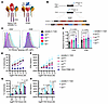 Figure 4
Higher-affinity STARs have increased tetramer binding but reduced antigen sensitivity. (A) Illustration of the patient-derived TCR AV6/BV11 compared with the H2 STAR, where the H2 variable heavy (VH) and variable light (VL) chains are linked to the TCRα constant domain (TRAC) and TCRβ constant domain (TRBC), respectively. (B) Transgenic T cells were generated by CRISPR knock-out at TRBC1/TRBC2 and knockin at TRAC using dsDNA homology-directed repair templates (HDRTs). HA, homology arm. (C) T cells expressing STARs were characterized by flow cytometry. Representative histograms of p53 R175H/HLA-A2 tetramer bound to edited (CD3+ tetramer) CD4+ and CD8+ T cells are shown. (D) Tetramer staining on CD4+ and CD8+ T cells was compared across n = 5 independent experiments by normalizing tetramer MFI to the tetramer MFI for CD4+ AV6/BV11 TCR T cells for each experiment (2-way ANOVA with Dunnett’s multiple comparisons to H229.5). (E–H) T cell activation at varying antigen densities was tested in a peptide-pulsing coculture. 8 × 10³ edited T cells were cocultured with 4 × 10⁴ peptide-pulsed T2 cells for 24 hours. (E) T cell–mediated cytotoxicity was measured by CellTiterGlo viability assay. (F) IFN-γ production. (G) Cytotoxicity and (H) IFN-γ were compared at selected concentrations from E and F (1-way ANOVA with Dunnett’s multiple comparisons, n = 3 biological replicates, representative of 3 independent experiments). All data are shown as the mean ± SD.
To confirm that the variant STAR receptors were able to bind to the p53 R175H MANA, we measured p53 R175H/A2 tetramer binding by flow cytometry. Relative tetramer binding increased with increasing receptor affinity for each variant in both CD4+ and CD8+ subsets, with greater differences in tetramer binding observed in CD4+ T cells than in CD8+ T cells (Figure 4, C and D, and Supplemental Figure 8, A–F). There was no difference in receptor expression, as indicated by CD3 staining (Supplemental Figure 8, B and E). All H2 STAR variants had increased tetramer binding compared with the patient-derived TCR AV6/BV11, which is consistent with TCRs’ lower affinity compared with antibodies (44, 50). We also assessed the purity of edited STAR T cells after bead-based positive selection for T cells expressing truncated nerve growth factor receptor (tNGFR; a marker for transgene expression) and showed that edited cells did not have differences in CD4+ or CD8+ T cell frequencies (Supplemental Figure 8, C and D).
Increasing CAR affinity does not improve STAR T cell function. In a coculture with peptide-pulsed T2 target cells, H2 STAR T cell–mediated cytotoxicity and IFN-γ production decreased as receptor affinity increased (Figure 4, E and F). This decreased T cell activity with increasing CAR affinity was seen both at supraphysiologic antigen densities (1E-5 M peptide) and at low antigen density (1E-9 M peptide, tens of copies per cell) (16). The lower-affinity AV6/BV11 TCR T cells had the highest sensitivity and activity of all T cells tested (Figure 4, E–H).
We then tested the H2 variant STAR T cells’ activity against cell lines with the endogenous TP53R175H mutation. In cocultures with NALM6R175H cells, the higher-affinity H23.3 STAR T cells killed fewer target cells (Figure 5, A and B) and produced less IFN-γ (Figure 5C) than the lower-affinity H229.5 STAR T cells, again the opposite of the analogous bispecific antibodies (Figure 2, C–H). We additionally employed a multiple-stimulation assay (MSA) in which STAR T cells were rechallenged with KMS26 target cells every 2 days (Supplemental Figure 9A). The H23.3 STAR T cells did not reduce target cell numbers after the first rechallenge, unlike the lower-affinity H229.5 STAR T cells or the AV6/BV11 TCR cells, which continued to kill target cells upon multiple challenges (Figure 5D). Similar results were observed in an MSA with NALM6 target cells (Supplemental Figure 9B).
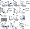 Figure 5
STAR activity decreases with increasing receptor affinity. (A) 5 × 10⁴ edited T cells were cocultured with 1 × 10⁵ GFP-expressing NALM6R175H or NALM6WT target cells for 2 days. (B and C) 1.2 × 10⁴ edited T cells were cocultured with 2.4 × 10⁴ KMS26 target cells for 24 hours. (B) Percent cytotoxicity by SteadyGlo assay compared with the TRAC/TRBC-KO condition. (C) IFN-γ production (n = 3 per condition, 2 independent experiments, 2-way ANOVA with Dunnett’s multiple comparisons). (D–F) STAR T cell multiple stimulation assay. 4 × 10³ edited T cells were cocultured with 1.6 × 10⁴ KMS26 target cells in replicate plates and rechallenged with 3.2 × 10⁴ target cells or assayed by flow cytometry on days 2, 4, and 6. (D) Live GFP+ target cells; (E) CellTrace Violet (CTV) dilution, indicative of cell division, in NGFR+-edited T cells relative to day 2; and (F) counts of NGFR+ T cells (n = 3 per condition, 3 independent experiments). (G–I) T cell killing of KLE target cells was observed by live-cell imaging (aggregate of 3 independent experiments per T cell type, Brown-Forsythe and Welch’s ANOVA with Dunnett’s T3 multiple comparisons to H23.3). (G) Time from T cell interaction to target cell blebbing (arrows indicate T cells). (H) Time from target cell blebbing to T cell detachment. (I) Total duration of interaction. (J–L) 5 × 10⁴ edited T cells were cocultured with 1 × 10⁵ NALM6R175H or NALM6WT target cells for 17 hours and assayed by flow cytometry (n = 3 per condition, 2 independent experiments). (J) Percent CD69+ CD137+ of live T cells (2-way ANOVA with Dunnett’s multiple comparisons to H229.5). (K) Percent IFN-γ+ of live T cells (2-way ANOVA with Dunnett’s multiple comparisons to H229.5). (L) MFI of IFN-γ in CD8+ T cells (ordinary 1-way ANOVA with Šidák’s multiple-comparison test to H229.5). All data are shown as the mean ± SD.
Proliferation analysis demonstrated that fewer higher-affinity STAR T cells divided over the 8-day MSA than did the lower-affinity STAR T cells (Figure 5, E and F, and Supplemental Figure 9, D–F) despite persistence of the H23.3 STAR T cells (Supplemental Figure 9C). Quantitatively, KMS26 cells stimulated only a 4.7-fold expansion in H23.3 STAR T cells compared with 32.7- and 22.1-fold expansions with AV6/BV11 TCR T cells and H229.5 STAR T cells, respectively (Supplemental Figure 9F). Finally, higher-affinity H23.3 STAR T cells produced less IFN-γ in both MSAs (Supplemental Figure 9G).
Investigations into the basis for the lower cytotoxicity of the higher-affinity STAR T cells. We sought to understand the mechanistic basis for the decreased cytotoxicity observed with the higher-affinity STAR T cells. One potential explanation for this decrease was less efficient target cell killing per T cell. To test this hypothesis, we performed time-lapse live-cell imaging of the H23.3 and H229.5 STAR T cells in coculture with KLE target cells expressing very low levels of the p53 R175H MANA (1.3 copies/cell) (5). KLE cells were chosen for these experiments because they are adherent and therefore more suitable for live-cell imaging than nonadherent cells, such as KMS26 or NALM6 cells. Similarly to KMS26 or NALM6 cells, KLE cells were less susceptible to killing by the high-affinity H23.3 STAR T cells than by the lower-affinity H229.5 STAR T cells (Supplemental Figure 10, A–C). There was no difference in CAR T cell activity against isogenic control KLE p53–/– cells compared with TRAC/TRBC-KO T cells (Supplemental Figure 10B). Additionally, after 6 days of coculture with KLE cells, the high-affinity H23.3 STAR T cells had less expression of the late activation marker HLA-DR and less proliferation than the lower-affinity H229.5 STAR T cells (Supplemental Figure 10, D–F). The relative order of the extent of cytotoxicity to target cells and cell division of T cells obtained with the various STAR T cells and the AV6/BV11 TCR were identical with all 3 target cell types (Supplemental Figure 10, A–F, Supplemental Figure 9, B–F, and Figure 5, A, B, and D–F).
We further examined the STAR T cell cytotoxic activity by manually tracking individual T cell interactions with KLE target cells with live-cell imaging. We observed T cell cytotoxic activity against KLE cells from T cell engagement with target cell–to–target cell blebbing (Figure 5G). There were no significant differences in time to target cell blebbing among H229.5 STAR, H23.3 STAR, and the AV6/BV11 TCR cells (Figure 5G). We also considered the possibility that the higher-affinity STAR T cells remained adhered to the target cells for longer, thereby limiting their ability to detach from one target cell and reform a synapse with another target cell. To test this possibility, we measured the interaction time between the various T cells and the target cells. We found no significant differences in time to detachment from dying cells or in total interaction time between the 3 T cell types and their target cell (Figure 5, H and I). We considered whether the higher-affinity STAR T cells themselves underwent cell death during or after target cell interactions, thereby limiting their killing potential, but we did not observe any instances of T cell apoptosis during or after a target cell interaction in the time-lapse experiments.
The data described in Figure 5, G–I, suggested that, once a T cell bound to a target cell, the resulting cytotoxic effect was similar regardless of receptor affinity. We therefore considered whether reduced activity of the high-affinity H23.3 STAR T cells was due to decreased activation. To assess this hypothesis, we measured T cell expression of activation markers CD69 and CD137 after coculture with NALM6 cells for 17 hours (Figure 5J). There was indeed a striking difference in activation, with the activation of the AV6/BV11 TCR and lower-affinity H229.5 STAR T cells much higher than that of the high-affinity H23.3 STAR T cells (Figure 5J). These differences were particularly evident in CD8+ T cells but were also observed in CD4+ T cells (Figure 5J). Little or no activation was observed when the same T cell types were cultured with NALM6R175WT cells, supporting the specificity of the CD69/CD137 marker upregulation (Figure 5J). Similarly, fewer H23.3 STAR T cells upregulated CD69 and CD25 in a 48-hour coculture with NALM6 target cells (Supplemental Figure 11, A, C, and E). In contrast, in parallel cocultures with scDbs, CD69/CD25 upregulation increased with increasing scDb affinity (Supplemental Figure 11, B and D).
As an additional measure of activation, we assessed the number of cells expressing IFN-γ in the same 17-hour cocultures. The fractions of AV6/BV11 TCR and lower-affinity H229.5 STAR CD8+ T cells that expressed IFN-γ were significantly higher than that of the high-affinity H23.3 STAR CD8+ T cells (Figure 5K). Not only was the fraction of IFN-γ–expressing cells lower, but the amount of IFN-γ per cell was significantly decreased (Figure 5L). Finally, though increasing receptor affinity had a greater effect on CD4+ T cell tetramer binding compared with CD8+ T cells (Figure 4, C and D, and Supplemental Figure 8, E and F), CD4+ cells secreted much lower levels of IFN-γ than the corresponding CD8+ cells, regardless of the receptor types (Figure 5K).
T cell activation-induced cell death (AICD) and exhaustion are 2 potential contributors to the decreased tumor cell killing and proliferation we observed in the higher-affinity STAR T cells. To examine this, we conducted an extended MSA where T cells were challenged with Nalm6R175H cells every 2 days and characterized by flow cytometry on day 14 (Supplemental Figure 12, A–C). As seen in single-stimulation experiments, fewer H23.3 STAR T cells expressed early activation markers (CD69 and CD25; Supplemental Figure 12D). There was no indication of increased AICD in the higher-affinity STAR T cells. Instead, H23.3 STAR T cells were less likely to have FasL expression (Supplemental Figure 12F). Across conditions, most T cells expressed Fas, which we attributed to exposure to IL-2 in the MSA and IL-2/IL-7 during T cell expansion (51). Finally, we assessed whether T cells that had been activated over the MSA had differing expression of inhibitory markers, such as Lag3, PD1, and Tim3, suggesting T cell exhaustion. We found that inhibitory marker expression corresponded with T cell activation, as expected. H23.3 cells were less likely to express HLA-DR or any inhibitory markers on day 14 of the MSA (Supplemental Figure 12, H and J). Even among HLA-DR+ cells, the higher-affinity H23.3 STAR T cells were less likely to express inhibitory markers (Supplemental Figure 12L), again suggesting that these cells were not activated to the same degree as the H229.5 STAR T cells or AV6/BV11 TCR. Importantly, we did not observe signs of tonic signaling causing T cell activation, or FasL or inhibitory marker expression in the T cell–only condition (Supplemental Figure 12, E, G, I, and K).


-
Discussion
Developing antibodies that recognize MANAs is challenging: the MANAbody must recognize a single amino acid difference in a 9– or 10–amino acid peptide embedded within a much larger HLA groove. It was therefore not clear that the affinity of the originally described H2 MANAbody directed against the p53 R175H/HLA-A2 complex could be improved without sacrificing specificity for the mutant peptide (24). Indeed, a standard affinity maturation approach, even when coupled with structural insights, was not successful. Fortunately, an approach described decades ago — employing thiocyanate elution — allowed us to develop higher-affinity variants. When incorporated into T cell–engaging bispecific antibody format, these high-affinity variants proved more potent in vitro and in mice. This is consistent with previous work showing that increasing bispecific antibody affinity improves potency (18, 24, 25). In this study, we used 2 hematologic models to differentiate our bispecific antibody affinity variants’ activity in vivo. How these antibodies perform against solid tumors remains of significant interest given p53 R175H’s frequency in common solid organ cancers and the additional challenges of targeting solid tumors with immunotherapies.
We expected the higher-affinity variants to lead to more potent engineered T cell therapies. This expectation was consistent with some studies, though others have shown that lower-affinity CAR T cells have improved performance (16, 38–40). It was difficult to extrapolate from these previous studies of CAR T cells employing various affinity antibodies because all were directed against cells bearing 100- to 1,000-fold higher densities of the target antigen. In fact, we have previously shown that standard CAR T cells cannot recognize cells with antigen densities as low as typically found in MANAs (37). To recognize the p53 R175H/A2 MANA, the CAR scFv had to be embedded within the endogenous TCR rather than expressed as a fusion with CD28 or other costimulatory domains (37). The hybrid TCR/antibody-based engineered T cells we developed in previous publications (37, 46, 47), described as STARs or HIT CARs (HIT: HLA-independent TCRs), enabled us to rigorously determine the relationship between affinity and function using cell-based and bispecific antibody-based therapeutic approaches.
The results of these experiments were definitive: higher affinity led to better function of the bispecific antibodies, as assessed by cytotoxicity, cytokine release, and T cell activation (Figures 1–3). Yet, higher affinity led to worse function of cell-based therapeutic approaches using the identical assays (Figures 4 and 5). Moreover, the specificity of the observed effects in both bispecific antibodies and T cells was meticulously controlled with isogenic target cells differing only in the single amino acid at codon 175 of the TP53 gene or with TP53 KO.
Several possible mechanisms could underlie the decreased potency of the STAR T cells bearing the higher-affinity antibodies. Differences in cell surface expression, epitope binding, or stability could potentially impact CAR function. It was therefore important to note that the variants differed by only 1 or 2 amino acids and had similar solubilities and melting temperatures (Supplemental Figure 3, B–E). We documented that the differences in potency were not due to decreased CAR expression levels on the cell surface (Figure 4C and Supplemental Figure 8, B–F). This similarity in expression across the different T cells compared in this study was expected given that they all were created through genetic knockin at the same locus rather than through methods leading to overexpression or varied levels of expression. We also demonstrated that the higher-affinity antibodies on the STAR T cells were properly folded, as evidenced by R175H/HLA-A2 tetramer binding (Figure 4C and Supplemental Figure 8, B–F).
Another possibility for the decreased potency was that higher-affinity CAR T cells had increased cell death. For example, receptor–ligand interactions could potentially result in trogocytosis — pulling target molecules from the target cell membrane to the T cell membrane — resulting in nonlethal target cell interactions and potential CAR T cell fratricide, though the limited activation seen with H23.3 argues against this mechanism (42, 52). Alternatively, tonic signaling or T cell exhaustion (due to high-affinity receptor interactions or tonic signaling) could limit the H23.3 T cells’ activity and lead to increased T cell death. In general, we did not observe signs of increased cell death in high-affinity CAR populations; high-affinity STARs persisted in extended cocultures and in multiple stimulation assays (Figure 5F, Supplemental Figure 9C, and Supplemental Figure 10D) and did not express markers of AICD (Supplemental Figure 12, F and G). We did not conclude that the higher-affinity STAR T cells were exhausted given that even HLA-DR+ H23.3 STAR T cells had less Lag3, PD1, and Tim3 expression than activated H229.5 or AV6/BV11 T cells.
To assess whether there was less efficient cell killing per T cell, we performed time-lapse live-cell imaging of the various STAR T cells in coculture with target cells. There were no significant differences noted in these experiments as far as the average time to target cell death (blebbing), the average interaction time between T cells and target cells, or the average time to T cell detachment from the target cells once killed (Figure 5, G–I, and Table 3). In fact, the only functional difference between the STAR T cells bearing the higher-affinity receptors was in their level of activation, as assessed by typical activation markers (Figure 5J and Supplemental Figures 10–12). These experiments support the idea that the decreased function in the higher-affinity STAR T cells is primarily related to decreased activation (or quality of activation; e.g., CD69 expression without CD25 upregulation or proliferation). Once a threshold for activation is met, we hypothesize that all the STAR T cells would then have similar abilities to kill target cells. According to this hypothesis, we expect that some T cells interacting with target cells would not form mature synapses or adhere to target cells because they weren’t sufficiently activated. We could not test this in our live-cell imaging experiments because the technique only allowed us to visualize and track relatively stable interactions between the T cells and target cells. Transient interactions, shorter than the 30-second interval between images or at the molecular level, would not have been observed in these experiments.
Future work to evaluate why higher-affinity receptors yield less frequent activation are planned, including testing how high-affinity STARs compare to TCRs as mechanoreceptors with single-molecule biophysical techniques (optical tweezers) (53, 54). Further transcriptomic or phosphoproteomic comparison of activated STAR T cells may yield additional insight into how affinity affects T cell function; however, these studies primarily address the cells’ response to activation rather than how receptor differences determine T cell activation.
These results have potentially important implications for immunotherapies based on MANAs or other low-antigen-density targets. Increasing affinity of the antibody for the target had predictable and expected effects on function when the immunotherapeutic agent was a soluble T cell–engaging bispecific antibody. This is consistent with decades of research showing that high-affinity drugs perform better than their lower-affinity counterparts, provided specificity is maintained (16, 18, 24–26). But for cellular immunotherapies, conclusions about relationship between performance and affinity have varied, as observed in our study and in previous studies of CAR T cells directed against cells expressing high levels of antigens (39–41, 55, 56). CAR T cells thereby present a Goldilocks situation, wherein the optimum antibody affinity to be used cannot be predicted — a much more challenging case for clinical development than encountered with soluble antibodies. Perhaps a better understanding of the complex interactions between CAR T cells and their targets, and the mechanisms through which activation and cytotoxicity is achieved (24, 55, 57–60), will address this quandary.


Copyright © 2026 American Society for Clinical Investigation
ISSN: 0021-9738 (print), 1558-8238 (online)