
Advertisement
Research ArticleAIDS/HIVImmunology
Open Access |  10.1172/JCI192575
A favorable follicular helper CD4+ T cell programming characterizes neutralization activity in chronic HIV infection
Eirini Moysi,1 Ashish A. Sharma,2 Sijy O’Dell,3 Spiros Georgakis,4 Perla Mariana Del Rio Estrada,2,5 Ghneim Khader,2 Alonso Arana,2 Fernanda Torres-Ruiz,5 Mauricio González Navarro,6 Yara Andrea Luna Villalobos,5 Santiago Avila Rios,5 Gustavo Reyes-Teran,7 Margaret H. Beddall,8 Sung Hee Ko,9 Frida Belinky,9 Michail Orfanakis,4 Laurence de Leval,4 Ana B. Enriquez,2 Clarisa M. Buckner,10 Susan Moir,10 Helen Lindsay,11 Raphael Gottardo,11 Nicole Doria-Rose,3 Eli A. Boritz,9 John R. Mascola,3,12 Rafick-Pierre Sekaly,2 Richard A. Koup,1 and Constantinos Petrovas1,4
1Tissue Analysis Core, Immunology Laboratory, Vaccine Research Center, National Institute of Allergy and Infectious Diseases (NIAID), NIH, Bethesda, Maryland, USA.
2Pathology Advanced Translational Research Unit, Department of Pathology, Emory University School of Medicine, Atlanta, Georgia, USA.
3Virology Laboratory, Vaccine Research Center, NIAID, NIH, Bethesda, Maryland, USA.
4Department of Laboratory Medicine and Pathology, Institute of Pathology, Lausanne University Hospital and University of Lausanne, Lausanne, Switzerland.
5Centro de Investigación en Enfermedades Infecciosas, Instituto Nacional de Enfermedades Respiratorias “Ismael Cosío Villegas,” Mexico City, Mexico.
6Centro de Investigación en Enfermedades Infecciosas, Instituto Nacional de Enfermedades Respiratorias “Ismael Cosío Villegas,” Subdireccion de Otorrinolaringologia, Instituto Nacional de Rehabilitación “Luis Guillermo Ibarra,” Mexico City, Mexico.
7Institutos Nacionales de Salud y Hospitales de Alta Especialidad, Secretaría de Salud de México, Mexico City, Mexico.
8ImmunoTechnology Section, Vaccine Research Center,
9Virus Persistence and Dynamics Section, Vaccine Research Center, and
10Laboratory of Immunoregulation, NIAID, NIH, Bethesda, Maryland, USA.
11Biomedical Data Science Center, Lausanne University Hospital and Lausanne University, Lausanne, Switzerland.
12ModeX Therapeutics, Weston, Massachusetts, USA.
Address correspondence to: Constantinos Petrovas, Department of Laboratory Medicine and Pathology, Institute of Pathology, Lausanne University Hospital and University of Lausanne, Bureau BU25-04-237, Rue du Bugnon 25, CH–1011 Lausanne, Switzerland. Phone: 301.761.6993; Email: Konstantinos.Petrovas@chuv.ch.
Find articles by Moysi, E. in: PubMed | Google Scholar
1Tissue Analysis Core, Immunology Laboratory, Vaccine Research Center, National Institute of Allergy and Infectious Diseases (NIAID), NIH, Bethesda, Maryland, USA.
2Pathology Advanced Translational Research Unit, Department of Pathology, Emory University School of Medicine, Atlanta, Georgia, USA.
3Virology Laboratory, Vaccine Research Center, NIAID, NIH, Bethesda, Maryland, USA.
4Department of Laboratory Medicine and Pathology, Institute of Pathology, Lausanne University Hospital and University of Lausanne, Lausanne, Switzerland.
5Centro de Investigación en Enfermedades Infecciosas, Instituto Nacional de Enfermedades Respiratorias “Ismael Cosío Villegas,” Mexico City, Mexico.
6Centro de Investigación en Enfermedades Infecciosas, Instituto Nacional de Enfermedades Respiratorias “Ismael Cosío Villegas,” Subdireccion de Otorrinolaringologia, Instituto Nacional de Rehabilitación “Luis Guillermo Ibarra,” Mexico City, Mexico.
7Institutos Nacionales de Salud y Hospitales de Alta Especialidad, Secretaría de Salud de México, Mexico City, Mexico.
8ImmunoTechnology Section, Vaccine Research Center,
9Virus Persistence and Dynamics Section, Vaccine Research Center, and
10Laboratory of Immunoregulation, NIAID, NIH, Bethesda, Maryland, USA.
11Biomedical Data Science Center, Lausanne University Hospital and Lausanne University, Lausanne, Switzerland.
12ModeX Therapeutics, Weston, Massachusetts, USA.
Address correspondence to: Constantinos Petrovas, Department of Laboratory Medicine and Pathology, Institute of Pathology, Lausanne University Hospital and University of Lausanne, Bureau BU25-04-237, Rue du Bugnon 25, CH–1011 Lausanne, Switzerland. Phone: 301.761.6993; Email: Konstantinos.Petrovas@chuv.ch.
Find articles by
Sharma, A.
in:
PubMed
|
Google Scholar
|

1Tissue Analysis Core, Immunology Laboratory, Vaccine Research Center, National Institute of Allergy and Infectious Diseases (NIAID), NIH, Bethesda, Maryland, USA.
2Pathology Advanced Translational Research Unit, Department of Pathology, Emory University School of Medicine, Atlanta, Georgia, USA.
3Virology Laboratory, Vaccine Research Center, NIAID, NIH, Bethesda, Maryland, USA.
4Department of Laboratory Medicine and Pathology, Institute of Pathology, Lausanne University Hospital and University of Lausanne, Lausanne, Switzerland.
5Centro de Investigación en Enfermedades Infecciosas, Instituto Nacional de Enfermedades Respiratorias “Ismael Cosío Villegas,” Mexico City, Mexico.
6Centro de Investigación en Enfermedades Infecciosas, Instituto Nacional de Enfermedades Respiratorias “Ismael Cosío Villegas,” Subdireccion de Otorrinolaringologia, Instituto Nacional de Rehabilitación “Luis Guillermo Ibarra,” Mexico City, Mexico.
7Institutos Nacionales de Salud y Hospitales de Alta Especialidad, Secretaría de Salud de México, Mexico City, Mexico.
8ImmunoTechnology Section, Vaccine Research Center,
9Virus Persistence and Dynamics Section, Vaccine Research Center, and
10Laboratory of Immunoregulation, NIAID, NIH, Bethesda, Maryland, USA.
11Biomedical Data Science Center, Lausanne University Hospital and Lausanne University, Lausanne, Switzerland.
12ModeX Therapeutics, Weston, Massachusetts, USA.
Address correspondence to: Constantinos Petrovas, Department of Laboratory Medicine and Pathology, Institute of Pathology, Lausanne University Hospital and University of Lausanne, Bureau BU25-04-237, Rue du Bugnon 25, CH–1011 Lausanne, Switzerland. Phone: 301.761.6993; Email: Konstantinos.Petrovas@chuv.ch.
Find articles by O’Dell, S. in: PubMed | Google Scholar
1Tissue Analysis Core, Immunology Laboratory, Vaccine Research Center, National Institute of Allergy and Infectious Diseases (NIAID), NIH, Bethesda, Maryland, USA.
2Pathology Advanced Translational Research Unit, Department of Pathology, Emory University School of Medicine, Atlanta, Georgia, USA.
3Virology Laboratory, Vaccine Research Center, NIAID, NIH, Bethesda, Maryland, USA.
4Department of Laboratory Medicine and Pathology, Institute of Pathology, Lausanne University Hospital and University of Lausanne, Lausanne, Switzerland.
5Centro de Investigación en Enfermedades Infecciosas, Instituto Nacional de Enfermedades Respiratorias “Ismael Cosío Villegas,” Mexico City, Mexico.
6Centro de Investigación en Enfermedades Infecciosas, Instituto Nacional de Enfermedades Respiratorias “Ismael Cosío Villegas,” Subdireccion de Otorrinolaringologia, Instituto Nacional de Rehabilitación “Luis Guillermo Ibarra,” Mexico City, Mexico.
7Institutos Nacionales de Salud y Hospitales de Alta Especialidad, Secretaría de Salud de México, Mexico City, Mexico.
8ImmunoTechnology Section, Vaccine Research Center,
9Virus Persistence and Dynamics Section, Vaccine Research Center, and
10Laboratory of Immunoregulation, NIAID, NIH, Bethesda, Maryland, USA.
11Biomedical Data Science Center, Lausanne University Hospital and Lausanne University, Lausanne, Switzerland.
12ModeX Therapeutics, Weston, Massachusetts, USA.
Address correspondence to: Constantinos Petrovas, Department of Laboratory Medicine and Pathology, Institute of Pathology, Lausanne University Hospital and University of Lausanne, Bureau BU25-04-237, Rue du Bugnon 25, CH–1011 Lausanne, Switzerland. Phone: 301.761.6993; Email: Konstantinos.Petrovas@chuv.ch.
Find articles by Georgakis, S. in: PubMed | Google Scholar
1Tissue Analysis Core, Immunology Laboratory, Vaccine Research Center, National Institute of Allergy and Infectious Diseases (NIAID), NIH, Bethesda, Maryland, USA.
2Pathology Advanced Translational Research Unit, Department of Pathology, Emory University School of Medicine, Atlanta, Georgia, USA.
3Virology Laboratory, Vaccine Research Center, NIAID, NIH, Bethesda, Maryland, USA.
4Department of Laboratory Medicine and Pathology, Institute of Pathology, Lausanne University Hospital and University of Lausanne, Lausanne, Switzerland.
5Centro de Investigación en Enfermedades Infecciosas, Instituto Nacional de Enfermedades Respiratorias “Ismael Cosío Villegas,” Mexico City, Mexico.
6Centro de Investigación en Enfermedades Infecciosas, Instituto Nacional de Enfermedades Respiratorias “Ismael Cosío Villegas,” Subdireccion de Otorrinolaringologia, Instituto Nacional de Rehabilitación “Luis Guillermo Ibarra,” Mexico City, Mexico.
7Institutos Nacionales de Salud y Hospitales de Alta Especialidad, Secretaría de Salud de México, Mexico City, Mexico.
8ImmunoTechnology Section, Vaccine Research Center,
9Virus Persistence and Dynamics Section, Vaccine Research Center, and
10Laboratory of Immunoregulation, NIAID, NIH, Bethesda, Maryland, USA.
11Biomedical Data Science Center, Lausanne University Hospital and Lausanne University, Lausanne, Switzerland.
12ModeX Therapeutics, Weston, Massachusetts, USA.
Address correspondence to: Constantinos Petrovas, Department of Laboratory Medicine and Pathology, Institute of Pathology, Lausanne University Hospital and University of Lausanne, Bureau BU25-04-237, Rue du Bugnon 25, CH–1011 Lausanne, Switzerland. Phone: 301.761.6993; Email: Konstantinos.Petrovas@chuv.ch.
Find articles by Del Rio Estrada, P. in: PubMed | Google Scholar
1Tissue Analysis Core, Immunology Laboratory, Vaccine Research Center, National Institute of Allergy and Infectious Diseases (NIAID), NIH, Bethesda, Maryland, USA.
2Pathology Advanced Translational Research Unit, Department of Pathology, Emory University School of Medicine, Atlanta, Georgia, USA.
3Virology Laboratory, Vaccine Research Center, NIAID, NIH, Bethesda, Maryland, USA.
4Department of Laboratory Medicine and Pathology, Institute of Pathology, Lausanne University Hospital and University of Lausanne, Lausanne, Switzerland.
5Centro de Investigación en Enfermedades Infecciosas, Instituto Nacional de Enfermedades Respiratorias “Ismael Cosío Villegas,” Mexico City, Mexico.
6Centro de Investigación en Enfermedades Infecciosas, Instituto Nacional de Enfermedades Respiratorias “Ismael Cosío Villegas,” Subdireccion de Otorrinolaringologia, Instituto Nacional de Rehabilitación “Luis Guillermo Ibarra,” Mexico City, Mexico.
7Institutos Nacionales de Salud y Hospitales de Alta Especialidad, Secretaría de Salud de México, Mexico City, Mexico.
8ImmunoTechnology Section, Vaccine Research Center,
9Virus Persistence and Dynamics Section, Vaccine Research Center, and
10Laboratory of Immunoregulation, NIAID, NIH, Bethesda, Maryland, USA.
11Biomedical Data Science Center, Lausanne University Hospital and Lausanne University, Lausanne, Switzerland.
12ModeX Therapeutics, Weston, Massachusetts, USA.
Address correspondence to: Constantinos Petrovas, Department of Laboratory Medicine and Pathology, Institute of Pathology, Lausanne University Hospital and University of Lausanne, Bureau BU25-04-237, Rue du Bugnon 25, CH–1011 Lausanne, Switzerland. Phone: 301.761.6993; Email: Konstantinos.Petrovas@chuv.ch.
Find articles by Khader, G. in: PubMed | Google Scholar
1Tissue Analysis Core, Immunology Laboratory, Vaccine Research Center, National Institute of Allergy and Infectious Diseases (NIAID), NIH, Bethesda, Maryland, USA.
2Pathology Advanced Translational Research Unit, Department of Pathology, Emory University School of Medicine, Atlanta, Georgia, USA.
3Virology Laboratory, Vaccine Research Center, NIAID, NIH, Bethesda, Maryland, USA.
4Department of Laboratory Medicine and Pathology, Institute of Pathology, Lausanne University Hospital and University of Lausanne, Lausanne, Switzerland.
5Centro de Investigación en Enfermedades Infecciosas, Instituto Nacional de Enfermedades Respiratorias “Ismael Cosío Villegas,” Mexico City, Mexico.
6Centro de Investigación en Enfermedades Infecciosas, Instituto Nacional de Enfermedades Respiratorias “Ismael Cosío Villegas,” Subdireccion de Otorrinolaringologia, Instituto Nacional de Rehabilitación “Luis Guillermo Ibarra,” Mexico City, Mexico.
7Institutos Nacionales de Salud y Hospitales de Alta Especialidad, Secretaría de Salud de México, Mexico City, Mexico.
8ImmunoTechnology Section, Vaccine Research Center,
9Virus Persistence and Dynamics Section, Vaccine Research Center, and
10Laboratory of Immunoregulation, NIAID, NIH, Bethesda, Maryland, USA.
11Biomedical Data Science Center, Lausanne University Hospital and Lausanne University, Lausanne, Switzerland.
12ModeX Therapeutics, Weston, Massachusetts, USA.
Address correspondence to: Constantinos Petrovas, Department of Laboratory Medicine and Pathology, Institute of Pathology, Lausanne University Hospital and University of Lausanne, Bureau BU25-04-237, Rue du Bugnon 25, CH–1011 Lausanne, Switzerland. Phone: 301.761.6993; Email: Konstantinos.Petrovas@chuv.ch.
Find articles by Arana, A. in: PubMed | Google Scholar
1Tissue Analysis Core, Immunology Laboratory, Vaccine Research Center, National Institute of Allergy and Infectious Diseases (NIAID), NIH, Bethesda, Maryland, USA.
2Pathology Advanced Translational Research Unit, Department of Pathology, Emory University School of Medicine, Atlanta, Georgia, USA.
3Virology Laboratory, Vaccine Research Center, NIAID, NIH, Bethesda, Maryland, USA.
4Department of Laboratory Medicine and Pathology, Institute of Pathology, Lausanne University Hospital and University of Lausanne, Lausanne, Switzerland.
5Centro de Investigación en Enfermedades Infecciosas, Instituto Nacional de Enfermedades Respiratorias “Ismael Cosío Villegas,” Mexico City, Mexico.
6Centro de Investigación en Enfermedades Infecciosas, Instituto Nacional de Enfermedades Respiratorias “Ismael Cosío Villegas,” Subdireccion de Otorrinolaringologia, Instituto Nacional de Rehabilitación “Luis Guillermo Ibarra,” Mexico City, Mexico.
7Institutos Nacionales de Salud y Hospitales de Alta Especialidad, Secretaría de Salud de México, Mexico City, Mexico.
8ImmunoTechnology Section, Vaccine Research Center,
9Virus Persistence and Dynamics Section, Vaccine Research Center, and
10Laboratory of Immunoregulation, NIAID, NIH, Bethesda, Maryland, USA.
11Biomedical Data Science Center, Lausanne University Hospital and Lausanne University, Lausanne, Switzerland.
12ModeX Therapeutics, Weston, Massachusetts, USA.
Address correspondence to: Constantinos Petrovas, Department of Laboratory Medicine and Pathology, Institute of Pathology, Lausanne University Hospital and University of Lausanne, Bureau BU25-04-237, Rue du Bugnon 25, CH–1011 Lausanne, Switzerland. Phone: 301.761.6993; Email: Konstantinos.Petrovas@chuv.ch.
Find articles by Torres-Ruiz, F. in: PubMed | Google Scholar
1Tissue Analysis Core, Immunology Laboratory, Vaccine Research Center, National Institute of Allergy and Infectious Diseases (NIAID), NIH, Bethesda, Maryland, USA.
2Pathology Advanced Translational Research Unit, Department of Pathology, Emory University School of Medicine, Atlanta, Georgia, USA.
3Virology Laboratory, Vaccine Research Center, NIAID, NIH, Bethesda, Maryland, USA.
4Department of Laboratory Medicine and Pathology, Institute of Pathology, Lausanne University Hospital and University of Lausanne, Lausanne, Switzerland.
5Centro de Investigación en Enfermedades Infecciosas, Instituto Nacional de Enfermedades Respiratorias “Ismael Cosío Villegas,” Mexico City, Mexico.
6Centro de Investigación en Enfermedades Infecciosas, Instituto Nacional de Enfermedades Respiratorias “Ismael Cosío Villegas,” Subdireccion de Otorrinolaringologia, Instituto Nacional de Rehabilitación “Luis Guillermo Ibarra,” Mexico City, Mexico.
7Institutos Nacionales de Salud y Hospitales de Alta Especialidad, Secretaría de Salud de México, Mexico City, Mexico.
8ImmunoTechnology Section, Vaccine Research Center,
9Virus Persistence and Dynamics Section, Vaccine Research Center, and
10Laboratory of Immunoregulation, NIAID, NIH, Bethesda, Maryland, USA.
11Biomedical Data Science Center, Lausanne University Hospital and Lausanne University, Lausanne, Switzerland.
12ModeX Therapeutics, Weston, Massachusetts, USA.
Address correspondence to: Constantinos Petrovas, Department of Laboratory Medicine and Pathology, Institute of Pathology, Lausanne University Hospital and University of Lausanne, Bureau BU25-04-237, Rue du Bugnon 25, CH–1011 Lausanne, Switzerland. Phone: 301.761.6993; Email: Konstantinos.Petrovas@chuv.ch.
Find articles by González Navarro, M. in: PubMed | Google Scholar
1Tissue Analysis Core, Immunology Laboratory, Vaccine Research Center, National Institute of Allergy and Infectious Diseases (NIAID), NIH, Bethesda, Maryland, USA.
2Pathology Advanced Translational Research Unit, Department of Pathology, Emory University School of Medicine, Atlanta, Georgia, USA.
3Virology Laboratory, Vaccine Research Center, NIAID, NIH, Bethesda, Maryland, USA.
4Department of Laboratory Medicine and Pathology, Institute of Pathology, Lausanne University Hospital and University of Lausanne, Lausanne, Switzerland.
5Centro de Investigación en Enfermedades Infecciosas, Instituto Nacional de Enfermedades Respiratorias “Ismael Cosío Villegas,” Mexico City, Mexico.
6Centro de Investigación en Enfermedades Infecciosas, Instituto Nacional de Enfermedades Respiratorias “Ismael Cosío Villegas,” Subdireccion de Otorrinolaringologia, Instituto Nacional de Rehabilitación “Luis Guillermo Ibarra,” Mexico City, Mexico.
7Institutos Nacionales de Salud y Hospitales de Alta Especialidad, Secretaría de Salud de México, Mexico City, Mexico.
8ImmunoTechnology Section, Vaccine Research Center,
9Virus Persistence and Dynamics Section, Vaccine Research Center, and
10Laboratory of Immunoregulation, NIAID, NIH, Bethesda, Maryland, USA.
11Biomedical Data Science Center, Lausanne University Hospital and Lausanne University, Lausanne, Switzerland.
12ModeX Therapeutics, Weston, Massachusetts, USA.
Address correspondence to: Constantinos Petrovas, Department of Laboratory Medicine and Pathology, Institute of Pathology, Lausanne University Hospital and University of Lausanne, Bureau BU25-04-237, Rue du Bugnon 25, CH–1011 Lausanne, Switzerland. Phone: 301.761.6993; Email: Konstantinos.Petrovas@chuv.ch.
Find articles by Luna Villalobos, Y. in: PubMed | Google Scholar
1Tissue Analysis Core, Immunology Laboratory, Vaccine Research Center, National Institute of Allergy and Infectious Diseases (NIAID), NIH, Bethesda, Maryland, USA.
2Pathology Advanced Translational Research Unit, Department of Pathology, Emory University School of Medicine, Atlanta, Georgia, USA.
3Virology Laboratory, Vaccine Research Center, NIAID, NIH, Bethesda, Maryland, USA.
4Department of Laboratory Medicine and Pathology, Institute of Pathology, Lausanne University Hospital and University of Lausanne, Lausanne, Switzerland.
5Centro de Investigación en Enfermedades Infecciosas, Instituto Nacional de Enfermedades Respiratorias “Ismael Cosío Villegas,” Mexico City, Mexico.
6Centro de Investigación en Enfermedades Infecciosas, Instituto Nacional de Enfermedades Respiratorias “Ismael Cosío Villegas,” Subdireccion de Otorrinolaringologia, Instituto Nacional de Rehabilitación “Luis Guillermo Ibarra,” Mexico City, Mexico.
7Institutos Nacionales de Salud y Hospitales de Alta Especialidad, Secretaría de Salud de México, Mexico City, Mexico.
8ImmunoTechnology Section, Vaccine Research Center,
9Virus Persistence and Dynamics Section, Vaccine Research Center, and
10Laboratory of Immunoregulation, NIAID, NIH, Bethesda, Maryland, USA.
11Biomedical Data Science Center, Lausanne University Hospital and Lausanne University, Lausanne, Switzerland.
12ModeX Therapeutics, Weston, Massachusetts, USA.
Address correspondence to: Constantinos Petrovas, Department of Laboratory Medicine and Pathology, Institute of Pathology, Lausanne University Hospital and University of Lausanne, Bureau BU25-04-237, Rue du Bugnon 25, CH–1011 Lausanne, Switzerland. Phone: 301.761.6993; Email: Konstantinos.Petrovas@chuv.ch.
Find articles by Avila Rios, S. in: PubMed | Google Scholar
1Tissue Analysis Core, Immunology Laboratory, Vaccine Research Center, National Institute of Allergy and Infectious Diseases (NIAID), NIH, Bethesda, Maryland, USA.
2Pathology Advanced Translational Research Unit, Department of Pathology, Emory University School of Medicine, Atlanta, Georgia, USA.
3Virology Laboratory, Vaccine Research Center, NIAID, NIH, Bethesda, Maryland, USA.
4Department of Laboratory Medicine and Pathology, Institute of Pathology, Lausanne University Hospital and University of Lausanne, Lausanne, Switzerland.
5Centro de Investigación en Enfermedades Infecciosas, Instituto Nacional de Enfermedades Respiratorias “Ismael Cosío Villegas,” Mexico City, Mexico.
6Centro de Investigación en Enfermedades Infecciosas, Instituto Nacional de Enfermedades Respiratorias “Ismael Cosío Villegas,” Subdireccion de Otorrinolaringologia, Instituto Nacional de Rehabilitación “Luis Guillermo Ibarra,” Mexico City, Mexico.
7Institutos Nacionales de Salud y Hospitales de Alta Especialidad, Secretaría de Salud de México, Mexico City, Mexico.
8ImmunoTechnology Section, Vaccine Research Center,
9Virus Persistence and Dynamics Section, Vaccine Research Center, and
10Laboratory of Immunoregulation, NIAID, NIH, Bethesda, Maryland, USA.
11Biomedical Data Science Center, Lausanne University Hospital and Lausanne University, Lausanne, Switzerland.
12ModeX Therapeutics, Weston, Massachusetts, USA.
Address correspondence to: Constantinos Petrovas, Department of Laboratory Medicine and Pathology, Institute of Pathology, Lausanne University Hospital and University of Lausanne, Bureau BU25-04-237, Rue du Bugnon 25, CH–1011 Lausanne, Switzerland. Phone: 301.761.6993; Email: Konstantinos.Petrovas@chuv.ch.
Find articles by Reyes-Teran, G. in: PubMed | Google Scholar
1Tissue Analysis Core, Immunology Laboratory, Vaccine Research Center, National Institute of Allergy and Infectious Diseases (NIAID), NIH, Bethesda, Maryland, USA.
2Pathology Advanced Translational Research Unit, Department of Pathology, Emory University School of Medicine, Atlanta, Georgia, USA.
3Virology Laboratory, Vaccine Research Center, NIAID, NIH, Bethesda, Maryland, USA.
4Department of Laboratory Medicine and Pathology, Institute of Pathology, Lausanne University Hospital and University of Lausanne, Lausanne, Switzerland.
5Centro de Investigación en Enfermedades Infecciosas, Instituto Nacional de Enfermedades Respiratorias “Ismael Cosío Villegas,” Mexico City, Mexico.
6Centro de Investigación en Enfermedades Infecciosas, Instituto Nacional de Enfermedades Respiratorias “Ismael Cosío Villegas,” Subdireccion de Otorrinolaringologia, Instituto Nacional de Rehabilitación “Luis Guillermo Ibarra,” Mexico City, Mexico.
7Institutos Nacionales de Salud y Hospitales de Alta Especialidad, Secretaría de Salud de México, Mexico City, Mexico.
8ImmunoTechnology Section, Vaccine Research Center,
9Virus Persistence and Dynamics Section, Vaccine Research Center, and
10Laboratory of Immunoregulation, NIAID, NIH, Bethesda, Maryland, USA.
11Biomedical Data Science Center, Lausanne University Hospital and Lausanne University, Lausanne, Switzerland.
12ModeX Therapeutics, Weston, Massachusetts, USA.
Address correspondence to: Constantinos Petrovas, Department of Laboratory Medicine and Pathology, Institute of Pathology, Lausanne University Hospital and University of Lausanne, Bureau BU25-04-237, Rue du Bugnon 25, CH–1011 Lausanne, Switzerland. Phone: 301.761.6993; Email: Konstantinos.Petrovas@chuv.ch.
Find articles by Beddall, M. in: PubMed | Google Scholar
1Tissue Analysis Core, Immunology Laboratory, Vaccine Research Center, National Institute of Allergy and Infectious Diseases (NIAID), NIH, Bethesda, Maryland, USA.
2Pathology Advanced Translational Research Unit, Department of Pathology, Emory University School of Medicine, Atlanta, Georgia, USA.
3Virology Laboratory, Vaccine Research Center, NIAID, NIH, Bethesda, Maryland, USA.
4Department of Laboratory Medicine and Pathology, Institute of Pathology, Lausanne University Hospital and University of Lausanne, Lausanne, Switzerland.
5Centro de Investigación en Enfermedades Infecciosas, Instituto Nacional de Enfermedades Respiratorias “Ismael Cosío Villegas,” Mexico City, Mexico.
6Centro de Investigación en Enfermedades Infecciosas, Instituto Nacional de Enfermedades Respiratorias “Ismael Cosío Villegas,” Subdireccion de Otorrinolaringologia, Instituto Nacional de Rehabilitación “Luis Guillermo Ibarra,” Mexico City, Mexico.
7Institutos Nacionales de Salud y Hospitales de Alta Especialidad, Secretaría de Salud de México, Mexico City, Mexico.
8ImmunoTechnology Section, Vaccine Research Center,
9Virus Persistence and Dynamics Section, Vaccine Research Center, and
10Laboratory of Immunoregulation, NIAID, NIH, Bethesda, Maryland, USA.
11Biomedical Data Science Center, Lausanne University Hospital and Lausanne University, Lausanne, Switzerland.
12ModeX Therapeutics, Weston, Massachusetts, USA.
Address correspondence to: Constantinos Petrovas, Department of Laboratory Medicine and Pathology, Institute of Pathology, Lausanne University Hospital and University of Lausanne, Bureau BU25-04-237, Rue du Bugnon 25, CH–1011 Lausanne, Switzerland. Phone: 301.761.6993; Email: Konstantinos.Petrovas@chuv.ch.
Find articles by Ko, S. in: PubMed | Google Scholar
1Tissue Analysis Core, Immunology Laboratory, Vaccine Research Center, National Institute of Allergy and Infectious Diseases (NIAID), NIH, Bethesda, Maryland, USA.
2Pathology Advanced Translational Research Unit, Department of Pathology, Emory University School of Medicine, Atlanta, Georgia, USA.
3Virology Laboratory, Vaccine Research Center, NIAID, NIH, Bethesda, Maryland, USA.
4Department of Laboratory Medicine and Pathology, Institute of Pathology, Lausanne University Hospital and University of Lausanne, Lausanne, Switzerland.
5Centro de Investigación en Enfermedades Infecciosas, Instituto Nacional de Enfermedades Respiratorias “Ismael Cosío Villegas,” Mexico City, Mexico.
6Centro de Investigación en Enfermedades Infecciosas, Instituto Nacional de Enfermedades Respiratorias “Ismael Cosío Villegas,” Subdireccion de Otorrinolaringologia, Instituto Nacional de Rehabilitación “Luis Guillermo Ibarra,” Mexico City, Mexico.
7Institutos Nacionales de Salud y Hospitales de Alta Especialidad, Secretaría de Salud de México, Mexico City, Mexico.
8ImmunoTechnology Section, Vaccine Research Center,
9Virus Persistence and Dynamics Section, Vaccine Research Center, and
10Laboratory of Immunoregulation, NIAID, NIH, Bethesda, Maryland, USA.
11Biomedical Data Science Center, Lausanne University Hospital and Lausanne University, Lausanne, Switzerland.
12ModeX Therapeutics, Weston, Massachusetts, USA.
Address correspondence to: Constantinos Petrovas, Department of Laboratory Medicine and Pathology, Institute of Pathology, Lausanne University Hospital and University of Lausanne, Bureau BU25-04-237, Rue du Bugnon 25, CH–1011 Lausanne, Switzerland. Phone: 301.761.6993; Email: Konstantinos.Petrovas@chuv.ch.
Find articles by Belinky, F. in: PubMed | Google Scholar
1Tissue Analysis Core, Immunology Laboratory, Vaccine Research Center, National Institute of Allergy and Infectious Diseases (NIAID), NIH, Bethesda, Maryland, USA.
2Pathology Advanced Translational Research Unit, Department of Pathology, Emory University School of Medicine, Atlanta, Georgia, USA.
3Virology Laboratory, Vaccine Research Center, NIAID, NIH, Bethesda, Maryland, USA.
4Department of Laboratory Medicine and Pathology, Institute of Pathology, Lausanne University Hospital and University of Lausanne, Lausanne, Switzerland.
5Centro de Investigación en Enfermedades Infecciosas, Instituto Nacional de Enfermedades Respiratorias “Ismael Cosío Villegas,” Mexico City, Mexico.
6Centro de Investigación en Enfermedades Infecciosas, Instituto Nacional de Enfermedades Respiratorias “Ismael Cosío Villegas,” Subdireccion de Otorrinolaringologia, Instituto Nacional de Rehabilitación “Luis Guillermo Ibarra,” Mexico City, Mexico.
7Institutos Nacionales de Salud y Hospitales de Alta Especialidad, Secretaría de Salud de México, Mexico City, Mexico.
8ImmunoTechnology Section, Vaccine Research Center,
9Virus Persistence and Dynamics Section, Vaccine Research Center, and
10Laboratory of Immunoregulation, NIAID, NIH, Bethesda, Maryland, USA.
11Biomedical Data Science Center, Lausanne University Hospital and Lausanne University, Lausanne, Switzerland.
12ModeX Therapeutics, Weston, Massachusetts, USA.
Address correspondence to: Constantinos Petrovas, Department of Laboratory Medicine and Pathology, Institute of Pathology, Lausanne University Hospital and University of Lausanne, Bureau BU25-04-237, Rue du Bugnon 25, CH–1011 Lausanne, Switzerland. Phone: 301.761.6993; Email: Konstantinos.Petrovas@chuv.ch.
Find articles by Orfanakis, M. in: PubMed | Google Scholar
1Tissue Analysis Core, Immunology Laboratory, Vaccine Research Center, National Institute of Allergy and Infectious Diseases (NIAID), NIH, Bethesda, Maryland, USA.
2Pathology Advanced Translational Research Unit, Department of Pathology, Emory University School of Medicine, Atlanta, Georgia, USA.
3Virology Laboratory, Vaccine Research Center, NIAID, NIH, Bethesda, Maryland, USA.
4Department of Laboratory Medicine and Pathology, Institute of Pathology, Lausanne University Hospital and University of Lausanne, Lausanne, Switzerland.
5Centro de Investigación en Enfermedades Infecciosas, Instituto Nacional de Enfermedades Respiratorias “Ismael Cosío Villegas,” Mexico City, Mexico.
6Centro de Investigación en Enfermedades Infecciosas, Instituto Nacional de Enfermedades Respiratorias “Ismael Cosío Villegas,” Subdireccion de Otorrinolaringologia, Instituto Nacional de Rehabilitación “Luis Guillermo Ibarra,” Mexico City, Mexico.
7Institutos Nacionales de Salud y Hospitales de Alta Especialidad, Secretaría de Salud de México, Mexico City, Mexico.
8ImmunoTechnology Section, Vaccine Research Center,
9Virus Persistence and Dynamics Section, Vaccine Research Center, and
10Laboratory of Immunoregulation, NIAID, NIH, Bethesda, Maryland, USA.
11Biomedical Data Science Center, Lausanne University Hospital and Lausanne University, Lausanne, Switzerland.
12ModeX Therapeutics, Weston, Massachusetts, USA.
Address correspondence to: Constantinos Petrovas, Department of Laboratory Medicine and Pathology, Institute of Pathology, Lausanne University Hospital and University of Lausanne, Bureau BU25-04-237, Rue du Bugnon 25, CH–1011 Lausanne, Switzerland. Phone: 301.761.6993; Email: Konstantinos.Petrovas@chuv.ch.
Find articles by
de Leval, L.
in:
PubMed
|
Google Scholar
|
1Tissue Analysis Core, Immunology Laboratory, Vaccine Research Center, National Institute of Allergy and Infectious Diseases (NIAID), NIH, Bethesda, Maryland, USA.
2Pathology Advanced Translational Research Unit, Department of Pathology, Emory University School of Medicine, Atlanta, Georgia, USA.
3Virology Laboratory, Vaccine Research Center, NIAID, NIH, Bethesda, Maryland, USA.
4Department of Laboratory Medicine and Pathology, Institute of Pathology, Lausanne University Hospital and University of Lausanne, Lausanne, Switzerland.
5Centro de Investigación en Enfermedades Infecciosas, Instituto Nacional de Enfermedades Respiratorias “Ismael Cosío Villegas,” Mexico City, Mexico.
6Centro de Investigación en Enfermedades Infecciosas, Instituto Nacional de Enfermedades Respiratorias “Ismael Cosío Villegas,” Subdireccion de Otorrinolaringologia, Instituto Nacional de Rehabilitación “Luis Guillermo Ibarra,” Mexico City, Mexico.
7Institutos Nacionales de Salud y Hospitales de Alta Especialidad, Secretaría de Salud de México, Mexico City, Mexico.
8ImmunoTechnology Section, Vaccine Research Center,
9Virus Persistence and Dynamics Section, Vaccine Research Center, and
10Laboratory of Immunoregulation, NIAID, NIH, Bethesda, Maryland, USA.
11Biomedical Data Science Center, Lausanne University Hospital and Lausanne University, Lausanne, Switzerland.
12ModeX Therapeutics, Weston, Massachusetts, USA.
Address correspondence to: Constantinos Petrovas, Department of Laboratory Medicine and Pathology, Institute of Pathology, Lausanne University Hospital and University of Lausanne, Bureau BU25-04-237, Rue du Bugnon 25, CH–1011 Lausanne, Switzerland. Phone: 301.761.6993; Email: Konstantinos.Petrovas@chuv.ch.
Find articles by Enriquez, A. in: PubMed | Google Scholar
1Tissue Analysis Core, Immunology Laboratory, Vaccine Research Center, National Institute of Allergy and Infectious Diseases (NIAID), NIH, Bethesda, Maryland, USA.
2Pathology Advanced Translational Research Unit, Department of Pathology, Emory University School of Medicine, Atlanta, Georgia, USA.
3Virology Laboratory, Vaccine Research Center, NIAID, NIH, Bethesda, Maryland, USA.
4Department of Laboratory Medicine and Pathology, Institute of Pathology, Lausanne University Hospital and University of Lausanne, Lausanne, Switzerland.
5Centro de Investigación en Enfermedades Infecciosas, Instituto Nacional de Enfermedades Respiratorias “Ismael Cosío Villegas,” Mexico City, Mexico.
6Centro de Investigación en Enfermedades Infecciosas, Instituto Nacional de Enfermedades Respiratorias “Ismael Cosío Villegas,” Subdireccion de Otorrinolaringologia, Instituto Nacional de Rehabilitación “Luis Guillermo Ibarra,” Mexico City, Mexico.
7Institutos Nacionales de Salud y Hospitales de Alta Especialidad, Secretaría de Salud de México, Mexico City, Mexico.
8ImmunoTechnology Section, Vaccine Research Center,
9Virus Persistence and Dynamics Section, Vaccine Research Center, and
10Laboratory of Immunoregulation, NIAID, NIH, Bethesda, Maryland, USA.
11Biomedical Data Science Center, Lausanne University Hospital and Lausanne University, Lausanne, Switzerland.
12ModeX Therapeutics, Weston, Massachusetts, USA.
Address correspondence to: Constantinos Petrovas, Department of Laboratory Medicine and Pathology, Institute of Pathology, Lausanne University Hospital and University of Lausanne, Bureau BU25-04-237, Rue du Bugnon 25, CH–1011 Lausanne, Switzerland. Phone: 301.761.6993; Email: Konstantinos.Petrovas@chuv.ch.
Find articles by Buckner, C. in: PubMed | Google Scholar
1Tissue Analysis Core, Immunology Laboratory, Vaccine Research Center, National Institute of Allergy and Infectious Diseases (NIAID), NIH, Bethesda, Maryland, USA.
2Pathology Advanced Translational Research Unit, Department of Pathology, Emory University School of Medicine, Atlanta, Georgia, USA.
3Virology Laboratory, Vaccine Research Center, NIAID, NIH, Bethesda, Maryland, USA.
4Department of Laboratory Medicine and Pathology, Institute of Pathology, Lausanne University Hospital and University of Lausanne, Lausanne, Switzerland.
5Centro de Investigación en Enfermedades Infecciosas, Instituto Nacional de Enfermedades Respiratorias “Ismael Cosío Villegas,” Mexico City, Mexico.
6Centro de Investigación en Enfermedades Infecciosas, Instituto Nacional de Enfermedades Respiratorias “Ismael Cosío Villegas,” Subdireccion de Otorrinolaringologia, Instituto Nacional de Rehabilitación “Luis Guillermo Ibarra,” Mexico City, Mexico.
7Institutos Nacionales de Salud y Hospitales de Alta Especialidad, Secretaría de Salud de México, Mexico City, Mexico.
8ImmunoTechnology Section, Vaccine Research Center,
9Virus Persistence and Dynamics Section, Vaccine Research Center, and
10Laboratory of Immunoregulation, NIAID, NIH, Bethesda, Maryland, USA.
11Biomedical Data Science Center, Lausanne University Hospital and Lausanne University, Lausanne, Switzerland.
12ModeX Therapeutics, Weston, Massachusetts, USA.
Address correspondence to: Constantinos Petrovas, Department of Laboratory Medicine and Pathology, Institute of Pathology, Lausanne University Hospital and University of Lausanne, Bureau BU25-04-237, Rue du Bugnon 25, CH–1011 Lausanne, Switzerland. Phone: 301.761.6993; Email: Konstantinos.Petrovas@chuv.ch.
Find articles by
Moir, S.
in:
PubMed
|
Google Scholar
|
1Tissue Analysis Core, Immunology Laboratory, Vaccine Research Center, National Institute of Allergy and Infectious Diseases (NIAID), NIH, Bethesda, Maryland, USA.
2Pathology Advanced Translational Research Unit, Department of Pathology, Emory University School of Medicine, Atlanta, Georgia, USA.
3Virology Laboratory, Vaccine Research Center, NIAID, NIH, Bethesda, Maryland, USA.
4Department of Laboratory Medicine and Pathology, Institute of Pathology, Lausanne University Hospital and University of Lausanne, Lausanne, Switzerland.
5Centro de Investigación en Enfermedades Infecciosas, Instituto Nacional de Enfermedades Respiratorias “Ismael Cosío Villegas,” Mexico City, Mexico.
6Centro de Investigación en Enfermedades Infecciosas, Instituto Nacional de Enfermedades Respiratorias “Ismael Cosío Villegas,” Subdireccion de Otorrinolaringologia, Instituto Nacional de Rehabilitación “Luis Guillermo Ibarra,” Mexico City, Mexico.
7Institutos Nacionales de Salud y Hospitales de Alta Especialidad, Secretaría de Salud de México, Mexico City, Mexico.
8ImmunoTechnology Section, Vaccine Research Center,
9Virus Persistence and Dynamics Section, Vaccine Research Center, and
10Laboratory of Immunoregulation, NIAID, NIH, Bethesda, Maryland, USA.
11Biomedical Data Science Center, Lausanne University Hospital and Lausanne University, Lausanne, Switzerland.
12ModeX Therapeutics, Weston, Massachusetts, USA.
Address correspondence to: Constantinos Petrovas, Department of Laboratory Medicine and Pathology, Institute of Pathology, Lausanne University Hospital and University of Lausanne, Bureau BU25-04-237, Rue du Bugnon 25, CH–1011 Lausanne, Switzerland. Phone: 301.761.6993; Email: Konstantinos.Petrovas@chuv.ch.
Find articles by Lindsay, H. in: PubMed | Google Scholar
1Tissue Analysis Core, Immunology Laboratory, Vaccine Research Center, National Institute of Allergy and Infectious Diseases (NIAID), NIH, Bethesda, Maryland, USA.
2Pathology Advanced Translational Research Unit, Department of Pathology, Emory University School of Medicine, Atlanta, Georgia, USA.
3Virology Laboratory, Vaccine Research Center, NIAID, NIH, Bethesda, Maryland, USA.
4Department of Laboratory Medicine and Pathology, Institute of Pathology, Lausanne University Hospital and University of Lausanne, Lausanne, Switzerland.
5Centro de Investigación en Enfermedades Infecciosas, Instituto Nacional de Enfermedades Respiratorias “Ismael Cosío Villegas,” Mexico City, Mexico.
6Centro de Investigación en Enfermedades Infecciosas, Instituto Nacional de Enfermedades Respiratorias “Ismael Cosío Villegas,” Subdireccion de Otorrinolaringologia, Instituto Nacional de Rehabilitación “Luis Guillermo Ibarra,” Mexico City, Mexico.
7Institutos Nacionales de Salud y Hospitales de Alta Especialidad, Secretaría de Salud de México, Mexico City, Mexico.
8ImmunoTechnology Section, Vaccine Research Center,
9Virus Persistence and Dynamics Section, Vaccine Research Center, and
10Laboratory of Immunoregulation, NIAID, NIH, Bethesda, Maryland, USA.
11Biomedical Data Science Center, Lausanne University Hospital and Lausanne University, Lausanne, Switzerland.
12ModeX Therapeutics, Weston, Massachusetts, USA.
Address correspondence to: Constantinos Petrovas, Department of Laboratory Medicine and Pathology, Institute of Pathology, Lausanne University Hospital and University of Lausanne, Bureau BU25-04-237, Rue du Bugnon 25, CH–1011 Lausanne, Switzerland. Phone: 301.761.6993; Email: Konstantinos.Petrovas@chuv.ch.
Find articles by
Gottardo, R.
in:
PubMed
|
Google Scholar
|
1Tissue Analysis Core, Immunology Laboratory, Vaccine Research Center, National Institute of Allergy and Infectious Diseases (NIAID), NIH, Bethesda, Maryland, USA.
2Pathology Advanced Translational Research Unit, Department of Pathology, Emory University School of Medicine, Atlanta, Georgia, USA.
3Virology Laboratory, Vaccine Research Center, NIAID, NIH, Bethesda, Maryland, USA.
4Department of Laboratory Medicine and Pathology, Institute of Pathology, Lausanne University Hospital and University of Lausanne, Lausanne, Switzerland.
5Centro de Investigación en Enfermedades Infecciosas, Instituto Nacional de Enfermedades Respiratorias “Ismael Cosío Villegas,” Mexico City, Mexico.
6Centro de Investigación en Enfermedades Infecciosas, Instituto Nacional de Enfermedades Respiratorias “Ismael Cosío Villegas,” Subdireccion de Otorrinolaringologia, Instituto Nacional de Rehabilitación “Luis Guillermo Ibarra,” Mexico City, Mexico.
7Institutos Nacionales de Salud y Hospitales de Alta Especialidad, Secretaría de Salud de México, Mexico City, Mexico.
8ImmunoTechnology Section, Vaccine Research Center,
9Virus Persistence and Dynamics Section, Vaccine Research Center, and
10Laboratory of Immunoregulation, NIAID, NIH, Bethesda, Maryland, USA.
11Biomedical Data Science Center, Lausanne University Hospital and Lausanne University, Lausanne, Switzerland.
12ModeX Therapeutics, Weston, Massachusetts, USA.
Address correspondence to: Constantinos Petrovas, Department of Laboratory Medicine and Pathology, Institute of Pathology, Lausanne University Hospital and University of Lausanne, Bureau BU25-04-237, Rue du Bugnon 25, CH–1011 Lausanne, Switzerland. Phone: 301.761.6993; Email: Konstantinos.Petrovas@chuv.ch.
Find articles by
Doria-Rose, N.
in:
PubMed
|
Google Scholar
|
1Tissue Analysis Core, Immunology Laboratory, Vaccine Research Center, National Institute of Allergy and Infectious Diseases (NIAID), NIH, Bethesda, Maryland, USA.
2Pathology Advanced Translational Research Unit, Department of Pathology, Emory University School of Medicine, Atlanta, Georgia, USA.
3Virology Laboratory, Vaccine Research Center, NIAID, NIH, Bethesda, Maryland, USA.
4Department of Laboratory Medicine and Pathology, Institute of Pathology, Lausanne University Hospital and University of Lausanne, Lausanne, Switzerland.
5Centro de Investigación en Enfermedades Infecciosas, Instituto Nacional de Enfermedades Respiratorias “Ismael Cosío Villegas,” Mexico City, Mexico.
6Centro de Investigación en Enfermedades Infecciosas, Instituto Nacional de Enfermedades Respiratorias “Ismael Cosío Villegas,” Subdireccion de Otorrinolaringologia, Instituto Nacional de Rehabilitación “Luis Guillermo Ibarra,” Mexico City, Mexico.
7Institutos Nacionales de Salud y Hospitales de Alta Especialidad, Secretaría de Salud de México, Mexico City, Mexico.
8ImmunoTechnology Section, Vaccine Research Center,
9Virus Persistence and Dynamics Section, Vaccine Research Center, and
10Laboratory of Immunoregulation, NIAID, NIH, Bethesda, Maryland, USA.
11Biomedical Data Science Center, Lausanne University Hospital and Lausanne University, Lausanne, Switzerland.
12ModeX Therapeutics, Weston, Massachusetts, USA.
Address correspondence to: Constantinos Petrovas, Department of Laboratory Medicine and Pathology, Institute of Pathology, Lausanne University Hospital and University of Lausanne, Bureau BU25-04-237, Rue du Bugnon 25, CH–1011 Lausanne, Switzerland. Phone: 301.761.6993; Email: Konstantinos.Petrovas@chuv.ch.
Find articles by Boritz, E. in: PubMed | Google Scholar
1Tissue Analysis Core, Immunology Laboratory, Vaccine Research Center, National Institute of Allergy and Infectious Diseases (NIAID), NIH, Bethesda, Maryland, USA.
2Pathology Advanced Translational Research Unit, Department of Pathology, Emory University School of Medicine, Atlanta, Georgia, USA.
3Virology Laboratory, Vaccine Research Center, NIAID, NIH, Bethesda, Maryland, USA.
4Department of Laboratory Medicine and Pathology, Institute of Pathology, Lausanne University Hospital and University of Lausanne, Lausanne, Switzerland.
5Centro de Investigación en Enfermedades Infecciosas, Instituto Nacional de Enfermedades Respiratorias “Ismael Cosío Villegas,” Mexico City, Mexico.
6Centro de Investigación en Enfermedades Infecciosas, Instituto Nacional de Enfermedades Respiratorias “Ismael Cosío Villegas,” Subdireccion de Otorrinolaringologia, Instituto Nacional de Rehabilitación “Luis Guillermo Ibarra,” Mexico City, Mexico.
7Institutos Nacionales de Salud y Hospitales de Alta Especialidad, Secretaría de Salud de México, Mexico City, Mexico.
8ImmunoTechnology Section, Vaccine Research Center,
9Virus Persistence and Dynamics Section, Vaccine Research Center, and
10Laboratory of Immunoregulation, NIAID, NIH, Bethesda, Maryland, USA.
11Biomedical Data Science Center, Lausanne University Hospital and Lausanne University, Lausanne, Switzerland.
12ModeX Therapeutics, Weston, Massachusetts, USA.
Address correspondence to: Constantinos Petrovas, Department of Laboratory Medicine and Pathology, Institute of Pathology, Lausanne University Hospital and University of Lausanne, Bureau BU25-04-237, Rue du Bugnon 25, CH–1011 Lausanne, Switzerland. Phone: 301.761.6993; Email: Konstantinos.Petrovas@chuv.ch.
Find articles by Mascola, J. in: PubMed | Google Scholar
1Tissue Analysis Core, Immunology Laboratory, Vaccine Research Center, National Institute of Allergy and Infectious Diseases (NIAID), NIH, Bethesda, Maryland, USA.
2Pathology Advanced Translational Research Unit, Department of Pathology, Emory University School of Medicine, Atlanta, Georgia, USA.
3Virology Laboratory, Vaccine Research Center, NIAID, NIH, Bethesda, Maryland, USA.
4Department of Laboratory Medicine and Pathology, Institute of Pathology, Lausanne University Hospital and University of Lausanne, Lausanne, Switzerland.
5Centro de Investigación en Enfermedades Infecciosas, Instituto Nacional de Enfermedades Respiratorias “Ismael Cosío Villegas,” Mexico City, Mexico.
6Centro de Investigación en Enfermedades Infecciosas, Instituto Nacional de Enfermedades Respiratorias “Ismael Cosío Villegas,” Subdireccion de Otorrinolaringologia, Instituto Nacional de Rehabilitación “Luis Guillermo Ibarra,” Mexico City, Mexico.
7Institutos Nacionales de Salud y Hospitales de Alta Especialidad, Secretaría de Salud de México, Mexico City, Mexico.
8ImmunoTechnology Section, Vaccine Research Center,
9Virus Persistence and Dynamics Section, Vaccine Research Center, and
10Laboratory of Immunoregulation, NIAID, NIH, Bethesda, Maryland, USA.
11Biomedical Data Science Center, Lausanne University Hospital and Lausanne University, Lausanne, Switzerland.
12ModeX Therapeutics, Weston, Massachusetts, USA.
Address correspondence to: Constantinos Petrovas, Department of Laboratory Medicine and Pathology, Institute of Pathology, Lausanne University Hospital and University of Lausanne, Bureau BU25-04-237, Rue du Bugnon 25, CH–1011 Lausanne, Switzerland. Phone: 301.761.6993; Email: Konstantinos.Petrovas@chuv.ch.
Find articles by Sekaly, R. in: PubMed | Google Scholar
1Tissue Analysis Core, Immunology Laboratory, Vaccine Research Center, National Institute of Allergy and Infectious Diseases (NIAID), NIH, Bethesda, Maryland, USA.
2Pathology Advanced Translational Research Unit, Department of Pathology, Emory University School of Medicine, Atlanta, Georgia, USA.
3Virology Laboratory, Vaccine Research Center, NIAID, NIH, Bethesda, Maryland, USA.
4Department of Laboratory Medicine and Pathology, Institute of Pathology, Lausanne University Hospital and University of Lausanne, Lausanne, Switzerland.
5Centro de Investigación en Enfermedades Infecciosas, Instituto Nacional de Enfermedades Respiratorias “Ismael Cosío Villegas,” Mexico City, Mexico.
6Centro de Investigación en Enfermedades Infecciosas, Instituto Nacional de Enfermedades Respiratorias “Ismael Cosío Villegas,” Subdireccion de Otorrinolaringologia, Instituto Nacional de Rehabilitación “Luis Guillermo Ibarra,” Mexico City, Mexico.
7Institutos Nacionales de Salud y Hospitales de Alta Especialidad, Secretaría de Salud de México, Mexico City, Mexico.
8ImmunoTechnology Section, Vaccine Research Center,
9Virus Persistence and Dynamics Section, Vaccine Research Center, and
10Laboratory of Immunoregulation, NIAID, NIH, Bethesda, Maryland, USA.
11Biomedical Data Science Center, Lausanne University Hospital and Lausanne University, Lausanne, Switzerland.
12ModeX Therapeutics, Weston, Massachusetts, USA.
Address correspondence to: Constantinos Petrovas, Department of Laboratory Medicine and Pathology, Institute of Pathology, Lausanne University Hospital and University of Lausanne, Bureau BU25-04-237, Rue du Bugnon 25, CH–1011 Lausanne, Switzerland. Phone: 301.761.6993; Email: Konstantinos.Petrovas@chuv.ch.
Find articles by
Koup, R.
in:
PubMed
|
Google Scholar
|
1Tissue Analysis Core, Immunology Laboratory, Vaccine Research Center, National Institute of Allergy and Infectious Diseases (NIAID), NIH, Bethesda, Maryland, USA.
2Pathology Advanced Translational Research Unit, Department of Pathology, Emory University School of Medicine, Atlanta, Georgia, USA.
3Virology Laboratory, Vaccine Research Center, NIAID, NIH, Bethesda, Maryland, USA.
4Department of Laboratory Medicine and Pathology, Institute of Pathology, Lausanne University Hospital and University of Lausanne, Lausanne, Switzerland.
5Centro de Investigación en Enfermedades Infecciosas, Instituto Nacional de Enfermedades Respiratorias “Ismael Cosío Villegas,” Mexico City, Mexico.
6Centro de Investigación en Enfermedades Infecciosas, Instituto Nacional de Enfermedades Respiratorias “Ismael Cosío Villegas,” Subdireccion de Otorrinolaringologia, Instituto Nacional de Rehabilitación “Luis Guillermo Ibarra,” Mexico City, Mexico.
7Institutos Nacionales de Salud y Hospitales de Alta Especialidad, Secretaría de Salud de México, Mexico City, Mexico.
8ImmunoTechnology Section, Vaccine Research Center,
9Virus Persistence and Dynamics Section, Vaccine Research Center, and
10Laboratory of Immunoregulation, NIAID, NIH, Bethesda, Maryland, USA.
11Biomedical Data Science Center, Lausanne University Hospital and Lausanne University, Lausanne, Switzerland.
12ModeX Therapeutics, Weston, Massachusetts, USA.
Address correspondence to: Constantinos Petrovas, Department of Laboratory Medicine and Pathology, Institute of Pathology, Lausanne University Hospital and University of Lausanne, Bureau BU25-04-237, Rue du Bugnon 25, CH–1011 Lausanne, Switzerland. Phone: 301.761.6993; Email: Konstantinos.Petrovas@chuv.ch.
Find articles by Petrovas, C. in: PubMed | Google Scholar
Published July 14, 2026 - More info
J Clin Invest. 2026;136(17):e192575. https://doi.org/10.1172/JCI192575.
© 2026 Moysi et al. This work is licensed under the Creative Commons Attribution 4.0 International License. To view a copy of this license, visit http://creativecommons.org/licenses/by/4.0/.
Received: February 19, 2025; Accepted: July 7, 2026

-
Results
Determination of plasma neutralizing activity in study participants. To determine whether differences exist between LNs, particularly follicles and germinal centers, with neutralizing and non-NAbs, we performed an analysis of cross-neutralizing activity in the serum of a cohort of 147 ART-naive people living with chronic HIV infection. Given the false-positive outcome of the applied neutralization assay for ART samples (26), we avoided using samples from treated PLWH. Participants were identified at the time of diagnosis, and tissues were promptly collected before starting ART. Sera were initially screened against a mini-panel (Supplemental Figure 1A; supplemental material available online with this article; https://doi.org/10.1172/JCI192575DS1) that consisted of the following 6 isolates: Q259.17 (A); Q461.e2 (AD); TH976.17 (AE); YU2.DG (B); CH070.1 (BC); and 0013095-2.11 (C), and findings were then validated for a subset of sera (n = 62) with an extended panel of 20 HIV isolates from clades A, AD, AE, AG, B, BC, C, and D. We observed a range of cross-neutralizing activities in the mini-panel screenings. Cross-neutralization of 50% of isolates (3 or more) in 23% of sera was observed, whereas 76% of sera had either low (16%–33% of isolates) or no measurable activity, and we observed a similar pattern in the extended panel screenings. Cross-neutralization of 45% of isolates or more at an ID50 of greater than 40 was observed in 25% of sera (high activity), while 26% of sera could cross-neutralize 20%–40% of isolates (medium activity) and 50% of sera could cross-neutralize less than 15% of isolates (low or no activity) (Figure 1A).
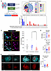 Figure 1
Cross-neutralization is associated with GCs displaying preserved features of maturation. (A) Diagram depicting the strategy employed for neutralization screening (left) as well as the distribution of cross-neutralization breadth in the population of study, defined as the percentage of the number of isolates recognized with an ID50 of greater than 40 out of 20 isolates tested, in the cohort (pie chart) and among study participants (bar graph). (B) Representative confocal images showing the distribution of CD20 (cyan) and Ki67 (magenta) in NNs and Ns. Total B cell follicle area quantification (CD20hi/dim) is shown as a superplot of both individual (faint background symbols) and mean ± SEM (bold foreground symbols) follicular measurements in control tissues (n = 6, gray), NN tissues (n = 6, blue), and N tissues (n = 6, red), as measured by quantitative imaging analysis and histocytometry. The different symbols represent different donors. For statistical comparisons using the mean measurements, a Kruskal-Wallis 1-way ANOVA was applied, followed by Dunn’s post hoc test to correct for multiple comparisons. P = 0.0798. Original magnification, ×40 (NA 1.3). Scale bars: 500 μm. (C) Superplots showing the number of total cells and GC/F ratios (areas) of both individual (faint background symbols) and mean ± SEM (bold foreground symbols) follicular measurements in NNs (n = 6, blue) and Ns (n = 6, red), as measured by quantitative imaging analysis and histocytometry. Data for each follicle represent total counts of JOJO-1+ (a fluorescent nucleic acid stain) events (total cells) normalized (Norm) to the total area (mm2) of follicles screened. The different symbols represent different donors. For statistical comparisons using the mean measurements, a Mann-Whitney U test was applied. P = 0.93; P = 0,0649. (D) Representative confocal images showing the distribution of FDC (red) in B cell follicles (CD20dim/hi areas, cyan) in NNs (n = 6) versus Ns (n = 6). Original magnification, ×40. Scale bars: 100 μm), and a Superplot summarizing the FDC areas (mm2) measurements of both individual (faint background symbols) and mean ± SEM (bold foreground symbols) follicular measurements in NNs (n = 6, blue) and Ns (n = 6, red), as measured by quantitative imaging analysis and histocytometry. Different symbols represent different donors. For statistical comparisons using the mean measurements, a Mann-Whitney U test was applied. P = 0.81.
On the basis of these findings and on overall tissue and LN cell suspension sample availability, a total of 33 participants with (n = 17) or without (n = 16) cross-neutralizing activity were selected for further study (Table 1 and Supplemental Tables 1 and 2). Participants were categorized as Ns if their serum neutralizing activity in the extended panel was medium or high (>20% of isolates), or as NNs if their serum neutralization activity was less than 5% (Figure 1A and Supplemental Figure 1B). Since neutralization breadth has been previously shown to depend on the nature of bNAb lineages involved (27), we sought to determine whether known bNAb lineages were represented in the Ns. We performed computational fingerprinting analysis using a neutralization fingerprinting algorithm (NFP) that predicts the epitopes targeted by neutralizing serum by matching patterns of neutralization to those of known bNAbs (28). In the sera from Ns, we detected signal (cutoff 0.25) (29) for 1 or more of the following: VRC01-, 8ANC195-, PGT18- 10E8-, and PGT151-like Abs (Supplemental Figure 1C). Therefore, known bNAb lineages were represented in the serum neutralizing activity in Ns.
Cross-neutralization is associated with a greater degree of B cell follicle preservation and GC formation. HIV acquisition causes morphological changes in the microarchitecture of LNs that affect the ability of B cell follicles to perform their function over time (30). Whether reduced cross-neutralization could be associated with anatomical changes is not well understood. Therefore, we examined the features of B cell follicle anatomy that were differentially represented in participants with and without serum cross-neutralizing activity. First, the areas of follicular regions of interest (ROIs), defined by the expression of CD20 (Figure 1B and Supplemental Figure 1D), were evaluated in our cohort and compared with benign HIV-negative participants’ LNs that were characterized by mature follicles (follicular hyperplasia) (Supplemental Table 1). We found that the calculated areas were substantially (P = 0.0798) higher in the control and NN participants compared with the N participants, indicating a higher degree of follicular hyperplasia in NNs, in line with previous data (4) (Figure 1B and Supplemental Figure 1D). Although not significant, we found a trend for higher circularity of follicular ROIs in N participants compared with NN participants (Supplemental Figure 1D).
We then evaluated additional elements associated with HIV disease including the overall B cell follicle cellularity, extent of Ki67hi/loCD20hi/dim areas as a surrogate of GCs, and FDC network preservation (31, 32). FDC networks were identified using the anti-CNA.42 Ab, which is highly specific for a fixative-resistant, evolutionarily conserved, glycosylated antigen that is expressed mainly on FDCs (33). We found a similar degree of overall cellularity — defined as the total number of nucleus-positive cells measured within CD20hi/dim B cell follicles — in Ns as compared with NNs (Figure 1C). Next, we examined the extent of GC preservation among tissues. For quantitation, we focused on Ki67, a marker of proliferation and cell-cycle progression (34), as a lack of Ki67 polarization in HIV GCs has previously been shown to associate with GC disorganization (35). Analysis of the area occupied by CD20hi/dimKi67hi B cells (denoted as GCs) versus the CD20hi/dimKi67hi/lo B cell area (denoted here as total follicle [F]) revealed a substantially (P = 0.0649) higher ratio of GC/F in Ns than in NNs (P = 0.00115) (Figure 1C and Supplemental Figure 1E). Next, we extended our analysis to FDC networks and observed a trend for higher FDC areas in NNs as compared with Ns (Figure 1D) and a reversed trend for the FDC/F ratios (Supplemental Figure 1, F and G). Together, these findings suggest that neutralization activity in chronic HIV is associated with better-preserved follicular structures.
Cross-neutralization is associated with distinct phenotypic signatures of Tfh cells. Having observed morphological differences in the LNs of NNs and Ns, we next wanted to assess the cellular composition of the LNs in these 2 groups. We performed flow cytometry on single-cell suspensions of LNs and enumerated the frequencies of the following immune subsets: naive (CD27hiCD45ROlo) and non-naive (CD27hiCD45ROhi, CD27loCD45ROhi) CD4+ T cell subsets and PD-1hiCXCR5hi Tfh cells (Figure 2A and Supplemental Figure 2A). We found a significantly higher percentage of non-naive CD27hi/loCD45ROhi T cells in N LNs than in NN LNs (P = 0.032) (Figure 2B). This was primarily driven by an increase in non-naive CD27loCD45ROhi cells (uO = 0.005) (Figure 2B). Between the latter frequencies and the recorded neutralization potencies, we observed a positive association which, although not significant (R2 = 0.1654, P = 0.1052), was suggestive of correlated changes between these 2 factors (Figure 2B).
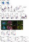 Figure 2
Cross-neutralization is associated with an expansion of the LN CD4+ T cell memory compartment and a distinct Tfh cell phenotypic profiling. (A) Flow cytometry plots depicting the frequencies of naive and non-naive CD4+ T cell subsets in NNs and Ns within the CD3hiCD4hi gated populations. (B) Graphs showing the relative frequencies of naive and non-naive CD4+ T cell subsets in Ns and NNs (left panel) and the positive association (Pearson correlation) between the percentage of non-naive CD27loCD45ROhi CD4+ T cells and microneutralization ID50 titers as assayed against a 20 HIV isolate panel. (C) Cumulative flow cytometry plots showing the frequencies of the receptors TIGIT and ICOS, as well as of the CD57 epitope and CXCR3 within the PD-1hiCXCR5hi Tfh cell compartment in NNs and Ns, and frequencies of combined CXCR3 and CD57 expression. TIGIT, P = 0.0075; ICOS, P = 0.0160; CD57; P = 0.0225; CXCR3, P = 0.0420 (Mann-Whitney U test); NN CXCR5loCD57hi versus NN CXCR3hiCD57lo, P < 0.0001 (1-way ANOVA). ****P < 0.0001. (D) Confocal images of follicles (CD20hi/dim) and GCs (CD20hiKi67hi) in NNs and N showing the distribution of CD4 (green), Tfh (CD4: green, PD-1hi: red) and Bcl-6+ Tfh (CD4: green, PD-1hi: red, Bcl-6hi: yellow) within the Ki67 follicular areas (magenta) and Superplots showing total GC-CD4, GC Bcl-6hi Tfh cell densities (cells per mm2) and GC Bcl-6hi Tfh/CD4 ratios of both individual (faint background symbols) and mean (bold foreground symbols) follicular measurements (± SEM) in NNs (n = 6 blue) versus Ns (n = 6 red) as measured by quantitative imaging analysis and histocytometry. Follicular area borders were determined based on the density of CD20 expression, whereas extrafollicular areas (T cell zone) were determined based on the density of CD4. Different symbols represent different donors. For statistical comparisons using the mean measurements, a Mann-Whitney U test was applied. P = 0.48; P = 0.132; P = 0.026. Original magnification, ×20. Scale bars: 60 μm left column; 30 μm right columns. (E) Regression plots showing the association between bulk Tfh and Bcl-6hi Tfh cells (normalized cell counts adjusted for mm2 of area screened) as quantified by confocal imaging analysis and histocytometry, and the breadth of neutralization expressed as the number of HIV isolates recognized among the 20 isolates tested. EF, extrafollicular area; F, follicular area. Mean ± SD values are shown in B and C and median values in D.
We found a balanced representation (percentage) of non-naive CD4+ T cell subsets defined by the expression of PD-1 and CXCR5 in Ns, while a gradual decrease from non-Tfh cells (PD-1loCXCR5lo) to Tfh cells (PD-1hiCXCR5hi) was observed in NNs (Supplemental Figure 2B). Furthermore, flow cytometric phenotyping of the Tfh cell subset revealed significantly higher expression of several coinhibitory/costimulatory receptors (36–38) in Ns compared with NNs (Figure 2C and Supplemental Figure 2C). With respect to CD57, a marker of highly differentiated GC Tfh cells (39, 40) and CXCR3, a chemokine associated with a Th1-like Tfh cell signature, we found significantly higher frequencies of CD57loCXCR3hi Tfh cells compared with CD57hi CXCR3lo Tfh cells in NNs (P < 0.0001), while a trend for higher frequencies of CD57lo CXCR3hi Tfh cells in NNs as compared with Ns was also notable (Figure 2C). Similar findings were also obtained when data from samples with sufficiently representative Tfh cell numbers from NNs and Ns were dissected by t-distributed stochastic neighbor embedding (t-SNE) analysis (Supplemental Figure 2D). We found a trend for higher representation of CD57loCXCR3hi Tfh cells within the TIGIThiICOShiCD95hi phenotypic compartment in NNs compared with Ns (cluster/population 5) (Supplemental Figure 2D). On the other hand, a trend for a higher frequency of the cluster bearing the CD57hiCXCR3lo signature was seen in Ns (cluster/population 6) (Supplemental Figure 2D). Taken together, our data suggest an altered differentiation profile of Tfh cells in NNs that was characterized by an accumulation of a less differentiated (CD57lo), Th1-like (CXCR3hi) phenotype in NNs compared with Ns.
We next analyzed the prevalence of Tfh cells within CD20hi/dim follicular areas of the LNs in situ. Using immunofluorescence, we found higher normalized numbers of bulk follicular CD4+ T cells in NNs than in Ns (Figure 2D and Supplemental Figure 3A). In contrast to total CD4+ T cells, PD-1hiBcl-6hi Tfh cells were substantially (P = 0.1320) increased in Ns compared with NNs (Figure 2D and Supplemental Figure 3B). Furthermore, the ratio of Tfh to CD4+ T cells within the B cell follicles of Ns was significantly higher than that for NNs (Figure 2D and Supplemental Figure 3B). We also examined whether a correlation existed between the normalized Tfh cell counts and the breadth of serum cross-neutralization. Contrary to PD1hi Tfh cells, we found a significant correlation between PD-1hiBcl-6hi Tfh cells and the cross-neutralization breadth in all participants (R2 = 0.66, P = 0.0012) (Figure 2E). We observed a similar profile when the group of Ns was analyzed separately (R2 = 0.79, P = 0.0173), which also extended to PD-1hi Tfh cells (R2 = 0.88, P = 0.0052) (Figure 2E). Overall, these results show that the development of cross-neutralizing breadth was associated with a higher in situ prevalence of PD-1hi and “effector” Bcl-6hi Tfh cells.
Cross-neutralization is associated with a molecular profile favoring the development and function of Tfh cells. To identify molecular effector pathways that could trigger the functional differences observed when comparing Tfh cells from N and NN groups, we performed 10X scRNA-seq on LN-derived cells (Figure 3A). Analysis of lineage biomarkers enabled the identification of the main immune cell types (Supplemental Figure 4A). Further clustering was performed on the basis of the expression of CD4+ T cell–specific genes (Supplemental Figure 4, B and C). We observed a significantly higher frequency of Tfh cells in Ns, whereas the frequency of naive CD4+ T cells was significantly higher in NNs than in Ns (Figure 3B), which corroborates our flow cytometric data (Figure 2). We then focused on characterizing the Tfh cell compartment. We observed higher transcription of genes encoding critical regulators that promote Tfh cell differentiation (e.g., MAF, IL6ST, PDCD1, ICOS, and TOX2), stemness (TCF7), and interaction with GC B cells (CXCL13) in Ns compared with NNs (Figure 3C). Interestingly, Tfh cells from Ns had a significant upregulation of Wnt signaling pathways as well as of targets that are downstream of TCF7L2, SMAD3,4 and STAT3, whereas NN Tfh cells were associated with significant upregulation of type I and II IFN signaling and STAT6 target genes. All shown pathways were significantly different between the groups (P < 0.05) (Figure 3C). Of note, Tfh cells from Ns exhibited significantly lower expression of the hallmark apoptosis gene set (normalized enrichment score [NES] = 1.32, P = 0.05), driven by leading-edge genes such as TXNIP and ANXA1, supporting an enhanced survival potential of these cells in Ns (Supplemental Figure 4, C and E). As a reference, analysis of tonsillar single cells showed differential expression of relevant genes among CD4+ T cell subsets (Supplemental Figure 4F). With respect to the potential CD4+ Treg transcriptomics profile, genes like FOXP1, a cofactor for FoxP3 (41), and IRF1, an inducer of type 1 regulatory Tr1 cells (42) and negative regulator of Tfh cell differentiation (43), were upregulated in NN compared with N participants (Figure 3D). Furthermore, downstream targets for several established Treg inducers including STAT3, STAT5, STAT6 (44–47), MYC (48), SMAD3 (49), vitamin D receptor (VDR) (50), and FOXP3/FOXP1 were upregulated in NNs compared with Ns (Figure 3D). Our data indicate that, despite the significantly higher frequency of FOXP3hi CD4+ T cells in Ns (Figure 3B), CD4+ T cells from NNs were characterized by a molecular profile favoring the development of potential suppressor CD4+ T cells. When we validated molecules revealed by scRNA-seq analysis with flow cytometry, we also observed significantly higher levels of combined TCF1hiAHRhiCAV1hiCD130hi expression in Tfh cell subsets (PD-1hiCD57lo/hi) compared with naive CD4+ T cells (4.04% ± 3.65% vs. 0.21% ± 0.13%, P = 0.0261) and compared with the PD-1loCD57lo CD4+ T cell subset (4.04% ± 3.65% vs. 0.17% ± 0.17%, P = 0.0244) in tonsillar cell suspensions (Supplemental Figure 5A). In sum, our data suggest that Tfh cells from Ns expressed a molecular program that favored their differentiation and stemness.
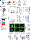 Figure 3
Tfh cells are characterized by a molecular profile favoring their differentiation and longevity in cross-neutralization participants. (A) scRNA analysis workflow. DEG, differentially expressed gene. (B) UMAP projections of total CD4+ T cells with color-coded cell populations and CD4+ T cell subpopulations identified and their percentages in NNs versus Ns. (C and D) Volcano plots and dot plots are shown to illustrate transcriptomic differences and pathway enrichment between Ns (Neut) and NNs (NonNeut). DEGs were identified using the MAST (hurdle model–based) test implemented in the FindMarkers function of the Seurat package in R. P values were adjusted by the Benjamini-Hochberg method, and genes meeting an adjusted P < 0.05 and |log2fold change [FC]| > 0.5 are highlighted in the volcano plots. Blue and red dots in the volcano plots represent genes that are significantly downregulated or upregulated in Ns, respectively. The accompanying dot plots depict average scaled expression (z scores) and the percentage of cells expressing each gene within Tfh or Treg subsets. Color intensity reflects relative expression, while dot size indicates the fraction of expressing cells. NESs from the GSEA are shown separately for selected pathways and transcription factor target gene sets. (E) Representative confocal images showing FOXP3hi CD4+ T cells in extrafollicular and follicular areas in NNs versus Ns. FoxP3hi CD4+ T cells (white arrows) were defined by means of concurrent CD4 (green) and FoxP3 (red) expression within CD20 (cyan) and Ki67 (yellow) double-positive areas. Original magnification, ×40; scale bars: 100 μm and 20 μm (enlarged images corresponding to the white dotted enclosures). Superplots showing the normalized per area imaged FoxP3hi CD4+ T cell numbers of both individual (faint background symbols) and mean (bold foreground symbols) follicular measurements (± SEM) in NNs (n = 6, blue) compared with Ns (n = 6, red), as measured by quantitative imaging analysis and histocytometry. The different symbols represent different donors. For statistical comparisons using the mean measurements, a Mann-Whitney U test was used. P = 0.1797. Superplots of CD25hiFoxP3hi (F, right) and IL-10hiCD25hiFoxP3hi (G, right) CD4+ T cells of both individual (faint background symbols) and mean (bold foreground symbols) follicular measurements in NNs (n = 6, blue) versus Ns (n = 5, red), as measured by quantitative imaging analysis and histocytometry. The different symbols represent different donors. For statistical comparisons using the mean measurements, a Mann-Whitney U test was applied (P = 0.426, P = 0.0823). Dot plots for extrafollicular (F, left) and whole tissue (G, left) data, where each participant tissue is represented by a circle of a different color. Mean values are shown in dot plots (E–G) and quartiles in box plots (F and G).
Cross-neutralization is associated with a lower cell density of IL-10hiCD25hiFoxP3hi follicular regulatory CD4+ T cells. Our transcriptomics analysis revealed a significantly higher frequency of FoxP3hi cells in N compared with NN tissues (Figure 3B). We then used an in situ approach to quantify FoxP3hi CD4+ T cells in extrafollicular and follicular LN areas. In line with our scRNA analysis (Figure 3B), we found a substantially (P = 0.1797) higher number of normalized FoxP3hi CD4+ T cells within the follicular areas in Ns compared with NNs (Figure 3E, Supplemental Figure 3A, and Supplemental Figure 5B). We further investigated the FoxP3hi CD4+ T cell compartment using CD25 (51) and IL-10, a cytokine that correlates with increased Treg functionality and suppressive capacity (52) (Supplemental Figure 6A). Analysis of CD25hiFoxP3hi CD4+ T cells revealed comparable normalized numbers of extrafollicular CD25hiFoxP3hi CD4+ T cells among the 2 categories (Figure 3F). However, there was a trend for lower numbers of follicular CD25hiFoxP3hi CD4+ T cells (Tfr) and substantially lower cell density (P = 0.0823) of follicular IL-10hiCD25hiFoxP3hi CD4+ T cells in the N tissues compared with the NN tissues (Figure 3, F and G). We found a significantly higher (P = 0.01691) ratio of FoxP3hi/Tfh CD4+ T cells in Ns compared with NNs, whereas the opposite was the case when we calculated the CD25hiFoxP3hi/Tfh CD4+ T cell ratio (data not shown). Our data suggest a GC-specific effect, rather than a global effect, of increased numbers of functional IL-10–producing Tfr cells in the follicles of NNs, which may contribute to limited GC Tfh cell responses.
Cross-neutralization is associated with a higher degree of viral evolution. To explore relationships between the aforementioned CD4+ T cell profiles and virus dynamics, we compared NN and N groups according to the plasma viral load (pVL), the prevalence of HIV RNA+ cells quantified by ISH, and the intra-host diversity of HIV env gene sequences. A trend, although not significant, for higher plasma viremia was found in NN LNs compared with N LNs (Figure 4A). The plasma viremia, however, did not correlate with the breadth of NAbs (Figure 4A). When the in situ expression of HIV mRNA was analyzed, we found significantly more HIV RNA+ cells (P = 0.0232) and a trend for higher levels of total viral RNA (P = 0.0704) in NN compared with N LNs (Supplemental Figure 6B). In line with the in situ analysis, more LN CD4+ T cells harboring HIV mRNA, analyzed by scRNA-seq, were detected in NNs compared with Ns (Supplemental Figure 6C). A strong association between the FDC area and RNA+ virions was found both in Ns and NNs (Supplemental Figure 6D). We detected a higher intra-host env diversity in Ns than in NNs, based on entropy analysis using high-throughput, single-genome amplification and sequencing (HT-SGS) (53) (Figure 4B and Supplemental Figure 7A). Intra-host diversity of env sequences in1 individual with a broad Ab neutralizing profile was among the highest entropy profiles (Supplemental Figure 7A). Of note, although this association could be consistent with positive selection by env-specific Abs in Ns, we also observed higher intra-host env sequence entropy in Ns when considering only synonymous variation (Supplemental Figure 7B). Moreover, several of the env gene regions with particularly high entropy in Ns did not correspond to known bNAb epitopes (Figure 4B). Taken together, these findings suggest higher cumulative levels of HIV evolution in Ns than in NNs.
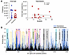 Figure 4
Higher viral evolution is associated with a lower in situ prevalence of actively transcribed virus in cross-neutralization participants. (A) pVLs in all study participants and association of VLs with neutralization breadth (number of isolates recognized out of the 20 isolates tested). Median values are shown. (B) Entropy-H(x) plot showing the location of positions with residue mismatches on alignment to the reference Env HIV sequence and the distribution of those mismatches over different HIV structural regions. Color shades denote distinct structural regions, and orange arcs the locations of gp120 variable loops.
Cross-neutralizing activity is associated with a higher in situ prevalence of BC B cells expressing a molecular profile favoring GC development. Finally, we sought to assess whether HIV neutralization could be linked to a specific GC B cell profile. To this end, we analyzed the numbers of GC B cells and total B cells (CD20hi/dim) in situ. Despite the comparable total numbers of B cells between the 2 groups, we found a trend for higher normalized numbers (P = 0.2403) of CD20hiKi67+Bcl-6hi B cells in LNs of Ns compared with NNs (Figure 5A and Supplemental Figure 8A). Furthermore, a significantly higher Tfh/B cell ratio (P < 0.0001) and a trend for higher Bcl-6hi Tfh/Bcl-6hiKi67hi B cell ratios were found in Ns compared with NNs (Supplemental Figure 8B). We further performed scRNA-seq on B cells to elucidate their transcriptomic profile. Further clustering based on the expression of B cell–specific genes (Supplemental Figure 8C) revealed significantly higher GC B cell frequencies and lower naive B cell frequencies in N LNs than NN LNs (Figure 5B). This observation was further supported by the higher expression of CXCR5 and CXCR4 in N LNs than NN LNs (Figure 5C). Moreover, we found increased expression of factors promoting GC B cell differentiation (BACH1, STAT6), as well as the development of the dark zone (DZ) B cell compartment (FOXO1, TGFB/SMAD2/4) and the response to local osmotic stress (NFAT5) in N B cells compared with NN B cells (Figure 5C). NN B cells were found to upregulate type I and type II IFN and mTOR signaling, which could have a detrimental effect on GC B cell development (Figure 5C). Together, these data show that Ns were programmed for the development and maintenance of GC B cells. Furthermore, possible molecular targets that could mediate the interaction between GC B cells and Tfh cells were investigated across all 8 participants using the NicheNet package in R. Our analysis revealed novel putative “molecular pairs” that could mediate such interaction, including macrophage migration inhibitory factor (MIF) (ligand for CXCR4 and CD74 and potential regulator of B cells at trafficking, survival, and antigen presentation level) (54); integrin-β 8 (ITGB8) (unit of the avb8 integrin, a critical activator of TGF-β) (55); and TGFB1/SMAD3 (Figure 5D). Therefore, our analysis reveals molecules that could serve as targets for in vivo interventions aiming to strengthen the Tfh–B cell interaction and bNAb development.
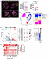 Figure 5
GC B cells express a molecular profile favoring GC development in cross-neutralization LNs. (A) Representative confocal images showing the distribution of BCL-6 (yellow) and Ki67 (magenta) within the GCs of NNs and Ns. Original magnification, ×40 with 1% zoom; scale bars: 100 μm. Superplots show normalized CD20hi and Bcl-6hiKi67hiCD20hi B cell numbers of both individual (faint background symbols) and mean ± SEM (bold foreground symbols) follicular measurements in NNs (n = 6, blue) versus Ns (n = 6, red), as measured by quantitative imaging analysis and histocytometry. The different symbols represent different donors. For statistical comparisons using mean measurements, a Mann-Whitney U test was applied (P = 0.588, P = 0.2403). (B) UMAP projections of B cell populations identified by scRNA analysis in total l ymph node mononuclear cells, color-coded by B cell category. (C) Volcano plots and dot plots of DEGs and NESs from GSEA for selection of B cell–specific ligands and transcription factors. The DEGs were calculated using the MAST (Hurdle model–based) test via the FindMarkers function using the Seurat package in R. P values were corrected using the Benjamini-Hochberg method and genes meeting an adjusted *P < 0.05 and (log2FC) > 0.5 are highlighted on the volcano plot. Dots in the volcano plots represent genes that were significantly downregulated (blue) or upregulated (red) in Ns. (D) Heatmap of pairwise Tfh–GC B cell interactions as identified through ligand-target analysis. Darker colors denote interactions corresponding to an increased regulatory potential.


Copyright © 2026 American Society for Clinical Investigation
ISSN: 0021-9738 (print), 1558-8238 (online)