
Advertisement
Research ArticleImmunologyInfectious disease
Open Access |  10.1172/JCI192370
Human monoclonal antibodies targeting α-Gal restrict IgE engagement of α-Gal syndrome allergens
Hyeseon Cho,1 Youngsil Seo,1 Haewon Sohn,1 Shailesh K. Choudhary,2 Jeff Skinner,1 Ming Zhao,3 Ludmila Krymskaya,4 Weizhi Zhong,5 Justin Lack,6 Shanping Li,1 Boubacar Traore,7 Joshua Tan,5 Scott P. Commins,2 and Peter D. Crompton1
1Malaria Infection Biology and Immunity Section, Laboratory of Immunogenetics, Division of Intramural Research, National Institute of Allergy and Infectious Diseases, NIH, Rockville, Maryland, USA.
2Division of Allergy and Immunology, University of North Carolina at Chapel Hill, Chapel Hill, North Carolina, USA.
3Protein and Chemistry Section, Research Technologies Branch, National Institute of Allergy and Infectious Diseases,
4Laboratory of Immunogenetics, Division of Intramural Research, National Institute of Allergy and Infectious Diseases,
5Antibody Biology Unit, Laboratory of Immunogenetics, Division of Intramural Research, National Institute of Allergy and Infectious Diseases, NIH, Rockville, Maryland, USA.
6Integrated Data Sciences Section, Research Technologies Branch, National Institute of Allergy and Infectious Diseases, NIH, Bethesda, Maryland, USA.
7Mali International Center of Excellence in Research; Malaria Research and Training Center, University of Sciences, Techniques and Technologies of Bamako, Bamako, Mali.
Address correspondence to: Scott P. Commins, Division of Allergy and Immunology, University of North Carolina at Chapel Hill, 6013 Farrington Road, Chapel Hill, North Carolina, 27517, USA. Phone: 919.537.3306; Email: scommins@email.unc.edu. Or to: Peter D. Crompton, Malaria Infection Biology and Immunity Section, Laboratory of Immunogenetics, National Institute of Allergy and Infectious Diseases, 5625 Fishers Lane, Rockville, Maryland, 20852, USA. Phone: 301.761.5042; Email: pcrompton@niaid.nih.gov.
Authorship note: HC and YS contributed equally to this work.
Find articles by Cho, H. in: PubMed | Google Scholar
1Malaria Infection Biology and Immunity Section, Laboratory of Immunogenetics, Division of Intramural Research, National Institute of Allergy and Infectious Diseases, NIH, Rockville, Maryland, USA.
2Division of Allergy and Immunology, University of North Carolina at Chapel Hill, Chapel Hill, North Carolina, USA.
3Protein and Chemistry Section, Research Technologies Branch, National Institute of Allergy and Infectious Diseases,
4Laboratory of Immunogenetics, Division of Intramural Research, National Institute of Allergy and Infectious Diseases,
5Antibody Biology Unit, Laboratory of Immunogenetics, Division of Intramural Research, National Institute of Allergy and Infectious Diseases, NIH, Rockville, Maryland, USA.
6Integrated Data Sciences Section, Research Technologies Branch, National Institute of Allergy and Infectious Diseases, NIH, Bethesda, Maryland, USA.
7Mali International Center of Excellence in Research; Malaria Research and Training Center, University of Sciences, Techniques and Technologies of Bamako, Bamako, Mali.
Address correspondence to: Scott P. Commins, Division of Allergy and Immunology, University of North Carolina at Chapel Hill, 6013 Farrington Road, Chapel Hill, North Carolina, 27517, USA. Phone: 919.537.3306; Email: scommins@email.unc.edu. Or to: Peter D. Crompton, Malaria Infection Biology and Immunity Section, Laboratory of Immunogenetics, National Institute of Allergy and Infectious Diseases, 5625 Fishers Lane, Rockville, Maryland, 20852, USA. Phone: 301.761.5042; Email: pcrompton@niaid.nih.gov.
Authorship note: HC and YS contributed equally to this work.
Find articles by Seo, Y. in: PubMed | Google Scholar
1Malaria Infection Biology and Immunity Section, Laboratory of Immunogenetics, Division of Intramural Research, National Institute of Allergy and Infectious Diseases, NIH, Rockville, Maryland, USA.
2Division of Allergy and Immunology, University of North Carolina at Chapel Hill, Chapel Hill, North Carolina, USA.
3Protein and Chemistry Section, Research Technologies Branch, National Institute of Allergy and Infectious Diseases,
4Laboratory of Immunogenetics, Division of Intramural Research, National Institute of Allergy and Infectious Diseases,
5Antibody Biology Unit, Laboratory of Immunogenetics, Division of Intramural Research, National Institute of Allergy and Infectious Diseases, NIH, Rockville, Maryland, USA.
6Integrated Data Sciences Section, Research Technologies Branch, National Institute of Allergy and Infectious Diseases, NIH, Bethesda, Maryland, USA.
7Mali International Center of Excellence in Research; Malaria Research and Training Center, University of Sciences, Techniques and Technologies of Bamako, Bamako, Mali.
Address correspondence to: Scott P. Commins, Division of Allergy and Immunology, University of North Carolina at Chapel Hill, 6013 Farrington Road, Chapel Hill, North Carolina, 27517, USA. Phone: 919.537.3306; Email: scommins@email.unc.edu. Or to: Peter D. Crompton, Malaria Infection Biology and Immunity Section, Laboratory of Immunogenetics, National Institute of Allergy and Infectious Diseases, 5625 Fishers Lane, Rockville, Maryland, 20852, USA. Phone: 301.761.5042; Email: pcrompton@niaid.nih.gov.
Authorship note: HC and YS contributed equally to this work.
Find articles by Sohn, H. in: PubMed | Google Scholar
1Malaria Infection Biology and Immunity Section, Laboratory of Immunogenetics, Division of Intramural Research, National Institute of Allergy and Infectious Diseases, NIH, Rockville, Maryland, USA.
2Division of Allergy and Immunology, University of North Carolina at Chapel Hill, Chapel Hill, North Carolina, USA.
3Protein and Chemistry Section, Research Technologies Branch, National Institute of Allergy and Infectious Diseases,
4Laboratory of Immunogenetics, Division of Intramural Research, National Institute of Allergy and Infectious Diseases,
5Antibody Biology Unit, Laboratory of Immunogenetics, Division of Intramural Research, National Institute of Allergy and Infectious Diseases, NIH, Rockville, Maryland, USA.
6Integrated Data Sciences Section, Research Technologies Branch, National Institute of Allergy and Infectious Diseases, NIH, Bethesda, Maryland, USA.
7Mali International Center of Excellence in Research; Malaria Research and Training Center, University of Sciences, Techniques and Technologies of Bamako, Bamako, Mali.
Address correspondence to: Scott P. Commins, Division of Allergy and Immunology, University of North Carolina at Chapel Hill, 6013 Farrington Road, Chapel Hill, North Carolina, 27517, USA. Phone: 919.537.3306; Email: scommins@email.unc.edu. Or to: Peter D. Crompton, Malaria Infection Biology and Immunity Section, Laboratory of Immunogenetics, National Institute of Allergy and Infectious Diseases, 5625 Fishers Lane, Rockville, Maryland, 20852, USA. Phone: 301.761.5042; Email: pcrompton@niaid.nih.gov.
Authorship note: HC and YS contributed equally to this work.
Find articles by Choudhary, S. in: PubMed | Google Scholar
1Malaria Infection Biology and Immunity Section, Laboratory of Immunogenetics, Division of Intramural Research, National Institute of Allergy and Infectious Diseases, NIH, Rockville, Maryland, USA.
2Division of Allergy and Immunology, University of North Carolina at Chapel Hill, Chapel Hill, North Carolina, USA.
3Protein and Chemistry Section, Research Technologies Branch, National Institute of Allergy and Infectious Diseases,
4Laboratory of Immunogenetics, Division of Intramural Research, National Institute of Allergy and Infectious Diseases,
5Antibody Biology Unit, Laboratory of Immunogenetics, Division of Intramural Research, National Institute of Allergy and Infectious Diseases, NIH, Rockville, Maryland, USA.
6Integrated Data Sciences Section, Research Technologies Branch, National Institute of Allergy and Infectious Diseases, NIH, Bethesda, Maryland, USA.
7Mali International Center of Excellence in Research; Malaria Research and Training Center, University of Sciences, Techniques and Technologies of Bamako, Bamako, Mali.
Address correspondence to: Scott P. Commins, Division of Allergy and Immunology, University of North Carolina at Chapel Hill, 6013 Farrington Road, Chapel Hill, North Carolina, 27517, USA. Phone: 919.537.3306; Email: scommins@email.unc.edu. Or to: Peter D. Crompton, Malaria Infection Biology and Immunity Section, Laboratory of Immunogenetics, National Institute of Allergy and Infectious Diseases, 5625 Fishers Lane, Rockville, Maryland, 20852, USA. Phone: 301.761.5042; Email: pcrompton@niaid.nih.gov.
Authorship note: HC and YS contributed equally to this work.
Find articles by Skinner, J. in: PubMed | Google Scholar
1Malaria Infection Biology and Immunity Section, Laboratory of Immunogenetics, Division of Intramural Research, National Institute of Allergy and Infectious Diseases, NIH, Rockville, Maryland, USA.
2Division of Allergy and Immunology, University of North Carolina at Chapel Hill, Chapel Hill, North Carolina, USA.
3Protein and Chemistry Section, Research Technologies Branch, National Institute of Allergy and Infectious Diseases,
4Laboratory of Immunogenetics, Division of Intramural Research, National Institute of Allergy and Infectious Diseases,
5Antibody Biology Unit, Laboratory of Immunogenetics, Division of Intramural Research, National Institute of Allergy and Infectious Diseases, NIH, Rockville, Maryland, USA.
6Integrated Data Sciences Section, Research Technologies Branch, National Institute of Allergy and Infectious Diseases, NIH, Bethesda, Maryland, USA.
7Mali International Center of Excellence in Research; Malaria Research and Training Center, University of Sciences, Techniques and Technologies of Bamako, Bamako, Mali.
Address correspondence to: Scott P. Commins, Division of Allergy and Immunology, University of North Carolina at Chapel Hill, 6013 Farrington Road, Chapel Hill, North Carolina, 27517, USA. Phone: 919.537.3306; Email: scommins@email.unc.edu. Or to: Peter D. Crompton, Malaria Infection Biology and Immunity Section, Laboratory of Immunogenetics, National Institute of Allergy and Infectious Diseases, 5625 Fishers Lane, Rockville, Maryland, 20852, USA. Phone: 301.761.5042; Email: pcrompton@niaid.nih.gov.
Authorship note: HC and YS contributed equally to this work.
Find articles by Zhao, M. in: PubMed | Google Scholar
1Malaria Infection Biology and Immunity Section, Laboratory of Immunogenetics, Division of Intramural Research, National Institute of Allergy and Infectious Diseases, NIH, Rockville, Maryland, USA.
2Division of Allergy and Immunology, University of North Carolina at Chapel Hill, Chapel Hill, North Carolina, USA.
3Protein and Chemistry Section, Research Technologies Branch, National Institute of Allergy and Infectious Diseases,
4Laboratory of Immunogenetics, Division of Intramural Research, National Institute of Allergy and Infectious Diseases,
5Antibody Biology Unit, Laboratory of Immunogenetics, Division of Intramural Research, National Institute of Allergy and Infectious Diseases, NIH, Rockville, Maryland, USA.
6Integrated Data Sciences Section, Research Technologies Branch, National Institute of Allergy and Infectious Diseases, NIH, Bethesda, Maryland, USA.
7Mali International Center of Excellence in Research; Malaria Research and Training Center, University of Sciences, Techniques and Technologies of Bamako, Bamako, Mali.
Address correspondence to: Scott P. Commins, Division of Allergy and Immunology, University of North Carolina at Chapel Hill, 6013 Farrington Road, Chapel Hill, North Carolina, 27517, USA. Phone: 919.537.3306; Email: scommins@email.unc.edu. Or to: Peter D. Crompton, Malaria Infection Biology and Immunity Section, Laboratory of Immunogenetics, National Institute of Allergy and Infectious Diseases, 5625 Fishers Lane, Rockville, Maryland, 20852, USA. Phone: 301.761.5042; Email: pcrompton@niaid.nih.gov.
Authorship note: HC and YS contributed equally to this work.
Find articles by Krymskaya, L. in: PubMed | Google Scholar
1Malaria Infection Biology and Immunity Section, Laboratory of Immunogenetics, Division of Intramural Research, National Institute of Allergy and Infectious Diseases, NIH, Rockville, Maryland, USA.
2Division of Allergy and Immunology, University of North Carolina at Chapel Hill, Chapel Hill, North Carolina, USA.
3Protein and Chemistry Section, Research Technologies Branch, National Institute of Allergy and Infectious Diseases,
4Laboratory of Immunogenetics, Division of Intramural Research, National Institute of Allergy and Infectious Diseases,
5Antibody Biology Unit, Laboratory of Immunogenetics, Division of Intramural Research, National Institute of Allergy and Infectious Diseases, NIH, Rockville, Maryland, USA.
6Integrated Data Sciences Section, Research Technologies Branch, National Institute of Allergy and Infectious Diseases, NIH, Bethesda, Maryland, USA.
7Mali International Center of Excellence in Research; Malaria Research and Training Center, University of Sciences, Techniques and Technologies of Bamako, Bamako, Mali.
Address correspondence to: Scott P. Commins, Division of Allergy and Immunology, University of North Carolina at Chapel Hill, 6013 Farrington Road, Chapel Hill, North Carolina, 27517, USA. Phone: 919.537.3306; Email: scommins@email.unc.edu. Or to: Peter D. Crompton, Malaria Infection Biology and Immunity Section, Laboratory of Immunogenetics, National Institute of Allergy and Infectious Diseases, 5625 Fishers Lane, Rockville, Maryland, 20852, USA. Phone: 301.761.5042; Email: pcrompton@niaid.nih.gov.
Authorship note: HC and YS contributed equally to this work.
Find articles by Zhong, W. in: PubMed | Google Scholar
1Malaria Infection Biology and Immunity Section, Laboratory of Immunogenetics, Division of Intramural Research, National Institute of Allergy and Infectious Diseases, NIH, Rockville, Maryland, USA.
2Division of Allergy and Immunology, University of North Carolina at Chapel Hill, Chapel Hill, North Carolina, USA.
3Protein and Chemistry Section, Research Technologies Branch, National Institute of Allergy and Infectious Diseases,
4Laboratory of Immunogenetics, Division of Intramural Research, National Institute of Allergy and Infectious Diseases,
5Antibody Biology Unit, Laboratory of Immunogenetics, Division of Intramural Research, National Institute of Allergy and Infectious Diseases, NIH, Rockville, Maryland, USA.
6Integrated Data Sciences Section, Research Technologies Branch, National Institute of Allergy and Infectious Diseases, NIH, Bethesda, Maryland, USA.
7Mali International Center of Excellence in Research; Malaria Research and Training Center, University of Sciences, Techniques and Technologies of Bamako, Bamako, Mali.
Address correspondence to: Scott P. Commins, Division of Allergy and Immunology, University of North Carolina at Chapel Hill, 6013 Farrington Road, Chapel Hill, North Carolina, 27517, USA. Phone: 919.537.3306; Email: scommins@email.unc.edu. Or to: Peter D. Crompton, Malaria Infection Biology and Immunity Section, Laboratory of Immunogenetics, National Institute of Allergy and Infectious Diseases, 5625 Fishers Lane, Rockville, Maryland, 20852, USA. Phone: 301.761.5042; Email: pcrompton@niaid.nih.gov.
Authorship note: HC and YS contributed equally to this work.
Find articles by Lack, J. in: PubMed | Google Scholar
1Malaria Infection Biology and Immunity Section, Laboratory of Immunogenetics, Division of Intramural Research, National Institute of Allergy and Infectious Diseases, NIH, Rockville, Maryland, USA.
2Division of Allergy and Immunology, University of North Carolina at Chapel Hill, Chapel Hill, North Carolina, USA.
3Protein and Chemistry Section, Research Technologies Branch, National Institute of Allergy and Infectious Diseases,
4Laboratory of Immunogenetics, Division of Intramural Research, National Institute of Allergy and Infectious Diseases,
5Antibody Biology Unit, Laboratory of Immunogenetics, Division of Intramural Research, National Institute of Allergy and Infectious Diseases, NIH, Rockville, Maryland, USA.
6Integrated Data Sciences Section, Research Technologies Branch, National Institute of Allergy and Infectious Diseases, NIH, Bethesda, Maryland, USA.
7Mali International Center of Excellence in Research; Malaria Research and Training Center, University of Sciences, Techniques and Technologies of Bamako, Bamako, Mali.
Address correspondence to: Scott P. Commins, Division of Allergy and Immunology, University of North Carolina at Chapel Hill, 6013 Farrington Road, Chapel Hill, North Carolina, 27517, USA. Phone: 919.537.3306; Email: scommins@email.unc.edu. Or to: Peter D. Crompton, Malaria Infection Biology and Immunity Section, Laboratory of Immunogenetics, National Institute of Allergy and Infectious Diseases, 5625 Fishers Lane, Rockville, Maryland, 20852, USA. Phone: 301.761.5042; Email: pcrompton@niaid.nih.gov.
Authorship note: HC and YS contributed equally to this work.
Find articles by Li, S. in: PubMed | Google Scholar
1Malaria Infection Biology and Immunity Section, Laboratory of Immunogenetics, Division of Intramural Research, National Institute of Allergy and Infectious Diseases, NIH, Rockville, Maryland, USA.
2Division of Allergy and Immunology, University of North Carolina at Chapel Hill, Chapel Hill, North Carolina, USA.
3Protein and Chemistry Section, Research Technologies Branch, National Institute of Allergy and Infectious Diseases,
4Laboratory of Immunogenetics, Division of Intramural Research, National Institute of Allergy and Infectious Diseases,
5Antibody Biology Unit, Laboratory of Immunogenetics, Division of Intramural Research, National Institute of Allergy and Infectious Diseases, NIH, Rockville, Maryland, USA.
6Integrated Data Sciences Section, Research Technologies Branch, National Institute of Allergy and Infectious Diseases, NIH, Bethesda, Maryland, USA.
7Mali International Center of Excellence in Research; Malaria Research and Training Center, University of Sciences, Techniques and Technologies of Bamako, Bamako, Mali.
Address correspondence to: Scott P. Commins, Division of Allergy and Immunology, University of North Carolina at Chapel Hill, 6013 Farrington Road, Chapel Hill, North Carolina, 27517, USA. Phone: 919.537.3306; Email: scommins@email.unc.edu. Or to: Peter D. Crompton, Malaria Infection Biology and Immunity Section, Laboratory of Immunogenetics, National Institute of Allergy and Infectious Diseases, 5625 Fishers Lane, Rockville, Maryland, 20852, USA. Phone: 301.761.5042; Email: pcrompton@niaid.nih.gov.
Authorship note: HC and YS contributed equally to this work.
Find articles by Traore, B. in: PubMed | Google Scholar
1Malaria Infection Biology and Immunity Section, Laboratory of Immunogenetics, Division of Intramural Research, National Institute of Allergy and Infectious Diseases, NIH, Rockville, Maryland, USA.
2Division of Allergy and Immunology, University of North Carolina at Chapel Hill, Chapel Hill, North Carolina, USA.
3Protein and Chemistry Section, Research Technologies Branch, National Institute of Allergy and Infectious Diseases,
4Laboratory of Immunogenetics, Division of Intramural Research, National Institute of Allergy and Infectious Diseases,
5Antibody Biology Unit, Laboratory of Immunogenetics, Division of Intramural Research, National Institute of Allergy and Infectious Diseases, NIH, Rockville, Maryland, USA.
6Integrated Data Sciences Section, Research Technologies Branch, National Institute of Allergy and Infectious Diseases, NIH, Bethesda, Maryland, USA.
7Mali International Center of Excellence in Research; Malaria Research and Training Center, University of Sciences, Techniques and Technologies of Bamako, Bamako, Mali.
Address correspondence to: Scott P. Commins, Division of Allergy and Immunology, University of North Carolina at Chapel Hill, 6013 Farrington Road, Chapel Hill, North Carolina, 27517, USA. Phone: 919.537.3306; Email: scommins@email.unc.edu. Or to: Peter D. Crompton, Malaria Infection Biology and Immunity Section, Laboratory of Immunogenetics, National Institute of Allergy and Infectious Diseases, 5625 Fishers Lane, Rockville, Maryland, 20852, USA. Phone: 301.761.5042; Email: pcrompton@niaid.nih.gov.
Authorship note: HC and YS contributed equally to this work.
Find articles by Tan, J. in: PubMed | Google Scholar
1Malaria Infection Biology and Immunity Section, Laboratory of Immunogenetics, Division of Intramural Research, National Institute of Allergy and Infectious Diseases, NIH, Rockville, Maryland, USA.
2Division of Allergy and Immunology, University of North Carolina at Chapel Hill, Chapel Hill, North Carolina, USA.
3Protein and Chemistry Section, Research Technologies Branch, National Institute of Allergy and Infectious Diseases,
4Laboratory of Immunogenetics, Division of Intramural Research, National Institute of Allergy and Infectious Diseases,
5Antibody Biology Unit, Laboratory of Immunogenetics, Division of Intramural Research, National Institute of Allergy and Infectious Diseases, NIH, Rockville, Maryland, USA.
6Integrated Data Sciences Section, Research Technologies Branch, National Institute of Allergy and Infectious Diseases, NIH, Bethesda, Maryland, USA.
7Mali International Center of Excellence in Research; Malaria Research and Training Center, University of Sciences, Techniques and Technologies of Bamako, Bamako, Mali.
Address correspondence to: Scott P. Commins, Division of Allergy and Immunology, University of North Carolina at Chapel Hill, 6013 Farrington Road, Chapel Hill, North Carolina, 27517, USA. Phone: 919.537.3306; Email: scommins@email.unc.edu. Or to: Peter D. Crompton, Malaria Infection Biology and Immunity Section, Laboratory of Immunogenetics, National Institute of Allergy and Infectious Diseases, 5625 Fishers Lane, Rockville, Maryland, 20852, USA. Phone: 301.761.5042; Email: pcrompton@niaid.nih.gov.
Authorship note: HC and YS contributed equally to this work.
Find articles by
Commins, S.
in:
PubMed
|
Google Scholar
|

1Malaria Infection Biology and Immunity Section, Laboratory of Immunogenetics, Division of Intramural Research, National Institute of Allergy and Infectious Diseases, NIH, Rockville, Maryland, USA.
2Division of Allergy and Immunology, University of North Carolina at Chapel Hill, Chapel Hill, North Carolina, USA.
3Protein and Chemistry Section, Research Technologies Branch, National Institute of Allergy and Infectious Diseases,
4Laboratory of Immunogenetics, Division of Intramural Research, National Institute of Allergy and Infectious Diseases,
5Antibody Biology Unit, Laboratory of Immunogenetics, Division of Intramural Research, National Institute of Allergy and Infectious Diseases, NIH, Rockville, Maryland, USA.
6Integrated Data Sciences Section, Research Technologies Branch, National Institute of Allergy and Infectious Diseases, NIH, Bethesda, Maryland, USA.
7Mali International Center of Excellence in Research; Malaria Research and Training Center, University of Sciences, Techniques and Technologies of Bamako, Bamako, Mali.
Address correspondence to: Scott P. Commins, Division of Allergy and Immunology, University of North Carolina at Chapel Hill, 6013 Farrington Road, Chapel Hill, North Carolina, 27517, USA. Phone: 919.537.3306; Email: scommins@email.unc.edu. Or to: Peter D. Crompton, Malaria Infection Biology and Immunity Section, Laboratory of Immunogenetics, National Institute of Allergy and Infectious Diseases, 5625 Fishers Lane, Rockville, Maryland, 20852, USA. Phone: 301.761.5042; Email: pcrompton@niaid.nih.gov.
Authorship note: HC and YS contributed equally to this work.
Find articles by
Crompton, P.
in:
PubMed
|
Google Scholar
|
Published September 1, 2026 - More info
J Clin Invest. 2026;136(17):e192370. https://doi.org/10.1172/JCI192370.
© 2026 Cho et al. This work is licensed under the Creative Commons Attribution 4.0 International License. To view a copy of this license, visit http://creativecommons.org/licenses/by/4.0/.
Received: February 13, 2025; Accepted: July 7, 2026


-
Results
Isolation of anti-α-Gal mAbs from malaria-exposed individuals. In a study initially designed to identify mAbs that target α-Gal on P. falciparum sporozoites (15), we analyzed 1,053 plasma samples from a cohort study in Mali (Supplemental Figure 1; supplemental material available online with this article; https://doi.org/10.1172/JCI192370DS1) where intense malaria transmission occurs during the 6-month rainy season from July to December (38). Using a multiplex bead assay, we determined plasma levels of polyclonal α-Gal–specific IgG, IgM, and IgA in May at the end of the 6-month dry season (n = 550 participants) and in October at the peak of malaria transmission (n = 503 participants). α-Gal-specific IgM was the most prevalent isotype and was detectable in nearly all participants, in contrast to α-Gal–specific IgA and IgG, which were not detectable in approximately 10% and 30% of individuals, respectively. In a paired analysis, the median value of all individual median fluorescence intensities (median MFI) of IgA was approximately 2.8-fold higher in October than in May while the median MFI of IgG was approximately 1.7-fold higher (Supplemental Figure 1A). IgM also trended higher in October (Supplemental Figure 1A). Increases in IgA and IgG from May to October were the most pronounced in infants with 3.7- and 493-fold increases, respectively, who generally had low α-Gal-specific IgA and IgG responses in May at the end of the dry season (Supplemental Figure 1B). Ab responses specific to α-Gal generally showed a positive correlation with age, except for a statistically significant decrease in IgM median MFI from the middle to oldest age group at the May time point (Supplemental Figure 1B).
Next, we isolated α-Gal–specific mAbs from B cells of the 18 participants with the highest polyclonal antibody responses to α-Gal (Supplemental Table 1). Using synthetic α-Gal conjugated to HSA as a probe, 17,388 B cells were sorted from PBMCs of these 18 participants (Supplemental Figure 2) and distributed onto 384-well plates (average 1–2 cells per well). After 2 weeks of B cell activation, which allowed memory B cells to proliferate and differentiate into antibody-producing cells, the supernatants were screened for secretion of α-Gal–binding Abs using a multiplex bead assay. We identified 85 α-Gal–specific B cell clones from which RT-PCR was performed to amplify genes encoding the heavy and light immunoglobulin chains. The resulting PCR products were sequenced, and, based on the sequence analysis using the International Immunogenetics Information System (IMGT) database (39), a panel of 42 fully human, α-Gal–specific recombinant mAbs were generated. All IgG and IgM clones were initially expressed as human IgG1 recombinant mAbs, while IgA clones were expressed as IgA1 or IgA2 monomers without the J chain. Binding of these mAbs to synthetic α-Gal beads was confirmed (Supplemental Figure 3A). Subsequently, 7 IgM clones (AG030, AG032, AG062, AG068, AG069, AG081, and AG082) in addition to one IgG (AG008) and 2 IgA (AG027 and AG028) clones were reexpressed as IgM with J chain, based on their binding activities to various α-Gal–expressing pathogens (data not shown). All 10 IgM mAbs exhibited enhanced binding to α-Gal beads compared with their IgG and IgA counterparts, likely due to increased avidity (Supplemental Figure 3B).
The gene usage and mutation frequency of human memory B cell–derived α-Gal–specific mAbs. The 42 memory B cell–derived mAbs specific for α-Gal utilized various VH and VL gene families, with a predominant use of the IGHV3 gene family for VH, consistent with a prior report (Figure 1A) (40). Also of note, a previous study found that anti-α-Gal B cells preferentially use the IGHV3-7 gene, particularly within the memory compartment (41). This gene encodes tryptophan at Kabat position 33 in the heavy chain complementarity-determining region 1 (CDRH1), which is crucial for the recognition of α-Gal (41). In the present study, we found that IGHV3-7 was the most frequently observed VH gene, with a prevalence of 31% (13 clones) (Figure 1A); and, moreover, 12 of the 13 IGHV3-7 clones encoded tryptophan at Kabat position 33 in CDRH1 (Supplemental Table 2). Overall, among all 42 mAbs, tryptophan and alanine were the most common residues at Kabat position 33 in CDRH1, with frequencies of 35.7% and 28.6%, respectively, while only one clone contained glycine, in contrast with a previous study that identified glycine as one of the 4 most common amino acids (Figure 1B) (41).
 Figure 1
Gene usage, isotype distribution, and mutation frequencies among the 42 human anti-α-Gal mAbs. (A) VH gene usage. The 2 predominant genes, IGVH3-7 and IGVH3-23, are highlighted with nonneutral colors. (B) Amino acid frequency at Kabat position 33 of CDRH1. (C) VK and VL gene usage. Frequently observed VK and VL genes are highlighted with nonneutral colors. (D) Isotype distribution of antibody heavy chains. (E) Mutation frequencies of VH genes in IgG, IgA, and IgM isotypes. (F) Mutation frequencies of VK and VL light chain genes. Mutation percentages were compared using Kruskal-Wallis and Dunn’s post hoc tests. *P < 0.05 (G) The CDR3 amino acid length of anti-α-Gal heavy chains. Numbers in parentheses indicate the exact number of clones.
Approximately 74% of the 42 mAbs utilized the κ light chain, with no predominant gene usage observed for IGKV or IGLV (Figure 1C). Among the 42 mAbs, 31 were isolated as IgM (73.8%), 7 as IgA (16.7%), and 4 as IgG (9.5%) (Figure 1D and Supplemental Table 2), which is consistent with a previous study showing that α-Gal–specific Abs are predominantly IgM (41). The α-Gal–specific mAbs exhibited a wide range of mutation frequencies, with heavy and light chain gene mutation frequencies ranging from 0%–12.1% and 0%–7.9%, respectively (Figure 1, E and F). The mutation frequencies of IgM VH varied the most, from 0%–11.5%, with a mean mutation frequency of 5.7%. The amino acid length of CDRH3 ranged from 6–21 with a mean of 14.8 (Figure 1G).
α-Gal–specific mAbs do not bind strongly to Plasmodium sporozoites. We screened the panel of 42 α-Gal–specific mAbs and ten mAbs reexpressed as IgM with J chain, for their ability to bind to live Plasmodium sporozoites. Unexpectedly, all mAbs exhibited minimal to very modest binding to the sporozoites, with the exception of AG062 IgM, which showed strong binding (Supplemental Figure 4, A–C). PfCSP is the most abundant and immunodominant protein on the sporozoite surface (42). Previous research by Yilmaz et al. investigated whether PfCSP had an α-Gal modification, but their findings were inconclusive (15). To determine if AG062 IgM could bind to PfCSP, we performed a multiplex bead assay using recombinant PfCSP and chemically synthesized peptides containing various regions of PfCSP, including the immunodominant NANP repeat, as described in a prior study (43). Surprisingly, AG062 IgM bound to the PfCSP NANP repeat, which does not contain α-Gal. This suggests that the binding of AG062 IgM to sporozoites is likely due to cross-reactivity with the NANP repeat of PfCSP (Supplemental Figure 4D). Yilmaz et al. also demonstrated that sera from α1,3-GT and activation-induced cytidine deaminase–deficient mice immunized with rabbit red blood cell membranes (polyclonal IgM), as well as an α-Gal–specific mouse mAb in IgG2b and IgG3 isotypes, bound to Plasmodium sporozoites and conferred protection against Plasmodium infection in α1,3-GT-deficient mice (15). This suggests the existence of multiple configurations of the α-Gal epitope, and that the α-Gal–specific mAbs isolated in this study have low-affinity binding to the form of α-Gal on P. falciparum sporozoites. Therefore, we explored whether these mAbs could bind to AGS allergens.
Human α-Gal–specific mAbs isolated from malaria-exposed individuals bind AGS allergens. We evaluated whether the anti-α-Gal mAbs, which showed higher binding to synthetic α-Gal conjugated to BSA, could bind other AGS allergens. In addition to synthetic α-Gal conjugated to BSA, 3 naturally derived AGS allergens were included. ACE and AP-N, purified from porcine kidney, have been identified as major IgE-reactive α-Gal–containing glycoproteins and are known as potent triggers of red meat–induced delayed anaphylaxis in patients with AGS. Cetuximab, a therapeutic IgG1 chimeric antibody consisting of human constant regions and mouse variable regions, is used in colorectal cancer treatment. Since cetuximab is expressed in the murine SP2/0 cell line, it can similarly induce severe hypersensitivity reactions through α-Gal epitopes present on the Fab portion of its heavy chain (4, 5). The concentration-dependent binding of anti-α-Gal mAbs to ACE, AP-N, and cetuximab was evaluated using allergen binding titration curves (Figure 2A). The overall extent of this binding was then quantified and visualized with AUC heat maps (Figure 2B). To avoid false positives from the secondary antibody recognizing the human IgG1 Fc region of cetuximab, we digested cetuximab with pepsin and purified the resulting F(ab′)2 fragments for use in the binding assays.
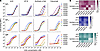 Figure 2
Binding of anti-α-Gal mAbs to ACE, AP-N, synthetic α-Gal–conjugated to BSA, and cetuximab. Titration curves (A) and AUC heat maps (B) of 17 anti-α-Gal mAbs (derived from 13 anti-α-Gal mAb clones) binding to the AGS allergens ACE, AP-N, synthetic α-Gal, and cetuximab. For the heat maps, the color scale ranges from dark [highest log10(AUC) values in each isotype] to white [log10(AUC) = 0]. For IgG1 mAbs binding to cetuximab, the F(ab′)2 fragment of cetuximab was used.
Analysis of the panel, comprising 13 mAbs and 4 reexpressed IgM mAbs, revealed 3 distinct binding patterns across the tested allergens. First, AG030 (IgG1 and IgM isotypes) and AG056 (IgG1) had higher activity against synthetic α-Gal than natural allergen sources, suggesting a preference for structural conformations that are more abundant in synthetic constructs. Second, AG041 (IgG1), AG082 (IgG1 and IgM isotypes), and AG032 (IgM) had higher affinity for natural allergen sources over synthetic α-Gal, suggesting recognition of extended epitopes or conformational features unique to native glycoproteins. Third, AG028 (IgA2 and IgM isotypes) and AG050 (IgA1) demonstrated broad cross reactivity, with consistently high binding activity across all allergens tested. This pattern suggests recognition of a core α-Gal structural determinant that is conserved regardless of its presentation context. Collectively, these results not only confirm the binding of these mAbs to AGS-relevant allergens but also reveal heterogeneity in their recognition patterns, which is dependent on the specific mAb and the nature of the allergen source.
IgE and IgG1–4 responses of patients with AGS to known AGS allergens. Next, with the aim of evaluating the potential functional relevance of the 13 α-Gal–specific mAbs and 4 reexpressed IgM mAbs that bound to AGS allergens, we characterized the serologic profiles of serum samples collected from a cohort of patients with AGS. In total, 88 serum samples from patients with AGS at the University of North Carolina (UNC), Chapel Hill allergy clinic, and 31 plasma samples from healthy adult controls at the UNC clinic and National Institutes of Health, were analyzed. Detailed characteristics of the patients with AGS, including previously determined total and α-Gal–specific IgE levels, are provided in Supplemental Table 3. Serum of patients with AGS had α-Gal–specific IgE levels ranging from 0.1 to > 100 international units (IU)/mL (Supplemental Table 3).
We first profiled the serum samples of this cohort using a multiplex bead assay to determine polyclonal antibody levels to ACE, AP-N, and cetuximab. The multiplex assay also included the α-Gal–containing milk allergens lactoferrin (LF), lactoperoxidase (LPO), and bovine γ-globulin (BGG) (44), which are considered weak AGS inducers, given that approximately 80%–90% of patients with AGS do not react to milk or milk products (45). We also examined antibody responses to bovine and porcine gelatin, since it was unclear if gelatin could trigger an AGS-related reaction (46). Synthetic α-Gal (α-Gal trisaccharide linked to BSA) and human thyroglobulin, which is highly glycosylated but lacks α-Gal, were used as positive and negative controls, respectively (47).
Compared to healthy controls, patients with AGS had elevated IgE reactivities to ACE, AP-N, and cetuximab, but not to milk proteins (Figure 3A and Supplemental Figure 5). Their IgE responses to ACE, AP-N, and cetuximab were similar to their IgE responses to synthetic α-Gal (Figure 3A).
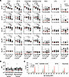 Figure 3
Antibody responses to AGS allergens in serum of patients with AGS. (A) Assessment of serum IgE and IgG1–4 responses of patients with AGS and healthy US individuals to ACE, AP-N, cetuximab, BGG, LF, LPO, and synthetic α-Gal–conjugated to BSA as determined by a multiplex beads assay. 88 AGS serum samples are divided into 3 groups based on the α-Gal–specific IgE levels: ≥ 50 IU (n = 12), 10–49 IU (n = 22), and 0.1–9.9 IU (n = 52). The serum samples of participant UNC0209 were used at 2 different time points due to different α-Gal–specific IgE levels. Healthy US individuals, n=31. For IgG1 reactivity, the F(ab′)2 of cetuximab was used. The MFI of human thyroglobulin (the negative control) was subtracted from each value. (B) AGS patient serum antibody responses to bovine and porcine gelatin. ANOVA multiple comparisons: *P < 0.05; **P < 0.01; ***P < 0.001; ****P < 0.0001. (C) IgE levels of UNC0383.1 serum to allergens in the absence (−) or presence (+α-Gal) of 10 mM α-Gal trisaccharide, and the secondary antibody–only control (2nd Ab).
Next, we assessed IgG1–4 responses to the AGS allergens, since prior studies reported that α-Gal–specific IgG levels were elevated in patients with AGS compared with healthy individuals and could be used as a potential marker of AGS risk (48, 49). As mentioned earlier, we used a cetuximab F(ab′)2 fragment to assess IgG1 mAbs. Patients with AGS showed higher IgG1–3, but not IgG4 binding to ACE, AP-N, synthetic α-Gal, and cetuximab compared with healthy individuals (Figure 3A). IgG1–3 levels to the milk allergens were similar between patients with AGS and healthy controls and tended to be lower than responses to ACE, AP-N, and cetuximab (Figure 3A). Among patients with AGS, we found little to no IgE and IgG1–4 responses to bovine and porcine gelatin except for 1 individual who had high IgG1 levels to bovine and porcine gelatin as well as milk allergens (Figure 3, A and B).
Next, we tested if soluble α-Gal could abolish serum IgE binding to the α-Gal–containing allergen beads to determine whether the IgE responses were specific to the α-Gal moieties. In the presence of 10 mM soluble Gal-α1,3-Gal-β1,4-GlcNAc trisaccharide (soluble α-Gal), the 4 AGS serum samples (UNC0146.2, UNC0147.1, UNC0209.1, and UNC0383.1) with the highest IgE levels to ACE, AP-N, cetuximab, and synthetic α-Gal lost IgE binding to these antigens, confirming that the IgE responses are mainly directed to α-Gal (Figure 3C and Supplemental Figure 5B).
As expected, we found a strong correlation between the MFI of IgE reactive to ACE, AP-N, and cetuximab measured by multiplex bead assay and the α-Gal–specific IgE levels (IU) shown in Supplemental Table 3 (Figure 4A). In contrast, bead-assay measurements of serum antibody reactivities to ACE, AP-N, and cetuximab showed no or only weak correlations between IgE and IgG1–3 as well as between the individual IgG subclasses (Figure 4, B–D). These findings suggested that ACE, AP-N, and cetuximab could be used as representative AGS allergens to further characterize the panel of α-Gal–specific mAbs described above.
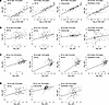 Figure 4
Correlation of reactivities to ACE, AP-N, synthetic α-Gal–conjugated to BSA, and cetuximab between IgE and IgG subclasses in serum from patients with AGS. Graphs were plotted MFIs of 88 patient serum antibodies reactive to the allergen-specific IgE and IgG subclass measured in Figure 3 and α-Gal–specific IgE levels in Supplemental Table 3 were analyzed for correlation. (A) Correlations between α-Gal–specific IgE levels and IgE MFIs specific to ACE, AP-N, cetuximab, and synthetic α-Gal. (B) Correlations between IgE MFIs and IgG subclass MFIs for the indicated antigens. (C) Correlations between IgG2 and IgG subclass MFIs for the indicated antigens. (D) Correlations between IgG3 and IgG subclass MFIs for the indicated antigens. Correlation coefficients (r) and P values were determined by linear fitting using JMP software. Only statistically significant correlations with r2 > 0.05 are shown.
Anti-α-Gal mAbs block IgE binding to ACE and AP-N. We used serum samples of patients with AGS to determine if the 13 α-Gal–specific mAbs and 4 reexpressed IgM mAbs that bound to AGS allergens could block IgE-binding to ACE, AP-N, and cetuximab. For the initial screen, due to limited availability of serum samples from patients with AGS, we only tested inhibition with the serum sample of patient UNC0209.1 (1 of the 4 serum samples that showed the highest IgE reactivity to AGS allergens). We preincubated the α-Gal–specific mAbs with the allergen-bound beads, added serum from AGS patient UNC0209.1, and then assessed the degree of inhibition of serum IgE-binding to AGS allergens (Figure 5A). The mAb AG028, both as an IgA2 and an IgM, interfered with IgE-binding to all 3 allergens (approximately 60%–97% inhibition), whereas AG050 IgA1 inhibited IgE-binding only to ACE and AP-N (approximately 81%–91% inhibition). The remaining mAbs showed low-to-modest inhibition (less than 60% inhibition).
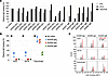 Figure 5
Anti-α-Gal mAbs interfere with polyclonal IgE binding of serum from patients with AGS to ACE, AP-N, and cetuximab. (A) Screening of anti-α-Gal mAbs generated in CHO cells for blocking UNC0209.1 AGS serum IgE binding to ACE, AP-N, and cetuximab. (B) Inhibition by AG028 IgA2, AG050 IgA1, and AG028 IgM mAbs produced in Expi293F cells (50 μg/mL) of polyclonal IgE binding of 4 diluted AGS serum samples (UNC0146.2, UNC0147.1, UNC0209.1, and UNC0383.1). Mean ± SD. (C) Soluble α-Gal competition with anti-α-Gal mAbs for binding to ACE, AP-N, and cetuximab. 10 mM of α-Gal trisaccharide was preincubated with mAbs before adding the allergen beads.
The recombinant mAbs were initially generated in Chinese hamster ovary (CHO) cells which express α1,3-GT. Although it has been reported that anti-α-Gal IgE from patients with AGS is not reactive to therapeutic mAbs generated in CHO cells (50), we reexpressed AG028 IgA2, AG050 IgA1, and AG028 IgM in human Expi293F cells that lack α1,3-GT activity. Since different cell culture conditions can result in variability in mAb characteristics, potentially through posttranslational modification (51), we compared CHO cell– versus Expi293F cell–generated mAbs for allergen binding and interference with AGS serum IgE binding. mAbs expressed in Expi293F cells compared with those expressed in CHO cells exhibited similar binding to the allergens and comparable inhibition of AGS serum IgE binding to allergens (Supplemental Figure 6).
Next, we used 4 serum samples from patients with AGS (UNC0146.2, UNC0147.1, UNC0209.1, and UNC0383.1) with the highest IgE reactivities to AGS allergens to assess whether mAb AG028, as an IgA2 and an IgM, and AG050 IgA1 showed similar inhibition with different AGS serum samples. Across all 4 serum samples, all 3 mAbs inhibited AGS serum IgE binding to ACE and AP-N (Figure 5B). AG028 IgA2 and AG050 IgA1 showed minimal inhibition of IgE binding to cetuximab, compared with approximately 70% inhibition by AG028 IgM (Figure 5B). The 2 mAbs that most effectively inhibited IgE binding to ACE and AP-N were AG028 IgA2/IgM and AG050 IgA1. To confirm that AG028 IgA2, AG050 IgA1, and AG028 IgM bind to α-Gal on ACE and AP-N, we incubated these mAbs with soluble α-Gal, which resulted in a loss of binding to ACE, AP-N, and cetuximab (Figure 5C).
Competition and binding affinity analysis of anti-α-Gal mAbs. Next, we performed competition assays to determine whether the α-Gal–specific mAbs bind overlapping or distinct epitopes and consequently compete with one another for antigen binding. In addition to AG028 and AG050 IgAs, 2 additional α-Gal–specific antibodies were included: the mouse IgM M86 and AG056 IgG1, each with distinct binding properties. M86, originally isolated against rabbit red blood cell membranes, has been widely used to study the structural basis of α-Gal–specific Ab responses (41, 52). AG056 IgG1 exhibited minimal inhibition of serum IgE binding (Figures 5A).
Competition assays were performed in which Alexa Fluor 647–conjugated (AF647-conjugated) AG028 IgA2 or AF647–AG050 IgA1 was competed with unlabeled AG028 IgA2, AG050 IgA1, AG056 IgG1, or M86 IgM for binding to ACE, AP-N, or synthetic α-Gal, and IC50 values were determined for each unlabeled mAb (Figure 6A). Binding of both AF647–AG028 and AF647–AG050 to all α-Gal allergens was strongly competed by M86. In contrast, AG056 showed no competition for ACE or AP-N, but competition for synthetic α-Gal was comparable with AG028. Together, these findings indicate that AG028 and AG050 recognize overlapping α-Gal epitopes similar to M86, rather than epitopes with a preference for synthetic α-Gal. In direct competition between AG028 and AG050, IC50 values were consistently lower when AF647–AG028 was competed with unlabeled AG050 than when AF647–AG050 was competed with unlabeled AG028 across all allergens, indicating that AG050 may bind the allergens with higher apparent affinity than AG028 (Figure 6A).
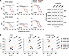 Figure 6
Competition and binding kinetics of AG028, AG050, AG056, and M86 antibodies to AGS allergens. (A) Anti-α-Gal mAbs binding competition assay to allergen-conjugated beads. AF647-AG028 IgA2 (1 μg/mL) or AF647-AG050 IgA1 (4 μg/mL) was competed for binding to allergen-conjugated beads with increasing concentrations (0.01 ng/mL to 100 μg/mL; 1:4 serial dilution) of the unlabeled human mAbs AG028 IgA2, AG050 IgA1, and AG056 IgG1, and the unlabeled mouse mAb M86 IgM. IC50 values were calculated using Prism software with a 4-parameter logistic (4PL) sigmoidal model (X = concentration); curves with R2 > 0.95 are shown; those with R2 < 0.95 were designated as not determined (ND). (B) Two-dimensional isoaffinity kinetic plots show the association (kon) and dissociation (koff) rate constants for antibody binding to ACE, AP-N, and synthetic α-Gal. Each allergen was immobilized on streptavidin biosensors, and binding kinetics were measured using the Octet system. Dashed diagonal lines indicate equilibrium dissociation constants (KD) to facilitate visualization of affinity distributions. The avidities of multivalent antibodies (AG028 IgA2, AG050 IgA1, and AG028 IgM) and affinities of corresponding monovalent Fab fragments (AG028, AG050, AG056, and M86) are shown. Binding kinetics of 4 Fabs for ACE and M86 Fab for AP-N were evaluated in 2 independent experiments, while the remaining samples were performed once.
To determine whether the observed serum IgE inhibition and competitive properties were driven by differences in binding affinity, we measured the binding affinities of 4 Abs in Fab format, as well as the apparent affinities of AG028 IgA2/M and AG050 IgA1 for ACE, AP-N, and synthetic α-Gal using biolayer interferometry (BLI) (Figure 6B, Supplemental Figures 7 and 8, and Supplemental Table 4). In contrast with IgA and IgM Abs, which exhibited strong apparent affinities due to their bi- and multivalent nature (KD range: 28.95–239.4 nM), Fab fragments showed substantially weaker binding affinities, with KD values ranging from 14.29 to 1,846 μM. Although the Fab affinities were generally too weak to allow precise quantitative comparisons, AG028 Fab consistently displayed the lowest binding affinity to all α-Gal allergens, despite AG028 exhibiting strong allergen binding in its IgA or IgM formats. Taken together, the BLI data indicate that differences in serum IgE inhibition and competitive behavior among anti-α-Gal mAbs cannot be explained solely by binding affinity.
Anti-α-Gal mAbs suppress ACE-mediated activation of basophils sensitized with serum from patients with AGS. The α-Gal allergenicity of ACE and AP-N was previously evaluated by the skin prick test and basophil activation test (BAT), which showed that ACE and AP-N at concentrations of 0.1–10 mg/ml activate basophils from patients with AGS, but not healthy individuals (53). To determine whether AG028 IgA2, AG050 IgA1, and AG028 IgM could inhibit AGS allergen-mediated basophil activation, we used an indirect BAT that quantifies the frequency of cells expressing the activation marker CD63+, as described previously (54). Basophils within PBMCs from healthy individuals were subjected to lactic acid stripping of membrane-bound IgE and sensitized with serum from 2 patients with AGS (P1, UNC0445.1 and P2, UNC0512.1). The sensitized cells were then stimulated with ACE or AP-N at a final concentration of 1mg/mL, based on a prior study reporting that this concentration gave near maximal activation with serum samples from 4 patients with AGS (53). CD63 expression on lineage-negative, HLA-DR negative, CD123/CD203c-positive basophils were assessed by flow cytometry in the presence or absence of AG028 IgA2, AG050 IgA1, and AG028 IgM (Figure 7 and Supplemental Figure 10A). VRC01LS, a human IgG1 mAb targeting the CD4-binding site of the HIV-1 envelope glycoprotein (55), does not bind to AGS allergens and was used as a negative control (Supplemental Figure 9).
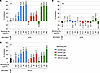 Figure 7
Indirect basophil activation test. Basophils within PBMCs from healthy individuals were stripped of IgE with cold lactic acid and sensitized overnight with serum samples of 2 patients with AGS (P1, UNC0445.1 and P2, UNC0512.1). The sensitized cells were stimulated with 2 ng/mL of IL-3 and 1 mg/ml of ACE or AP-N for 30 minutes at 37°C in the presence or absence of AG028 IgA2, AG050 IgA1, and AG028 IgM at concentrations of 3.12, 12.5, 50, and 200 mg/mL, and then the percentage of lineage-negative, HLA-DR negative, CD123/CD203c-positive basophils that were CD63+ was assessed by flow cytometry. The values above or below the bars indicate the corresponding percent inhibition relative to the allergen-stimulated, nonblocking mAb control. Percent inhibition (% inhibition) was calculated as: [1 — (sample — no stimulator)/(no blocking Ab control)] ′ 100. (A and B) Basophils sensitized with P1 (A) or P2 (B) serum and then stimulated with ACE. (C) Basophils sensitized with P1 serum and stimulated with AP-N. The anti-human IgE heavy chain Ab (α-IgE) was used as a positive control for basophil activation. VRC01LS IgG was used as a negative control. The P2 samples were run in triplicate (B). Due to the limited availability, P1 samples were run once in A and C.
Following stimulation with ACE alone, 55.7% of basophils sensitized with P1 serum (single replicate) were CD63+ (Supplemental Figure 10, B and C), and an average of 71.1% of basophils sensitized with P2 serum (triplicate) were CD63+ (Supplemental Figure 10D). Following stimulation with AP-N alone, 67.3% of basophils sensitized with P1 serum (single replicate) were CD63+ (Supplemental Figure 10, B and E). For basophils sensitized with P1 serum, both AG028 IgA2 and AG028 IgM inhibited ACE-mediated basophil activation, achieving 86.2% and 78.8% inhibition, respectively, at the highest concentration tested (Figure 7A). AG028 IgA2 and AG028 IgM inhibited AP-N–mediated basophil activation by 60.1% and 68.8%, respectively, at the highest concentration tested, which was lower than the inhibition observed for ACE-mediated activation (Figure 7C). AG050 IgA1 showed little effect on ACE- or AP-N-mediated basophil activation (Figure 7, A and C) despite near-complete blocking of polyclonal IgE binding to ACE in Figure 5B. For basophils sensitized with P2 serum, the 3 mAbs showed little inhibition of ACE-mediated basophil activation (Figure 7B).
To confirm that inhibition of basophil activation correlates with mAbs blocking IgE binding to ACE and APN, we repeated the IgE blocking assay using the same P1 and P2 sera (Supplemental Figure 11). With P1 serum, AG028 IgA2, AG050 IgA1, and AG028 IgM reduced IgE binding to ACE by approximately 30%, 40%, and 80%, respectively, with similar inhibition observed for APN. Interestingly, the AG028 clone showed higher functional inhibition regardless of Ig isotypes, IgA or IgM, or IgE blocking ability in P1 serum. In contrast, with P2 serum, only AG028 IgM partially inhibited IgE binding to ACE (45%). Collectively, IgE-binding blockade to ACE/APN by these blocking mAbs and BAT inhibition were not concordant across serum.
Comparison of the sequences of α-Gal-specific mAbs isolated from malaria-exposed individuals versus patients with AGS. Langley et al. reported approximately 400 heavy chain variable region (VH) sequences from single-sorted B cells of 4 individuals with a history of mammalian meat allergy, using AF647-conjugated α-Gal-BSA as a probe (41). Among these sequences, 3 antibodies—HKB7, HKD8, and JEC1—were confirmed for their expression and binding to α-Gal. To determine whether the 42 mAbs isolated from malaria-exposed individuals in this study exhibited any similarity to HKB7, HKD8, JEC1, and the murine mAb M86, we compared their VH sequences. Abalign, a sequence alignment platform for B cell receptor immune repertoire analysis (56), was used to align VH sequences. Subsequently, Geneious Prime was employed to generate an unrooted distance tree to illustrate the evolutionary relationships between VH sequences. Among the α-Gal–specific mAbs compared, the majority of mAbs isolated in this study clustered with the previously identified mAbs (Figure 8A). Notably, AG028, 1 of the 2 mAbs that interfered with serum IgE binding to AGS allergens, appeared on a long branch diverging from the main cluster of other VH sequences, indicating an evolutionary divergence from the main group of VH sequences (Figure 8A). Additionally, we compared the VH sequences of AG028 and AG050, both of which interfered with serum IgE binding to AGS allergens, and AG056, which did not, with those of HKB7, HKD8, JEC1, and M86. AG028 was the most divergent from other α-Gal antibodies in the unrooted distance tree (Figure 8B). The CDR3 length of AG028 was 17 amino acids, longer than the average CDR3 length of 13.1 observed across 42 α-Gal mAbs and substantially longer than the 4 reported α-Gal antibodies, which ranged from 9–11 amino acids (Figure 8C). Additionally, AG028 harbors serine at VH Kabat position 33, a residue that is relatively rare among α-Gal antibodies (40) (Figure 8C). These sequence characteristics of AG028 suggest that this antibody may utilize a distinct binding mechanism to interfere with serum IgE binding to AGS allergens.
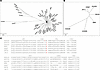 Figure 8
Comparison of VH sequences of α-Gal–specific mAbs derived from malaria-exposed individuals and patients with AGS. Abalign and Geneious Prime were used to align the VH sequences and to generate unrooted distance trees, respectively. (A) An unrooted distance tree that includes the 42 mAbs isolated from malaria-exposed individuals in this study (beginning with “AG”) along with the previously identified α-Gal–specific mAbs from patients with AGS (HKB7, HKD8, JEC1) and the murine mAb M86. (B) An unrooted distance tree that includes AG028, AG050, AG056, HKB7, HKD8, JEC1, and M86. (C) VH sequence alignment of AG028, AG050, AG056, HKB7, HKD8, JEC1, and M86, with CDRs shown in bold and residues at Kabat position 33 highlighted in red. FR; framework region.


Copyright © 2026 American Society for Clinical Investigation
ISSN: 0021-9738 (print), 1558-8238 (online)