
Advertisement
Research ArticleDermatologyImmunologyInfectious disease
Open Access |  10.1172/JCI188827
CARD9-dependent macrophage plasticity regulates effective fungal clearance
Lu Zhang,1,2,3,4 Zhichun Tang,5 Yi Zhang,1,2,3,4 Wenjie Liu,1,2,3,4 Haitao Jiang,6,7 Li Yu,6,7 Kexin Lei,1,2,3,4 Yubo Ma,1,2,3,4 Yang-Xin Fu,6,7 Ruoyu Li,1,2,3,4 Wenyan Wang,6,7 Fan Bai,5,8,9,10 and Xiaowen Wang1,2,3,4
1Department of Dermatology and Venerology, Peking University First Hospital, Beijing, China.
2Research Center for Medical Mycology, Peking University, Beijing, China.
3Beijing Key Laboratory of Molecular Diagnosis on Dermatoses, Beijing, China.
4National Clinical Research Center for Skin and Immune Diseases, Beijing, China.
5Biomedical Pioneering Innovation Center (BIOPIC) and School of Life Sciences, Peking University, Beijing, China.
6School of Basic Medical Sciences and
7State Key Laboratory of Molecular Oncology, Tsinghua University, Beijing, China.
8Peking-Tsinghua Center for Life Sciences (CLS), Peking University, Beijing, China.
9State Key Laboratory of Metabolic Dysregulation and Prevention and Treatment of Esophageal Cancer, BIOPIC, Peking University, Beijing, China.
10Peking University Beijing-Tianjin-Hebei Biomedical Pioneering Innovation Center, Tianjin, China.
Address correspondence to: Xiaowen Wang, Department of Dermatology and Venereology, Peking University First Hospital, Research Center for Medical Mycology, Peking University, 8 Xishiku Street, Xicheng District, 100034 Beijing, China, xiaowenpku@126.com. Or to: Bai Fan, Biomedical Pioneering Innovation Center (BIOPIC), Peking-Tsinghua Center for Life Sciences (CLS), School of Life Sciences, Peking University, Beijing 100871, China, fbai@pku.edu.cn. Or to: Wenyan Wang, School of Basic Medical Sciences, State Key Laboratory of Molecular Oncology, Tsinghua University, Beijing 100084, China, wywang2022@tsinghua.edu.cn.
Authorship note: LZ, ZT, YZ, and WL contributed equally to this work. WW, FB, and XW contributed equally to this work.
Find articles by Zhang, L. in: PubMed | Google Scholar
1Department of Dermatology and Venerology, Peking University First Hospital, Beijing, China.
2Research Center for Medical Mycology, Peking University, Beijing, China.
3Beijing Key Laboratory of Molecular Diagnosis on Dermatoses, Beijing, China.
4National Clinical Research Center for Skin and Immune Diseases, Beijing, China.
5Biomedical Pioneering Innovation Center (BIOPIC) and School of Life Sciences, Peking University, Beijing, China.
6School of Basic Medical Sciences and
7State Key Laboratory of Molecular Oncology, Tsinghua University, Beijing, China.
8Peking-Tsinghua Center for Life Sciences (CLS), Peking University, Beijing, China.
9State Key Laboratory of Metabolic Dysregulation and Prevention and Treatment of Esophageal Cancer, BIOPIC, Peking University, Beijing, China.
10Peking University Beijing-Tianjin-Hebei Biomedical Pioneering Innovation Center, Tianjin, China.
Address correspondence to: Xiaowen Wang, Department of Dermatology and Venereology, Peking University First Hospital, Research Center for Medical Mycology, Peking University, 8 Xishiku Street, Xicheng District, 100034 Beijing, China, xiaowenpku@126.com. Or to: Bai Fan, Biomedical Pioneering Innovation Center (BIOPIC), Peking-Tsinghua Center for Life Sciences (CLS), School of Life Sciences, Peking University, Beijing 100871, China, fbai@pku.edu.cn. Or to: Wenyan Wang, School of Basic Medical Sciences, State Key Laboratory of Molecular Oncology, Tsinghua University, Beijing 100084, China, wywang2022@tsinghua.edu.cn.
Authorship note: LZ, ZT, YZ, and WL contributed equally to this work. WW, FB, and XW contributed equally to this work.
Find articles by Tang, Z. in: PubMed | Google Scholar
1Department of Dermatology and Venerology, Peking University First Hospital, Beijing, China.
2Research Center for Medical Mycology, Peking University, Beijing, China.
3Beijing Key Laboratory of Molecular Diagnosis on Dermatoses, Beijing, China.
4National Clinical Research Center for Skin and Immune Diseases, Beijing, China.
5Biomedical Pioneering Innovation Center (BIOPIC) and School of Life Sciences, Peking University, Beijing, China.
6School of Basic Medical Sciences and
7State Key Laboratory of Molecular Oncology, Tsinghua University, Beijing, China.
8Peking-Tsinghua Center for Life Sciences (CLS), Peking University, Beijing, China.
9State Key Laboratory of Metabolic Dysregulation and Prevention and Treatment of Esophageal Cancer, BIOPIC, Peking University, Beijing, China.
10Peking University Beijing-Tianjin-Hebei Biomedical Pioneering Innovation Center, Tianjin, China.
Address correspondence to: Xiaowen Wang, Department of Dermatology and Venereology, Peking University First Hospital, Research Center for Medical Mycology, Peking University, 8 Xishiku Street, Xicheng District, 100034 Beijing, China, xiaowenpku@126.com. Or to: Bai Fan, Biomedical Pioneering Innovation Center (BIOPIC), Peking-Tsinghua Center for Life Sciences (CLS), School of Life Sciences, Peking University, Beijing 100871, China, fbai@pku.edu.cn. Or to: Wenyan Wang, School of Basic Medical Sciences, State Key Laboratory of Molecular Oncology, Tsinghua University, Beijing 100084, China, wywang2022@tsinghua.edu.cn.
Authorship note: LZ, ZT, YZ, and WL contributed equally to this work. WW, FB, and XW contributed equally to this work.
Find articles by Zhang, Y. in: PubMed | Google Scholar
1Department of Dermatology and Venerology, Peking University First Hospital, Beijing, China.
2Research Center for Medical Mycology, Peking University, Beijing, China.
3Beijing Key Laboratory of Molecular Diagnosis on Dermatoses, Beijing, China.
4National Clinical Research Center for Skin and Immune Diseases, Beijing, China.
5Biomedical Pioneering Innovation Center (BIOPIC) and School of Life Sciences, Peking University, Beijing, China.
6School of Basic Medical Sciences and
7State Key Laboratory of Molecular Oncology, Tsinghua University, Beijing, China.
8Peking-Tsinghua Center for Life Sciences (CLS), Peking University, Beijing, China.
9State Key Laboratory of Metabolic Dysregulation and Prevention and Treatment of Esophageal Cancer, BIOPIC, Peking University, Beijing, China.
10Peking University Beijing-Tianjin-Hebei Biomedical Pioneering Innovation Center, Tianjin, China.
Address correspondence to: Xiaowen Wang, Department of Dermatology and Venereology, Peking University First Hospital, Research Center for Medical Mycology, Peking University, 8 Xishiku Street, Xicheng District, 100034 Beijing, China, xiaowenpku@126.com. Or to: Bai Fan, Biomedical Pioneering Innovation Center (BIOPIC), Peking-Tsinghua Center for Life Sciences (CLS), School of Life Sciences, Peking University, Beijing 100871, China, fbai@pku.edu.cn. Or to: Wenyan Wang, School of Basic Medical Sciences, State Key Laboratory of Molecular Oncology, Tsinghua University, Beijing 100084, China, wywang2022@tsinghua.edu.cn.
Authorship note: LZ, ZT, YZ, and WL contributed equally to this work. WW, FB, and XW contributed equally to this work.
Find articles by Liu, W. in: PubMed | Google Scholar
1Department of Dermatology and Venerology, Peking University First Hospital, Beijing, China.
2Research Center for Medical Mycology, Peking University, Beijing, China.
3Beijing Key Laboratory of Molecular Diagnosis on Dermatoses, Beijing, China.
4National Clinical Research Center for Skin and Immune Diseases, Beijing, China.
5Biomedical Pioneering Innovation Center (BIOPIC) and School of Life Sciences, Peking University, Beijing, China.
6School of Basic Medical Sciences and
7State Key Laboratory of Molecular Oncology, Tsinghua University, Beijing, China.
8Peking-Tsinghua Center for Life Sciences (CLS), Peking University, Beijing, China.
9State Key Laboratory of Metabolic Dysregulation and Prevention and Treatment of Esophageal Cancer, BIOPIC, Peking University, Beijing, China.
10Peking University Beijing-Tianjin-Hebei Biomedical Pioneering Innovation Center, Tianjin, China.
Address correspondence to: Xiaowen Wang, Department of Dermatology and Venereology, Peking University First Hospital, Research Center for Medical Mycology, Peking University, 8 Xishiku Street, Xicheng District, 100034 Beijing, China, xiaowenpku@126.com. Or to: Bai Fan, Biomedical Pioneering Innovation Center (BIOPIC), Peking-Tsinghua Center for Life Sciences (CLS), School of Life Sciences, Peking University, Beijing 100871, China, fbai@pku.edu.cn. Or to: Wenyan Wang, School of Basic Medical Sciences, State Key Laboratory of Molecular Oncology, Tsinghua University, Beijing 100084, China, wywang2022@tsinghua.edu.cn.
Authorship note: LZ, ZT, YZ, and WL contributed equally to this work. WW, FB, and XW contributed equally to this work.
Find articles by Jiang, H. in: PubMed | Google Scholar
1Department of Dermatology and Venerology, Peking University First Hospital, Beijing, China.
2Research Center for Medical Mycology, Peking University, Beijing, China.
3Beijing Key Laboratory of Molecular Diagnosis on Dermatoses, Beijing, China.
4National Clinical Research Center for Skin and Immune Diseases, Beijing, China.
5Biomedical Pioneering Innovation Center (BIOPIC) and School of Life Sciences, Peking University, Beijing, China.
6School of Basic Medical Sciences and
7State Key Laboratory of Molecular Oncology, Tsinghua University, Beijing, China.
8Peking-Tsinghua Center for Life Sciences (CLS), Peking University, Beijing, China.
9State Key Laboratory of Metabolic Dysregulation and Prevention and Treatment of Esophageal Cancer, BIOPIC, Peking University, Beijing, China.
10Peking University Beijing-Tianjin-Hebei Biomedical Pioneering Innovation Center, Tianjin, China.
Address correspondence to: Xiaowen Wang, Department of Dermatology and Venereology, Peking University First Hospital, Research Center for Medical Mycology, Peking University, 8 Xishiku Street, Xicheng District, 100034 Beijing, China, xiaowenpku@126.com. Or to: Bai Fan, Biomedical Pioneering Innovation Center (BIOPIC), Peking-Tsinghua Center for Life Sciences (CLS), School of Life Sciences, Peking University, Beijing 100871, China, fbai@pku.edu.cn. Or to: Wenyan Wang, School of Basic Medical Sciences, State Key Laboratory of Molecular Oncology, Tsinghua University, Beijing 100084, China, wywang2022@tsinghua.edu.cn.
Authorship note: LZ, ZT, YZ, and WL contributed equally to this work. WW, FB, and XW contributed equally to this work.
Find articles by Yu, L. in: PubMed | Google Scholar
1Department of Dermatology and Venerology, Peking University First Hospital, Beijing, China.
2Research Center for Medical Mycology, Peking University, Beijing, China.
3Beijing Key Laboratory of Molecular Diagnosis on Dermatoses, Beijing, China.
4National Clinical Research Center for Skin and Immune Diseases, Beijing, China.
5Biomedical Pioneering Innovation Center (BIOPIC) and School of Life Sciences, Peking University, Beijing, China.
6School of Basic Medical Sciences and
7State Key Laboratory of Molecular Oncology, Tsinghua University, Beijing, China.
8Peking-Tsinghua Center for Life Sciences (CLS), Peking University, Beijing, China.
9State Key Laboratory of Metabolic Dysregulation and Prevention and Treatment of Esophageal Cancer, BIOPIC, Peking University, Beijing, China.
10Peking University Beijing-Tianjin-Hebei Biomedical Pioneering Innovation Center, Tianjin, China.
Address correspondence to: Xiaowen Wang, Department of Dermatology and Venereology, Peking University First Hospital, Research Center for Medical Mycology, Peking University, 8 Xishiku Street, Xicheng District, 100034 Beijing, China, xiaowenpku@126.com. Or to: Bai Fan, Biomedical Pioneering Innovation Center (BIOPIC), Peking-Tsinghua Center for Life Sciences (CLS), School of Life Sciences, Peking University, Beijing 100871, China, fbai@pku.edu.cn. Or to: Wenyan Wang, School of Basic Medical Sciences, State Key Laboratory of Molecular Oncology, Tsinghua University, Beijing 100084, China, wywang2022@tsinghua.edu.cn.
Authorship note: LZ, ZT, YZ, and WL contributed equally to this work. WW, FB, and XW contributed equally to this work.
Find articles by Lei, K. in: PubMed | Google Scholar
1Department of Dermatology and Venerology, Peking University First Hospital, Beijing, China.
2Research Center for Medical Mycology, Peking University, Beijing, China.
3Beijing Key Laboratory of Molecular Diagnosis on Dermatoses, Beijing, China.
4National Clinical Research Center for Skin and Immune Diseases, Beijing, China.
5Biomedical Pioneering Innovation Center (BIOPIC) and School of Life Sciences, Peking University, Beijing, China.
6School of Basic Medical Sciences and
7State Key Laboratory of Molecular Oncology, Tsinghua University, Beijing, China.
8Peking-Tsinghua Center for Life Sciences (CLS), Peking University, Beijing, China.
9State Key Laboratory of Metabolic Dysregulation and Prevention and Treatment of Esophageal Cancer, BIOPIC, Peking University, Beijing, China.
10Peking University Beijing-Tianjin-Hebei Biomedical Pioneering Innovation Center, Tianjin, China.
Address correspondence to: Xiaowen Wang, Department of Dermatology and Venereology, Peking University First Hospital, Research Center for Medical Mycology, Peking University, 8 Xishiku Street, Xicheng District, 100034 Beijing, China, xiaowenpku@126.com. Or to: Bai Fan, Biomedical Pioneering Innovation Center (BIOPIC), Peking-Tsinghua Center for Life Sciences (CLS), School of Life Sciences, Peking University, Beijing 100871, China, fbai@pku.edu.cn. Or to: Wenyan Wang, School of Basic Medical Sciences, State Key Laboratory of Molecular Oncology, Tsinghua University, Beijing 100084, China, wywang2022@tsinghua.edu.cn.
Authorship note: LZ, ZT, YZ, and WL contributed equally to this work. WW, FB, and XW contributed equally to this work.
Find articles by Ma, Y. in: PubMed | Google Scholar
1Department of Dermatology and Venerology, Peking University First Hospital, Beijing, China.
2Research Center for Medical Mycology, Peking University, Beijing, China.
3Beijing Key Laboratory of Molecular Diagnosis on Dermatoses, Beijing, China.
4National Clinical Research Center for Skin and Immune Diseases, Beijing, China.
5Biomedical Pioneering Innovation Center (BIOPIC) and School of Life Sciences, Peking University, Beijing, China.
6School of Basic Medical Sciences and
7State Key Laboratory of Molecular Oncology, Tsinghua University, Beijing, China.
8Peking-Tsinghua Center for Life Sciences (CLS), Peking University, Beijing, China.
9State Key Laboratory of Metabolic Dysregulation and Prevention and Treatment of Esophageal Cancer, BIOPIC, Peking University, Beijing, China.
10Peking University Beijing-Tianjin-Hebei Biomedical Pioneering Innovation Center, Tianjin, China.
Address correspondence to: Xiaowen Wang, Department of Dermatology and Venereology, Peking University First Hospital, Research Center for Medical Mycology, Peking University, 8 Xishiku Street, Xicheng District, 100034 Beijing, China, xiaowenpku@126.com. Or to: Bai Fan, Biomedical Pioneering Innovation Center (BIOPIC), Peking-Tsinghua Center for Life Sciences (CLS), School of Life Sciences, Peking University, Beijing 100871, China, fbai@pku.edu.cn. Or to: Wenyan Wang, School of Basic Medical Sciences, State Key Laboratory of Molecular Oncology, Tsinghua University, Beijing 100084, China, wywang2022@tsinghua.edu.cn.
Authorship note: LZ, ZT, YZ, and WL contributed equally to this work. WW, FB, and XW contributed equally to this work.
Find articles by Fu, Y. in: PubMed | Google Scholar
1Department of Dermatology and Venerology, Peking University First Hospital, Beijing, China.
2Research Center for Medical Mycology, Peking University, Beijing, China.
3Beijing Key Laboratory of Molecular Diagnosis on Dermatoses, Beijing, China.
4National Clinical Research Center for Skin and Immune Diseases, Beijing, China.
5Biomedical Pioneering Innovation Center (BIOPIC) and School of Life Sciences, Peking University, Beijing, China.
6School of Basic Medical Sciences and
7State Key Laboratory of Molecular Oncology, Tsinghua University, Beijing, China.
8Peking-Tsinghua Center for Life Sciences (CLS), Peking University, Beijing, China.
9State Key Laboratory of Metabolic Dysregulation and Prevention and Treatment of Esophageal Cancer, BIOPIC, Peking University, Beijing, China.
10Peking University Beijing-Tianjin-Hebei Biomedical Pioneering Innovation Center, Tianjin, China.
Address correspondence to: Xiaowen Wang, Department of Dermatology and Venereology, Peking University First Hospital, Research Center for Medical Mycology, Peking University, 8 Xishiku Street, Xicheng District, 100034 Beijing, China, xiaowenpku@126.com. Or to: Bai Fan, Biomedical Pioneering Innovation Center (BIOPIC), Peking-Tsinghua Center for Life Sciences (CLS), School of Life Sciences, Peking University, Beijing 100871, China, fbai@pku.edu.cn. Or to: Wenyan Wang, School of Basic Medical Sciences, State Key Laboratory of Molecular Oncology, Tsinghua University, Beijing 100084, China, wywang2022@tsinghua.edu.cn.
Authorship note: LZ, ZT, YZ, and WL contributed equally to this work. WW, FB, and XW contributed equally to this work.
Find articles by Li, R. in: PubMed | Google Scholar
1Department of Dermatology and Venerology, Peking University First Hospital, Beijing, China.
2Research Center for Medical Mycology, Peking University, Beijing, China.
3Beijing Key Laboratory of Molecular Diagnosis on Dermatoses, Beijing, China.
4National Clinical Research Center for Skin and Immune Diseases, Beijing, China.
5Biomedical Pioneering Innovation Center (BIOPIC) and School of Life Sciences, Peking University, Beijing, China.
6School of Basic Medical Sciences and
7State Key Laboratory of Molecular Oncology, Tsinghua University, Beijing, China.
8Peking-Tsinghua Center for Life Sciences (CLS), Peking University, Beijing, China.
9State Key Laboratory of Metabolic Dysregulation and Prevention and Treatment of Esophageal Cancer, BIOPIC, Peking University, Beijing, China.
10Peking University Beijing-Tianjin-Hebei Biomedical Pioneering Innovation Center, Tianjin, China.
Address correspondence to: Xiaowen Wang, Department of Dermatology and Venereology, Peking University First Hospital, Research Center for Medical Mycology, Peking University, 8 Xishiku Street, Xicheng District, 100034 Beijing, China, xiaowenpku@126.com. Or to: Bai Fan, Biomedical Pioneering Innovation Center (BIOPIC), Peking-Tsinghua Center for Life Sciences (CLS), School of Life Sciences, Peking University, Beijing 100871, China, fbai@pku.edu.cn. Or to: Wenyan Wang, School of Basic Medical Sciences, State Key Laboratory of Molecular Oncology, Tsinghua University, Beijing 100084, China, wywang2022@tsinghua.edu.cn.
Authorship note: LZ, ZT, YZ, and WL contributed equally to this work. WW, FB, and XW contributed equally to this work.
Find articles by Wang, W. in: PubMed | Google Scholar
1Department of Dermatology and Venerology, Peking University First Hospital, Beijing, China.
2Research Center for Medical Mycology, Peking University, Beijing, China.
3Beijing Key Laboratory of Molecular Diagnosis on Dermatoses, Beijing, China.
4National Clinical Research Center for Skin and Immune Diseases, Beijing, China.
5Biomedical Pioneering Innovation Center (BIOPIC) and School of Life Sciences, Peking University, Beijing, China.
6School of Basic Medical Sciences and
7State Key Laboratory of Molecular Oncology, Tsinghua University, Beijing, China.
8Peking-Tsinghua Center for Life Sciences (CLS), Peking University, Beijing, China.
9State Key Laboratory of Metabolic Dysregulation and Prevention and Treatment of Esophageal Cancer, BIOPIC, Peking University, Beijing, China.
10Peking University Beijing-Tianjin-Hebei Biomedical Pioneering Innovation Center, Tianjin, China.
Address correspondence to: Xiaowen Wang, Department of Dermatology and Venereology, Peking University First Hospital, Research Center for Medical Mycology, Peking University, 8 Xishiku Street, Xicheng District, 100034 Beijing, China, xiaowenpku@126.com. Or to: Bai Fan, Biomedical Pioneering Innovation Center (BIOPIC), Peking-Tsinghua Center for Life Sciences (CLS), School of Life Sciences, Peking University, Beijing 100871, China, fbai@pku.edu.cn. Or to: Wenyan Wang, School of Basic Medical Sciences, State Key Laboratory of Molecular Oncology, Tsinghua University, Beijing 100084, China, wywang2022@tsinghua.edu.cn.
Authorship note: LZ, ZT, YZ, and WL contributed equally to this work. WW, FB, and XW contributed equally to this work.
Find articles by
Bai, F.
in:
PubMed
|
Google Scholar
|

1Department of Dermatology and Venerology, Peking University First Hospital, Beijing, China.
2Research Center for Medical Mycology, Peking University, Beijing, China.
3Beijing Key Laboratory of Molecular Diagnosis on Dermatoses, Beijing, China.
4National Clinical Research Center for Skin and Immune Diseases, Beijing, China.
5Biomedical Pioneering Innovation Center (BIOPIC) and School of Life Sciences, Peking University, Beijing, China.
6School of Basic Medical Sciences and
7State Key Laboratory of Molecular Oncology, Tsinghua University, Beijing, China.
8Peking-Tsinghua Center for Life Sciences (CLS), Peking University, Beijing, China.
9State Key Laboratory of Metabolic Dysregulation and Prevention and Treatment of Esophageal Cancer, BIOPIC, Peking University, Beijing, China.
10Peking University Beijing-Tianjin-Hebei Biomedical Pioneering Innovation Center, Tianjin, China.
Address correspondence to: Xiaowen Wang, Department of Dermatology and Venereology, Peking University First Hospital, Research Center for Medical Mycology, Peking University, 8 Xishiku Street, Xicheng District, 100034 Beijing, China, xiaowenpku@126.com. Or to: Bai Fan, Biomedical Pioneering Innovation Center (BIOPIC), Peking-Tsinghua Center for Life Sciences (CLS), School of Life Sciences, Peking University, Beijing 100871, China, fbai@pku.edu.cn. Or to: Wenyan Wang, School of Basic Medical Sciences, State Key Laboratory of Molecular Oncology, Tsinghua University, Beijing 100084, China, wywang2022@tsinghua.edu.cn.
Authorship note: LZ, ZT, YZ, and WL contributed equally to this work. WW, FB, and XW contributed equally to this work.
Find articles by Wang, X. in: PubMed | Google Scholar
Published December 2, 2025 - More info
J Clin Invest. 2026;136(3):e188827. https://doi.org/10.1172/JCI188827.
© 2025 Zhang et al. This work is licensed under the Creative Commons Attribution 4.0 International License. To view a copy of this license, visit http://creativecommons.org/licenses/by/4.0/.
Received: November 4, 2024; Accepted: November 25, 2025


-
Results
CARD9 is necessary for defense against subcutaneous dematiaceous fungal infection. Previous studies indicated that patients with CARD9 deficiency are susceptible to severe dematiaceous fungal infections; however, its underlying mechanisms remain poorly understood. Phialophora verrucosa is the most commonly identified causative fungus in patients with CARD9 deficiency with phaeohyphomycosis (32). To investigate the role of CARD9 in shaping host protective immunity against dematiaceous fungi, we first modeled subcutaneous phaeohyphomycosis with footpad inoculation of P. verrucosa in WT and Card9-knockout (Card9–/–) mice. After fungal inoculation, the 2 groups exhibited different patterns of footpad swelling. WT mice showed more pronounced swelling on day 3, with comparable levels between groups on day 7, followed by gradual resolution. However, Card9–/– mice experienced progressive deterioration, with increased swelling on day 10 and more pronounced swelling on day 14 and thereafter (Figure 1, A and B).
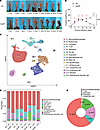 Figure 1
CARD9 is necessary for defense against subcutaneous dematiaceous fungal infection. (A) Natural course of subcutaneous infection with P. verrucosa in WT and Card9–/– mice. (B) Footpad swelling of P. verrucosa–infected WT and Card9–/– mice at different time points after infection (n = 3). (C) The uniform manifold approximation and projection (UMAP) plot presents the projection of 59,396 high-quality cells from 8 scRNA-seq samples, comprising 4 samples each from WT and Card9–/– group. Each point on the plot represents a single cell, with colors varying according to distinct cell types. (D) The stacked bar chart shows the percentage distribution of 10 immune cell types across all samples. The colors representing each cell type are consistent with those shown in C. (E) The pie chart depicts the distribution of Card9+ cells among immune cell subsets within lesional skin. Data were integrated from all samples. Data are representative of 3 independent experiments and are shown as the mean ± SD. ****P < 0.0001, by 2-way ANOVA (B).
To elucidate the cellular and molecular mechanisms underlying CARD9-mediated antifungal immune responses during subcutaneous P. verrucosa infection, we conducted scRNA-seq of total cells in mouse footpads at 3, 7, 10, and 14 days after infection (Figure 1B). After data preprocessing and quality control, we partitioned the cells into 15 major clusters and labeled them based on representative marker genes, including 10 immune cell clusters and 5 nonimmune cells (Figure 1C). Overall, on day 3 after infection, neutrophils, monocytes, and macrophages were the predominant immune cells in both mouse strains, with Card9–/– mice having fewer neutrophils and more monocytes and macrophages than did WT control mice (Figure 1D). On day 7 after infection, the proportions of T and NK cells increased in the skin lesions of WT mice and remained elevated through days 10 and 14 (Figure 1D). In contrast, Card9–/– mice had reduced T and NK cell infiltration but an increased proportion of eosinophils in the lesions compared with WT mice (Figure 1D).
To elucidate the role of CARD9 in modulating the recruitment and function of local immune cells in lesions, we analyzed the distribution of Card9-expressing cells in this model. Macrophages constituted the predominant population, comprising 61.69% of Card9+ cells (Figure 1E). This predominant expression underscores macrophages as the pivotal cellular subset for subsequent in-depth analysis.
TREM2hi macrophages display antiinflammatory signatures and are increased in Card9–/– mice. Macrophages, a pivotal cell type in antifungal immunity, are among the main expressers of CARD9. We conducted further subpopulation analyses and identified 5 distinct macrophage subsets: Cxcl3hi, Ccl5hi, Trem2hi, Il10hi, and Mrc1hi macrophages (Figure 2A). The proportions of Cxcl3hi macrophage and Ccl5hi macrophage were higher in WT mice, whereas the TREM2hi macrophage subset was considerably more abundant in Card9–/– murine lesions (Figure 2B). Notably, the TREM2hi macrophage subset was characterized by high expression of Trem2, Lgals3, and Spp1 (Figure 2C), a transcriptional profile consistent with the gene signature previously described for skin monocyte–derived macrophages (39). In addition, the analysis of monocyte-associated gene programs across macrophage subsets provided further indications that this population represents a monocyte-derived macrophage lineage (Supplemental Figure 1A; supplemental material available online with this article; https://doi.org/10.1172/JCI188827DS1).
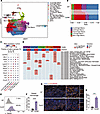 Figure 2
TREM2hi macrophages display antiinflammatory signatures and are increased in Card9–/– mice. (A) UMAP of myeloid cells. Each dot represents a single cell, with colors varying according to distinct cell subpopulations. (B) The Sankey diagram illustrates the proportional differences between the major macrophage subpopulations across the 2 groups. (C) The bubble chart shows the mean relative expression of signature genes across the major macrophage subpopulations. (D) The heatmap illustrates the differences in HALLMARK gene set scores among the major macrophage subpopulations, with scores calculated via the irGSEA R package (https://github.com/chuiqin/irGSEA/). Only gene set scores exhibiting differences across subpopulations are presented. (E and F) Representative flow cytometry histogram plots for TREM2 staining (E) and frequency of TREM2+ macrophage subsets (F) in murine footpad lesions at day 10 after infection. One data point denotes a result from 1 mouse (n = 5). RRA, robust rank aggregation. (G and H) Staining of TREM2+ macrophages (TREM2 and F4/80) in murine lesions. Scale bars: 100 μm (left) and 20 μm (right). Bar plots show the quantification results (H). One data point represents the statistical result of 1 field of view (n = 3 fields analyzed per condition). Data are shown as the mean ± SD. ***P < 0.001 and ***P < 0.0001, by 2-tailed Student’s t test (F and H).
HALLMARK gene set scoring among the major macrophage subsets revealed that the TREM2hi macrophages exhibited notably reduced activities in the NF-κB signaling pathway and pathways related to proinflammatory cytokines, such as TNF-α, IFN-γ, and IL-6 (Figure 2D). Further flow cytometry analysis demonstrated that the proportion of TREM2+ macrophages among all macrophages was markedly higher in Card9–/– murine lesions than in WT murine lesions on day 10 after infection (Figures 2, E and F), whereas no differences were observed prior to infection (Supplemental Figure 1B). Multiplex immunofluorescence (mIHC) experiments also showed higher TREM2 expression and its colocalization with the macrophage marker F4/80 in lesions of Card9–/– mice (Figure 2, G and H).
The collective findings indicate CARD9 deficiency does not notably affect macrophage recruitment to the infection site but rather substantially alters macrophage phenotypes in response to subcutaneous P. verrucosa infection. A similar pattern of increased recruitment but impaired functional responses has been reported in CARD9-deficient mice challenged with C. albicans (18). Together, these observations suggest CARD9 regulates macrophage plasticity, potentially impairing antifungal effector functions.
Neutrophils are another type of immune cell that mainly expresses CARD9 and plays a critical role in antifungal immunity. We performed a detailed subpopulation analysis of neutrophils to investigate their heterogeneity, identifying 8 subsets (Supplemental Figure 1C). Neu-C1, characterized by high expression of S100a9 and Nfkb1, and Neu-C2, enriched for Tnf and Il23a, were predominantly enriched in the WT mice, whereas Neu-C4, marked by elevated Apoe and Il10, was more abundant in the Card9–/– mice. (Supplemental Figure 1, D–F). Subsequent comparative analyses further revealed that multiple proinflammatory cytokines and chemokines, including Il1b, Tnf, Ccl5, Cxcl9, and Cxcl10, were downregulated in the Card9–/– group (Supplemental Figure 1G). Gene ontology (GO) enrichment analysis consistently indicated that key pathways associated with cytokine production, ROS response, and other immune-related processes were suppressed in Card9–/– neutrophils (Supplemental Figure 1H).
Collectively, these findings are consistent with those of previous studies, supporting the notion that CARD9 is essential for maintaining neutrophil function in antifungal immunity. Future studies may help to elucidate its detailed regulatory mechanisms.
Exhaustion-like Th1 cells are more pronounced in Card9–/– mice and correlate with antiinflammatory macrophages. CARD9 has been shown to regulate the activation of innate immune cells and the production of cytokines, thereby shaping adaptive T cell responses (7). Due to the altered macrophage phenotypes and reduction in T cells observed in Card9–/– mice infected with P. verrucosa, we conducted a subpopulation analysis of T cells (Figure 3A). We found Card9–/– mice had a higher frequency of Tregs and Th2 cells but a lower proportion of Th1 cells than did WT mice (Figure 3B and Supplemental Figure 2A).
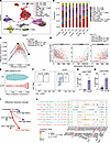 Figure 3
Increased abundance of exhaustion-like Th1 cells in Card9–/– mice. (A) UMAP of 9 T cell subsets. Each dot represents a single cell, colored according to the specific cell type. (B) The stacked bar chart shows the percentage distribution of 9 T cell subsets across all samples. (C) The diffusion map illustrates the developmental trajectory of Th1 cells, with the direction of development indicated by arrows. This process was implemented by using the R package destiny (https://github.com/theislab/destiny). (D) Scatter plots show the expression of naive and exhaustion markers throughout the pseudotime of the Th1 cell development process. The dashed line represented the fitted trend of changes. Each point represented a Th1 cell, with its color corresponding to that in C. (E) Scatter plot shows the distribution of Th1 cells in the WT group and the Card9–/– group. Each point represents a Th1 cell, with its x-axis corresponding to the ordinal value of diffusion component 1, arranged from smallest to largest in Supplemental Figure 3C. (F–H) Representative flow cytometry plots for TIM3 and LAG3 staining and frequency of LAG3+TIM3+CD4+ T cell subsets (G) and LAG3–TIM3+CD4+ T cell subsets (H) in murine footpad lesion at day 10 after infection. One data point denotes a result from 1 mouse. (I) The circle plot demonstrates the enhanced interaction strength between the major macrophage subpopulations and exhaustion-like Th1 cells in the Card9–/– group. (J) The bubble plot shows the main ligand-receptor pair between the major macrophage subpopulations and exhaustion-like Th1 cells in WT and Card9–/– group. Commun. Prob., communication probability. Data are shown as the mean ± SD. **P < 0.01 and ***P < 0.001, by 2-tailed Student’s t test (G and H), and pairwise Wilcoxon rank sum test (J).
To gain further insight into the functional status of the predominant T cell subsets, we performed a pseudotime trajectory analysis of Th1 cells (Figure 3C). The results revealed a progressive shift from a naive state through an effector phenotype and, ultimately, to an exhausted state during infection (Figure 3, C and D). Notably, Card9–/– mice had a greater accumulation of exhaustion-like Th1 cells within the lesions on days 10 and 14 after infection (Figure 3E and Supplemental Figure 2, B and C). Flow cytometry analysis confirmed that CD4+ T cells in Card9–/– murine lesions exhibited markedly higher expression of immune checkpoints than those in WT control mice on day 10 after infection (Figure 3, F–H), whereas no differences were observed prior to infection (Supplemental Figure 2D). Furthermore, mIHC revealed a greater degree of colocalization between these immune checkpoints and the CD4+ T cell population in lesions of Card9–/– mice on day 10 after infection (Supplemental Figure 2, E and F).
Collectively, these results suggest CARD9 plays a critical role in regulating T cell recruitment and function during dematiaceous fungal infections. Specifically, CARD9 deficiency promotes the accumulation of immunosuppressive Treg cells and exhaustion-like Th1 cells.
Given that CARD9 is predominantly expressed in myeloid rather than lymphoid cells, the observed alteration in T cells is likely to be a secondary effect of altered myeloid cell function. The interactome analysis of primary immune cell populations revealed that macrophages exhibited the most obvious interactions with T cells (Supplemental Figure 2G). Previous studies have shown that certain macrophage subsets can promote T cell exhaustion (40, 41). We further examined the interactions between major macrophage subpopulations and exhaustion-like Th1 cells; the results indicated that TREM2hi macrophages displayed higher interactions with exhaustion-like Th1 cells (Figure 3I). Additionally, we analyzed the ligand-receptor interactions between macrophage subpopulations and exhausted Th1 cells. Macrophages from the Card9–/– group demonstrated stronger interactions with exhausted Th1 cells compared with those from the WT group. In particular, ligand-receptor pairs such as Tgfb1-(Tgfbr1+Tgfbr2), Lgals3-Lag3, and Cd274-Pdcd1 exhibited a marked increase in signaling strength, suggesting these enhanced interactions may contribute to the promotion of Th1 cell exhaustion in the Card9-deficient condition (Figure 3J). These findings further support the notion that CARD9 deficiency promotes the accumulation of antiinflammatory macrophages, which may contribute to the impaired T cell responses observed in lesions of Card9–/– mice.
TREM2hi macrophages in lesions of patients with CARD9 deficiency who have dematiaceous fungal infection. Building on the murine infection model findings, we validated our observations in a patient with CARD9 deficiency who had a subcutaneous dematiaceous fungal infection. Three distinct macrophage subpopulations were identified (Figure 4A). The most abundant subset was TREM2hi macrophages, whose transcriptional profile closely resembled those in murine lesions (Figure 4, A and B). In contrast, analysis of skin tissues from 3 healthy control study participants revealed minimal TREM2 expression in macrophages (Figure 4C). Further, mIHC analysis confirmed the high TREM2 expression and its colocalization with the macrophage marker CD68 in skin lesions from 3 patients with CARD9 deficiency who had dematiaceous fungal infections, compared with lesions from 3 healthy control participants (Figure 4, D and E).
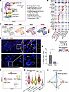 Figure 4
Antiinflammatory TREM2hi macrophages in lesions from a patient with CARD9 deficiency and phaeohyphomycosis. (A) UMAP of macrophages and dendritic cells in the samples from the patient with CARD9 deficiency. Each point represents an individual cell, with the proportion of each subset within the total cell population annotated in the graph. (B) The bubble chart shows the mean relative expression of signature genes across the macrophage and dendritic cell subsets. (C) A UMAP plot shows the expression of TREM2 in macrophages within skin tissues obtained from healthy individuals (n = 3). (D and E) Representative staining of TREM2+ macrophages (TREM2 and CD68) in lesions of patients with CARD9 deficiency and control participants. Scale bars: 100 μm (left) and 20 μm (right). The bar plots show the quantification results (E). One data point represents the statistical result of 1 sample (n = 3). HC, healthy control. (F) A UMAP projection displays the cellular distribution of the CD4+ T cell subset, CD8+T_GZMK subset, CD8+T_HAVCR2 subset, and memory T cell subset as depicted in Supplemental Figure 2A; these were categorized into naive, cytotoxic, and exhaustion states based on the expression of marker genes. (G) Violin plots show the exhaustion score (defined by 5 genes: LAG3, TIGIT, PDCD1, CTLA4, and HAVCR2) of 5 T cell subsets. Box plots overlaid on the violins depict the interquartile range and median score for each subset. (H) The bubble chart shows the interactions between the major macrophage subsets and exhaustion-like Th1 cells in the patient with CARD9 deficiency. Data are shown as the mean ± SD. *P < 0.05 and ***P < 0.001, by 2-tailed Student’s t test (E), or by 1-way ANOVA with Tukey’s test (G).
T cell analysis showed Tregs were the largest CD4+ T cell subset (Supplemental Figure 3, A and B), with a notable proportion of the remaining CD4+ and CD8+ T cells exhibiting signs of exhaustion, characterized by a high immune checkpoint expression and elevated exhaustion scores (Figure 4, F and G). Using mIHC, we further confirmed that the immune checkpoints TIM3 and LAG3 were notably colocalized with CD4 in the skin lesions of 3 patients with CARD9 deficiency with subcutaneous dematiaceous fungal infection, in comparison with those of 3 healthy control participants (Supplemental Figure 3C). Moreover, we analyzed the receptor-ligand interactions between macrophage subsets and exhaustion-like CD4+ T cells. This analysis corroborated our findings in the mouse model, demonstrating that the TREM2hi macrophage subset had the most prominent interactions with exhaustion-like CD4+ cells (Figure 4H). In conclusion, these findings strongly validate our findings in a mouse infection model, indicating that CARD9 deficiency leads to the differentiation of antiinflammatory macrophages, which may contribute to impaired T cell responses in human skin lesions of dematiaceous fungal infection.
CARD9 deficiency induces high TREM2 expression in macrophages and impairs antifungal infection. To further determine the mechanisms underlying the accumulation of TREM2hi macrophages in CARD9-deficient skin lesions, we conducted RNA-seq to analyze the transcriptional profile of bone marrow–derived macrophages (BMDMs) derived from WT and Card9–/– mice stimulated with heat-killed P. verrucosa for 24 hours. Several genes exhibited differential expression patterns between WT and Card9–/– BMDMs (Figure 5A). The upregulated genes in Card9–/– BMDMs included Trem2, Lgals3, and Apoe, which are characteristics of the TREM2hi macrophage subset identified in the in vivo infection model (Figure 5A and Figure 2D). Additionally, the expression of antiinflammatory cytokine, including Tgfb1, Tgfb3, and Il10, was also higher in Card9–/– BMDMs (Figure 5A). In contrast, the upregulated genes in WT BMDMs included Ccl5 and Cxcl3, which were specifically expressed in Cxcl3hi macrophage and Ccl5hi macrophage subsets observed in vivo (Figure 5A and Figure 2D). Furthermore, the expression of proinflammatory cytokines and chemokines, such as Il1b, Tnf, Cxcl1, and Cxcl2, was higher in WT BMDMs (Figure 5A).
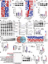 Figure 5
CARD9 deficiency induces higher TREM2 expression in macrophages and impairs antifungal infection. (A–F) Heatmap of selected gene expression from RNA-seq and Western blot with densitometric analysis of TREM2 in BMDMs (A–C) and THP-1 cells (D–F) stimulated with P. verrucosa for 24 hours (n = 3). (G and H) Western blot (G) and densitometric (H) analyses of phosphorylated and total P65, AKT, GSK3β, and CREB in BMDMs stimulated with P. verrucosa. (I–L) Western blot and densitometric analyses of phosphorylated CREB (I and J) and TREM2 (K and L) in WT BMDMs with or without JSH-23 pretreatment, after P. verrucosa stimulation. (M–O) BMDMs were transfected with a small interfering negative control (si-NC) or si-TREM2 and stimulated with P. verrucosa for 24 hours. Venn diagram (M) and heatmap (N) show the overlap between genes downregulated in Card9–/– si-NC versus WT si-NC and those upregulated in Card9–/– si-TREM2 versus Card9–/– si-NC. The bubble plot shows GO enrichment of the overlap genes in Card9–/– si-TREM2 BMDMs (O). (P and Q) Killing efficacy analysis (P) and ROS production of TREM2-overexressing RAW 264.7 cells and controls with P. verrucosa stimulation for 60 minutes (Q). (R and S) Western blot (R) and densitometric (S) analysis of IL-10 and TGF-β in BMDMs stimulated with P. verrucosa for 72 hours. In A, D, and M, columns represent replicates from independent culture wells (n = 3). In C, F, H, J, L, and S, each point represents an independent replicate. Data are shown as mean ± SD. *P < 0.05, **P < 0.01, ***P < 0.001, and ****P < 0.0001, by 1-way ANOVA with Tukey’s multiple-comparison test (C, F, L, Q, and S), 2-way ANOVA (H), and multiple unpaired t tests with Holm-Šídák correction (J and P). All stimulations used heat-killed P. verrucosa at MOI 10. OE, overexpressing; PV, Phialophora verrucosa; US, unstimulated.
Western blotting was performed to validate the differential expression of TREM2 in BMDMs. Card9–/– BMDMs had substantially higher TREM2 levels than WT BMDMs after heat-killed P. verrucosa simulation (Figure 5, B and C). Additionally, ELISA analysis of culture supernatants revealed no difference in the levels of soluble TREM2, suggesting the increased expression observed by Western blot primarily reflected the membrane-bound form, rather than enhanced secretion (Supplemental Figure 4A). Similarly, the knockdown of endogenous CARD9 in THP-1 cells resulted in consistent differential expression patterns. RNA-seq and immunoblotting confirmed P. verrucosa–induced higher expression of TREM2 in CARD9-knockdown THP-1 cells (Figure 5, D–F).
Subsequently, we conducted a Kyoto Encyclopedia of Genes and Genomes (KEGG) enrichment analysis to compare the activation of P. verrucosa–induced signaling molecules in WT and Card9–/– BMDMs. These results demonstrated markedly diminished activation of NF-κB signaling in Card9–/– BMDMs (Supplemental Figure 4B). By contrast, activation of the PI3K/AKT signaling pathway was greater in Card9–/– BMDMs than in WT BMDMs (Supplemental Figure 4B), suggesting CARD9 played a pivotal role in regulating the equilibrium between NF-κB and PI3K/AKT signaling pathways.
Previous studies revealed that TLRs trigger antiinflammatory signaling via the PI3K/AKT/GSK3β pathways in macrophages, which converge to activate CREB (42, 43). The relative amounts of active nuclear CREB and NF-κB p65 determine subsequent association with the nuclear coactivator CBP/p300, thereby regulating the proinflammatory and antiinflammatory responses in macrophages (43, 44). To elucidate whether the antiinflammatory phenotype of Card9–/– BMDMs is associated with alterations in NF-κB and PI3K/AKT signaling, we conducted immunoblotting assays to measure the activation of key molecules in these pathways. Upon P. verrucosa stimulation, Card9–/– BMDMs had lower phosphorylation of the NF-κB p65 subunit and higher phosphorylation of Akt, GSK3β, and CREB in comparison with WT BMDMs (Figure 5, G and H). Furthermore, pretreatment of WT BMDMs with NF-κB inhibitor followed by P. verrucosa stimulation resulted in a notable increase in the phosphorylation of CREB (Figure 5, I and J, and Supplemental Figure 4C), as well as the expression of TREM2 (Figure 5, K and L). These findings provide further evidence that, in conditions of impaired NF-κB signaling, CREB is activated and contributes to the transcriptional upregulation of TREM2.
To further elucidate the role of TREM2 upregulation in CARD9-deficient macrophage, we performed transcriptome sequencing of WT and Card9–/– BMDMs following TREM2 knockdown and 24-hour P. verrucosa stimulation. Notably, a subset of genes downregulated in Card9–/– BMDMs exhibited increased expression upon TREM2 knockdown compared with WT BMDMs (Figure 5M). These included proinflammatory factors, such as Il1α, Cxcl5, and Cxcl9, as well as the antimicrobial peptide S100A8 (Figure 5N). GO functional enrichment analysis revealed that the upregulated genes in the small interfering TREM2 (si-TREM2) group were enriched in biological processes, including defense response, response to stimulus, and inflammatory response (Figure 5O). Furthermore, KEGG pathway enrichment analysis indicated marked enrichment of upregulated genes in cytokine-cytokine receptor interactions and signaling pathways related to IL-17, TNF, and chemokines (Supplemental Figure 4D). To more directly assess the functional role of TREM2hi macrophages in antifungal immunity, we overexpressed TREM2 in RAW264.7 cells (Supplemental Figure 4, E and F). In comparison with control cells, TREM2-overexpressing macrophages demonstrated impaired fungicidal activity (Figure 5P) and decreased production of total ROS (Figure 5Q).
Single-cell interaction analysis suggested TREM2hi macrophages may contribute to T cell exhaustion under Card9-deficient conditions (Figure 3, I and J). Notably, Card9–/– BMDMs exhibited elevated expression of Il10 and Tgfb1, 2 immunoregulatory cytokines implicated in driving T cell exhaustion (41, 45, 46). Consistent with the transcriptional data, Western blot analysis confirmed increased protein levels of IL-10 and TGF-β in Card9–/– BMDMs under P. verrucosa stimulation (Figure 5, R and S), providing further support for a Card9–/– macrophage–mediated mechanism promoting T cell exhaustion.
Collectively, these data indicate that in CARD9-deficient macrophages, P. verrucosa–induced NF-κB signaling activation is constrained, leading to enhanced activation of CREB and the predominance of Trem2hi macrophages. Moreover, upregulation of TREM2 expression in CARD9-deficient hosts is associated with impaired innate and adaptive antifungal function.
CARD9 negatively regulates Trem2 expression by activating C/EBPβ. To further elucidate the mechanism by which the CARD9-related pathway regulates the expression of TREM2, we screened, using bioinformatics analysis, transcription factors that can directly bind to the Trem2 promoter in both mouse and human cells; this ultimately led to the identification of C/EBPβ as the most promising candidate (Figure 6A). Activated CREB promotes the expression and activation of C/EBPβ. Western blot analysis was performed to confirm the increased activation of C/EBPβ in Card9–/– BMDMs upon P. verrucosa stimulation in comparison with WT BMDMs (Figure 6, B and C). Previous studies also demonstrated that the activation of C/EBPβ can promote the antiinflammatory polarization of macrophages (44, 47).
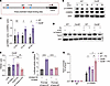 Figure 6
CARD9 negatively regulates Trem2 expression by activating C/EBPβ. (A) Promoter region of WT Trem2, showing 3 predicted C/EBPβ-binding sites at positions. (B and C) Western blot (B) and densitometric (C) analysis of phosphorylated (p-) and total C/EBPβ (left margin) in BMDMs isolated from WT and Card9–/– mice and stimulated for 0–30 minutes (above lanes) with heat-killed P. verrucosa conidia (MOI 10). Each data point represents an independent experimental replicate (n = 3). (D and E) Knockdown of endogenous C/EBPβ by RNA interference in BMDMs, which were transfected with siRNA against murine C/EBPβ and nontargeting control siRNA using Lipofectamine 3000 transfection reagent (Thermo Fisher). Cells were cultured for 48 hours after transfection and then stimulated with heat-killed P. verrucosa conidia (MOI 10) for 24 hours. Cell lysates were subjected to Western blot analysis using indicated antibodies (D) and then quantified using densitometric analysis in Card9–/– group (E). Each data point represents an independent experimental replicate (n = 3). (F) Firefly luciferase activity in human embryonic kidney 293T cells cotransfected with constructs for the overexpression of Cebpb and a construct containing various Trem2 promoter–driven firefly luciferase constructs together with an EF1α promoter–driven Renilla luciferase reporter; results were normalized to those of Renilla luciferase. Ctr, control construct lacking Cebpb cotransfected with a construct containing the Trem2 promoter. (G) Chromatin immunoprecipitation (with control IgG or anti-Cebpb) and PCR analysis of the binding of Cebpb to the Trem2 promoter in BMDMs obtained from WT mice and Card9–/– mice and left unstimulated or challenged for 4 hours in vitro with heat-killed P. verrucosa spores (MOI 10). Data are shown as the mean ± SD. *P < 0.05 and **P < 0.01, and ****P < 0.0001, by multiple unpaired t tests with Holm-Šídák correction (C), or 1-way ANOVA with Tukey’s multiple-comparison test (E–G). PV, Phialophora verrucosa; si-NC, small interfering negative control; mTrem2, mouse Trem2; MT, mutant type.
To ascertain whether C/EBPβ can regulate P. verrucosa–induced expression of TREM2 in macrophages, siRNA was used to knockdown C/EBPβ in BMDMs, which were then stimulated with P. verrucosa. Knockdown of C/EBPβ notably suppressed P. verrucosa–induced expression of TREM2 in Card9–/– BMDMs (Figure 6, D and E). To determine whether C/EBPβ directly dictates the Trem2 transcription, we also used a dual-luciferase reporter assay, which revealed that C/EBPβ overexpression in 293T human embryonic cells resulted in Trem2 promoter activation (Figure 6F). Similarly, we conducted chromatin immunoprecipitation assays and observed C/EBPβ binding to the promoter region of Trem2 in CARD9-deficient macrophages (Figure 6G). These results collectively indicate that augmented C/EBPβ signaling in CARD9-deficient macrophages can directly bind to the promoter region of Trem2, thereby exerting a positive regulatory effect on its expression.
Anti-TREM2 antibody improves the antifungal immune response in vivo and in vitro. To investigate the potential therapeutic benefits of targeting TREM2 in the treatment of phaeohyphomycosis, WT and Card9–/– mice were infected with P. verrucosa and treated with a blocking antibody of TREM2 (Figure 7A). The administration of anti-TREM2 antibody delayed disease progression and reduced the footpad swelling rate in Card9–/– mice compared with that in the control group (Figure 7B). However, no differences in footpad lesion phenotypes were observed between the antibody treatment and control groups in WT mice (Figure 7B). Histological examination of the footpad on day 21 after infection showed a marked reduction in inflammatory cell infiltration, smaller infectious granulomas, and a lower fungal burden (i.e., spores and hyphae) in the lesions of Card9–/– mice treated with anti-TREM2 antibody compared with that in nontreated Card9–/– mice (Figure 7C). Furthermore, treatment with an anti-TREM2 antibody resulted in a notable reduction in local fungal loads in Card9–/– mice on day 21 after infection (Figure 7D).
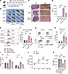 Figure 7
Anti-TREM2 antibody improves the antifungal immune response in vivo and in vitro. (A and B) WT and Card9–/– mice were subcutaneously injected with P. verrucosa and treated intraperitoneally with PBS or anti-TREM2 antibody. Experimental scheme (A) and natural course (B) of infection. (C) Histopathology of H&E and Grocott’s methenamine silver (GMS) staining of footpads from infected mice at day 14 after infection. Scale bars: 1 mm (H&E stain) and 100 μm (GMS stain). Arrows indicate fungal yeast and hyphae. (D) Fungal burden of footpad from WT and Card9–/– mice on day 14 after infection. (E and F) Representative flow cytometry histogram plots for TREM2 staining (E) and frequency of TREM2+ macrophage subsets (F) in murine lesions. One data point denotes a result from 1 mouse (n = 10, from 3 independent experiments). (G and H) Representative flow cytometry contour plots for TIM3 and LAG3 staining (G) and frequency of TIM3+ LAG3+CD4+ T cell subsets (H) in murine lesions. One data point denotes a result from 1 mouse (n = 10, from 3 independent experiments). (I–K) Knockdown of endogenous TREM2 by RNA interference in BMDMs. Cells were cultured for 48 hours after transfection and then stimulated with heat-killed P. verrucosa spores (MOI 10) for the indicated times. (I) Killing efficacy analysis of BMDMs of Card9–/– mice. (J and K) Total ROS production of WT and Card9–/– BMDMs at the indicated time point was measured by the Reactive Oxygen Assay kit (Beyotime). Data in B, C, and I–K are representative of 3 independent experiments. Data are shown as the mean ± SD. *P < 0.05, **P < 0.01, ***P < 0.001, and ***P < 0.001, by 1-way ANOVA with Tukey’s multiple-comparison test (D, F, H, I, and K), or 2-way ANOVA with Šídák’s multiple-comparison test (J). PV, Phialophora verrucosa; NC, negative control.
Flow cytometry was performed to elucidate the cellular mechanisms underlying anti-TREM2 antibody treatment. In WT mice, no differences were observed in the proportions of different macrophage subsets between the antibody-treated and control groups. However, in Card9–/– mice, the anti-TREM2 antibody treatment group had a substantially lower proportion of TREM2+ antiinflammatory macrophages than the control group (Figure 7, E and F). Pretreatment of TREM2-overexpressing RAW 264.7 cells with the blocking antibody did not affect subsequent detection by flow cytometry, confirming no epitope interference (Supplemental Figure 4E). Furthermore, the anti-TREM2 antibody–treated group displayed reduced expression of immune checkpoints (TIM3 and LAG3) on CD4+ T cells compared with the nontreated group. In contrast, both treatment and control groups of WT mice exhibited minimal expression of immune checkpoints on CD4+ T cells within the lesions (Figure 7, G and H). These results indicate anti-TREM2 antibody may serve as a potential therapeutic strategy for enhancing host innate and adaptive immunity against fungal infections in Card9–/– mice.
Previous studies have demonstrated that CARD9-deficient macrophages exhibit defects in killing P. verrucosa (48). To ascertain whether targeting TREM2 affects the fungal-killing ability of macrophages in vitro, siRNA was used to knock down the expression of TREM2. The knockdown of endogenous TREM2 restored the spore-killing ability of Card9–/– BMDMs, whereas it had no effect on WT BMDMs (Figure 7I). Moreover, the generation of ROS is a pivotal effector mechanism of macrophages in antifungal immunity. HALLMARK gene set scoring among the major macrophage subsets indicated the TREM2hi macrophage subsets had a reduction in ROS pathway function (Figure 2E), and overexpression of TREM2 in RAW 264.7 cells also showed impaired ROS production (Figure 5Q). Consequently, we investigated the impact of targeting TREM2 on total ROS production in macrophages. Card9–/– BMDMs exhibited a markedly diminished capacity to generate ROS upon stimulation with P. verrucosa compared with WT BMDMs (Figure 7J). Importantly, the knockdown of TREM2 considerably enhanced the ROS generation by Card9–/– BMDMs (Figure 7K).
These results provide further evidence for the crucial role of the TREM2-mediated antiinflammatory pathway in compromising antifungal immunity in the context of CARD9 deficiency. Modulation of TREM2 signaling represents a promising strategy to enhance the fungicidal ROS response and fungal killing by macrophages and improve host defense against dematiaceous fungal infections in CARD9-deficient settings.


Copyright © 2026 American Society for Clinical Investigation
ISSN: 0021-9738 (print), 1558-8238 (online)