Issue published August 15, 2001 Previous issue | Next issue
- Volume 108, Issue 4
Go to section:
-
Letter to the Editor
×
Abstract
Authors
Raffaele Marfella, Lisa Quagliaro, Francesco Nappo, Antonio Ceriello, Dario Giugliano


-
Research Articles
×
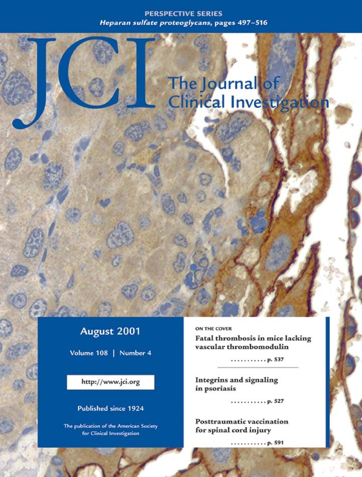
A role for mitogen-activated protein kinase activation by integrins in the pathogenesis of psoriasis
Abstract
In normal epidermis, β1 integrin expression is confined to the basal layer, whereas in hyperproliferative epidermis, integrins are also expressed in the suprabasal layers. Transgenic mice in which integrins are expressed suprabasally via the involucrin promoter have a sporadic psoriatic phenotype; however, the mechanism by which integrins contribute to the pathogenesis of psoriasis is unknown. We observed activation of mitogen-activated protein kinase (MAPK) in basal and suprabasal keratinocytes of human and transgenic mouse psoriatic lesions and healing mouse skin wounds, correlating in each case with suprabasal integrin expression. Phenotypically normal human and transgenic mouse epidermis did not contain activated MAPK. Transgene-positive keratinocytes produced more IL-1α than controls did, and keratinocyte MAPK could be activated by ligation of suprabasal integrins or treatment with IL-1α. Constitutive activation of MAPK increased the growth rate of human keratinocytes and delayed the onset of terminal differentiation, recreating many of the histological features of psoriatic epidermis. We propose that activation of MAPK by integrins, either directly or through increased IL-1α production, is responsible for epidermal hyperproliferation in psoriasis and wound healing, and that the sporadic phenotype of the transgenic mice may reflect the complex mechanisms by which IL-1 release and responsiveness are controlled in skin.
Authors
Ingo Haase, Robin M. Hobbs, M. Rosario Romero, Simon Broad, Fiona M. Watt
×Abstract
The thrombomodulin (TM) gene was ablated in mice in a cell type–restricted manner from vascular endothelium by Cre-recombinase–mediated excision controlled by the endothelial cell lineage–specific Tie2 promoter. Forty percent of mutant (TMLox-) mice display a distinct lethal embryonic phenotype not observed in completely TM-deficient embryos. The remaining 60% of TMLox mice survive beyond birth, but invariably succumb to a severe hypercoagulable state and massive thrombosis after 3 weeks, terminating in a lethal consumptive coagulopathy. The progression of thrombosis was age- and sex-dependent. Disruption of the TM/protein C pathway was not associated with a latent proinflammatory state. Disease onset and progression could be prevented by warfarin anticoagulation. These results show that in mice, loss of endothelial cell TM function causes spontaneous and fatal thrombosis in the arterial and venous circulation, resulting from unfettered activation of the coagulation system. The combination of complete disease penetrance, uniform disease onset at young age, large vessel thrombosis of the extremities and multiple organ systems, and consumptive coagulopathy as the disease end-point provides a unique mouse model of human thrombotic disease.
Authors
Berend Isermann, Sara B. Hendrickson, Mark Zogg, Mark Wing, Marjorie Cummiskey, Yaz Y. Kisanuki, Masashi Yanagisawa, Hartmut Weiler
×Abstract
Bronchiolitis obliterans syndrome (BOS) is the major limitation to survival after lung transplantation. Acute rejection, its main risk factor, is characterized by perivascular/bronchiolar leukocyte infiltration. BOS is characterized by persistent peribronchiolar leukocyte recruitment leading to airway fibrosis and obliteration. The specific mechanism(s) by which these leukocytes are recruited are unknown. Because MCP-1, acting through its receptor CCR2, is a potent mononuclear cell chemoattractant, we hypothesized that expression of this chemokine during an allogeneic-response promotes persistent recruitment of leukocytes and, ultimately, rejection. We found that elevated levels of biologically active MCP-1 in human bronchial lavage fluid (BALF) were associated with the continuum from acute to chronic allograft rejection. Translational studies in a murine model of BOS demonstrated increased MCP-1 expression paralleling mononuclear cell recruitment and CCR2 expression. Loss of MCP-1/CCR2 signaling, as seen in CCR2–/– mice or in WT mice treated with neutralizing antibodies to MCP-1, significantly reduced recruitment of mononuclear phagocytes following tracheal transplantation and led to attenuation of BOS. Lymphocyte infiltration was not reduced under these conditions. We suggest that MCP-1/CCR2 signaling plays an important role in recruitment of mononuclear phagocytes, a pivotal event in the pathogenesis of BOS.
Authors
John A. Belperio, Michael P. Keane, Marie D. Burdick, Joseph P. Lynch III, Ying Ying Xue, Aaron Berlin, David J. Ross, Steven L. Kunkel, Israel F. Charo, Robert M. Strieter
×Abstract
Direct in vivo evidence is still lacking for α4-integrin–mediated T cell interaction with VCAM-1 on blood-brain barrier–endothelium in experimental autoimmune encephalomyelitis (EAE). To investigate a possible α4-integrin–mediated interaction of encephalitogenic T cell blasts with VCAM-1 on the blood-brain barrier white matter endothelium in vivo, we have developed a novel spinal cord window preparation that enabled us to directly visualize CNS white matter microcirculation by intravital fluorescence videomicroscopy. Our study provides the first in vivo evidence that encephalitogenic T cell blasts interact with the spinal cord white matter microvasculature without rolling and that α4-integrin mediates the G protein–independent capture and subsequently the G protein–dependent adhesion strengthening of T cell blasts to microvascular VCAM-1.
Authors
Peter Vajkoczy, Melanie Laschinger, Britta Engelhardt
×Abstract
Ron receptor activation induces numerous cellular responses in vitro, including proliferation, dissociation, and migration. Ron is thought to be involved in blood cell development in vivo, as well as in many aspects of the immune response including macrophage activation, antigen presentation, and nitric oxide regulation. In previous studies to determine the function of Ron in vivo, mice were generated with a targeted deletion of the extracellular and transmembrane regions of this gene. Mice homologous for this deletion appear to die early during embryonic development. To ascertain the in vivo function of Ron in more detail, we have generated mice with a germline ablation of the tyrosine kinase domain. Strikingly, our studies indicate that this domain of Ron, and therefore Ron cytoplasmic signaling, is not essential for embryonic development. While mice deficient in this domain are overtly normal, mice lacking Ron signaling have an altered ability to regulate nitric oxide levels and, in addition, have enhanced tissue damage following acute and cell-mediated inflammatory responses.
Authors
Susan E. Waltz, Laura Eaton, Kenya Toney-Earley, Karla A. Hess, Belinda E. Peace, Jeffrey R. Ihlendorf, Ming-Hai Wang, Klaus H. Kaestner, Sandra J.F. Degen
×Abstract
The objective of this study was to investigate the contribution of secondary lymphoid organs in the generation and maintenance of experimental allergic airway inflammation. We employed a previously reported murine model of respiratory mucosal allergic sensitization, induced by repeated aerosolizations of ovalbumin in the context of a GM-CSF airway environment. We executed this protocol in wild-type (WT) and lymphotoxin-α–deficient mice (LTα-KO) mice, which are devoid of lymph nodes (LNs) and possess rudimentary spleen structures. Despite the lack of pulmonary LNs draining the airway compartment, LTα-KO mice were fully capable of mounting a robust inflammatory response in the airways, consisting of Th2 polarized CD4+ T cells and eosinophils. This was accompanied by IL-5, IL-13, and IFN-γ production by splenocytes and generation of ovalbumin-specific serum IgE. Exposure to the same antigen 7 weeks after complete resolution of airway inflammation once again induced a Th2 polarized infiltrate, demonstrating intact immunological memory. To investigate inherent plasticity in establishing antigen-specific immunity, mice were splenectomized before sensitization. Allergic sensitization was completely abrogated in splenectomized LTα-KO mice, compared with eusplenic LTα-KO controls. These data demonstrate that secondary lymphoid organs, either LN or spleen, are essential for the generation of allergic airway responses.
Authors
Beata U. Gajewska, David Alvarez, Mariana Vidric, Susanna Goncharova, Martin R. Stämpfli, Anthony J. Coyle, José-Carlos Gutierrez-Ramos, Manel Jordana
×Abstract
The clinical use of doxorubicin, an anthracycline chemotherapeutic agent, is limited by cardiotoxicity, particularly when combined with herceptin, an antibody that blocks the HER2 receptor. Doxorubicin induces cyclooxygenase–2 (COX-2) activity in rat neonatal cardiomyocytes. This expression of COX-2 limits doxorubicin-induced cardiac cell injury, raising the possibility that the administration of a prostaglandin may protect the heart during the in vivo administration of doxorubicin. Doxorubicin (15 mg/kg) administered to adult male Sprague Dawley rats induced COX-2 expression and activity in cardiac tissue. Prostacyclin generation measured as the excretion of 2,3-dinor-6-keto-PGF1α also increased, and this was blocked by a COX-2 inhibitor, SC236. In contrast, administration of a COX-1 inhibitor SC560 at a dose that reduced serum thromboxane B2 by more than 80% did not prevent the doxorubicin-induced increase in prostacyclin generation. Doxorubicin increased cardiac injury, detected as a rise in plasma cardiac troponin T, serum lactate dehydrogenase, and cardiomyocyte apoptosis; this was aggravated by coadministration of SC236 but not SC560. The degree of injury in animals treated with a combination of doxorubicin and SC236 was attenuated by prior administration of the prostacyclin analogue iloprost. These data raise the possibility of protecting the heart during the administration of doxorubicin by prior administration of prostacyclin.
Authors
Noreen P. Dowd, Michael Scully, Sharon R. Adderley, Anthony J. Cunningham, Desmond J. Fitzgerald
×Abstract
Spinal cord injury results in a massive loss of neurons, and thus of function. We recently reported that passive transfer of autoimmune T cells directed against myelin-associated antigens provides acutely damaged spinal cords with effective neuroprotection. The therapeutic time window for the passive transfer of T cells was found to be at least 1 week. Here we show that posttraumatic T cell–based active vaccination is also neuroprotective. Immunization with myelin-associated antigens such as myelin basic protein (MBP) significantly promoted recovery after spinal cord contusion injury in the rat model. To reduce the risk of autoimmune disease while retaining the benefit of the immunization, we vaccinated the rats immediately after severe incomplete spinal cord injury with MBP-derived altered peptide ligands. Immunization with these peptides resulted in significant protection from neuronal loss and thus in a reduced extent of paralysis, assessed by an open-field behavioral test. Retrograde labeling of the rubrospinal tracts and magnetic resonance imaging supported the behavioral results. Further optimization of nonpathogenic myelin-derived peptides can be expected to lead the way to the development of an effective therapeutic vaccination protocol as a strategy for the prevention of total paralysis after incomplete spinal cord injury.
Authors
Ehud Hauben, Eugenia Agranov, Amalia Gothilf, Uri Nevo, Avi Cohen, Igor Smirnov, Lawrence Steinman, Michal Schwartz
×Abstract
TGF-β1 functions as a negative regulator of T cell immune responses, signaling to target cells using the Smad family of proteins. We show here that Smad7, an inhibitor of TGF-β1 signaling, is overexpressed in inflammatory bowel disease (IBD) mucosa and purified mucosal T cells. Both whole tissue and isolated cells exhibit defective signaling through this pathway, as measured by phospho-Smad3 immunoreactivity. Specific antisense oligonucleotides for Smad7 reduce Smad7 protein expression in cells isolated from patients with IBD, permitting the cells to respond to exogenous TGF-β1. TGF-β1 cannot inhibit proinflammatory cytokine production in isolated lamina propria mononuclear cells from patients with Crohn disease (CD), but inhibition of Smad7 restores TGF-β1 signaling and enables TGF-β1 to inhibit cytokine production. In inflamed mucosal tissue explants from patients with CD, inhibition of Smad7 also restores p-Smad3 and decreases proinflammatory cytokine production, an effect that is partially blocked by anti–TGF-β1. These results show that Smad7 blockade of TGF-β1 signaling helps maintain the chronic production of proinflammatory cytokines that drives the inflammatory process in IBD and that inhibition of Smad7 enables endogenous TGF-β to downregulate this response.
Authors
Giovanni Monteleone, Andrea Kumberova, Nicholas M. Croft, Catriona McKenzie, Howard W. Steer, Thomas T. MacDonald
×Abstract
Clinical studies of hormone replacement therapy to prevent cardiovascular diseases have heightened interest in the cardiovascular effects of progestins. However, the role of the progesterone receptor (PR) in vascular biology has not been studied in vivo. We studied ovariectomized female PR knockout (PRKO) mice and their wild-type (WT) littermates using the mouse carotid artery injury model. Placebo-treated PRKO mice showed significantly greater vascular medial hypertrophy and vascular smooth muscle cell (VSMC) proliferation in response to vascular injury than did WT mice. Progesterone had no significant effect in the PRKO mice, but worsened the response to injury in WT mice. VSMCs cultured from PRKO mouse aortae were markedly hyperproliferative, and their growth was not affected by progesterone. In contrast to the in vivo findings, progesterone inhibited proliferation of WT-derived VSMCs. Furthermore, reintroduction of PR into PRKO-derived VSMCs using adenoviral methods restored progesterone-mediated inhibition of proliferation to these cells. This effect was reversed by the PR antagonist, RU 486. Thus, the effects of PR and progesterone differ markedly between cultured VSMCs and intact blood vessels. These data demonstrate a direct role for the PR in regulating the response to vascular injury and VSMC proliferation.
Authors
Richard H. Karas, Martin van Eickels, John P. Lydon, Sean Roddy, Moon Kwoun, Mark Aronovitz, Wendy E. Baur, Orla Conneely, Bert W. O’Malley, Michael E. Mendelsohn
×Abstract
Hemochromatosis is a progressive iron overload disorder that is prevalent among individuals of European descent. It is usually inherited in an autosomal-recessive pattern and associated with missense mutations in HFE, an atypical major histocompatibility class I gene. Recently, we described a large family with autosomal-dominant hemochromatosis not linked to HFE and distinguished by early iron accumulation in reticuloendothelial cells. Through analysis of a large pedigree, we have determined that this disease maps to 2q32. The gene encoding ferroportin (SLC11A3), a transmembrane iron export protein, lies within a candidate interval defined by highly significant lod scores. We show that the iron-loading phenotype in autosomal-dominant hemochromatosis is associated with a nonconservative missense mutation in the ferroportin gene. This missense mutation, converting alanine to aspartic acid at residue 77 (A77D), was not seen in samples from 100 unaffected control individuals. We propose that partial loss of ferroportin function leads to an imbalance in iron distribution and a consequent increase in tissue iron accumulation.
Authors
Giuliana Montosi, Adriana Donovan, Angela Totaro, Cinzia Garuti, Elisa Pignatti, Stefano Cassanelli, Cameron C. Trenor, Paolo Gasparini, Nancy C. Andrews, Antonello Pietrangelo
×Abstract
Melatonin is released from intestinal enterochromaffin cells and from the pineal gland, but its role in gastrointestinal function is largely unknown. Our aim was to study the involvement of intestinal and central nervous melatonin in the neurohumoral control of the duodenal mucosa-protective bicarbonate secretion. Working in anesthetized rats, we cannulated a 12-mm segment of duodenum with an intact blood supply and titrated the local bicarbonate secretion with pH-stat. Melatonin and receptor ligands were supplied to the duodenum by close intra-arterial infusion. Even at low doses, melatonin and the full agonist 2-iodo-N-butanoyl-5-methoxytryptamine increased duodenal bicarbonate secretion. Responses were inhibited by the predominantly MT2-selective antagonist luzindole but not by prazosin, acting at MT3 receptors. Also, luzindole almost abolished the marked rise in secretion induced by intracerebroventricular infusion of the adrenoceptor agonist phenylephrine. This response was also abolished by sublaryngeal ligation of all nerves around the carotid arteries. However, it was insensitive to truncal vagotomy alone or sympathectomy alone and was unaffected by removal of either the pineal gland or pituitary gland. Thus, melatonin stimulates duodenal bicarbonate secretion via action at enterocyte MT2-receptors and mediates neural stimulation of the secretion.
Authors
Markus Sjöblom, Gunilla Jedstedt, Gunnar Flemström


Copyright © 2026 American Society for Clinical Investigation
ISSN: 0021-9738 (print), 1558-8238 (online)