Citation Information: J Clin Invest. 2006;116(9):2378-2384. https://doi.org/10.1172/JCI28341.
Abstract
Neurofibromatosis type I (NF1; also known as von Recklinghausen’s disease) is a common autosomal-dominant condition primarily affecting neural crest–derived tissues. The disease gene, NF1, encodes neurofibromin, a protein of over 2,800 amino acids that contains a 216–amino acid domain with Ras–GTPase-activating protein (Ras-GAP) activity. Potential therapies for NF1 currently in development and being tested in clinical trials are designed to modify NF1 Ras-GAP activity or target downstream effectors of Ras signaling. Mice lacking the murine homolog (Nf1) have mid-gestation lethal cardiovascular defects due to a requirement for neurofibromin in embryonic endothelium. We sought to determine whether the GAP activity of neurofibromin is sufficient to rescue complete loss of function or whether other as yet unidentified functions of neurofibromin might also exist. Using cre-inducible ubiquitous and tissue-specific expression, we demonstrate that the isolated GAP-related domain (GRD) rescued cardiovascular development in Nf1–/– embryos, but overgrowth of neural crest–derived tissues persisted, leading to perinatal lethality. These results suggest that neurofibromin may possess activities outside of the GRD that modulate neural crest homeostasis and that therapeutic approaches solely aimed at targeting Ras activity may not be sufficient to treat tumors of neural crest origin in NF1.
Authors
Fraz A. Ismat, Junwang Xu, Min Min Lu, Jonathan A. Epstein
Citation Information: J Clin Invest. 2006;116(9):2552-2561. https://doi.org/10.1172/JCI28371.
Abstract
ROS are a risk factor of several cardiovascular disorders and interfere with NO/soluble guanylyl cyclase/cyclic GMP (NO/sGC/cGMP) signaling through scavenging of NO and formation of the strong oxidant peroxynitrite. Increased oxidative stress affects the heme-containing NO receptor sGC by both decreasing its expression levels and impairing NO-induced activation, making vasodilator therapy with NO donors less effective. Here we show in vivo that oxidative stress and related vascular disease states, including human diabetes mellitus, led to an sGC that was indistinguishable from the in vitro oxidized/heme-free enzyme. This sGC variant represents what we believe to be a novel cGMP signaling entity that is unresponsive to NO and prone to degradation. Whereas high-affinity ligands for the unoccupied heme pocket of sGC such as zinc–protoporphyrin IX and the novel NO-independent sGC activator 4-[((4-carboxybutyl){2-[(4-phenethylbenzyl)oxy]phenethyl}amino) methyl [benzoic]acid (BAY 58-2667) stabilized the enzyme, only the latter activated the NO-insensitive sGC variant. Importantly, in isolated cells, in blood vessels, and in vivo, BAY 58-2667 was more effective and potentiated under pathophysiological and oxidative stress conditions. This therapeutic principle preferentially dilates diseased versus normal blood vessels and may have far-reaching implications for the currently investigated clinical use of BAY 58-2667 as a unique diagnostic tool and highly innovative vascular therapy.
Authors
Johannes-Peter Stasch, Peter M. Schmidt, Pavel I. Nedvetsky, Tatiana Y. Nedvetskaya, Arun Kumar H.S., Sabine Meurer, Martin Deile, Ashraf Taye, Andreas Knorr, Harald Lapp, Helmut Müller, Yagmur Turgay, Christiane Rothkegel, Adrian Tersteegen, Barbara Kemp-Harper, Werner Müller-Esterl, Harald H.H.W. Schmidt
Citation Information: J Clin Invest. 2006;116(9):2510-2520. https://doi.org/10.1172/JCI29128.
Abstract
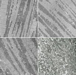
Cardiac calsequestrin (Casq2) is thought to be the key sarcoplasmic reticulum (SR) Ca2+ storage protein essential for SR Ca2+ release in mammalian heart. Human CASQ2 mutations are associated with catecholaminergic ventricular tachycardia. However, homozygous mutation carriers presumably lacking functional Casq2 display surprisingly normal cardiac contractility. Here we show that Casq2-null mice are viable and display normal SR Ca2+ release and contractile function under basal conditions. The mice exhibited striking increases in SR volume and near absence of the Casq2-binding proteins triadin-1 and junctin; upregulation of other Ca2+-binding proteins was not apparent. Exposure to catecholamines in Casq2-null myocytes caused increased diastolic SR Ca2+ leak, resulting in premature spontaneous SR Ca2+ releases and triggered beats. In vivo, Casq2-null mice phenocopied the human arrhythmias. Thus, while the unique molecular and anatomic adaptive response to Casq2 deletion maintains functional SR Ca2+ storage, lack of Casq2 also causes increased diastolic SR Ca2+ leak, rendering Casq2-null mice susceptible to catecholaminergic ventricular arrhythmias.
Authors
Björn C. Knollmann, Nagesh Chopra, Thinn Hlaing, Brandy Akin, Tao Yang, Kristen Ettensohn, Barbara E.C. Knollmann, Kenneth D. Horton, Neil J. Weissman, Izabela Holinstat, Wei Zhang, Dan M. Roden, Larry R. Jones, Clara Franzini-Armstrong, Karl Pfeifer
Citation Information: J Clin Invest. 2006;116(8):2218-2225. https://doi.org/10.1172/JCI16980.
Abstract
The carboxypeptidase ACE2 is a homologue of angiotensin-converting enzyme (ACE). To clarify the physiological roles of ACE2, we generated mice with targeted disruption of the Ace2 gene. ACE2-deficient mice were viable, fertile, and lacked any gross structural abnormalities. We found normal cardiac dimensions and function in ACE2-deficient animals with mixed or inbred genetic backgrounds. On the C57BL/6 background, ACE2 deficiency was associated with a modest increase in blood pressure, whereas the absence of ACE2 had no effect on baseline blood pressures in 129/SvEv mice. After acute Ang II infusion, plasma concentrations of Ang II increased almost 3-fold higher in ACE2-deficient mice than in controls. In a model of Ang II–dependent hypertension, blood pressures were substantially higher in the ACE2-deficient mice than in WT. Severe hypertension in ACE2-deficient mice was associated with exaggerated accumulation of Ang II in the kidney, as determined by MALDI-TOF mass spectrometry. Although the absence of functional ACE2 causes enhanced susceptibility to Ang II–induced hypertension, we found no evidence for a role of ACE2 in the regulation of cardiac structure or function. Our data suggest that ACE2 is a functional component of the renin-angiotensin system, metabolizing Ang II and thereby contributing to regulation of blood pressure.
Authors
Susan B. Gurley, Alicia Allred, Thu H. Le, Robert Griffiths, Lan Mao, Nisha Philip, Timothy A. Haystead, Mary Donoghue, Roger E. Breitbart, Susan L. Acton, Howard A. Rockman, Thomas M. Coffman
Citation Information: J Clin Invest. 2006;116(7):1865-1877. https://doi.org/10.1172/JCI27019.
Abstract
Clinical trials of bone marrow stem/progenitor cell therapy after myocardial infarction (MI) have shown promising results, but the mechanism of benefit is unclear. We examined the nature of endogenous myocardial repair that is dependent on the function of the c-kit receptor, which is expressed on bone marrow stem/progenitor cells and on recently identified cardiac stem cells. MI increased the number of c-kit+ cells in the heart. These cells were traced back to a bone marrow origin, using genetic tagging in bone marrow chimeric mice. The recruited c-kit+ cells established a proangiogenic milieu in the infarct border zone by increasing VEGF and by reversing the cardiac ratio of angiopoietin-1 to angiopoietin-2. These oscillations potentiated endothelial mitogenesis and were associated with the establishment of an extensive myofibroblast-rich repair tissue. Mutations in the c-kit receptor interfered with the mobilization of the cells to the heart, prevented angiogenesis, diminished myofibroblast-rich repair tissue formation, and led to precipitous cardiac failure and death. Replacement of the mutant bone marrow with wild-type cells rescued the cardiomyopathic phenotype. We conclude that, consistent with their documented role in tumorigenesis, bone marrow c-kit+ cells act as key regulators of the angiogenic switch in infarcted myocardium, thereby driving efficient cardiac repair.
Authors
Shafie Fazel, Massimo Cimini, Liwen Chen, Shuhong Li, Denis Angoulvant, Paul Fedak, Subodh Verma, Richard D. Weisel, Armand Keating, Ren-Ke Li
Citation Information: J Clin Invest. 2006;116(7):1853-1864. https://doi.org/10.1172/JCI27438.
Abstract
Class IIa histone deacetylases (HDACs) regulate a variety of cellular processes, including cardiac growth, bone development, and specification of skeletal muscle fiber type. Multiple serine/threonine kinases control the subcellular localization of these HDACs by phosphorylation of common serine residues, but whether certain class IIa HDACs respond selectively to specific kinases has not been determined. Here we show that calcium/calmodulin-dependent kinase II (CaMKII) signals specifically to HDAC4 by binding to a unique docking site that is absent in other class IIa HDACs. Phosphorylation of HDAC4 by CaMKII promotes nuclear export and prevents nuclear import of HDAC4, with consequent derepression of HDAC target genes. In cardiomyocytes, CaMKII phosphorylation of HDAC4 results in hypertrophic growth, which can be blocked by a signal-resistant HDAC4 mutant. These findings reveal a central role for HDAC4 in CaMKII signaling pathways and have implications for the control of gene expression by calcium signaling in a variety of cell types.
Authors
Johannes Backs, Kunhua Song, Svetlana Bezprozvannaya, Shurong Chang, Eric N. Olson
Citation Information: J Clin Invest. 2006;116(7):2012-2021. https://doi.org/10.1172/JCI27751.
Abstract
Arrhythmogenic right ventricular dysplasia/cardiomyopathy (ARVC) is a genetic disease caused by mutations in desmosomal proteins. The phenotypic hallmark of ARVC is fibroadipocytic replacement of cardiac myocytes, which is a unique phenotype with a yet-to-be-defined molecular mechanism. We established atrial myocyte cell lines expressing siRNA against desmoplakin (DP), responsible for human ARVC. We show suppression of DP expression leads to nuclear localization of the desmosomal protein plakoglobin and a 2-fold reduction in canonical Wnt/β-catenin signaling through Tcf/Lef1 transcription factors. The ensuing phenotype is increased expression of adipogenic and fibrogenic genes and accumulation of fat droplets. We further show that cardiac-restricted deletion of Dsp, encoding DP, impairs cardiac morphogenesis and leads to high embryonic lethality in the homozygous state. Heterozygous DP-deficient mice exhibited excess adipocytes and fibrosis in the myocardium, increased myocyte apoptosis, cardiac dysfunction, and ventricular arrhythmias, thus recapitulating the phenotype of human ARVC. We believe our results provide for a novel molecular mechanism for the pathogenesis of ARVC and establish cardiac-restricted DP-deficient mice as a model for human ARVC. These findings could provide for the opportunity to identify new diagnostic markers and therapeutic targets in patients with ARVC.
Authors
Eduardo Garcia-Gras, Raffaella Lombardi, Michael J. Giocondo, James T. Willerson, Michael D. Schneider, Dirar S. Khoury, Ali J. Marian
Citation Information: J Clin Invest. 2006;116(7):1913-1923. https://doi.org/10.1172/JCI27933.
Abstract
Adenosine has been described as playing a role in the control of inflammation, but it has not been certain which of its receptors mediate this effect. Here, we generated an A2B adenosine receptor–knockout/reporter gene–knock-in (A2BAR-knockout/reporter gene–knock-in) mouse model and showed receptor gene expression in the vasculature and macrophages, the ablation of which causes low-grade inflammation compared with age-, sex-, and strain-matched control mice. Augmentation of proinflammatory cytokines, such as TNF-α, and a consequent downregulation of IκB-α are the underlying mechanisms for an observed upregulation of adhesion molecules in the vasculature of these A2BAR-null mice. Intriguingly, leukocyte adhesion to the vasculature is significantly increased in the A2BAR-knockout mice. Exposure to an endotoxin results in augmented proinflammatory cytokine levels in A2BAR-null mice compared with control mice. Bone marrow transplantations indicated that bone marrow (and to a lesser extent vascular) A2BARs regulate these processes. Hence, we identify the A2BAR as a new critical regulator of inflammation and vascular adhesion primarily via signals from hematopoietic cells to the vasculature, focusing attention on the receptor as a therapeutic target.
Authors
Dan Yang, Ying Zhang, Hao G. Nguyen, Milka Koupenova, Anil K. Chauhan, Maria Makitalo, Matthew R. Jones, Cynthia St. Hilaire, David C. Seldin, Paul Toselli, Edward Lamperti, Barbara M. Schreiber, Haralambos Gavras, Denisa D. Wagner, Katya Ravid
Citation Information: J Clin Invest. 2006;116(6):1547-1560. https://doi.org/10.1172/JCI25397.
Abstract
For over a century, there has been intense debate as to the reason why some cardiac stresses are pathological and others are physiological. One long-standing theory is that physiological overloads such as exercise are intermittent, while pathological overloads such as hypertension are chronic. In this study, we hypothesized that the nature of the stress on the heart, rather than its duration, is the key determinant of the maladaptive phenotype. To test this, we applied intermittent pressure overload on the hearts of mice and tested the roles of duration and nature of the stress on the development of cardiac failure. Despite a mild hypertrophic response, preserved systolic function, and a favorable fetal gene expression profile, hearts exposed to intermittent pressure overload displayed pathological features. Importantly, intermittent pressure overload caused diastolic dysfunction, altered β-adrenergic receptor (βAR) function, and vascular rarefaction before the development of cardiac hypertrophy, which were largely normalized by preventing the recruitment of PI3K by βAR kinase 1 to ligand-activated receptors. Thus stress-induced activation of pathogenic signaling pathways, not the duration of stress or the hypertrophic growth per se, is the molecular trigger of cardiac dysfunction.
Authors
Cinzia Perrino, Sathyamangla V. Naga Prasad, Lan Mao, Takahisa Noma, Zhen Yan, Hyung-Suk Kim, Oliver Smithies, Howard A. Rockman
Citation Information: J Clin Invest. 2006;116(6):1696-1702. https://doi.org/10.1172/JCI27546.
Abstract
Functional and biochemical data have suggested a role for the cytochrome P450 arachidonate monooxygenases in the pathophysiology of hypertension, a leading cause of cardiovascular, cerebral, and renal morbidity and mortality. We show here that disruption of the murine cytochrome P450, family 4, subfamily a, polypeptide 10 (Cyp4a10) gene causes a type of hypertension that is, like most human hypertension, dietary salt sensitive. Cyp4a10–/– mice fed low-salt diets were normotensive but became hypertensive when fed normal or high-salt diets. Hypertensive Cyp4a10–/– mice had a dysfunctional kidney epithelial sodium channel and became normotensive when administered amiloride, a selective inhibitor of this sodium channel. These studies (a) establish a physiological role for the arachidonate monooxygenases in renal sodium reabsorption and blood pressure regulation, (b) demonstrate that a dysfunctional Cyp4a10 gene causes alterations in the gating activity of the kidney epithelial sodium channel, and (c) identify a conceptually novel approach for studies of the molecular basis of human hypertension. It is expected that these results could lead to new strategies for the early diagnosis and clinical management of this devastating disease.
Authors
Kiyoshi Nakagawa, Vijaykumar R. Holla, Yuan Wei, Wen-Hui Wang, Arnaldo Gatica, Shouzou Wei, Shaojun Mei, Crystal M. Miller, Dae Ryong Cha, Edward Price, Roy Zent, Ambra Pozzi, Matthew D. Breyer, Youfei Guan, John R. Falck, Michael R. Waterman, Jorge H. Capdevila


Copyright © 2025 American Society for Clinical Investigation
ISSN: 0021-9738 (print), 1558-8238 (online)


