Citation Information: J Clin Invest. 2007;117(10):2952-2961. https://doi.org/10.1172/JCI30639.
Abstract
The mechanisms by which exposure to particulate matter increases the risk of cardiovascular events are not known. Recent human and animal data suggest that particulate matter may induce alterations in hemostatic factors. In this study we determined the mechanisms by which particulate matter might accelerate thrombosis. We found that mice treated with a dose of well characterized particulate matter of less than 10 μM in diameter exhibited a shortened bleeding time, decreased prothrombin and partial thromboplastin times (decreased plasma clotting times), increased levels of fibrinogen, and increased activity of factor II, VIII, and X. This prothrombotic tendency was associated with increased generation of intravascular thrombin, an acceleration of arterial thrombosis, and an increase in bronchoalveolar fluid concentration of the prothrombotic cytokine IL-6. Knockout mice lacking IL-6 were protected against particulate matter–induced intravascular thrombin formation and the acceleration of arterial thrombosis. Depletion of macrophages by the intratracheal administration of liposomal clodronate attenuated particulate matter–induced IL-6 production and the resultant prothrombotic tendency. Our findings suggest that exposure to particulate matter triggers IL-6 production by alveolar macrophages, resulting in reduced clotting times, intravascular thrombin formation, and accelerated arterial thrombosis. These results provide a potential mechanism linking ambient particulate matter exposure and thrombotic events.
Authors
Gökhan M. Mutlu, David Green, Amy Bellmeyer, Christina M. Baker, Zach Burgess, Nalini Rajamannan, John W. Christman, Nancy Foiles, David W. Kamp, Andrew J. Ghio, Navdeep S. Chandel, David A. Dean, Jacob I. Sznajder, G.R. Scott Budinger
Citation Information: J Clin Invest. 2007;117(10):2812-2824. https://doi.org/10.1172/JCI30804.
Abstract
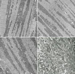
Marked sarcomere disorganization is a well-documented characteristic of cardiomyocytes in the failing human myocardium. Myosin regulatory light chain 2, ventricular/cardiac muscle isoform (MLC2v), which is involved in the development of human cardiomyopathy, is an important structural protein that affects physiologic cardiac sarcomere formation and heart development. Integrated cDNA expression analysis of failing human myocardia uncovered a novel protein kinase, cardiac-specific myosin light chain kinase (cardiac-MLCK), which acts on MLC2v. Expression levels of cardiac-MLCK were well correlated with the pulmonary arterial pressure of patients with heart failure. In cultured cardiomyocytes, knockdown of cardiac-MLCK by specific siRNAs decreased MLC2v phosphorylation and impaired epinephrine-induced activation of sarcomere reassembly. To further clarify the physiologic roles of cardiac-MLCK in vivo, we cloned the zebrafish ortholog z–cardiac-MLCK. Knockdown of z–cardiac-MLCK expression using morpholino antisense oligonucleotides resulted in dilated cardiac ventricles and immature sarcomere structures. These results suggest a significant role for cardiac-MLCK in cardiogenesis.
Authors
Osamu Seguchi, Seiji Takashima, Satoru Yamazaki, Masanori Asakura,, Yoshihiro Asano, Yasunori Shintani, Masakatsu Wakeno, Tetsuo Minamino, Hiroya Kondo, Hidehiko Furukawa, Kenji Nakamaru, Asuka Naito,, Tomoko Takahashi, Toshiaki Ohtsuka, Koichi Kawakami, Tadashi Isomura,, Soichiro Kitamura, Hitonobu Tomoike, Naoki Mochizuki, Masafumi Kitakaze
Citation Information: J Clin Invest. 2007;117(10):2974-2982. https://doi.org/10.1172/JCI31344.
Abstract
T lymphocyte responses promote proatherogenic inflammatory events, which are influenced by costimulatory molecules of the B7 family. Effects of negative regulatory members of the B7 family on atherosclerosis have not been described. Programmed death–ligand 1 (PD-L1) and PD-L2 are B7 family members expressed on several cell types, which inhibit T cell activation via binding to programmed death–1 (PD-1) on T cells. In order to test whether the PD-1/PD-L pathway regulates proatherogenic T cell responses, we compared atherosclerotic lesion burden and phenotype in hypercholesterolemic PD-L1/2–/–LDLR–/– mice and LDLR–/– controls. PD-L1/2 deficiency led to significantly increased atherosclerotic burden throughout the aorta and increased numbers of lesional CD4+ and CD8+ T cells. Compared with controls, PD-L1/2–/–LDLR–/– mice had iliac lymphadenopathy and increased numbers of activated CD4+ T cells. Serum levels of TNF-α were higher in PD-L1/2–/–LDLR–/– mice than in controls. PD-L1/2–deficient APCs were more effective than control APCs in activating CD4+ T cells in vitro, with or without cholesterol loading. Freshly isolated APCs from hypercholesterolemic PD-L1/2–/–LDLR–/– mice stimulated greater T cell responses than did APCs from hypercholesterolemic controls. Our findings indicate that the PD-1/PD-L pathway has an important role in downregulating proatherogenic T cell response and atherosclerosis by limiting APC-dependent T cell activation.
Authors
Israel Gotsman, Nir Grabie, Rosa Dacosta, Galina Sukhova, Arlene Sharpe, Andrew H. Lichtman
Citation Information: J Clin Invest. 2007;117(10):2802-2811. https://doi.org/10.1172/JCI32308.
Abstract
Myotonic dystrophy type 1 (DM1) is caused by a CTG trinucleotide expansion in the 3′ untranslated region (3′ UTR) of DM protein kinase (DMPK). The key feature of DM1 pathogenesis is nuclear accumulation of RNA, which causes aberrant alternative splicing of specific pre-mRNAs by altering the functions of CUG-binding proteins (CUGBPs). Cardiac involvement occurs in more than 80% of individuals with DM1 and is responsible for up to 30% of disease-related deaths. We have generated an inducible and heart-specific DM1 mouse model expressing expanded CUG RNA in the context of DMPK 3′ UTR that recapitulated pathological and molecular features of DM1 including dilated cardiomyopathy, arrhythmias, systolic and diastolic dysfunction, and misregulated alternative splicing. Combined in situ hybridization and immunofluorescent staining for CUGBP1 and CUGBP2, the 2 CUGBP1 and ETR-3 like factor (CELF) proteins expressed in heart, demonstrated elevated protein levels specifically in nuclei containing foci of CUG repeat RNA. A time-course study demonstrated that colocalization of MBNL1 with RNA foci and increased CUGBP1 occurred within hours of induced expression of CUG repeat RNA and coincided with reversion to embryonic splicing patterns. These results indicate that CUGBP1 upregulation is an early and primary response to expression of CUG repeat RNA.
Authors
Guey-Shin Wang, Debra L. Kearney, Mariella De Biasi, George Taffet, Thomas A. Cooper
Citation Information: J Clin Invest. 2007;117(9):2692-2701. https://doi.org/10.1172/JCI29134.
Abstract
Transgenic mice with cardiac-restricted overexpression of secretable TNF (MHCsTNF) develop progressive LV wall thinning and dilation accompanied by an increase in cardiomyocyte apoptosis and a progressive loss of cytoprotective Bcl-2. To test whether cardiac-restricted overexpression of Bcl-2 would prevent adverse cardiac remodeling, we crossed MHCsTNF mice with transgenic mice harboring cardiac-restricted overexpression of Bcl-2. Sustained TNF signaling resulted in activation of the intrinsic cell death pathway, leading to increased cytosolic levels of cytochrome c, Smac/Diablo and Omi/HtrA2, and activation of caspases -3 and -9. Cardiac-restricted overexpression of Bcl-2 blunted activation of the intrinsic pathway and prevented LV wall thinning; however, Bcl-2 only partially attenuated cardiomyocyte apoptosis. Subsequent studies showed that c-FLIP was degraded, that caspase-8 was activated, and that Bid was cleaved to t-Bid, suggesting that the extrinsic pathway was activated concurrently in MHCsTNF hearts. As expected, cardiac Bcl-2 overexpression had no effect on extrinsic signaling. Thus, our results suggest that sustained inflammation leads to activation of multiple cell death pathways that contribute to progressive cardiomyocyte apoptosis; hence the extent of such programmed myocyte cell death is a critical determinant of adverse cardiac remodeling.
Authors
Sandra B. Haudek, George E. Taffet, Michael D. Schneider, Douglas L. Mann
Citation Information: J Clin Invest. 2007;117(9):2496-2505. https://doi.org/10.1172/JCI29838.
Abstract
Clinical use of prostaglandin synthase–inhibiting NSAIDs is associated with the development of hypertension; however, the cardiovascular effects of antagonists for individual prostaglandin receptors remain uncharacterized. The present studies were aimed at elucidating the role of prostaglandin E2 (PGE2) E-prostanoid receptor subtype 1 (EP1) in regulating blood pressure. Oral administration of the EP1 receptor antagonist SC51322 reduced blood pressure in spontaneously hypertensive rats. To define whether this antihypertensive effect was caused by EP1 receptor inhibition, an EP1-null mouse was generated using a “hit-and-run” strategy that disrupted the gene encoding EP1 but spared expression of protein kinase N (PKN) encoded at the EP1 locus on the antiparallel DNA strand. Selective genetic disruption of the EP1 receptor blunted the acute pressor response to Ang II and reduced chronic Ang II–driven hypertension. SC51322 blunted the constricting effect of Ang II on in vitro–perfused preglomerular renal arterioles and mesenteric arteriolar rings. Similarly, the pressor response to EP1-selective agonists sulprostone and 17-phenyltrinor PGE2 were blunted by SC51322 and in EP1-null mice. These data support the possibility of targeting the EP1 receptor for antihypertensive therapy.
Authors
Youfei Guan, Yahua Zhang, Jing Wu, Zhonghua Qi, Guangrui Yang, Dou Dou, Yuansheng Gao, Lihong Chen, Xiaoyan Zhang, Linda S. Davis, Mingfeng Wei, Xuefeng Fan, Monica Carmosino, Chuanming Hao, John D. Imig, Richard M. Breyer, Matthew D. Breyer
Citation Information: J Clin Invest. 2007;117(9):2431-2444. https://doi.org/10.1172/JCI31060.
Abstract
Loss of cardiac myocytes in heart failure is thought to occur largely through an apoptotic process. Here we show that heart failure can also be precipitated through myocyte necrosis associated with Ca2+ overload. Inducible transgenic mice with enhanced sarcolemmal L-type Ca2+ channel (LTCC) activity showed progressive myocyte necrosis that led to pump dysfunction and premature death, effects that were dramatically enhanced by acute stimulation of β-adrenergic receptors. Enhanced Ca2+ influx–induced cellular necrosis and cardiomyopathy was prevented with either LTCC blockers or β-adrenergic receptor antagonists, demonstrating a proximal relationship among β-adrenergic receptor function, Ca2+ handling, and heart failure progression through necrotic cell loss. Mechanistically, loss of cyclophilin D, a regulator of the mitochondrial permeability transition pore that underpins necrosis, blocked Ca2+ influx–induced necrosis of myocytes, heart failure, and isoproterenol-induced premature death. In contrast, overexpression of the antiapoptotic factor Bcl-2 was ineffective in mitigating heart failure and death associated with excess Ca2+ influx and acute β-adrenergic receptor stimulation. This paradigm of mitochondrial- and necrosis-dependent heart failure was also observed in other mouse models of disease, which supports the concept that heart failure is a pleiotropic disorder that involves not only apoptosis, but also necrotic loss of myocytes in association with dysregulated Ca2+ handling and β-adrenergic receptor signaling.
Authors
Hiroyuki Nakayama, Xiongwen Chen, Christopher P. Baines, Raisa Klevitsky, Xiaoying Zhang, Hongyu Zhang, Naser Jaleel, Balvin H.L. Chua, Timothy E. Hewett, Jeffrey Robbins, Steven R. Houser, Jeffery D. Molkentin
Citation Information: J Clin Invest. 2007;117(9):2486-2495. https://doi.org/10.1172/JCI32827.
Abstract
Maintenance of skeletal and cardiac muscle structure and function requires precise control of the synthesis, assembly, and turnover of contractile proteins of the sarcomere. Abnormalities in accumulation of sarcomere proteins are responsible for a variety of myopathies. However, the mechanisms that mediate turnover of these long-lived proteins remain poorly defined. We show that muscle RING finger 1 (MuRF1) and MuRF3 act as E3 ubiquitin ligases that cooperate with the E2 ubiquitin–conjugating enzymes UbcH5a, -b, and -c to mediate the degradation of β/slow myosin heavy chain (β/slow MHC) and MHCIIa via the ubiquitin proteasome system (UPS) in vivo and in vitro. Accordingly, mice deficient for MuRF1 and MuRF3 develop a skeletal muscle myopathy and hypertrophic cardiomyopathy characterized by subsarcolemmal MHC accumulation, myofiber fragmentation, and diminished muscle performance. These findings identify MuRF1 and MuRF3 as key E3 ubiquitin ligases for the UPS-dependent turnover of sarcomeric proteins and reveal a potential basis for myosin storage myopathies.
Authors
Jens Fielitz, Mi-Sung Kim, John M. Shelton, Shuaib Latif, Jeffrey A. Spencer, David J. Glass, James A. Richardson, Rhonda Bassel-Duby, Eric N. Olson
Citation Information: J Clin Invest. 2007;117(8):2123-2132. https://doi.org/10.1172/JCI30756.
Abstract
Noonan syndrome (NS) is an autosomal dominant disorder characterized by a wide spectrum of defects, which most frequently include proportionate short stature, craniofacial anomalies, and congenital heart disease (CHD). NS is the most common nonchromosomal cause of CHD, and 80%–90% of NS patients have cardiac involvement. Mutations within the protein tyrosine phosphatase Src homology region 2, phosphatase 2 (SHP2) are responsible for approximately 50% of the cases of NS with cardiac involvement. To understand the developmental stage– and cell type–specific consequences of the NS SHP2 gain-of-function mutation, Q79R, we generated transgenic mice in which the mutated protein was expressed during gestation or following birth in cardiomyocytes. Q79R SHP2 embryonic hearts showed altered cardiomyocyte cell cycling, ventricular noncompaction, and ventricular septal defects, while, in the postnatal cardiomyocyte, Q79R SHP2 expression was completely benign. Fetal expression of Q79R led to the specific activation of the ERK1/2 pathway, and breeding of the Q79R transgenics into ERK1/2-null backgrounds confirmed the pathway’s necessity and sufficiency in mediating mutant SHP2’s effects. Our data establish the developmental stage–specific effects of Q79R cardiac expression in NS; show that ablation of subsequent ERK1/2 activation prevents the development of cardiac abnormalities; and suggest that ERK1/2 modulation could have important implications for developing therapeutic strategies in CHD.
Authors
Tomoki Nakamura, Melissa Colbert, Maike Krenz, Jeffery D. Molkentin, Harvey S. Hahn, Gerald W. Dorn II, Jeffrey Robbins
Citation Information: J Clin Invest. 2007;117(8):2337-2346. https://doi.org/10.1172/JCI31909.
Abstract
Liver X receptors (LXRs) α and β are transcriptional regulators of cholesterol homeostasis and potential targets for the development of antiatherosclerosis drugs. However, the specific roles of individual LXR isotypes in atherosclerosis and the pharmacological effects of synthetic agonists remain unclear. Previous work has shown that mice lacking LXRα accumulate cholesterol in the liver but not in peripheral tissues. In striking contrast, we demonstrate here that LXRα–/–apoE–/– mice exhibit extreme cholesterol accumulation in peripheral tissues, a dramatic increase in whole-body cholesterol burden, and accelerated atherosclerosis. The phenotype of these mice suggests that the level of LXR pathway activation in macrophages achieved by LXRβ and endogenous ligand is unable to maintain homeostasis in the setting of hypercholesterolemia. Surprisingly, however, a highly efficacious synthetic agonist was able to compensate for the loss of LXRα. Treatment of LXRα–/–apoE–/– mice with synthetic LXR ligand ameliorates the cholesterol overload phenotype and reduces atherosclerosis. These observations indicate that LXRα has an essential role in maintaining peripheral cholesterol homeostasis in the context of hypercholesterolemia and provide in vivo support for drug development strategies targeting LXRβ.
Authors
Michelle N. Bradley, Cynthia Hong, Mingyi Chen, Sean B. Joseph, Damien C. Wilpitz, Xuping Wang, Aldons J. Lusis, Allan Collins, Willa A. Hseuh, Jon L. Collins, Rajendra K. Tangirala, Peter Tontonoz


Copyright © 2025 American Society for Clinical Investigation
ISSN: 0021-9738 (print), 1558-8238 (online)


