Go to JCI Insight
  • About
  • Editors
  • Consulting Editors
  • For authors
  • Publication ethics
  • Alerts
  • Advertising
  • Job board
  • Subscribe
  • Contact
  • Current issue
  • Past issues
  • By specialty
    • COVID-19
    • Cardiology
    • Gastroenterology
    • Immunology
    • Metabolism
    • Nephrology
    • Neuroscience
    • Oncology
    • Pulmonology
    • Vascular biology
    • All ...
  • Videos
    • Conversations with Giants in Medicine
    • Author's Takes
  • Reviews
    • View all reviews ...
    • Aging (Upcoming)
    • Next-Generation Sequencing in Medicine (Jun 2022)
    • New Therapeutic Targets in Cardiovascular Diseases (Mar 2022)
    • Immunometabolism (Jan 2022)
    • Circadian Rhythm (Oct 2021)
    • Gut-Brain Axis (Jul 2021)
    • Tumor Microenvironment (Mar 2021)
    • View all review series ...
  • Viewpoint
  • Collections
    • In-Press Preview
    • Commentaries
    • Concise Communication
    • Editorials
    • Viewpoint
    • Top read articles
  • Clinical Medicine
  • JCI This Month
    • Current issue
    • Past issues

  • Current issue
  • Past issues
  • Specialties
  • Reviews
  • Review series
  • Conversations with Giants in Medicine
  • Author's Takes
  • In-Press Preview
  • Commentaries
  • Concise Communication
  • Editorials
  • Viewpoint
  • Top read articles
  • About
  • Editors
  • Consulting Editors
  • For authors
  • Publication ethics
  • Alerts
  • Advertising
  • Job board
  • Subscribe
  • Contact
X-linked macrocytic dyserythropoietic anemia in females with an ALAS2 mutation
Vijay G. Sankaran, … , David F. Bishop, David P. Steensma
Published February 23, 2015
Citation Information: J Clin Invest. 2015;125(4):1665-1669. https://doi.org/10.1172/JCI78619.
View: Text | PDF | Corrigendum
Brief Report Hematology

X-linked macrocytic dyserythropoietic anemia in females with an ALAS2 mutation

  • Text
  • PDF
Abstract

Macrocytic anemia with abnormal erythropoiesis is a common feature of megaloblastic anemias, congenital dyserythropoietic anemias, and myelodysplastic syndromes. Here, we characterized a family with multiple female individuals who have macrocytic anemia. The proband was noted to have dyserythropoiesis and iron overload. After an extensive diagnostic evaluation that did not provide insight into the cause of the disease, whole-exome sequencing of multiple family members revealed the presence of a mutation in the X chromosomal gene ALAS2, which encodes 5′-aminolevulinate synthase 2, in the affected females. We determined that this mutation (Y365C) impairs binding of the essential cofactor pyridoxal 5′-phosphate to ALAS2, resulting in destabilization of the enzyme and consequent loss of function. X inactivation was not highly skewed in wbc from the affected individuals. In contrast, and consistent with the severity of the ALAS2 mutation, there was a complete skewing toward expression of the WT allele in mRNA from reticulocytes that could be recapitulated in primary erythroid cultures. Together, the results of the X inactivation and mRNA studies illustrate how this X-linked dominant mutation in ALAS2 can perturb normal erythropoiesis through cell-nonautonomous effects. Moreover, our findings highlight the value of whole-exome sequencing in diagnostically challenging cases for the identification of disease etiology and extension of the known phenotypic spectrum of disease.

Authors

Vijay G. Sankaran, Jacob C. Ulirsch, Vassili Tchaikovskii, Leif S. Ludwig, Aoi Wakabayashi, Senkottuvelan Kadirvel, R. Coleman Lindsley, Rafael Bejar, Jiahai Shi, Scott B. Lovitch, David F. Bishop, David P. Steensma

×

Figure 3

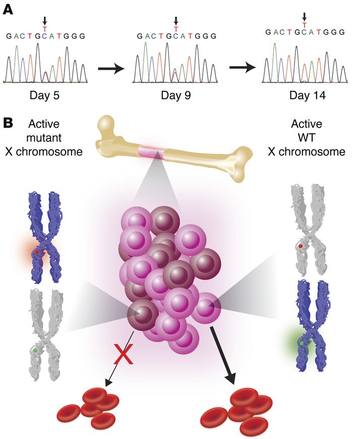
X-linked dominant LOF ALAS2 mutations can result in macrocytosis and dyserythropoiesis through cell-nonautonomous effects.

Options: View larger image (or click on image) Download as PowerPoint
X-linked dominant LOF ALAS2 mutations can result in macrocytosis and dys...
(A) Sequencing of cDNA derived from cultured erythroid cells from the proband showed skewing toward the WT ALAS2 allele at the late stages of erythroid differentiation. Cells began as progenitors (day 5), became intermediate erythroblasts (day 9), and transitioned to orthochromatic erythroblasts (day 14). By late erythropoiesis, only a trace amount of the mutant allele was detectable. (B) Model showing how developing erythroid progenitors and precursors compete for space within the BM. The active X chromosome expressed in each group of cells (brown and pink for the mutant and WT, respectively) is shown in blue, with some cells expressing the mutant (red) and others expressing the WT (green) allele.

Copyright © 2022 American Society for Clinical Investigation
ISSN: 0021-9738 (print), 1558-8238 (online)

Sign up for email alerts