Go to JCI Insight
  • About
  • Editors
  • Consulting Editors
  • For authors
  • Publication ethics
  • Alerts
  • Advertising
  • Job board
  • Subscribe
  • Contact
  • Current issue
  • Past issues
  • By specialty
    • COVID-19
    • Cardiology
    • Gastroenterology
    • Immunology
    • Metabolism
    • Nephrology
    • Neuroscience
    • Oncology
    • Pulmonology
    • Vascular biology
    • All ...
  • Videos
    • Conversations with Giants in Medicine
    • Author's Takes
  • Reviews
    • View all reviews ...
    • Aging (Upcoming)
    • Next-Generation Sequencing in Medicine (Jun 2022)
    • New Therapeutic Targets in Cardiovascular Diseases (Mar 2022)
    • Immunometabolism (Jan 2022)
    • Circadian Rhythm (Oct 2021)
    • Gut-Brain Axis (Jul 2021)
    • Tumor Microenvironment (Mar 2021)
    • View all review series ...
  • Viewpoint
  • Collections
    • In-Press Preview
    • Commentaries
    • Concise Communication
    • Editorials
    • Viewpoint
    • Top read articles
  • Clinical Medicine
  • JCI This Month
    • Current issue
    • Past issues

  • Current issue
  • Past issues
  • Specialties
  • Reviews
  • Review series
  • Conversations with Giants in Medicine
  • Author's Takes
  • In-Press Preview
  • Commentaries
  • Concise Communication
  • Editorials
  • Viewpoint
  • Top read articles
  • About
  • Editors
  • Consulting Editors
  • For authors
  • Publication ethics
  • Alerts
  • Advertising
  • Job board
  • Subscribe
  • Contact
Marfan syndrome and mitral valve prolapse
Arthur E. Weyman, Marielle Scherrer-Crosbie
Published December 1, 2004
Citation Information: J Clin Invest. 2004;114(11):1543-1546. https://doi.org/10.1172/JCI23701.
View: Text | PDF
Commentary

Marfan syndrome and mitral valve prolapse

  • Text
  • PDF
Abstract

Mitral valve prolapse (MVP), an abnormal displacement into the left atrium of a thickened and redundant mitral valve during systole, is a relatively frequent abnormality in humans and may be associated with serious complications. A recent study implicates fibrillin-1, a component of extracellular matrix microfibrils, in the pathogenesis of a murine model of MVP. This investigation represents an initial step toward understanding the mechanisms involved in human MVP disease and the development of potential treatments.

Authors

Arthur E. Weyman, Marielle Scherrer-Crosbie

×

Figure 1

Options: View larger image (or click on image) Download as PowerPoint
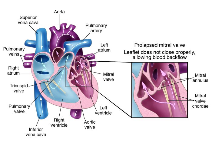
MVP. Mitral valve leaflets, shaped like parachutes, are attached to the ...
MVP. Mitral valve leaflets, shaped like parachutes, are attached to the inner wall of the left ventricle by a series of strings called chordae. When the ventricles contract, the mitral valve leaflets close snugly to prevent backflow of blood from the left ventricle into the left atrium. Upon ventricle relaxation, the valves open to allow oxygenated blood from the lungs to fill the left ventricle. In patients with MVP, myxomatous degeneration of the leaflets and chordae results in leaflet thickening and redundancy, causing the leaflets to prolapse, or flop backwards, into the left atrium (detail). This sometimes allows leakage of blood through the mitral valve (mitral regurgitation). Severe mitral regurgitation can lead to heart failure and abnormal heart rhythms.

Copyright © 2022 American Society for Clinical Investigation
ISSN: 0021-9738 (print), 1558-8238 (online)

Sign up for email alerts