
Advertisement
Review
Open Access |  10.1172/JCI207999
The gut microbiome and allograft outcomes: implications for heart transplantation
Ivan Ðuran,1,2 W.H. Wilson Tang,3,4 and Petra Mamic5,6,7
1University of Zagreb School of Medicine, Zagreb, Croatia.
2Public Health Center (Dom Zdravlja) Zagreb-Centar, Zagreb, Croatia.
3Kaufman Center for Heart Failure Treatment and Recovery; Heart, Vascular and Thoracic Institute; and
4Department of Cardiovascular and Metabolic Sciences, Lerner Research Institute, Cleveland Clinic, Cleveland, Ohio, USA.
5Division of Cardiovascular Medicine, Department of Medicine;
6Department of Genetics; and
7Stanford Cardiovascular Institute, Stanford University School of Medicine, Stanford, California, USA.
Address correspondence to: Petra Mamic, 870 Quarry Rd., Falk Cardiovascular Research Building (CV-261), Stanford University School of Medicine, Stanford, California 94305, USA. Email: mamic@stanford.edu, pmamic1@gmail.com.
Find articles by Ðuran, I. in: PubMed | Google Scholar
1University of Zagreb School of Medicine, Zagreb, Croatia.
2Public Health Center (Dom Zdravlja) Zagreb-Centar, Zagreb, Croatia.
3Kaufman Center for Heart Failure Treatment and Recovery; Heart, Vascular and Thoracic Institute; and
4Department of Cardiovascular and Metabolic Sciences, Lerner Research Institute, Cleveland Clinic, Cleveland, Ohio, USA.
5Division of Cardiovascular Medicine, Department of Medicine;
6Department of Genetics; and
7Stanford Cardiovascular Institute, Stanford University School of Medicine, Stanford, California, USA.
Address correspondence to: Petra Mamic, 870 Quarry Rd., Falk Cardiovascular Research Building (CV-261), Stanford University School of Medicine, Stanford, California 94305, USA. Email: mamic@stanford.edu, pmamic1@gmail.com.
Find articles by Tang, W. in: PubMed | Google Scholar
1University of Zagreb School of Medicine, Zagreb, Croatia.
2Public Health Center (Dom Zdravlja) Zagreb-Centar, Zagreb, Croatia.
3Kaufman Center for Heart Failure Treatment and Recovery; Heart, Vascular and Thoracic Institute; and
4Department of Cardiovascular and Metabolic Sciences, Lerner Research Institute, Cleveland Clinic, Cleveland, Ohio, USA.
5Division of Cardiovascular Medicine, Department of Medicine;
6Department of Genetics; and
7Stanford Cardiovascular Institute, Stanford University School of Medicine, Stanford, California, USA.
Address correspondence to: Petra Mamic, 870 Quarry Rd., Falk Cardiovascular Research Building (CV-261), Stanford University School of Medicine, Stanford, California 94305, USA. Email: mamic@stanford.edu, pmamic1@gmail.com.
Find articles by Mamic, P. in: PubMed | Google Scholar
Published August 17, 2026 - More info
J Clin Invest. 2026;136(16):e207999. https://doi.org/10.1172/JCI207999.
© 2026 Ðuran et al. This work is licensed under the Creative Commons Attribution 4.0 International License. To view a copy of this license, visit http://creativecommons.org/licenses/by/4.0/.

-
Allograft injury mechanisms and post-HT morbidity drivers
Heart allograft injury, one of the leading culprits of post-HT morbidity, occurs by multiple mechanisms, starting with ischemia/reperfusion injury (IRI). Blood flow cessation and restoration between organ procurement and implant incite sterile inflammation, endothelial injury, platelet adhesion, and complement activation (3). This initial tissue injury primes the immune system for subsequent alloimmune injury occurring via acute cellular or antibody-mediated rejection (ACR and AMR, respectively). In ACR, recipient T cells recognize donor HLAs, triggering recruitment of cytotoxic T cells, macrophages, and NK cells that attack allograft parenchyma and endothelium (4). In AMR, donor-specific antibodies (DSAs) target donor HLA on allograft endothelium to trigger complement-mediated endothelial activation, cytokine upregulation, macrophage infiltration, increased vascular permeability, and microvascular thrombosis (5). Cardiac allograft vasculopathy (CAV), a leading cause of late post-transplant morbidity and mortality (6, 7), is a form of chronic rejection causing diffuse coronary artery intimal hyperplasia.
Obligatory immunosuppression used to mitigate these alloimmune complications predisposes to other morbid complications. Infections, some of the most common and serious post-HT complications, affect over half of recipients within the first post-transplant year, causing 20% of post-HT deaths (8–10). Malignancy affects over 20% of HT recipients by 10 years and is the leading cause of mortality beyond 3–5 years post-HT (11, 12). Immunosuppression also predisposes to chronic renal dysfunction and several metabolic complications (diabetes, dyslipidemia, etc.) (13, 14). With its dense connections with the host immune system and metabolism, the gut microbiome plays an important modulator role in many of these processes (Figure 1).
 Figure 1
Multidimensional interactions between the gut microbiome and post-HT complications. HT-associated gut dysbiosis is characterized by reduced α-diversity, depletion of SCFA-producing bacteria, and enrichment of pathobionts. These changes bidirectionally interact with major domains affecting transplant outcomes. Allograft injury and rejection encompass ischemia/reperfusion injury, acute cellular and antibody-mediated rejection, and coronary allograft vasculopathy and are modulated by microbiome-derived metabolites and immune signaling. Infectious complications arise from gut barrier disruption and loss of colonization resistance, increasing susceptibility to bacterial, viral, and fungal pathogens. Metabolic complications including glucose intolerance, diabetes, dyslipidemia, and chronic kidney disease are influenced by alterations in microbial metabolite production and bile acid metabolism. Immunosuppressant pharmacology demonstrates bidirectional interactions, with drugs (tacrolimus, MMF, corticosteroids, mTORi) affecting microbiome composition, while microbial metabolism of immunosuppressants occurs in the luminal compartment, with microbial metabolites affecting epithelial and hepatic compartments influencing drug bioavailability and efficacy. The immune system integrates signals across all domains. Bidirectional arrows emphasize the dynamic, modifiable nature of these interactions, highlighting the gut microbiome as a therapeutic target to optimize post-transplant outcomes. MMF, mycophenolate mofetil; mTORi, mammalian target of rapamycin inhibitors; SCFA, short-chain fatty acid.

-
Environment-microbiome-host crosstalk: a powerful modulator of host physiology
A complex and highly personalized ecosystem, the gut microbiome interfaces closely with the host to influence physiology (15, 16). Gut microbial metabolites, comprising nearly 10% of circulating metabolites in mammals (17), modulate both local and distant organs, including the heart, kidneys, and brain (18, 19). Extrinsic factors, particularly diet (20–22) and medications (including immunosuppressants) (23–26), overwhelm host genetics in shaping the microbiome composition (27) (Figure 2A), with profound physiologic effects, including in the context of HF and HT.
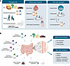 Figure 2
Gut microbiome modulators across the HT continuum and microbiome-targeted therapeutic strategies. (A) Exposures throughout life, including diet and lifestyle choices, shape the gut microbiome. Prior to HT, several factors influence the gut microbiome, including HF progression, medications, and hospitalizations, as well as left ventricular assist device–related (LVAD-related) complications. With HT, new influences are introduced, including surgical stress, critical illness, and intensive medication regimens comprising antibiotics and immunosuppressants, all of which contribute to further alterations of the gut microbiome. (B) The gut microbiome’s malleability presents therapeutic opportunities to restore microbial homeostasis and mitigate transplant-related complications. Interventions include diet, supplementation with prebiotics (fiber and other complex carbohydrates selectively digestible by the gut microbiota), probiotics (live microbes that when administered in adequate amounts confer a health benefit on the host), postbiotics (preparation of microbial metabolic products and/or inactivated microbial cells or cell components that confer a health benefit), synbiotics (mixtures of prebiotics and probiotics), fecal microbiota transplant, and targeted gut microbial enzyme modulation. These strategies aim to reduce complications in the pretransplant period (e.g., HF progression and LVAD-related infections) and post-HT (e.g., rejection, infection, allograft vasculopathy).
Diet is a principal determinant of microbiome composition. Fiber-rich diets, including Mediterranean and plant-based diets, promote antiinflammatory microbes (e.g., Lactobacilli, Bifidobacteria, Faecalibacterium), increasing SCFA production (28, 29). These by-products of gut microbial fermentation of dietary fiber strengthen gut barrier integrity (30, 31), reduce systemic inflammation (32, 33), enhance insulin sensitivity (34), and protect against hypertension and cardiorenal damage (35–38). Conversely, red meat and animal products abound in precursors of microbial metabolites harmful to cardiovascular health. For example, l-carnitine, phosphatidylcholine, and choline are metabolized by gut microbes into trimethylamine, which hepatic enzymes convert to trimethylamine N-oxide (TMAO). Nearly undetectable in vegans, vegetarians, or omnivores after red meat cessation (39), TMAO increases gut permeability, promotes inflammation and endothelial dysfunction, impairs cholesterol metabolism, and enhances platelet activation, driving atherothrombosis (39–41). These findings establish the gut microbiome as a diet-responsive metabolic interface linking environmental exposures to host physiology, with direct implications for HT outcomes.
-
HF to HT: compounded insults to gut homeostasis
Chronic HF, which precedes HT, is associated with gut dysbiosis (42–50) (Figure 2B), which parallels HF severity and predicts clinical outcomes (43, 44, 48, 51). HF-associated α-diversity loss (42–44, 48, 51) reflects depletion of beneficial SCFA producers, including Bifidobacterium, Faecalibacterium, and Lachnospira (44, 45, 48), and overgrowth of pathobionts, including Shigella and Salmonella (47). Functionally, this results in reduced microbiome SCFA biosynthetic capacity, with loss of acetate, butyrate, and propionate, metabolites critical for regulating systemic inflammation, cardiorenal fibrosis and repair, and HF energy metabolism (35, 52, 53). Other beneficial metabolites are similarly depleted — indole-3-propionic acid, a microbial metabolite of dietary tryptophan that strengthens the gut barrier, improves insulin sensitivity, reduces inflammation, and exerts direct cardioprotective effects (54–60), is reduced in chronic HF and associated with milder HF (48, 56, 61). For a comprehensive discussion of HF-specific microbiome alterations, we direct readers to focused reviews (50, 62).
Due to limited organ availability, nearly half of US HT recipients are bridged to transplant with a durable LVAD (63), which introduces additional microbiome perturbations. These include infection-related antibiotic exposure, nonpulsatile blood flow–related gastrointestinal architectural changes, and right-sided HF. LVAD-related infections, among the most common complications (64), predispose to post-HT infections and higher mortality, likely in part through antibiotic-induced dysbiosis (65, 66).
Post-HT gut microbiome dysregulation likely reflects the composite effects of surgical stress and critical illness, and the complex immunosuppressant and antibiotic regimens, superimposed on preexisting HF-associated dysbiosis (Figure 2C). However, clinical studies characterizing the microbiome in HT recipients are limited, with a single longitudinal investigation evaluating changes from pre- to post-transplant (Table 1). In that single-center, prospective cohort of 48 recent HT recipients, α-diversity was markedly reduced compared with healthy controls (67). Multiorgan transplant recipients and patients receiving broad-spectrum antibiotics had the lowest diversity. This reflected loss of antiinflammatory Lachnospiraceae and expansion of potentially pathogenic, pro-inflammatory Enterobacteriaceae and Enterococcaceae, which correlated with reduced fecal SCFAs and secondary bile acids (67). Both of these metabolite classes are immunomodulatory (68–73), pointing to potential pathways by which the microbiome may influence post-transplant adaptive immunity. No significant pre- to post-HT microbiome shifts were observed, though the longitudinal cohort comprised just 17 recipients (67).
Microbial diversity loss post-HT was confirmed in a cross-sectional, single-center study of 86 HT recipients (median 4.2 years post-HT), revealing diversity comparable to advanced HF and inversely correlated with systemic immune activation (51). While inflammation and oxidative stress markers were lower post-transplant than in advanced HF, endotoxemia markers remained elevated compared with patients with mild HF, suggesting ongoing gut dysbiosis (51). Taxonomically, both HF and HT shared loss of Bacteroidetes, suggesting this feature persists across the disease spectrum.
Subsequent cross-sectional, single-center evaluation of 266 HT recipients, some profiled early post-transplant, revealed diversity nadir at 1 week post-transplant, gradual recovery by 3 months, and stabilization by 1 year (74). Immediately post-HT, diversity loss appeared driven by Firmicutes depletion and Proteobacteria enrichment, with temporal recovery toward HF-like composition (74). Consistent with Firmicutes recovery, enrichment of Lachnospiraceae (depleted early post-HT, ref. 67) was observed in long-term HT recipients, though high interindividual variability limited broader taxonomic interpretation. In this study, sarcopenia index, a marker of robust skeletal muscle mass, was independently associated with α-diversity and abundance of Roseburia inulinivorans, a prominent SCFA producer, and inversely associated with circulating C-reactive protein, IL-6, and TNF-α (74). These findings align with preclinical evidence supporting a relationship between the microbiome and skeletal musculature, termed the gut/muscle axis, partially mediated by systemic inflammation (75).
A prospective observational cohort study of over 1,300 Dutch SOT (including 67 HT) recipients demonstrated that gut dysbiosis (defined by reduced α-diversity, dissimilarity from healthy microbiome, and increased antibiotic resistance and virulence factor genes) was associated with all-cause and cause-specific mortality (76). Increased mortality risk was associated with Ruminococcus gnavus abundance, while decreased risk was associated with Bifidobacterium longum and Bifidobacterium adolescentis. A balance between 19 bacterial species predicted all-cause mortality (76), suggesting a potential risk stratification biomarker and a potentially modifiable therapeutic target in this population.
Other studies of HT recipients have focused on circulating gut microbial metabolites, which are implicated in post-transplant physiology (Supplemental Table 1; supplemental material available online with this article; https://doi.org/10.1172/JCI207999DS1). A study of TMAO and its association with inflammation, endotoxemia, and oxidative stress in HT recipients and patients with HF and LVAD found that while TMAO initially decreased post-transplant, it progressively increased to levels comparable to those in patients with symptomatic HF (77). Concurrently, inflammation markers declined post-transplant, but endotoxemia and oxidative stress markers remained elevated. After adjustment for baseline characteristics and renal function, TMAO was no longer associated with inflammation and oxidative stress. Interestingly, TMAO was not correlated with any of the metagenomic features (77), though lack of dietary data may have obscured these relationships.
TMAO, related metabolite γ-butyrobetaine (γ-BB), and their precursors trimethyllysine (TML) and l-carnitine were assessed longitudinally in 62 HT recipients in the SCandinavian HEart transplant everolimus De novo stUdy with earLy calcineurin inhibitor avoidancE (SCHEDULE), a prospective, multicenter, randomized controlled trial (RCT) comparing early everolimus- versus cyclosporine-based immunosuppression (78). These metabolites are implicated in atherothrombotic disease and HF (39, 79, 80). Baseline levels of l-carnitine, TMAO, and TML were elevated in HT recipients compared with healthy controls. Over time, TMAO and TML decreased, though they remained mildly elevated. In contrast, γ-BB and l-carnitine increased steadily post-transplant and were independently associated with future risk of acute rejection and CAV. No immunosuppressant-specific metabolite patterns emerged (78), though the cohort was likely underpowered.
Collectively, these studies demonstrated profound post-HT gut dysbiosis characterized by reduced microbial diversity, pro-inflammatory shifts, and altered metabolite profiles associated with immune dysfunction, gut barrier impairment, frailty, and increased mortality. Notably, many of these features, including loss of α-diversity and depletion of SCFA producers like Lachnospiraceae, coupled with impaired pathogen colonization resistance and expansion of pathobionts including Enterobacteriaceae and Enterococcus, appear shared across SOT populations. Whether organ-specific microbial signatures exist remains unclear; while some studies suggest potential distinctions (e.g., Escherichia enrichment in kidney transplant, KT; ref. 81), direct cross-organ taxonomic comparisons are limited by heterogeneous study design, technical variability, variable immunosuppressive and antibiotic regimens, and, particularly for HT, small cohort sizes. These confounders underscore the need for larger, standardized, multicenter studies with harmonized protocols to definitively distinguish shared versus organ-specific dysbiotic patterns and their clinical implication. Nevertheless, consistent demonstration of dysbiosis and its association with adverse outcomes across SOT populations suggest that targeting the microbiome throughout the peritransplant period may be a promising therapeutic avenue to improve long-term outcomes in transplant recipients (Figure 2D).

-
Gut microbiome and allograft rejection
Modulation of the immune system and alloimmunity. The gut microbiome plays an integral role in shaping the immune system (82), initially by providing a diverse antigen pool to train the developing immune system and later by directing local and systemic immune responses. Gut microbes regulate intra-epithelial lymphocyte proliferation, lymphoid tissue function, and expression of critical Toll-like receptors required for systemic pathogen recognition (83, 84). Segmented filamentous bacteria in the gut also regulate production of immunoglobulin A, the predominant antibody class in humans, through direct interaction with the gut epithelium and indirectly by regulating Th17 cells (85, 86). Other microbes, such as Coprobacillus, stimulate antiinflammatory IL-10–producing T cells in the small intestine (87). Gut microbiome disruption activates pro-inflammatory pathways, contributing to chronic diseases characterized by systemic inflammation, including chronic HF (43).
The gut microbiome has also been implicated in alloimmunity, the host immune response against transplanted tissues (Figure 3). Both preclinical and clinical studies show greater post-transplant survival with “sterile” organs, such as the heart and kidney, compared with colonized organs, like the lungs and intestine (88). In mouse models, genetically identical animals with distinct gut microbiota rejected skin allografts at different rates, with genus Alistipes linked to slower rejection (89). Moreover, cohousing of mice with different rejection kinetics allowed for microbiome intermixing and reduced rejection rates across groups (89). Antibiotic-driven microbiome modulation pretransplant has been shown to improve allograft survival in both skin and HT mouse models, an effect mediated by attenuated type I IFN signaling in antigen-presenting cells and diminished alloreactive T cell priming (90). Additionally, the gut microbiome has been implicated in homeostatic proliferation, the process by which new memory cells emerge to fill the immunologic niche left by induction therapy. Microbiome-derived antigens partly determine which memory cells will emerge (91, 92), thus modulating overall rejection risk.
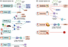 Figure 3
The mechanisms of gut microbiome modulation of alloimmunity and post-transplant rejection risk. (A) A tolerogenic environment is promoted by high-fiber diet and downstream production of SCFAs like acetate and butyrate by the gut microbiome. Acetate and butyrate promote Treg differentiation. Butyrate additionally stimulates regulatory B cells and suppresses pro-inflammatory responses by inhibiting histone deacetylase in macrophages. Beneficial bacteria, such as Bifidobacterium and Alistipes, support immune tolerance by enhancing Treg function, increasing antiinflammatory cytokines (IL-10, TGF-β), and reducing pro-inflammatory cytokines (TNF-α, IL-6). Select probiotics can further induce Tregs. (B) Increased rejection risk is related to heightened inflammatory tone associated with pro-inflammatory microbes such as Desulfovibrio, which increases inflammatory cytokines while suppressing Tregs and antiinflammatory IL-10. Unhealthy dietary habits, such as a high-salt diet, can inhibit protective bacteria, leading to the expansion of pro-inflammatory Th17 cells. High-fat diet is also associated with heightened T cell stimulation and increased rejection risk. Full lines indicate host effects demonstrated in transplant models, with colors signifying protective (green) or detrimental (red) alloimmune effects. Arrows represent stimulatory, and blunt-ended lines inhibitory, relationships. Organ icons refer to the transplanted organ (heart, kidney, skin, or aorta). Dotted lines represent immune effects demonstrated in nontransplant models, of potential relevance for alloimmunity.
The gut microbiome modulates alloimmunity through both local and systemic facets of innate and adaptive immunity. In a fully mismatched HT mouse model, fecal microbiota transplant (FMT) from pregnant, functionally immunosuppressed mice reduced allograft inflammation and fibrosis compared with FMT from colitis-afflicted or healthy mice, improving allograft survival (93). Notably, transfer of monoculture of Bifidobacterium pseudolongum, enriched in the pregnant mouse microbiome, conferred a similar protective effect, suggesting that even a single species may meaningfully modulate rejection risk. These changes corresponded to increased circulating antiinflammatory IL-10 and CCL19, decreased pro-inflammatory TNF-α and IL-6, and an immunosuppressive shift in the mesenteric lymph node architecture. In contrast, transfer of Desulfovibrio desulfuricans, enriched in colitis-afflicted mice, promoted pro-inflammatory lymph node remodeling and a macrophage and dendritic cell inflammatory cytokine response (93).
Mechanistic studies also highlight the concept of “functional redundancy” among microbial species, whereby distinct microbial species fulfill similar physiological roles (e.g., succession between Bifidobacterium species) (94). Concurrently, strain-specific immune influences are evident, with divergent effects of Bifidobacterium pseudolongum strains in mice hypothesized to relate to their host origins, as the mouse isolate appeared to modulate Treg function, whereas the porcine-tropic strain enhanced phagocytosis and dendritic cell proliferation (95). Notably, some immune effects seem independent of long-term microbial engraftment, a phenomenon observed for both protective probiotics and pro-inflammatory Desulfovibrio, which promoted allograft fibrosis despite lack of persistent colonization (94). Supporting the protective role of Bifidobacterium, multistrain probiotic supplementation in a rat liver transplant model increased intestinal Tregs and circulating TGF-β, lowered the CD4/CD8 ratio, and ameliorated rejection-induced liver injury (96).
Gut microbial metabolites of dietary fiber, SCFAs, have also been implicated in alloimmunity. In mouse kidney and aortic transplant models, increasing SCFA levels, through either high-fiber diets or direct acetate supplementation, mitigated allograft rejection and injury while improving allograft longevity (68, 97). These effects were mediated partly through GPR43 receptor and subsequent stimulation of CD25+ Tregs (68). SCFAs also induce epigenetic changes; for instance, butyrate inhibits histone deacetylase in intestinal macrophages and diminishes pro-inflammatory cytokine production (98). Butyrate additionally activates regulatory, IL-10–producing B cells (99).
Clinical implications of gut microbiome–related alloimmunity modulation. Clinical observational studies have evaluated microbiome readouts as potential predictive biomarkers for alloimmunity. As noted above, post-transplant increases in metabolites γ-BB and l-carnitine were associated with acute rejection in HT recipients, even after adjustments for relevant clinical covariates (78). In KT recipients, risk of rejection was associated with a pretransplant Firmicutes/Bacteroidetes imbalance, reduced post-transplant α-diversity (100–102), loss of butyrate-producing taxa (e.g., Clostridiales, Bifidobacterium), and expansion of pro-inflammatory pathobionts (e.g., Enterococcus, Escherichia-Shigella) across multiple observational studies (25, 100, 101, 103). Functional dysbiosis, including enrichment of endotoxin biosynthetic pathways and reduced SCFAs, was also associated with higher rejection risk (101). Elevated circulating TMAO was also linked with kidney allograft failure and mortality, though a direct immunological mechanism remains unconfirmed (104, 105). Crucially, longitudinal studies in KT recipients demonstrate that microbiome alterations precede clinical rejection, normalizing following treatment (100). Moreover, in a prospective study of 97 KT recipients, microbiome signatures enhanced rejection prediction beyond clinical parameters alone (101).
The gut microbiome is also positioned to be a therapeutic target for mitigating rejection. Preclinical studies of kidney and aortic transplantation show reduced rejection and prolonged allograft survival with high-fiber diet (68, 97), offering a proof of concept for potential allograft protection through plant-based, fiber-rich diets (e.g., Mediterranean) with ample SCFA precursors. Fiber (prebiotic) supplementation may further enhance SCFA delivery. While these dietary approaches remain investigational in SOT recipients with no HT-specific data, an ongoing RCT in KT recipients is evaluating the feasibility and efficacy of early post-transplant prebiotic inulin fiber supplementation on systemic inflammation, immune function, and allograft outcomes (106).
Reducing red meat consumption, rich in precursors for TMAO, γ-BB, and other proatherogenic and pro-inflammatory microbial metabolites (39, 107), may be another protective intervention. Notably, while TMAO levels are elevated post-HT (77, 78), a clear association with systemic inflammation or rejection has not yet been established (78). γ-BB has been associated with increased risk of rejection and CAV post-HT (78), though it remains to be determined whether dietary red meat and animal product restriction would lower circulating γ-BB and mitigate rejection.
Other prominent features of a Western diet, such as high salt and saturated fat intake, also modulate alloimmunity. In a mouse HT model, a high-salt diet suppressed Treg proliferation and accelerated rejection via serum- and glucocorticoid-regulated kinase-1 (108). Mechanistically, a high-salt diet depleted gut Lactobacillus murinus, leading to Th17 cell proliferation and hypertension, effects abrogated by L. murinus probiotic supplementation both in mice and in a pilot study of healthy adults (109). Although dietary sodium restriction is routinely recommended post-HT, this recommendation is extrapolated from non-HT populations and hypertension, HF, and chronic kidney disease management guidelines. In KT at least one RCT has demonstrated marked blood pressure reduction with strict sodium control (<2.3 g/d) (110, 111). Similarly, high-fat diet in a mouse HT model enhanced antigen-presenting cell activity and increased IFN-γ production, accelerating rejection (112).
Conversely, fermented foods represent a promising avenue for beneficial host immunomodulation. Foods like yogurt, kefir, kombucha, sourdough, sauerkraut, tempeh, and others, produced through controlled and desirable microbial growth (113), represent a source of beneficial microbial metabolites (including SCFAs) and, in some cases, live microbes. In healthy adults, a fermented food–rich diet increased microbiome α-diversity and decreased circulating inflammatory markers (114). In a mouse HT model, treatment with a probiotic yogurt containing live lactic acid bacteria increased the number of CD4+Foxp3+ Tregs, extending heart allograft survival (115). While the risk of iatrogenic infection in immunosuppressed recipients requires careful consideration, fermented foods may offer net benefits, particularly as immunosuppression is tapered.
-
The gut microbiome and immunosuppressants
The relationship between the microbiome and medications is bidirectional and clinically meaningful in HT recipients. The microbiome modulates immunosuppressant pharmacokinetics, altering drug efficacy and toxicity, while immunosuppressants shift microbiome composition, contributing to their immunomodulatory effects. Elucidating this interplay is essential for personalizing immunosuppressive regimens and improving HT outcomes.
Tacrolimus. The calcineurin inhibitor (CNI) tacrolimus is the foundational immunosuppressant for HT recipients. Its narrow therapeutic window and high pharmacokinetic variability pose clinical challenges, risking underimmunosuppression and rejection, or overexposure and toxicity. The gut microbiome contributes to this variability through direct drug metabolism (Figure 4A). Common gut microbes, including Faecalibacterium prausnitzii, metabolize tacrolimus to less potent metabolites that crossreact with standard immunoassays, confounding therapeutic monitoring (167). This phenomenon can lead to overestimation of biologically active drug concentrations, potentially masking subtherapeutic immunosuppression. Clinically, higher abundances of Faecalibacterium prausnitzii and related Subdoligranulum have been associated with increased tacrolimus dose requirements in KT and HT recipients, respectively (168, 169).
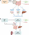 Figure 4
Bidirectional interactions between the gut microbiome and commonly used immunosuppressants after HT. (A) Gut microbes metabolize tacrolimus into its less potent metabolite, in addition to affecting transport of tacrolimus among the gut lumen, epithelium, and circulation. The gut microbiome affects expression of liver-metabolizing enzymes, which may also affect systemic tacrolimus levels. (B) MMF is converted by the host to its active form, mycophenolic acid (MPA). MPA is modified by the liver into inactive MPA-glucuronide (MPAG), which is either excreted by the kidneys or delivered back into the gut lumen, where gut microbes, via the β-glucuronidase (β-GUS) enzyme, metabolize it back to active MPA. MPA mediates MMF-related immunosuppressive effects as well as intestinal inflammation and diarrhea, common MMF side effects that are entirely absent in germ-free animals. MMF also alters gut microbiome composition by stimulating growth of microbes that produce β-GUS. Blue lines denote effects on drug metabolism. Green lines signify immunosuppression-related effects. Red lines refer to adverse effects.
The microbiome also modulates host metabolism, affecting tacrolimus disposition. Antibiotic-induced microbiome shifts increase mouse gut epithelium ABCB1 transporter expression, suppressing tacrolimus recirculation and reducing systemic tacrolimus levels (170). Gut microbial metabolites, particularly SCFAs, function as epigenetic regulators altering hepatic cytochrome P450 expression, influencing drug clearance. Additionally, microbiome-produced indoxyl sulfate modulated host CYP3A4 expression with interindividual variability and context dependence (171).
The microbiome-tacrolimus interaction is bidirectional. Tacrolimus increases gut permeability, potentially facilitating microbial product translocation that influences systemic immunity (172–174). In mice, tacrolimus enriched Allobaculum, Bacteroides, and Lactobacillus, shifting SCFA production (26). Critically, these changes mechanistically contribute to tacrolimus pharmacodynamic effects. For example, combination of low-dose tacrolimus and FMT from high-dose tacrolimus-treated mice fully recapitulated the high-dose tacrolimus treatment effects by expanding Treg populations, reducing pro-inflammatory cytokines, and prolonging skin allograft survival (26). These landmark findings established a paradigm wherein the gut microbiome is not a passive bystander but an active intermediary in the drug’s mechanism of action.
The microbiome also mediates tacrolimus adverse effects like post-transplant diabetes mellitus (PTDM) (175). Specific gut microbiome signatures are associated with PTDM risk in liver transplant and KT recipients (176, 177). Preclinical studies have demonstrated that tacrolimus upregulates bacterial β-glucuronidase (GUS) activity, alters bile acid metabolism, activates ileal farnesoid X receptor, and suppresses glucagon-like peptide-1 secretion, impairing glucose homeostasis (178). Importantly, inhibiting bacterial GUS with vancomycin or by supplementing the SCFA butyrate ameliorated tacrolimus-induced hyperglycemia in preclinical models (179, 180).
These studies highlight a complex triad involving tacrolimus, gut microbiome, and host immunity and metabolism. Defining these interactions in human cohorts is critical for optimizing the efficacy and safety of this essential immunosuppressant.
MMF. MMF, another key post-HT immunosuppressant, demonstrates bidirectional microbiome interactions that profoundly affect its efficacy and toxicity (Figure 4B). Host tissues metabolize MMF to active mycophenolic acid (MPA), which undergoes hepatic glucuronidation to inactive MPA-glucuronide (MPAG). MPAG is renally excreted or undergoes enterohepatic recirculation, where gut microbial β-glucuronidases reactivate it to MPA, accounting for 30%–40% of circulating MPA (181). Critically, MMF selectively expands GUS-expressing bacteria, creating a feed-forward loop that increases MPA levels (182).
These interactions have profound clinical consequences. In mice, colonic inflammation, diarrhea, and weight loss occur only in MMF-treated conventional but not germ-free animals, establishing that gastrointestinal toxicity, one of the most common MMF side effects, is entirely microbiome dependent (183). Multiple mechanisms contribute. First, MMF-mediated expansion of GUS-expressing bacteria elevates colonic MPA levels; in mice, vancomycin selectively eradicates these bacteria and prevents gastrointestinal toxicity (182). Second, MMF reduces α-diversity while enriching pathogenic Escherichia and Shigella, enhancing microbial endotoxin biosynthesis and increasing its fecal and circulating levels, driving severe colonic inflammation (183). Third, MMF reduces gut and systemic SCFA levels (184), with potential consequences for alloimmunity and metabolic homeostasis.
Clinical studies corroborate these preclinical findings. Among 139 KT recipients, MMF was the dominant driver of microbiome dysbiosis, causing α-diversity loss, depletion of antiinflammatory Actinobacteria and SCFA producers, and expansion of pro-inflammatory Proteobacteria (185). In 97 KT recipients, elevated fecal GUS activity was strongly associated with prolonged post-transplant diarrhea (186). Clinical microbiome-targeted interventions remain unexplored, though proof of concept exists in octyl gallate, a food additive with antioxidant and GUS-inhibitory properties, which prevents MMF gastrointestinal toxicity in mice (187). Whether MMF-mediated microbiome shifts contribute to other MMF adverse effects, including myelosuppression (188), warrants investigation.
Corticosteroids. Corticosteroids, another integral post-HT immunosuppressant drug class, demonstrate reciprocal microbiome interactions through active microbial metabolism and microbiome alterations. Gut Clostridium steroidoreducens expresses steroid reductases that inactivate exogenous steroids and reduce their bioavailability (189). One clinical study associated steroid treatment with gut enrichment of Streptococcus salivarius, typically an oral commensal, potentially signaling gut dysbiosis (190). However, human data on steroid-induced microbiome changes remain limited.
Mouse models provide mechanistic insight. Steroid treatment increased Anaerobacterium while depleting Eisenbergiella, Alistipes, and Clostridium XIVb, predicted to diminish SCFA production and compromise immune regulation (191). Comparative analysis in mice revealed the most pronounced microbiome shifts with prednisolone, characterized by reduced Bacteroidetes and expanded Firmicutes (192). This was accompanied by decreased gut antimicrobial defensins Reg3γ and IL-22, Escherichia coli overgrowth, and colonization with its uropathogenic strain. Notably, these microbiome effects appeared dependent on baseline microbiome profiles (192).
Intriguingly, some corticosteroid immunosuppressive effects may be microbiome mediated. In a lupus mouse model, prednisone’s systemic immunomodulatory effects were comparable to those elicited by FMT from prednisone-treated mice (193). These findings highlight critical knowledge gaps and raise questions about whether baseline microbiome profiles may influence steroid efficacy and toxicity.
mTOR inhibitors. Sirolimus (rapamycin) and everolimus, mTOR inhibitors, are frequently used post-HT to reduce malignancy risk and feature prominently in CNI-sparing regimens to mitigate renal dysfunction.
Multiple preclinical studies demonstrate that mTOR inhibitors profoundly alter gut microbiome composition with functional immunologic consequences. Sirolimus induces dysbiosis characterized by increased pro-inflammatory Proteobacteria and decreased antiinflammatory Akkermansia, with associated shifts in microbiome lipid metabolism and immune pathways (194). Sirolimus also compromises gut barrier integrity, increasing gut permeability and circulating pro-inflammatory cytokines (194). Time-dependent sirolimus-induced microbiome shifts include reduced α-diversity and increased Bacteroides/Firmicutes ratio with elevated gut luminal amino acid availability, paralleling early pro-inflammatory to protolerogenic transitions through modulated lymph node architecture and Treg distribution (195). mTOR inhibitor–induced shifts in Firmicutes, Acidobacteria, and Bacteroidetes correlated with Peyer’s patch and splenic T cell populations, some associated with prolonged lifespan (196).
The microbiome also mediates the metabolic complications associated with mTOR inhibitors. In mice fed high-fat diet, sirolimus induced microbiome alterations, worsening gut inflammation and glucose intolerance, which were mitigated by natural polyphenol antioxidant resveratrol (197). Similarly, probiotic Lactobacillus rhamnosus HN001 alleviated sirolimus-induced systemic inflammation, dyslipidemia, and insulin resistance (194), suggesting potential microbiome-targeted therapeutic interventions for managing mTOR inhibitor–associated metabolic toxicity in HT recipients.
Implications of gut microbiome–immunosuppressant interactions for HT. Immunosuppressants are indispensable after HT, yet emerging evidence reveals that they function within a tripartite drug-microbiome-host system. The microbiome actively determines immunosuppressant pharmacokinetics, pharmacodynamics, and toxicity.
These insights suggest translational opportunities for precision immunosuppression management to optimize allograft protection while minimizing adverse effects. However, current evidence is derived predominantly from preclinical models and non-HT populations, necessitating dedicated studies in HT recipients to validate these associations and establish clinical utility. Microbiome profiling, including quantifying gut Faecalibacterium prausnitzii (167–169) or GUS activity (186), could facilitate therapeutic drug monitoring and mitigate adverse effects. Targeted microbiome modulation offers a strategy to decouple efficacy from toxicity. Preclinical proof of concept includes the findings that butyrate supplementation prevents tacrolimus-associated hyperglycemia (180), GUS inhibition mitigates MMF-related diarrhea (182, 187), and probiotics ameliorate sirolimus-related metabolic complications (194). As noted above, FMT from tacrolimus-treated mice recapitulates immunosuppressive effects at lower drug exposure (26), suggesting microbiome-targeted therapies might enhance allograft tolerance while reducing drug burden and associated infectious and malignant complications.
Article tools
- Download citation information
- Send a comment
- Terms of use
- Standard abbreviations
- Need help? Email the journal
Metrics
Go to
- Top
- Abstract
- Introduction
- Allograft injury mechanisms and post-HT morbidity drivers
- Environment-microbiome-host crosstalk: a powerful modulator of host physiology
- HF to HT: compounded insults to gut homeostasis
- Gut microbiome and allograft rejection
- Gut microbiome and implications for post-transplant infections
- The gut microbiome and immunosuppressants
- Current challenges and a roadmap to clinical translation
- Conflict of interest
- Funding support
- Supplemental material
- Footnotes
- References
- Version history


Copyright © 2026 American Society for Clinical Investigation
ISSN: 0021-9738 (print), 1558-8238 (online)