
Advertisement
Research ArticleGeneticsNeuroscience
Open Access |  10.1172/JCI207160
Gene-specific machine learning model EpiPred identifies likely pathogenic variants in the epilepsy-related gene STXBP1
Jeffrey D. Calhoun,1 Chengbing Wang,2 Carina G. Biar,1 Jonathan R. Gunti,1 John S. Lee,3 Aaron M. Geller,3 Jung H. Hong,1 Santiago Schnell,4 Louis T. Dang,5 Yu Wang,6 Jack M. Parent,6 Lori L. Isom,6,7 Michael D. Uhler,8 Heather C. Mefford,9 M. Elizabeth Ross,2 Vanessa Aguiar-Pulido,2,10 and Gemma L. Carvill1,11
1Ken and Ruth Davee Department of Neurology, Northwestern Feinberg School of Medicine, Chicago, Illinois, USA.
2Center for Neurogenetics, Feil Family Brain and Mind Research Institute, Weill Cornell Medicine, New York, New York, USA.
3Northwestern IT Research Computing and Data Services, Evanston, Illinois, USA.
4Departments of Mathematics, Biochemistry & Cell Biology and Biomedical Data Science, Dartmouth College, Hanover, New Hampshire, USA.
5Department of Pediatrics,
6Department of Neurology,
7Department of Pharmacology, and
8Department of Biological Chemistry and Molecular Neuroscience Institute, University of Michigan, Ann Arbor, Michigan, USA.
9Center for Pediatric Neurological Disease Research, St. Jude Children’s Research Hospital, Memphis, Tennessee, USA.
10Department of Computer Science, University of Miami, Miami, Florida, USA.
11Department of Pharmacology, Northwestern Feinberg School of Medicine, Chicago, Illinois, USA.
Address for correspondence. Gemma L. Carvill, Ward 9-183, 303 E Chicago, Chicago, Illinois, 60610, USA. Phone: 312.503.6187; Email: gemma.carvill@northwestern.edu. Or: M. Elizabeth Ross, Belfer Research Building, 413 East 69th Street, New York, New York, 10021, USA. Phone: 646.962.6144; Email: mer2005@med.cornell.edu.
Authorship note: JDC, CW, MER, VAP, and GLC contributed equally to this work.
Find articles by Calhoun, J. in: PubMed | Google Scholar
1Ken and Ruth Davee Department of Neurology, Northwestern Feinberg School of Medicine, Chicago, Illinois, USA.
2Center for Neurogenetics, Feil Family Brain and Mind Research Institute, Weill Cornell Medicine, New York, New York, USA.
3Northwestern IT Research Computing and Data Services, Evanston, Illinois, USA.
4Departments of Mathematics, Biochemistry & Cell Biology and Biomedical Data Science, Dartmouth College, Hanover, New Hampshire, USA.
5Department of Pediatrics,
6Department of Neurology,
7Department of Pharmacology, and
8Department of Biological Chemistry and Molecular Neuroscience Institute, University of Michigan, Ann Arbor, Michigan, USA.
9Center for Pediatric Neurological Disease Research, St. Jude Children’s Research Hospital, Memphis, Tennessee, USA.
10Department of Computer Science, University of Miami, Miami, Florida, USA.
11Department of Pharmacology, Northwestern Feinberg School of Medicine, Chicago, Illinois, USA.
Address for correspondence. Gemma L. Carvill, Ward 9-183, 303 E Chicago, Chicago, Illinois, 60610, USA. Phone: 312.503.6187; Email: gemma.carvill@northwestern.edu. Or: M. Elizabeth Ross, Belfer Research Building, 413 East 69th Street, New York, New York, 10021, USA. Phone: 646.962.6144; Email: mer2005@med.cornell.edu.
Authorship note: JDC, CW, MER, VAP, and GLC contributed equally to this work.
Find articles by Wang, C. in: PubMed | Google Scholar
1Ken and Ruth Davee Department of Neurology, Northwestern Feinberg School of Medicine, Chicago, Illinois, USA.
2Center for Neurogenetics, Feil Family Brain and Mind Research Institute, Weill Cornell Medicine, New York, New York, USA.
3Northwestern IT Research Computing and Data Services, Evanston, Illinois, USA.
4Departments of Mathematics, Biochemistry & Cell Biology and Biomedical Data Science, Dartmouth College, Hanover, New Hampshire, USA.
5Department of Pediatrics,
6Department of Neurology,
7Department of Pharmacology, and
8Department of Biological Chemistry and Molecular Neuroscience Institute, University of Michigan, Ann Arbor, Michigan, USA.
9Center for Pediatric Neurological Disease Research, St. Jude Children’s Research Hospital, Memphis, Tennessee, USA.
10Department of Computer Science, University of Miami, Miami, Florida, USA.
11Department of Pharmacology, Northwestern Feinberg School of Medicine, Chicago, Illinois, USA.
Address for correspondence. Gemma L. Carvill, Ward 9-183, 303 E Chicago, Chicago, Illinois, 60610, USA. Phone: 312.503.6187; Email: gemma.carvill@northwestern.edu. Or: M. Elizabeth Ross, Belfer Research Building, 413 East 69th Street, New York, New York, 10021, USA. Phone: 646.962.6144; Email: mer2005@med.cornell.edu.
Authorship note: JDC, CW, MER, VAP, and GLC contributed equally to this work.
Find articles by Biar, C. in: PubMed | Google Scholar
1Ken and Ruth Davee Department of Neurology, Northwestern Feinberg School of Medicine, Chicago, Illinois, USA.
2Center for Neurogenetics, Feil Family Brain and Mind Research Institute, Weill Cornell Medicine, New York, New York, USA.
3Northwestern IT Research Computing and Data Services, Evanston, Illinois, USA.
4Departments of Mathematics, Biochemistry & Cell Biology and Biomedical Data Science, Dartmouth College, Hanover, New Hampshire, USA.
5Department of Pediatrics,
6Department of Neurology,
7Department of Pharmacology, and
8Department of Biological Chemistry and Molecular Neuroscience Institute, University of Michigan, Ann Arbor, Michigan, USA.
9Center for Pediatric Neurological Disease Research, St. Jude Children’s Research Hospital, Memphis, Tennessee, USA.
10Department of Computer Science, University of Miami, Miami, Florida, USA.
11Department of Pharmacology, Northwestern Feinberg School of Medicine, Chicago, Illinois, USA.
Address for correspondence. Gemma L. Carvill, Ward 9-183, 303 E Chicago, Chicago, Illinois, 60610, USA. Phone: 312.503.6187; Email: gemma.carvill@northwestern.edu. Or: M. Elizabeth Ross, Belfer Research Building, 413 East 69th Street, New York, New York, 10021, USA. Phone: 646.962.6144; Email: mer2005@med.cornell.edu.
Authorship note: JDC, CW, MER, VAP, and GLC contributed equally to this work.
Find articles by Gunti, J. in: PubMed | Google Scholar
1Ken and Ruth Davee Department of Neurology, Northwestern Feinberg School of Medicine, Chicago, Illinois, USA.
2Center for Neurogenetics, Feil Family Brain and Mind Research Institute, Weill Cornell Medicine, New York, New York, USA.
3Northwestern IT Research Computing and Data Services, Evanston, Illinois, USA.
4Departments of Mathematics, Biochemistry & Cell Biology and Biomedical Data Science, Dartmouth College, Hanover, New Hampshire, USA.
5Department of Pediatrics,
6Department of Neurology,
7Department of Pharmacology, and
8Department of Biological Chemistry and Molecular Neuroscience Institute, University of Michigan, Ann Arbor, Michigan, USA.
9Center for Pediatric Neurological Disease Research, St. Jude Children’s Research Hospital, Memphis, Tennessee, USA.
10Department of Computer Science, University of Miami, Miami, Florida, USA.
11Department of Pharmacology, Northwestern Feinberg School of Medicine, Chicago, Illinois, USA.
Address for correspondence. Gemma L. Carvill, Ward 9-183, 303 E Chicago, Chicago, Illinois, 60610, USA. Phone: 312.503.6187; Email: gemma.carvill@northwestern.edu. Or: M. Elizabeth Ross, Belfer Research Building, 413 East 69th Street, New York, New York, 10021, USA. Phone: 646.962.6144; Email: mer2005@med.cornell.edu.
Authorship note: JDC, CW, MER, VAP, and GLC contributed equally to this work.
Find articles by Lee, J. in: PubMed | Google Scholar
1Ken and Ruth Davee Department of Neurology, Northwestern Feinberg School of Medicine, Chicago, Illinois, USA.
2Center for Neurogenetics, Feil Family Brain and Mind Research Institute, Weill Cornell Medicine, New York, New York, USA.
3Northwestern IT Research Computing and Data Services, Evanston, Illinois, USA.
4Departments of Mathematics, Biochemistry & Cell Biology and Biomedical Data Science, Dartmouth College, Hanover, New Hampshire, USA.
5Department of Pediatrics,
6Department of Neurology,
7Department of Pharmacology, and
8Department of Biological Chemistry and Molecular Neuroscience Institute, University of Michigan, Ann Arbor, Michigan, USA.
9Center for Pediatric Neurological Disease Research, St. Jude Children’s Research Hospital, Memphis, Tennessee, USA.
10Department of Computer Science, University of Miami, Miami, Florida, USA.
11Department of Pharmacology, Northwestern Feinberg School of Medicine, Chicago, Illinois, USA.
Address for correspondence. Gemma L. Carvill, Ward 9-183, 303 E Chicago, Chicago, Illinois, 60610, USA. Phone: 312.503.6187; Email: gemma.carvill@northwestern.edu. Or: M. Elizabeth Ross, Belfer Research Building, 413 East 69th Street, New York, New York, 10021, USA. Phone: 646.962.6144; Email: mer2005@med.cornell.edu.
Authorship note: JDC, CW, MER, VAP, and GLC contributed equally to this work.
Find articles by Geller, A. in: PubMed | Google Scholar
1Ken and Ruth Davee Department of Neurology, Northwestern Feinberg School of Medicine, Chicago, Illinois, USA.
2Center for Neurogenetics, Feil Family Brain and Mind Research Institute, Weill Cornell Medicine, New York, New York, USA.
3Northwestern IT Research Computing and Data Services, Evanston, Illinois, USA.
4Departments of Mathematics, Biochemistry & Cell Biology and Biomedical Data Science, Dartmouth College, Hanover, New Hampshire, USA.
5Department of Pediatrics,
6Department of Neurology,
7Department of Pharmacology, and
8Department of Biological Chemistry and Molecular Neuroscience Institute, University of Michigan, Ann Arbor, Michigan, USA.
9Center for Pediatric Neurological Disease Research, St. Jude Children’s Research Hospital, Memphis, Tennessee, USA.
10Department of Computer Science, University of Miami, Miami, Florida, USA.
11Department of Pharmacology, Northwestern Feinberg School of Medicine, Chicago, Illinois, USA.
Address for correspondence. Gemma L. Carvill, Ward 9-183, 303 E Chicago, Chicago, Illinois, 60610, USA. Phone: 312.503.6187; Email: gemma.carvill@northwestern.edu. Or: M. Elizabeth Ross, Belfer Research Building, 413 East 69th Street, New York, New York, 10021, USA. Phone: 646.962.6144; Email: mer2005@med.cornell.edu.
Authorship note: JDC, CW, MER, VAP, and GLC contributed equally to this work.
Find articles by Hong, J. in: PubMed | Google Scholar
1Ken and Ruth Davee Department of Neurology, Northwestern Feinberg School of Medicine, Chicago, Illinois, USA.
2Center for Neurogenetics, Feil Family Brain and Mind Research Institute, Weill Cornell Medicine, New York, New York, USA.
3Northwestern IT Research Computing and Data Services, Evanston, Illinois, USA.
4Departments of Mathematics, Biochemistry & Cell Biology and Biomedical Data Science, Dartmouth College, Hanover, New Hampshire, USA.
5Department of Pediatrics,
6Department of Neurology,
7Department of Pharmacology, and
8Department of Biological Chemistry and Molecular Neuroscience Institute, University of Michigan, Ann Arbor, Michigan, USA.
9Center for Pediatric Neurological Disease Research, St. Jude Children’s Research Hospital, Memphis, Tennessee, USA.
10Department of Computer Science, University of Miami, Miami, Florida, USA.
11Department of Pharmacology, Northwestern Feinberg School of Medicine, Chicago, Illinois, USA.
Address for correspondence. Gemma L. Carvill, Ward 9-183, 303 E Chicago, Chicago, Illinois, 60610, USA. Phone: 312.503.6187; Email: gemma.carvill@northwestern.edu. Or: M. Elizabeth Ross, Belfer Research Building, 413 East 69th Street, New York, New York, 10021, USA. Phone: 646.962.6144; Email: mer2005@med.cornell.edu.
Authorship note: JDC, CW, MER, VAP, and GLC contributed equally to this work.
Find articles by Schnell, S. in: PubMed | Google Scholar
1Ken and Ruth Davee Department of Neurology, Northwestern Feinberg School of Medicine, Chicago, Illinois, USA.
2Center for Neurogenetics, Feil Family Brain and Mind Research Institute, Weill Cornell Medicine, New York, New York, USA.
3Northwestern IT Research Computing and Data Services, Evanston, Illinois, USA.
4Departments of Mathematics, Biochemistry & Cell Biology and Biomedical Data Science, Dartmouth College, Hanover, New Hampshire, USA.
5Department of Pediatrics,
6Department of Neurology,
7Department of Pharmacology, and
8Department of Biological Chemistry and Molecular Neuroscience Institute, University of Michigan, Ann Arbor, Michigan, USA.
9Center for Pediatric Neurological Disease Research, St. Jude Children’s Research Hospital, Memphis, Tennessee, USA.
10Department of Computer Science, University of Miami, Miami, Florida, USA.
11Department of Pharmacology, Northwestern Feinberg School of Medicine, Chicago, Illinois, USA.
Address for correspondence. Gemma L. Carvill, Ward 9-183, 303 E Chicago, Chicago, Illinois, 60610, USA. Phone: 312.503.6187; Email: gemma.carvill@northwestern.edu. Or: M. Elizabeth Ross, Belfer Research Building, 413 East 69th Street, New York, New York, 10021, USA. Phone: 646.962.6144; Email: mer2005@med.cornell.edu.
Authorship note: JDC, CW, MER, VAP, and GLC contributed equally to this work.
Find articles by Dang, L. in: PubMed | Google Scholar
1Ken and Ruth Davee Department of Neurology, Northwestern Feinberg School of Medicine, Chicago, Illinois, USA.
2Center for Neurogenetics, Feil Family Brain and Mind Research Institute, Weill Cornell Medicine, New York, New York, USA.
3Northwestern IT Research Computing and Data Services, Evanston, Illinois, USA.
4Departments of Mathematics, Biochemistry & Cell Biology and Biomedical Data Science, Dartmouth College, Hanover, New Hampshire, USA.
5Department of Pediatrics,
6Department of Neurology,
7Department of Pharmacology, and
8Department of Biological Chemistry and Molecular Neuroscience Institute, University of Michigan, Ann Arbor, Michigan, USA.
9Center for Pediatric Neurological Disease Research, St. Jude Children’s Research Hospital, Memphis, Tennessee, USA.
10Department of Computer Science, University of Miami, Miami, Florida, USA.
11Department of Pharmacology, Northwestern Feinberg School of Medicine, Chicago, Illinois, USA.
Address for correspondence. Gemma L. Carvill, Ward 9-183, 303 E Chicago, Chicago, Illinois, 60610, USA. Phone: 312.503.6187; Email: gemma.carvill@northwestern.edu. Or: M. Elizabeth Ross, Belfer Research Building, 413 East 69th Street, New York, New York, 10021, USA. Phone: 646.962.6144; Email: mer2005@med.cornell.edu.
Authorship note: JDC, CW, MER, VAP, and GLC contributed equally to this work.
Find articles by Wang, Y. in: PubMed | Google Scholar
1Ken and Ruth Davee Department of Neurology, Northwestern Feinberg School of Medicine, Chicago, Illinois, USA.
2Center for Neurogenetics, Feil Family Brain and Mind Research Institute, Weill Cornell Medicine, New York, New York, USA.
3Northwestern IT Research Computing and Data Services, Evanston, Illinois, USA.
4Departments of Mathematics, Biochemistry & Cell Biology and Biomedical Data Science, Dartmouth College, Hanover, New Hampshire, USA.
5Department of Pediatrics,
6Department of Neurology,
7Department of Pharmacology, and
8Department of Biological Chemistry and Molecular Neuroscience Institute, University of Michigan, Ann Arbor, Michigan, USA.
9Center for Pediatric Neurological Disease Research, St. Jude Children’s Research Hospital, Memphis, Tennessee, USA.
10Department of Computer Science, University of Miami, Miami, Florida, USA.
11Department of Pharmacology, Northwestern Feinberg School of Medicine, Chicago, Illinois, USA.
Address for correspondence. Gemma L. Carvill, Ward 9-183, 303 E Chicago, Chicago, Illinois, 60610, USA. Phone: 312.503.6187; Email: gemma.carvill@northwestern.edu. Or: M. Elizabeth Ross, Belfer Research Building, 413 East 69th Street, New York, New York, 10021, USA. Phone: 646.962.6144; Email: mer2005@med.cornell.edu.
Authorship note: JDC, CW, MER, VAP, and GLC contributed equally to this work.
Find articles by Parent, J. in: PubMed | Google Scholar
1Ken and Ruth Davee Department of Neurology, Northwestern Feinberg School of Medicine, Chicago, Illinois, USA.
2Center for Neurogenetics, Feil Family Brain and Mind Research Institute, Weill Cornell Medicine, New York, New York, USA.
3Northwestern IT Research Computing and Data Services, Evanston, Illinois, USA.
4Departments of Mathematics, Biochemistry & Cell Biology and Biomedical Data Science, Dartmouth College, Hanover, New Hampshire, USA.
5Department of Pediatrics,
6Department of Neurology,
7Department of Pharmacology, and
8Department of Biological Chemistry and Molecular Neuroscience Institute, University of Michigan, Ann Arbor, Michigan, USA.
9Center for Pediatric Neurological Disease Research, St. Jude Children’s Research Hospital, Memphis, Tennessee, USA.
10Department of Computer Science, University of Miami, Miami, Florida, USA.
11Department of Pharmacology, Northwestern Feinberg School of Medicine, Chicago, Illinois, USA.
Address for correspondence. Gemma L. Carvill, Ward 9-183, 303 E Chicago, Chicago, Illinois, 60610, USA. Phone: 312.503.6187; Email: gemma.carvill@northwestern.edu. Or: M. Elizabeth Ross, Belfer Research Building, 413 East 69th Street, New York, New York, 10021, USA. Phone: 646.962.6144; Email: mer2005@med.cornell.edu.
Authorship note: JDC, CW, MER, VAP, and GLC contributed equally to this work.
Find articles by
Isom, L.
in:
PubMed
|
Google Scholar
|

1Ken and Ruth Davee Department of Neurology, Northwestern Feinberg School of Medicine, Chicago, Illinois, USA.
2Center for Neurogenetics, Feil Family Brain and Mind Research Institute, Weill Cornell Medicine, New York, New York, USA.
3Northwestern IT Research Computing and Data Services, Evanston, Illinois, USA.
4Departments of Mathematics, Biochemistry & Cell Biology and Biomedical Data Science, Dartmouth College, Hanover, New Hampshire, USA.
5Department of Pediatrics,
6Department of Neurology,
7Department of Pharmacology, and
8Department of Biological Chemistry and Molecular Neuroscience Institute, University of Michigan, Ann Arbor, Michigan, USA.
9Center for Pediatric Neurological Disease Research, St. Jude Children’s Research Hospital, Memphis, Tennessee, USA.
10Department of Computer Science, University of Miami, Miami, Florida, USA.
11Department of Pharmacology, Northwestern Feinberg School of Medicine, Chicago, Illinois, USA.
Address for correspondence. Gemma L. Carvill, Ward 9-183, 303 E Chicago, Chicago, Illinois, 60610, USA. Phone: 312.503.6187; Email: gemma.carvill@northwestern.edu. Or: M. Elizabeth Ross, Belfer Research Building, 413 East 69th Street, New York, New York, 10021, USA. Phone: 646.962.6144; Email: mer2005@med.cornell.edu.
Authorship note: JDC, CW, MER, VAP, and GLC contributed equally to this work.
Find articles by Uhler, M. in: PubMed | Google Scholar
1Ken and Ruth Davee Department of Neurology, Northwestern Feinberg School of Medicine, Chicago, Illinois, USA.
2Center for Neurogenetics, Feil Family Brain and Mind Research Institute, Weill Cornell Medicine, New York, New York, USA.
3Northwestern IT Research Computing and Data Services, Evanston, Illinois, USA.
4Departments of Mathematics, Biochemistry & Cell Biology and Biomedical Data Science, Dartmouth College, Hanover, New Hampshire, USA.
5Department of Pediatrics,
6Department of Neurology,
7Department of Pharmacology, and
8Department of Biological Chemistry and Molecular Neuroscience Institute, University of Michigan, Ann Arbor, Michigan, USA.
9Center for Pediatric Neurological Disease Research, St. Jude Children’s Research Hospital, Memphis, Tennessee, USA.
10Department of Computer Science, University of Miami, Miami, Florida, USA.
11Department of Pharmacology, Northwestern Feinberg School of Medicine, Chicago, Illinois, USA.
Address for correspondence. Gemma L. Carvill, Ward 9-183, 303 E Chicago, Chicago, Illinois, 60610, USA. Phone: 312.503.6187; Email: gemma.carvill@northwestern.edu. Or: M. Elizabeth Ross, Belfer Research Building, 413 East 69th Street, New York, New York, 10021, USA. Phone: 646.962.6144; Email: mer2005@med.cornell.edu.
Authorship note: JDC, CW, MER, VAP, and GLC contributed equally to this work.
Find articles by Mefford, H. in: PubMed | Google Scholar
1Ken and Ruth Davee Department of Neurology, Northwestern Feinberg School of Medicine, Chicago, Illinois, USA.
2Center for Neurogenetics, Feil Family Brain and Mind Research Institute, Weill Cornell Medicine, New York, New York, USA.
3Northwestern IT Research Computing and Data Services, Evanston, Illinois, USA.
4Departments of Mathematics, Biochemistry & Cell Biology and Biomedical Data Science, Dartmouth College, Hanover, New Hampshire, USA.
5Department of Pediatrics,
6Department of Neurology,
7Department of Pharmacology, and
8Department of Biological Chemistry and Molecular Neuroscience Institute, University of Michigan, Ann Arbor, Michigan, USA.
9Center for Pediatric Neurological Disease Research, St. Jude Children’s Research Hospital, Memphis, Tennessee, USA.
10Department of Computer Science, University of Miami, Miami, Florida, USA.
11Department of Pharmacology, Northwestern Feinberg School of Medicine, Chicago, Illinois, USA.
Address for correspondence. Gemma L. Carvill, Ward 9-183, 303 E Chicago, Chicago, Illinois, 60610, USA. Phone: 312.503.6187; Email: gemma.carvill@northwestern.edu. Or: M. Elizabeth Ross, Belfer Research Building, 413 East 69th Street, New York, New York, 10021, USA. Phone: 646.962.6144; Email: mer2005@med.cornell.edu.
Authorship note: JDC, CW, MER, VAP, and GLC contributed equally to this work.
Find articles by Ross, M. in: PubMed | Google Scholar
1Ken and Ruth Davee Department of Neurology, Northwestern Feinberg School of Medicine, Chicago, Illinois, USA.
2Center for Neurogenetics, Feil Family Brain and Mind Research Institute, Weill Cornell Medicine, New York, New York, USA.
3Northwestern IT Research Computing and Data Services, Evanston, Illinois, USA.
4Departments of Mathematics, Biochemistry & Cell Biology and Biomedical Data Science, Dartmouth College, Hanover, New Hampshire, USA.
5Department of Pediatrics,
6Department of Neurology,
7Department of Pharmacology, and
8Department of Biological Chemistry and Molecular Neuroscience Institute, University of Michigan, Ann Arbor, Michigan, USA.
9Center for Pediatric Neurological Disease Research, St. Jude Children’s Research Hospital, Memphis, Tennessee, USA.
10Department of Computer Science, University of Miami, Miami, Florida, USA.
11Department of Pharmacology, Northwestern Feinberg School of Medicine, Chicago, Illinois, USA.
Address for correspondence. Gemma L. Carvill, Ward 9-183, 303 E Chicago, Chicago, Illinois, 60610, USA. Phone: 312.503.6187; Email: gemma.carvill@northwestern.edu. Or: M. Elizabeth Ross, Belfer Research Building, 413 East 69th Street, New York, New York, 10021, USA. Phone: 646.962.6144; Email: mer2005@med.cornell.edu.
Authorship note: JDC, CW, MER, VAP, and GLC contributed equally to this work.
Find articles by Aguiar-Pulido, V. in: PubMed | Google Scholar
1Ken and Ruth Davee Department of Neurology, Northwestern Feinberg School of Medicine, Chicago, Illinois, USA.
2Center for Neurogenetics, Feil Family Brain and Mind Research Institute, Weill Cornell Medicine, New York, New York, USA.
3Northwestern IT Research Computing and Data Services, Evanston, Illinois, USA.
4Departments of Mathematics, Biochemistry & Cell Biology and Biomedical Data Science, Dartmouth College, Hanover, New Hampshire, USA.
5Department of Pediatrics,
6Department of Neurology,
7Department of Pharmacology, and
8Department of Biological Chemistry and Molecular Neuroscience Institute, University of Michigan, Ann Arbor, Michigan, USA.
9Center for Pediatric Neurological Disease Research, St. Jude Children’s Research Hospital, Memphis, Tennessee, USA.
10Department of Computer Science, University of Miami, Miami, Florida, USA.
11Department of Pharmacology, Northwestern Feinberg School of Medicine, Chicago, Illinois, USA.
Address for correspondence. Gemma L. Carvill, Ward 9-183, 303 E Chicago, Chicago, Illinois, 60610, USA. Phone: 312.503.6187; Email: gemma.carvill@northwestern.edu. Or: M. Elizabeth Ross, Belfer Research Building, 413 East 69th Street, New York, New York, 10021, USA. Phone: 646.962.6144; Email: mer2005@med.cornell.edu.
Authorship note: JDC, CW, MER, VAP, and GLC contributed equally to this work.
Find articles by Carvill, G. in: PubMed | Google Scholar
Published September 17, 2026 - More info
J Clin Invest. 2026;136(21):e207160. https://doi.org/10.1172/JCI207160.
© 2026 Calhoun et al. This work is licensed under the Creative Commons Attribution 4.0 International License. To view a copy of this license, visit http://creativecommons.org/licenses/by/4.0/.
Received: March 26, 2026; Accepted: September 8, 2026

-
Introduction
Pathogenic loss-of-function variants in the gene STXBP1 are one of the most common monogenic causes of epilepsy and related neurodevelopmental disorders (NDDs). Clinically, disease-causing STXBP1 variants were first described in the Developmental and Epileptic Encephalopathies (DEEs) (1), but there has been a widening of the phenotypic spectrum to NDDs more broadly. However, epilepsy remains a prominent feature, present in 95% of patients (2). Two metaanalyses revealed STXBP1 as the 5th most common epilepsy-related gene and the 6th most commonly mutated gene relative to its size of all de novo variant containing genes (3, 4). STXBP1 has been included in gene panels since approximately 2011, and, as such, there are hundreds of variants of uncertain significance (VUS). For example, in a cohort of approximately 10,000 individuals sequenced for STXBP1, an almost equal number of pathogenic variants (0.48%) were identified as the number of individuals reported with an STXBP1 VUS (0.3%) (5). When we began this study, there were 150 missense VUS listed in ClinVar, the database for deposition of clinically relevant genetic variants. This number has subsequently risen to 222, an increase of 33% in just a few years, highlighting the need for VUS resolution. The emerging challenge of VUS is not limited to STXBP1, as there are currently over 1.5 million VUS in ClinVar accounting for over 90% of all variants in this collation (6). Moreover, roughly half of all clinically reported variants are VUS (7). Here, we built EpiPred, a supervised machine learning model designed to classify STXBP1 missense variants using both computational and biochemical characterization. This study forms part of the Epilepsy Multiplatform Variant Prediction (EpiMVP) study, an NIH-funded, multicenter collaborative effort to resolve epilepsy-related VUS through the integration of computational modeling and cellular and animal-based experimental assays.
The majority of pathogenic STXBP1 variants are truncations that arise de novo, consistent with a disease mechanism of haploinsufficiency. The 100 STXBP1 pathogenic missense variants are located throughout the protein. This lack of clustering into discrete functional domains limits VUS resolution strategies that have been shown to be effective in other epilepsy-related genes, such as the voltage-gated ion channels, where variants tend to aggregate in the pore-forming regions (8). Thus, we must leverage other key features of pathogenic missense variants to discriminate between pathogenic and benign STXBP1 variants. STXBP1, also known as UNC18/MUNC18, together with STX1, synaptobrevin and SNAP25 form the SNARE complex (9). This complex is a key component of neuronal function, as it facilitates fusion of the synaptic vesicle with the plasma membrane allowing neurotransmitter release into the synapse.
Fortunately, there is an increasing understanding of STXBP1 missense pathogenic variant effects. This includes reduced abundance of mutant STXBP1 proteins in HEK293 cells, Stxbp1 null mouse neurons (10, 11), induced pluripotent stem cells (iPSCs) (12), as well as engineered C. elegans models (13–15). Pathogenic missense variants also lead to reduced interaction with SNARE partner, STX1, and interactor Doc1 (10, 11). Finally, there is some evidence for protein aggregation (11, 15). Together, these studies converge on a common STXBP1 missense variant effect phenotype characterized by reduced protein abundance, stability, and solubility, along with disrupted STX1 interaction; features that enable the experimental resolution of missense VUS.
In addition to experimental assessment of variant effect, global and gene-specific variant effect predictors (VEPs) are increasingly used to predict variant effect. Global, or genome-wide, VEPs, such as REVEL, AlphaMissense, ClinPred, VARITY, VESM, gMVP are often used as supporting evidence when determining variant pathogenicity, per the American College of Medical Genetics (ACMG) guidelines (16–21). These global predictors are the most common and generalize reasonably well due to the large datasets available for training. However, there are increasing efforts to develop both disease (cancer, cardiac conditions) (22, 23) and gene-specific (BRCA1/2) classifiers (24–27). These gene-specific classifiers are necessarily narrower in scope, but they can exploit gene-level biology, curated clinical labels, and mechanistic functional data to improve clinically relevant calibration. This includes predicting STXBP1-related disorder (PRESR), a gene-specific predictor benchmarked primarily against pathogenic variants alone (13).
Here, we generated an STXBP1-specific VEP, EpiPred (Epilepsy Prediction), using a supervised machine learning approach. We benchmarked EpiPred-STXBP1 against commonly used global VEPs and tested our predictions using known readouts of STXBP1 variant effect, including reduced protein abundance, stability, and solubility, and disrupted STX1 interaction (Figure 1A).
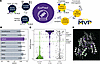 Figure 1
Supervised learning facilitates the generation of an STXBP1-specific variant effect predictor, EpiPred. (A) Overview of the approach adopted for the generation of the EpiPred model, including the input datasets (blue) and the variant effect testing of the EpiPred predictions using 3 biochemical assays (stability, abundance, solubility) and STX1 interaction in an overexpression model system (yellow). (B) Comparison of area under the receiver operator curve (AUROC) for EpiPred and a subset of high-performing globally trained VEPs, and the STXBP1-specific tool, PRESR, across the full truth set. Error bars give the 95% confidence interval. (C) EpiPred Probability of PLP (EpiPred-PPLP) score distributions in the full truth set separated by class as well as the full set of VUS. EpiPred-PPLP scores of 1 suggest pathogenicity, while scores of 0 suggest benignity (D) EpiPred-PPLP output for known PLP and BLB variants used in the truth set mapped onto hSTXBP1 predicted structure (AlphaFold structure: AF-P61764-F1-model_v4), EpiPred-PPLP scores close to 1 are shown in purple, while scores close to 0 appear in green.
Collectively, EpiPred-STXBP1 exhibited improved performance over global VEPs and had high concordance with experimental analyses across 20 variants, including equal distribution of known positive (pathogenic) and negative (benign) controls to better validate the dynamic range of predictive power. We also generated predictions for every possible missense variant in STXBP1 and demonstrated that the core of the STXBP1 protein is susceptible to perturbation by missense variants and likely underpins reduced abundance of STXBP1. Importantly, the reduced abundance of STXBP1 can be rescued in both animal and cellular models using 4-phenylbutyrate (PBA), an FDA-approved medication commonly used to lower serum ammonia levels in individuals with urea cycle disorders (15). A small clinical trial of PBA in individuals with STXBP1-related neurodevelopmental disorders suggests that the treatment may reduce seizure frequency and improve EEG abnormalities (28). Thus, collectively, our study highlights the power of integrating computational and experimental characterization of STXBP1 missense variants to inform precision diagnostics and empower precision clinical care, and suggests similar gene-specific calibration of VEPs, or gene-specific models can improve in silico variant prediction broadly in genetic conditions.
-
Results
EpiPred-STXBP1 accurately distinguishes between pathogenic and benign variants. We used our curated dataset of STXBP1 missense variants, which includes 80 BLB and 96 PLP variants annotated with 42 distinct features, including other VEPs, to develop the single-gene ensemble classifier, EpiPred-STXBP1 (Supplemental Tables 1–3; supplemental material available online with this article; https://doi.org/10.1172/JCI207160DS1). We excluded any global VEPs that were trained on ClinVar data, given that this was a primary source for variants used to train EpiPred. We built a random forest model, which performed as a robust gene-specific classifier for our Truth Set (TS) (Supplemental Figure 1).
Despite being trained on a substantially smaller dataset, EpiPred outperformed all global VEPs with an AUROC value of 98%, with the next 2 closest competitors having AUROC values of 97% (Varity) and 96% (ClinPred) (Figure 1B and Supplemental Table 4). Indeed, EpiPred outperformed the global VEPs and the STXBP1-specific tool, PRESR, on sensitivity (EpiPred: 96%; PRESR: 90%), specificity (EpiPred: 96%; PRESR: 93%), and accuracy (EpiPred: 96%; PRESR: 92%) (Supplemental Table 4 and Supplemental Figure 2). Several global VEPs that were not trained on ClinVar data, including AlphaMissense and VEST4, were prevalent features used in EpiPred, as assessed by SHAP, a game theory–based method to reveal feature impact on model decisions (29) (Supplemental Figure 3). Other features that impact model predictions range from evolutionary conservation (PhyloP; 470way mammalian), residue solvent accessibility (RSA), and in silico prediction of protein stability (MAESTRO). These latter features strongly suggested that stability may be a key defining feature of STXBP1 PLP missense variants, aligning with the decreased abundance and stability observed for missense variants in animal and cell models (12–15).
We used this model to score all variants with a Probability of PLP (EpiPred-PPLP score) (Figure 1, C and D, and Supplemental Table 1). Using a Gaussian mixture model, we defined the PLP threshold as an EpiPred-PPLP score greater than 0.871 and the BLB threshold as an EpiPred-PPLP score less than 0.625, with variants between these thresholds classified as ‘neutral’ (Figure 1C).
In general, gnomAD variants present greater than thrice in the dataset had EpiPred-PPLP scores below the PLP threshold (77/80 – 96% accuracy). The 3 exceptions were Phe153Ser and Arg536His, both predicted PLP and Ala553Thr in the ambiguous range (Supplemental Table 1 and Supplemental Figure 4). In the PLP TS, 81% (78/96) of variants exceeded the EpiPred PLP threshold, with 9 variants in the ambiguous range and the remaining 10 PLP variants more likely to be benign by EpiPred (Supplemental Table 1 and Supplemental Figure 4). VUS (n = 150) values were scored broadly across the range, with 23% (35/150) in the PLP range and 55% (83/150) in the BLB range (Figure 1, C and D, and Supplemental Table 2). We mapped these conflicting variants (10 PLP, 5 BLB) onto the 3D structure of the protein and assessed the SHAP outputs for each variant, but we were unable to detect any common features that could consistently explain this misclassification (Supplemental Figures 4 and 5).
Missense variant effect aligns with EpiPred predictions. We selected 20 variants for experimental validation of EpiPred performance in a cellular system, including 10 BLB, 5 PLP, and 5 VUS. Of these VUS, 3 (p.Ala202Val, p.Trp288Leu, and p.Ile576Thr) were predicted PLP by EpiPred and 2 (p.Gln301Leu, p.Lys333Glu) were predicted BLB by EpiPred (Figure 1C and Supplemental Data Files 1 and 2). We used 4 readouts of variant effect that had been previously reported in models of STXBP1 pathogenic missense variants. These included STXBP1 protein abundance (Figure 2A), solubility (Figure 2B), stability (Figure 2C), and the protein-protein interaction (PPI) between STXBP1 and STX1 (Figure 2D) (Supplemental Data Files 1 and 2) (10, 11).
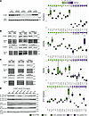 Figure 2
Biochemical assays and syntaxin 1 interaction distinguish pathogenic from benign STXBP1 missense variants. Data from representative Western blots (left) and quantification (right) are plotted for the WT STXBP1 and 20 missense variants. Each assay is plotted separately: (A) Abundance, (B) Stability, (C) Solubility, and (D) STX1 PPI. Scores are normalized to the WT reference allele score. The quantified data is sorted, left to right, by EpiPred-PPLP score from lowest (likely benign) to highest (likely pathogenic) and colored by class (WT, PLP, BLB, VUS predicted PLP/BLB). n = 3, *P < 0.05 (2-tailed Student’s t test); all raw data and P values in Supplemental Data Files 1–2.
Individually, these assays differentiated pathogenic from benign variants to some extent (Figure 2). In particular, the best discriminator was abundance; all of the PLP variants (n = 5) showed statistically significant reduced abundance, with a mean reduction to 14% (range 4%–42%) of WT levels, while only 5 of 10 BLB variants showed a significant change, with 4 variants reduced to approximately 71% (range 50%–73%) of WT, and one with 28% more protein than WT (Figure 2A). Similarly, protein stability was a good predictor of pathogenicity; all 5 PLP variants had statistically significant reduced stability (mean: 55%, range 38–64%), compared to only 3 of the BLB variants (n = 3/10) with a smaller reduction (mean 80%, range 75%–81%) (Figure 2C). There was a statistically significant correlation between stability and abundance (R = 0.73, P = 0.0005), as expected given the relatedness of these 2 assays. All but one of the PLP variants (p.Leu41Gln) were less soluble (mean 41%, range 32%–45%), compared with only one of the BLB variants (p.Val84Ile, 56% of WT).
The STX1:STXBP1 interactions were more complex. Two of the PLP variants (p.Arg292His and p.Arg406His) had statistically significantly reduced interactions (33% and 36% respectively); similarly, the predicted PLP variant p.Trp288Leu interacted with STX1 at only 6% of WT levels. However, one PLP variant (p.Ala101Pro) showed a greater than 2-fold increased interaction with STX1, as did 3 BLB variants, with a mean increase of 1.7-fold compared with WT (range 1.4–2.2). Overall, our VUS predictions by EpiPred were correct and the VUS that were predicted as either PLP or BLB clustered with known variants of each class.
In addition to these individual correlations, we considered the effects of the 4 readouts of variant effect in combination and generated a combined variant effect score using principal component analysis (Supplemental Table 4). The first principal component accounted for 56% of the total variance and effectively distinguished between a BLB and a PLP cluster of variants (Figure 3A). All assays contributed to this distinction, with stability and abundance among the largest contributors, and STX1 interaction at the least. Moreover, EpiPred had the strongest correlation with the combined variant effect score (0.72), compared with all other features, including global VEPs trained on ClinVar that were not used for EpiPred (Figure 3B, Supplemental Figures 6–8, and Supplemental Table 5).
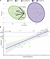 Figure 3
Integrating 4 variant effect assays with principal component analysis. (A) Biplot showing distinct BLB and PLP clusters within the dataset across 2 principal components, with PC1 explaining most of the variance in the data. (B) Scatterplot of EpiPred-PPLP scores and combined variant effect score (PC1). EpiPred-PPLP scores are highly correlated with the combined variant effect score. Variant and colored by class: PLP, dark purple; BLB, dark green; VUS predicted PLP, light purple; VUS predicted BLB, light green. (r = 0.73; P = 0.00024 Pearson).
Using this combined variant effect score, the p.Arg536His variant clustered with the benign variants. This BLB variant, present in gnomAD (n = 30), was predicted as pathogenic by EpiPred, PRESR, and most global VEPs, and had reduced abundance levels similar to PLP variants, with minimal effects on solubility and stability (Figure 2). However, p.Arg536His also demonstrated a greater than 2-fold increased interaction with STX1, which led to a combined variant effect score in the BLB range. This example highlights the importance of leveraging multiple assays as a readout of missense STXBP1 variant effect, as the detrimental impacts of a missense variant in one assay may potentially be ‘rescued’ by a different property of that variant in another.
Evaluation of EpiPred in a holdout dataset demonstrates the ability to detect misclassified disease-relevant missense variants. Leveraging the rapid pace of sequencing in population cohorts and individuals with epilepsy, we were able to establish a holdout set consisting of 43 unique PLP missense variants, primarily from a large STXBP1 phenotyping study (30), as well as 78 unique missense BLB variants (Regeneron exome server and gnomAD v4) (Supplemental Table 6) (31). EpiPred, and indeed all VEPs investigated, exhibited reduced performance on the Holdout set relative to the truth set (Figure 4A, Supplemental Figures 9–13, and Supplemental Table 7). All top performers in the truth set, EpiPred (98%), Varity (97%), and ClinPred (96%), had reduced AUROCs in the holdout set: EpiPred (89%), Varity (88%), and ClinPred (80%) (Supplemental Table 7). To explore this reduced performance, we evaluated the accuracy of the model for PLP and BLB variants in EpiPred. In the BLB holdout set, EpiPred had a 92% (72/78) accuracy, with 3 variants predicted ambiguous and 3 predicted PLP, thus performing similarly to the truth set (96% accuracy). Moreover, ultra-rare variants present in GnomAD with an allele count less than 3 were also largely (85%) predicted to be benign (257/304), and only 10 variants (3%) predicted as PLP, with the remainder in the ambiguous range (Figure 4B). In contrast, In the PLP holdout set accuracy diminished from 96% accuracy in the truth set, to 67% (29/43) in the holdout set. This included 5 variants in the ambiguous range, and the remaining 9 PLP variants predicted more likely to be benign by EpiPred (Figure 4B). Thus, these findings revealed a potential misclassification of PLP variants in the cohort study used to derive the holdout set. The criteria for PLP designation were described in the publication as “a modified annotation pipeline and only included variants that were either pathogenic or likely pathogenic by ACMG criteria or de novo variants in the coding region of STXBP1”. Moreover, we found good agreement between EpiPred and ClinPred/Varity predictions, with both global VEPs predicting ‘PLP’ missense variants as benign (n = 8) or ‘ambiguous’ (n = 1) (Supplemental Figures 11 and 12, and Supplemental Table 8). We reevaluated the classification of these variants and determined that most would not meet the ACMG criteria for PLP classification; indeed 2 (p.Asn398Ser and p.Ser253Asn) are classified as VUS in ClinVar. More specifically, the inheritance of these variants was not reported, 3 were found in individuals without epilepsy or in atypical phenotypes (e.g., focal cortical dysplasia), and these amino acid locations (though different substitutions) were present in the general population (gnomAD). Finally, 2 variants, p.Asp207Gly and p.Asn398Lys, showed no change in any of the biochemical assays or STX1 interactions that were used to assess the core STXBP1 phenotype (Figure 4D and Supplemental Data Files 1 and 2).
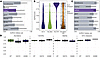 Figure 4
EpiPred classification of a holdout dataset. (A) Comparison of area under the ROC curve (AUROC) for EpiPred and a subset of high-performing globally trained VEPs and PRESR across the holdout dataset demonstrates reduced performance of EpiPred, error bars show the 95% CI. (B) EpiPred-PPLP score distributions in the full holdout data set show the conserved performance for BLB variants, but a larger percentage of PLP variants with EpiPred-PPLP scores in the benign/neutral range. Ultra-rare variants in GnomAD (allele count < 3) have scores more similar to BLB variants, while simulated variants occur across the range. EpiPred-PPLP scores of 1 suggest pathogenicity, while scores of 0 suggest benignity. (C) Comparison of area under the ROC curve (AUROC) for EpiPred, PRESR, and a subset of high-performing globally trained VEPs across the holdout dataset with reclassified variants, including the exclusion of 9 VUS from the PLP set, demonstrates improved EpiPred performance, on par with AlphaMissense, error bars show the 95% CI. (D) Modeling of 2 misclassified variants, reclassified to VUS, which demonstrate properties similar to WT for abundance, stability, solubility and syntaxin interaction. n = 6, *P < 0.05 (2-tailed Student’s t test); all raw data and P values in Supplemental Data Files 1–2.
We thus reclassified these 9 variants as VUS and reassessed EpiPred classification performance with 34 PLP (from n = 43 originally) and the same 78 BLB variants. We observed increased AUROC performance in both EpiPred (97.8%) and global VEPs, Varity (95%),and ClinPred (93%) (Figure 4C, Supplemental Figure 10, and Supplemental Table 9). The AUROC for all VEPs in this modified holdout set was also less variable, suggesting that reclassification led to more stable VEP predictions (Figure 4C). PRESR had reduced performance compared with EpiPred in the holdout set across all metrics (Figure 4, A and C, and Supplemental Figure 10). Both EpiPred, PRESR, and global VEPs (ClinPred/Varity) were still marginally outperformed by AlphaMissense (AUROC – 98.3%). This result suggests strong generalizability of AlphaMissense in the holdout set; however, EpiPred retained greater sensitivity, which is better aligned with the clinical utility goals of this VEP to identify individuals with pathogenic variants for clinical trial eligibility. In this context, sensitivity is particularly important because it reflects the ability to capture true positives rather than miss potentially eligible individuals. Collectively, while the holdout dataset had lower performance than the truth set, EpiPred generalized well and still outperformed global VEPs and PRESR. This analysis also highlights the power of EpiPred and global VEPs to accurately classify variants and identify false positives (and potentially misdiagnoses) in the literature.
Gene-wide EpiPred simulation reveals specific regions susceptible to perturbation. To facilitate dissemination of EpiPred to the scientific and epilepsy community, we generated EpiPred scores for all possible missense STXBP1 variants (n = 3,939 total, n = 3,492 simulated, and n = 447 observed) resulting from a single nucleotide change (Supplemental Table 10, Figure 4B, and Figure 5A). The simulated variants exhibit a broad distribution of scores across the range, including 13% (466/3,492) predicted as PLP. This is expected, as a variant that has never been observed could indeed be pathogenic if it were to arise de novo in an individual with epilepsy. We also collated data from the literature that characterized missense variants (n = 26) using both cellular and animal model systems and generated an aggregate variant effect score for each variant (Supplemental Table 11) (10–15). This variant effect score, essentially a measure of how often a tested missense variant exhibited a phenotype, correlated reasonably well with EpiPred and PRESR, though EpiPred outperformed PRESR across metrics (Supplemental Figures 14–16, and Supplemental Table 12).
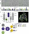 Figure 5
EpiPred-PPLP scores for every possible STXBP1 missense variant and comparison with published experimental data. (A) EpiPred-PPLP variant calls for every possible SNV in the STXBP1 gene represented as a heatmap (top). Variants called BLB are shown in green and those called PLP are shown in purple. Variants called ambiguous are displayed in grey. Distribution of all known BLB and PLP variants across STXBP1 (bottom) (B) Distribution of EpiPred scores for latest version of ClinVar (accessed 8-7-2025) (C) EpiPred-PPLP scores for every possible missense variant (mean per residue) were mapped onto the AlphaFold structure using PyMol. (D) Potential workflow for deploying EpiPred in reclassification of all classes of variants. For instance, those VUS EpiPred predicted as pathogenic, or PLP variants predicted benign are the highest priority for further characterization to confirm pathogenicity. Conversely, those VUS and PLP variants predicted benign by EpiPred should be reevaluated in terms of the ACMG criteria for classification, as these may not be contributory.
Using our comprehensive STXBP1 EpiPred simulation, we scored all VUS in the latest ClinVar dataset, which now includes an expanded set of 222 variants, up from 150 when we initiated this study (Figure 5B and Supplemental Tables 13 and 14). This includes 133 VUS from the original dataset, as well as 32 simulation or ‘new’ variants, which had comparable median EpiPred-PPLP scores, 0.25 for new VUS, and 0.44 for previously identified VUS. There were, however, more VUS that were predicted to be pathogenic in the previously identified VUS (31/133–23%) as compared to the newly identified VUS (3/32–9%), though this difference was not statistically significant (Fisher’s Exact Test, Odds Ratio: 0.34, P-value of 0.092). There were also multiple VUS that were previously classified as BLB due to presence in gnomAD more than thrice, this group had a lower median EpiPred-PPLP score (0.09). In addition, only 1 variant in this BLB group is predicted to be pathogenic (1/48–2%), which is far less than all VUS (40/222–18%) (Fisher’s Exact Test, Odds Ratio: 0.097, P-value of 0.0032). Finally, there were also 9 variants that were previously classified as PLP and have subsequently been reclassified as VUS. This includes 4 variants that were classified as benign by EpiPred, 2 variants that we reclassified from the holdout set (p.Asn398Ser, p.Ser253Ans) and 2 variants from the truth set p.Lys7Glu (EpiPred-PPLP score 0.53) and p.Ser80Pro (EpiPred-PPLP score 0.43) (Supplemental Figure 5 and Supplemental Table 10).
Overall, this reclassification analysis further demonstrates the utility of EpiPred to reclassify STXBP1 missense variants. To facilitate this, we developed a webapp, EpiPred (https://carvill-lab.shinyapps.io/epipred-shiny/) a Shiny-based user interface for clinicians, families and researchers to access our data and use the model to aid interpretation of STXBP1 missense VUS. We envision a workflow whereby EpiPred can assist in the reclassification of variants from clinical genetic testing, prioritizing high yield variants for experimental characterization in the instance of predicted pathogenicity, and revisiting the ACMG classification criteria in the instance of predicted benignity (Figure 5D).
To visualize whether predicted pathogenic/benign variants cluster in specific regions of STXBP1, we aggregated EpiPred scores at each variant position and projected these onto the STXBP1 3D structure (Figure 5C). Rather than clustering within specific domains, which STXBP1 lacks, we found that known and predicted PLP variants tend to localize within the central core of the protein. This localization may underlie the reduced abundance and stability observed in STXBP1 proteins with pathogenic missense variants; highlighting a subset of variants and patients that could benefit from protein-stabilizing–based therapies such as PBA.


Copyright © 2026 American Society for Clinical Investigation
ISSN: 0021-9738 (print), 1558-8238 (online)