
Advertisement
Clinical Research and Public HealthClinical ResearchImmunologyInflammation
Open Access |  10.1172/JCI202867
Aberrant STAT signaling and T cell dysregulation define a targetable pediatric sepsis endotype
Robert B. Lindell,1,2 Samir U. Sayed,3 Jose S. Campos Duran,3 Sydney A. Sheetz,3,4 Apoorva Babu,5 Montana S. Knight,5 Andrea A. Mauracher,3 Ceire A. Hay,3 Peyton E. Conrey,3 Julie C. Fitzgerald,1 Nadir Yehya,1 Stephen T. Famularo III,4 Teresa Arroyo,4 Richard Tustin III,3 Hossein Fazelinia,5 Edward M. Behrens,6 David T. Teachey,7 Lisa R. Forbes Satter,8 Alexandra F. Freeman,9 Jenna R.E. Bergerson,9 Steven M. Holland,9 Jennifer W. Leiding,10 Scott L. Weiss,11 Mark W. Hall,12 Deanne M. Taylor,13,14 Rui Feng,15 E. John Wherry,2,16 Nuala J. Meyer,2,17 and Sarah E. Henrickson2,18,19
1Division of Critical Care Medicine, Department of Anesthesia and Critical Care, Children’s Hospital of Philadelphia and the Perelman School of Medicine, University of Pennsylvania, Philadelphia, Pennsylvania, USA.
2Institute for Immunology and Immune Health, Perelman School of Medicine, University of Pennsylvania, Philadelphia, Pennsylvania, USA.
3Division of Allergy and Immunology, Department of Pediatrics,
4Division of Critical Care Medicine, Department of Anesthesia and Critical Care, and
5Department of Biomedical and Health Informatics, Children’s Hospital of Philadelphia, Philadelphia, Pennsylvania, USA.
6Division of Rheumatology, Department of Pediatrics and
7Division of Oncology, Department of Pediatrics, Children’s Hospital of Philadelphia and the Perelman School of Medicine, University of Pennsylvania, Philadelphia, Pennsylvania, USA.
8Division of Immunology, Allergy, and Retrovirology, Department of Pediatrics, Texas Children’s Hospital and Baylor College of Medicine, Houston, Texas, USA.
9Laboratory of Clinical Immunology and Microbiology, National Institute of Allergy and Infectious Diseases, NIH, Bethesda, Maryland, USA.
10Division of Allergy and Immunology, Department of Pediatrics, Johns Hopkins University School of Medicine and Johns Hopkins All Children’s Hospital, St. Petersburg, Florida, USA.
11Division of Critical Care, Department of Pediatrics, Nemours Children’s Health and Sidney Kimmel Medical College at Thomas Jefferson University, Wilmington, Delaware, USA.
12Division of Critical Care, Department of Pediatrics, Nationwide Children’s Hospital and The Ohio State University College of Medicine, Columbus, Ohio, USA.
13Department of Biomedical and Health Informatics, Children’s Hospital of Philadelphia, Philadelphia, Pennsylvania, USA.
14Department of Pediatrics,
15Department of Biostatistics, Epidemiology, and Informatics,
16Department of Systems Pharmacology and Translational Therapeutics, and
17Division of Pulmonary, Allergy, and Critical Care Medicine, Department of Medicine, Perelman School of Medicine, University of Pennsylvania, Philadelphia, Pennsylvania, USA.
18Division of Allergy and Immunology, Department of Pediatrics, Children’s Hospital of Philadelphia and the Perelman School of Medicine, University of Pennsylvania, Philadelphia, Pennsylvania, USA.
19Department of Microbiology, Perelman School of Medicine, University of Pennsylvania, Philadelphia, Pennsylvania, USA.
Address correspondence to: Robert B. Lindell, Children’s Hospital of Philadelphia, 3401 Civic Center Blvd, Philadelphia, Pennsylvania 19104, USA. Phone: 215.590.1000; Email: LindellR@chop.edu.
Find articles by Lindell, R. in: PubMed | Google Scholar
1Division of Critical Care Medicine, Department of Anesthesia and Critical Care, Children’s Hospital of Philadelphia and the Perelman School of Medicine, University of Pennsylvania, Philadelphia, Pennsylvania, USA.
2Institute for Immunology and Immune Health, Perelman School of Medicine, University of Pennsylvania, Philadelphia, Pennsylvania, USA.
3Division of Allergy and Immunology, Department of Pediatrics,
4Division of Critical Care Medicine, Department of Anesthesia and Critical Care, and
5Department of Biomedical and Health Informatics, Children’s Hospital of Philadelphia, Philadelphia, Pennsylvania, USA.
6Division of Rheumatology, Department of Pediatrics and
7Division of Oncology, Department of Pediatrics, Children’s Hospital of Philadelphia and the Perelman School of Medicine, University of Pennsylvania, Philadelphia, Pennsylvania, USA.
8Division of Immunology, Allergy, and Retrovirology, Department of Pediatrics, Texas Children’s Hospital and Baylor College of Medicine, Houston, Texas, USA.
9Laboratory of Clinical Immunology and Microbiology, National Institute of Allergy and Infectious Diseases, NIH, Bethesda, Maryland, USA.
10Division of Allergy and Immunology, Department of Pediatrics, Johns Hopkins University School of Medicine and Johns Hopkins All Children’s Hospital, St. Petersburg, Florida, USA.
11Division of Critical Care, Department of Pediatrics, Nemours Children’s Health and Sidney Kimmel Medical College at Thomas Jefferson University, Wilmington, Delaware, USA.
12Division of Critical Care, Department of Pediatrics, Nationwide Children’s Hospital and The Ohio State University College of Medicine, Columbus, Ohio, USA.
13Department of Biomedical and Health Informatics, Children’s Hospital of Philadelphia, Philadelphia, Pennsylvania, USA.
14Department of Pediatrics,
15Department of Biostatistics, Epidemiology, and Informatics,
16Department of Systems Pharmacology and Translational Therapeutics, and
17Division of Pulmonary, Allergy, and Critical Care Medicine, Department of Medicine, Perelman School of Medicine, University of Pennsylvania, Philadelphia, Pennsylvania, USA.
18Division of Allergy and Immunology, Department of Pediatrics, Children’s Hospital of Philadelphia and the Perelman School of Medicine, University of Pennsylvania, Philadelphia, Pennsylvania, USA.
19Department of Microbiology, Perelman School of Medicine, University of Pennsylvania, Philadelphia, Pennsylvania, USA.
Address correspondence to: Robert B. Lindell, Children’s Hospital of Philadelphia, 3401 Civic Center Blvd, Philadelphia, Pennsylvania 19104, USA. Phone: 215.590.1000; Email: LindellR@chop.edu.
Find articles by
Sayed, S.
in:
PubMed
|
Google Scholar
|

1Division of Critical Care Medicine, Department of Anesthesia and Critical Care, Children’s Hospital of Philadelphia and the Perelman School of Medicine, University of Pennsylvania, Philadelphia, Pennsylvania, USA.
2Institute for Immunology and Immune Health, Perelman School of Medicine, University of Pennsylvania, Philadelphia, Pennsylvania, USA.
3Division of Allergy and Immunology, Department of Pediatrics,
4Division of Critical Care Medicine, Department of Anesthesia and Critical Care, and
5Department of Biomedical and Health Informatics, Children’s Hospital of Philadelphia, Philadelphia, Pennsylvania, USA.
6Division of Rheumatology, Department of Pediatrics and
7Division of Oncology, Department of Pediatrics, Children’s Hospital of Philadelphia and the Perelman School of Medicine, University of Pennsylvania, Philadelphia, Pennsylvania, USA.
8Division of Immunology, Allergy, and Retrovirology, Department of Pediatrics, Texas Children’s Hospital and Baylor College of Medicine, Houston, Texas, USA.
9Laboratory of Clinical Immunology and Microbiology, National Institute of Allergy and Infectious Diseases, NIH, Bethesda, Maryland, USA.
10Division of Allergy and Immunology, Department of Pediatrics, Johns Hopkins University School of Medicine and Johns Hopkins All Children’s Hospital, St. Petersburg, Florida, USA.
11Division of Critical Care, Department of Pediatrics, Nemours Children’s Health and Sidney Kimmel Medical College at Thomas Jefferson University, Wilmington, Delaware, USA.
12Division of Critical Care, Department of Pediatrics, Nationwide Children’s Hospital and The Ohio State University College of Medicine, Columbus, Ohio, USA.
13Department of Biomedical and Health Informatics, Children’s Hospital of Philadelphia, Philadelphia, Pennsylvania, USA.
14Department of Pediatrics,
15Department of Biostatistics, Epidemiology, and Informatics,
16Department of Systems Pharmacology and Translational Therapeutics, and
17Division of Pulmonary, Allergy, and Critical Care Medicine, Department of Medicine, Perelman School of Medicine, University of Pennsylvania, Philadelphia, Pennsylvania, USA.
18Division of Allergy and Immunology, Department of Pediatrics, Children’s Hospital of Philadelphia and the Perelman School of Medicine, University of Pennsylvania, Philadelphia, Pennsylvania, USA.
19Department of Microbiology, Perelman School of Medicine, University of Pennsylvania, Philadelphia, Pennsylvania, USA.
Address correspondence to: Robert B. Lindell, Children’s Hospital of Philadelphia, 3401 Civic Center Blvd, Philadelphia, Pennsylvania 19104, USA. Phone: 215.590.1000; Email: LindellR@chop.edu.
Find articles by Campos Duran, J. in: PubMed | Google Scholar
1Division of Critical Care Medicine, Department of Anesthesia and Critical Care, Children’s Hospital of Philadelphia and the Perelman School of Medicine, University of Pennsylvania, Philadelphia, Pennsylvania, USA.
2Institute for Immunology and Immune Health, Perelman School of Medicine, University of Pennsylvania, Philadelphia, Pennsylvania, USA.
3Division of Allergy and Immunology, Department of Pediatrics,
4Division of Critical Care Medicine, Department of Anesthesia and Critical Care, and
5Department of Biomedical and Health Informatics, Children’s Hospital of Philadelphia, Philadelphia, Pennsylvania, USA.
6Division of Rheumatology, Department of Pediatrics and
7Division of Oncology, Department of Pediatrics, Children’s Hospital of Philadelphia and the Perelman School of Medicine, University of Pennsylvania, Philadelphia, Pennsylvania, USA.
8Division of Immunology, Allergy, and Retrovirology, Department of Pediatrics, Texas Children’s Hospital and Baylor College of Medicine, Houston, Texas, USA.
9Laboratory of Clinical Immunology and Microbiology, National Institute of Allergy and Infectious Diseases, NIH, Bethesda, Maryland, USA.
10Division of Allergy and Immunology, Department of Pediatrics, Johns Hopkins University School of Medicine and Johns Hopkins All Children’s Hospital, St. Petersburg, Florida, USA.
11Division of Critical Care, Department of Pediatrics, Nemours Children’s Health and Sidney Kimmel Medical College at Thomas Jefferson University, Wilmington, Delaware, USA.
12Division of Critical Care, Department of Pediatrics, Nationwide Children’s Hospital and The Ohio State University College of Medicine, Columbus, Ohio, USA.
13Department of Biomedical and Health Informatics, Children’s Hospital of Philadelphia, Philadelphia, Pennsylvania, USA.
14Department of Pediatrics,
15Department of Biostatistics, Epidemiology, and Informatics,
16Department of Systems Pharmacology and Translational Therapeutics, and
17Division of Pulmonary, Allergy, and Critical Care Medicine, Department of Medicine, Perelman School of Medicine, University of Pennsylvania, Philadelphia, Pennsylvania, USA.
18Division of Allergy and Immunology, Department of Pediatrics, Children’s Hospital of Philadelphia and the Perelman School of Medicine, University of Pennsylvania, Philadelphia, Pennsylvania, USA.
19Department of Microbiology, Perelman School of Medicine, University of Pennsylvania, Philadelphia, Pennsylvania, USA.
Address correspondence to: Robert B. Lindell, Children’s Hospital of Philadelphia, 3401 Civic Center Blvd, Philadelphia, Pennsylvania 19104, USA. Phone: 215.590.1000; Email: LindellR@chop.edu.
Find articles by Sheetz, S. in: PubMed | Google Scholar
1Division of Critical Care Medicine, Department of Anesthesia and Critical Care, Children’s Hospital of Philadelphia and the Perelman School of Medicine, University of Pennsylvania, Philadelphia, Pennsylvania, USA.
2Institute for Immunology and Immune Health, Perelman School of Medicine, University of Pennsylvania, Philadelphia, Pennsylvania, USA.
3Division of Allergy and Immunology, Department of Pediatrics,
4Division of Critical Care Medicine, Department of Anesthesia and Critical Care, and
5Department of Biomedical and Health Informatics, Children’s Hospital of Philadelphia, Philadelphia, Pennsylvania, USA.
6Division of Rheumatology, Department of Pediatrics and
7Division of Oncology, Department of Pediatrics, Children’s Hospital of Philadelphia and the Perelman School of Medicine, University of Pennsylvania, Philadelphia, Pennsylvania, USA.
8Division of Immunology, Allergy, and Retrovirology, Department of Pediatrics, Texas Children’s Hospital and Baylor College of Medicine, Houston, Texas, USA.
9Laboratory of Clinical Immunology and Microbiology, National Institute of Allergy and Infectious Diseases, NIH, Bethesda, Maryland, USA.
10Division of Allergy and Immunology, Department of Pediatrics, Johns Hopkins University School of Medicine and Johns Hopkins All Children’s Hospital, St. Petersburg, Florida, USA.
11Division of Critical Care, Department of Pediatrics, Nemours Children’s Health and Sidney Kimmel Medical College at Thomas Jefferson University, Wilmington, Delaware, USA.
12Division of Critical Care, Department of Pediatrics, Nationwide Children’s Hospital and The Ohio State University College of Medicine, Columbus, Ohio, USA.
13Department of Biomedical and Health Informatics, Children’s Hospital of Philadelphia, Philadelphia, Pennsylvania, USA.
14Department of Pediatrics,
15Department of Biostatistics, Epidemiology, and Informatics,
16Department of Systems Pharmacology and Translational Therapeutics, and
17Division of Pulmonary, Allergy, and Critical Care Medicine, Department of Medicine, Perelman School of Medicine, University of Pennsylvania, Philadelphia, Pennsylvania, USA.
18Division of Allergy and Immunology, Department of Pediatrics, Children’s Hospital of Philadelphia and the Perelman School of Medicine, University of Pennsylvania, Philadelphia, Pennsylvania, USA.
19Department of Microbiology, Perelman School of Medicine, University of Pennsylvania, Philadelphia, Pennsylvania, USA.
Address correspondence to: Robert B. Lindell, Children’s Hospital of Philadelphia, 3401 Civic Center Blvd, Philadelphia, Pennsylvania 19104, USA. Phone: 215.590.1000; Email: LindellR@chop.edu.
Find articles by Babu, A. in: PubMed | Google Scholar
1Division of Critical Care Medicine, Department of Anesthesia and Critical Care, Children’s Hospital of Philadelphia and the Perelman School of Medicine, University of Pennsylvania, Philadelphia, Pennsylvania, USA.
2Institute for Immunology and Immune Health, Perelman School of Medicine, University of Pennsylvania, Philadelphia, Pennsylvania, USA.
3Division of Allergy and Immunology, Department of Pediatrics,
4Division of Critical Care Medicine, Department of Anesthesia and Critical Care, and
5Department of Biomedical and Health Informatics, Children’s Hospital of Philadelphia, Philadelphia, Pennsylvania, USA.
6Division of Rheumatology, Department of Pediatrics and
7Division of Oncology, Department of Pediatrics, Children’s Hospital of Philadelphia and the Perelman School of Medicine, University of Pennsylvania, Philadelphia, Pennsylvania, USA.
8Division of Immunology, Allergy, and Retrovirology, Department of Pediatrics, Texas Children’s Hospital and Baylor College of Medicine, Houston, Texas, USA.
9Laboratory of Clinical Immunology and Microbiology, National Institute of Allergy and Infectious Diseases, NIH, Bethesda, Maryland, USA.
10Division of Allergy and Immunology, Department of Pediatrics, Johns Hopkins University School of Medicine and Johns Hopkins All Children’s Hospital, St. Petersburg, Florida, USA.
11Division of Critical Care, Department of Pediatrics, Nemours Children’s Health and Sidney Kimmel Medical College at Thomas Jefferson University, Wilmington, Delaware, USA.
12Division of Critical Care, Department of Pediatrics, Nationwide Children’s Hospital and The Ohio State University College of Medicine, Columbus, Ohio, USA.
13Department of Biomedical and Health Informatics, Children’s Hospital of Philadelphia, Philadelphia, Pennsylvania, USA.
14Department of Pediatrics,
15Department of Biostatistics, Epidemiology, and Informatics,
16Department of Systems Pharmacology and Translational Therapeutics, and
17Division of Pulmonary, Allergy, and Critical Care Medicine, Department of Medicine, Perelman School of Medicine, University of Pennsylvania, Philadelphia, Pennsylvania, USA.
18Division of Allergy and Immunology, Department of Pediatrics, Children’s Hospital of Philadelphia and the Perelman School of Medicine, University of Pennsylvania, Philadelphia, Pennsylvania, USA.
19Department of Microbiology, Perelman School of Medicine, University of Pennsylvania, Philadelphia, Pennsylvania, USA.
Address correspondence to: Robert B. Lindell, Children’s Hospital of Philadelphia, 3401 Civic Center Blvd, Philadelphia, Pennsylvania 19104, USA. Phone: 215.590.1000; Email: LindellR@chop.edu.
Find articles by Knight, M. in: PubMed | Google Scholar
1Division of Critical Care Medicine, Department of Anesthesia and Critical Care, Children’s Hospital of Philadelphia and the Perelman School of Medicine, University of Pennsylvania, Philadelphia, Pennsylvania, USA.
2Institute for Immunology and Immune Health, Perelman School of Medicine, University of Pennsylvania, Philadelphia, Pennsylvania, USA.
3Division of Allergy and Immunology, Department of Pediatrics,
4Division of Critical Care Medicine, Department of Anesthesia and Critical Care, and
5Department of Biomedical and Health Informatics, Children’s Hospital of Philadelphia, Philadelphia, Pennsylvania, USA.
6Division of Rheumatology, Department of Pediatrics and
7Division of Oncology, Department of Pediatrics, Children’s Hospital of Philadelphia and the Perelman School of Medicine, University of Pennsylvania, Philadelphia, Pennsylvania, USA.
8Division of Immunology, Allergy, and Retrovirology, Department of Pediatrics, Texas Children’s Hospital and Baylor College of Medicine, Houston, Texas, USA.
9Laboratory of Clinical Immunology and Microbiology, National Institute of Allergy and Infectious Diseases, NIH, Bethesda, Maryland, USA.
10Division of Allergy and Immunology, Department of Pediatrics, Johns Hopkins University School of Medicine and Johns Hopkins All Children’s Hospital, St. Petersburg, Florida, USA.
11Division of Critical Care, Department of Pediatrics, Nemours Children’s Health and Sidney Kimmel Medical College at Thomas Jefferson University, Wilmington, Delaware, USA.
12Division of Critical Care, Department of Pediatrics, Nationwide Children’s Hospital and The Ohio State University College of Medicine, Columbus, Ohio, USA.
13Department of Biomedical and Health Informatics, Children’s Hospital of Philadelphia, Philadelphia, Pennsylvania, USA.
14Department of Pediatrics,
15Department of Biostatistics, Epidemiology, and Informatics,
16Department of Systems Pharmacology and Translational Therapeutics, and
17Division of Pulmonary, Allergy, and Critical Care Medicine, Department of Medicine, Perelman School of Medicine, University of Pennsylvania, Philadelphia, Pennsylvania, USA.
18Division of Allergy and Immunology, Department of Pediatrics, Children’s Hospital of Philadelphia and the Perelman School of Medicine, University of Pennsylvania, Philadelphia, Pennsylvania, USA.
19Department of Microbiology, Perelman School of Medicine, University of Pennsylvania, Philadelphia, Pennsylvania, USA.
Address correspondence to: Robert B. Lindell, Children’s Hospital of Philadelphia, 3401 Civic Center Blvd, Philadelphia, Pennsylvania 19104, USA. Phone: 215.590.1000; Email: LindellR@chop.edu.
Find articles by Mauracher, A. in: PubMed | Google Scholar
1Division of Critical Care Medicine, Department of Anesthesia and Critical Care, Children’s Hospital of Philadelphia and the Perelman School of Medicine, University of Pennsylvania, Philadelphia, Pennsylvania, USA.
2Institute for Immunology and Immune Health, Perelman School of Medicine, University of Pennsylvania, Philadelphia, Pennsylvania, USA.
3Division of Allergy and Immunology, Department of Pediatrics,
4Division of Critical Care Medicine, Department of Anesthesia and Critical Care, and
5Department of Biomedical and Health Informatics, Children’s Hospital of Philadelphia, Philadelphia, Pennsylvania, USA.
6Division of Rheumatology, Department of Pediatrics and
7Division of Oncology, Department of Pediatrics, Children’s Hospital of Philadelphia and the Perelman School of Medicine, University of Pennsylvania, Philadelphia, Pennsylvania, USA.
8Division of Immunology, Allergy, and Retrovirology, Department of Pediatrics, Texas Children’s Hospital and Baylor College of Medicine, Houston, Texas, USA.
9Laboratory of Clinical Immunology and Microbiology, National Institute of Allergy and Infectious Diseases, NIH, Bethesda, Maryland, USA.
10Division of Allergy and Immunology, Department of Pediatrics, Johns Hopkins University School of Medicine and Johns Hopkins All Children’s Hospital, St. Petersburg, Florida, USA.
11Division of Critical Care, Department of Pediatrics, Nemours Children’s Health and Sidney Kimmel Medical College at Thomas Jefferson University, Wilmington, Delaware, USA.
12Division of Critical Care, Department of Pediatrics, Nationwide Children’s Hospital and The Ohio State University College of Medicine, Columbus, Ohio, USA.
13Department of Biomedical and Health Informatics, Children’s Hospital of Philadelphia, Philadelphia, Pennsylvania, USA.
14Department of Pediatrics,
15Department of Biostatistics, Epidemiology, and Informatics,
16Department of Systems Pharmacology and Translational Therapeutics, and
17Division of Pulmonary, Allergy, and Critical Care Medicine, Department of Medicine, Perelman School of Medicine, University of Pennsylvania, Philadelphia, Pennsylvania, USA.
18Division of Allergy and Immunology, Department of Pediatrics, Children’s Hospital of Philadelphia and the Perelman School of Medicine, University of Pennsylvania, Philadelphia, Pennsylvania, USA.
19Department of Microbiology, Perelman School of Medicine, University of Pennsylvania, Philadelphia, Pennsylvania, USA.
Address correspondence to: Robert B. Lindell, Children’s Hospital of Philadelphia, 3401 Civic Center Blvd, Philadelphia, Pennsylvania 19104, USA. Phone: 215.590.1000; Email: LindellR@chop.edu.
Find articles by Hay, C. in: PubMed | Google Scholar
1Division of Critical Care Medicine, Department of Anesthesia and Critical Care, Children’s Hospital of Philadelphia and the Perelman School of Medicine, University of Pennsylvania, Philadelphia, Pennsylvania, USA.
2Institute for Immunology and Immune Health, Perelman School of Medicine, University of Pennsylvania, Philadelphia, Pennsylvania, USA.
3Division of Allergy and Immunology, Department of Pediatrics,
4Division of Critical Care Medicine, Department of Anesthesia and Critical Care, and
5Department of Biomedical and Health Informatics, Children’s Hospital of Philadelphia, Philadelphia, Pennsylvania, USA.
6Division of Rheumatology, Department of Pediatrics and
7Division of Oncology, Department of Pediatrics, Children’s Hospital of Philadelphia and the Perelman School of Medicine, University of Pennsylvania, Philadelphia, Pennsylvania, USA.
8Division of Immunology, Allergy, and Retrovirology, Department of Pediatrics, Texas Children’s Hospital and Baylor College of Medicine, Houston, Texas, USA.
9Laboratory of Clinical Immunology and Microbiology, National Institute of Allergy and Infectious Diseases, NIH, Bethesda, Maryland, USA.
10Division of Allergy and Immunology, Department of Pediatrics, Johns Hopkins University School of Medicine and Johns Hopkins All Children’s Hospital, St. Petersburg, Florida, USA.
11Division of Critical Care, Department of Pediatrics, Nemours Children’s Health and Sidney Kimmel Medical College at Thomas Jefferson University, Wilmington, Delaware, USA.
12Division of Critical Care, Department of Pediatrics, Nationwide Children’s Hospital and The Ohio State University College of Medicine, Columbus, Ohio, USA.
13Department of Biomedical and Health Informatics, Children’s Hospital of Philadelphia, Philadelphia, Pennsylvania, USA.
14Department of Pediatrics,
15Department of Biostatistics, Epidemiology, and Informatics,
16Department of Systems Pharmacology and Translational Therapeutics, and
17Division of Pulmonary, Allergy, and Critical Care Medicine, Department of Medicine, Perelman School of Medicine, University of Pennsylvania, Philadelphia, Pennsylvania, USA.
18Division of Allergy and Immunology, Department of Pediatrics, Children’s Hospital of Philadelphia and the Perelman School of Medicine, University of Pennsylvania, Philadelphia, Pennsylvania, USA.
19Department of Microbiology, Perelman School of Medicine, University of Pennsylvania, Philadelphia, Pennsylvania, USA.
Address correspondence to: Robert B. Lindell, Children’s Hospital of Philadelphia, 3401 Civic Center Blvd, Philadelphia, Pennsylvania 19104, USA. Phone: 215.590.1000; Email: LindellR@chop.edu.
Find articles by Conrey, P. in: PubMed | Google Scholar
1Division of Critical Care Medicine, Department of Anesthesia and Critical Care, Children’s Hospital of Philadelphia and the Perelman School of Medicine, University of Pennsylvania, Philadelphia, Pennsylvania, USA.
2Institute for Immunology and Immune Health, Perelman School of Medicine, University of Pennsylvania, Philadelphia, Pennsylvania, USA.
3Division of Allergy and Immunology, Department of Pediatrics,
4Division of Critical Care Medicine, Department of Anesthesia and Critical Care, and
5Department of Biomedical and Health Informatics, Children’s Hospital of Philadelphia, Philadelphia, Pennsylvania, USA.
6Division of Rheumatology, Department of Pediatrics and
7Division of Oncology, Department of Pediatrics, Children’s Hospital of Philadelphia and the Perelman School of Medicine, University of Pennsylvania, Philadelphia, Pennsylvania, USA.
8Division of Immunology, Allergy, and Retrovirology, Department of Pediatrics, Texas Children’s Hospital and Baylor College of Medicine, Houston, Texas, USA.
9Laboratory of Clinical Immunology and Microbiology, National Institute of Allergy and Infectious Diseases, NIH, Bethesda, Maryland, USA.
10Division of Allergy and Immunology, Department of Pediatrics, Johns Hopkins University School of Medicine and Johns Hopkins All Children’s Hospital, St. Petersburg, Florida, USA.
11Division of Critical Care, Department of Pediatrics, Nemours Children’s Health and Sidney Kimmel Medical College at Thomas Jefferson University, Wilmington, Delaware, USA.
12Division of Critical Care, Department of Pediatrics, Nationwide Children’s Hospital and The Ohio State University College of Medicine, Columbus, Ohio, USA.
13Department of Biomedical and Health Informatics, Children’s Hospital of Philadelphia, Philadelphia, Pennsylvania, USA.
14Department of Pediatrics,
15Department of Biostatistics, Epidemiology, and Informatics,
16Department of Systems Pharmacology and Translational Therapeutics, and
17Division of Pulmonary, Allergy, and Critical Care Medicine, Department of Medicine, Perelman School of Medicine, University of Pennsylvania, Philadelphia, Pennsylvania, USA.
18Division of Allergy and Immunology, Department of Pediatrics, Children’s Hospital of Philadelphia and the Perelman School of Medicine, University of Pennsylvania, Philadelphia, Pennsylvania, USA.
19Department of Microbiology, Perelman School of Medicine, University of Pennsylvania, Philadelphia, Pennsylvania, USA.
Address correspondence to: Robert B. Lindell, Children’s Hospital of Philadelphia, 3401 Civic Center Blvd, Philadelphia, Pennsylvania 19104, USA. Phone: 215.590.1000; Email: LindellR@chop.edu.
Find articles by Fitzgerald, J. in: PubMed | Google Scholar
1Division of Critical Care Medicine, Department of Anesthesia and Critical Care, Children’s Hospital of Philadelphia and the Perelman School of Medicine, University of Pennsylvania, Philadelphia, Pennsylvania, USA.
2Institute for Immunology and Immune Health, Perelman School of Medicine, University of Pennsylvania, Philadelphia, Pennsylvania, USA.
3Division of Allergy and Immunology, Department of Pediatrics,
4Division of Critical Care Medicine, Department of Anesthesia and Critical Care, and
5Department of Biomedical and Health Informatics, Children’s Hospital of Philadelphia, Philadelphia, Pennsylvania, USA.
6Division of Rheumatology, Department of Pediatrics and
7Division of Oncology, Department of Pediatrics, Children’s Hospital of Philadelphia and the Perelman School of Medicine, University of Pennsylvania, Philadelphia, Pennsylvania, USA.
8Division of Immunology, Allergy, and Retrovirology, Department of Pediatrics, Texas Children’s Hospital and Baylor College of Medicine, Houston, Texas, USA.
9Laboratory of Clinical Immunology and Microbiology, National Institute of Allergy and Infectious Diseases, NIH, Bethesda, Maryland, USA.
10Division of Allergy and Immunology, Department of Pediatrics, Johns Hopkins University School of Medicine and Johns Hopkins All Children’s Hospital, St. Petersburg, Florida, USA.
11Division of Critical Care, Department of Pediatrics, Nemours Children’s Health and Sidney Kimmel Medical College at Thomas Jefferson University, Wilmington, Delaware, USA.
12Division of Critical Care, Department of Pediatrics, Nationwide Children’s Hospital and The Ohio State University College of Medicine, Columbus, Ohio, USA.
13Department of Biomedical and Health Informatics, Children’s Hospital of Philadelphia, Philadelphia, Pennsylvania, USA.
14Department of Pediatrics,
15Department of Biostatistics, Epidemiology, and Informatics,
16Department of Systems Pharmacology and Translational Therapeutics, and
17Division of Pulmonary, Allergy, and Critical Care Medicine, Department of Medicine, Perelman School of Medicine, University of Pennsylvania, Philadelphia, Pennsylvania, USA.
18Division of Allergy and Immunology, Department of Pediatrics, Children’s Hospital of Philadelphia and the Perelman School of Medicine, University of Pennsylvania, Philadelphia, Pennsylvania, USA.
19Department of Microbiology, Perelman School of Medicine, University of Pennsylvania, Philadelphia, Pennsylvania, USA.
Address correspondence to: Robert B. Lindell, Children’s Hospital of Philadelphia, 3401 Civic Center Blvd, Philadelphia, Pennsylvania 19104, USA. Phone: 215.590.1000; Email: LindellR@chop.edu.
Find articles by Yehya, N. in: PubMed | Google Scholar
1Division of Critical Care Medicine, Department of Anesthesia and Critical Care, Children’s Hospital of Philadelphia and the Perelman School of Medicine, University of Pennsylvania, Philadelphia, Pennsylvania, USA.
2Institute for Immunology and Immune Health, Perelman School of Medicine, University of Pennsylvania, Philadelphia, Pennsylvania, USA.
3Division of Allergy and Immunology, Department of Pediatrics,
4Division of Critical Care Medicine, Department of Anesthesia and Critical Care, and
5Department of Biomedical and Health Informatics, Children’s Hospital of Philadelphia, Philadelphia, Pennsylvania, USA.
6Division of Rheumatology, Department of Pediatrics and
7Division of Oncology, Department of Pediatrics, Children’s Hospital of Philadelphia and the Perelman School of Medicine, University of Pennsylvania, Philadelphia, Pennsylvania, USA.
8Division of Immunology, Allergy, and Retrovirology, Department of Pediatrics, Texas Children’s Hospital and Baylor College of Medicine, Houston, Texas, USA.
9Laboratory of Clinical Immunology and Microbiology, National Institute of Allergy and Infectious Diseases, NIH, Bethesda, Maryland, USA.
10Division of Allergy and Immunology, Department of Pediatrics, Johns Hopkins University School of Medicine and Johns Hopkins All Children’s Hospital, St. Petersburg, Florida, USA.
11Division of Critical Care, Department of Pediatrics, Nemours Children’s Health and Sidney Kimmel Medical College at Thomas Jefferson University, Wilmington, Delaware, USA.
12Division of Critical Care, Department of Pediatrics, Nationwide Children’s Hospital and The Ohio State University College of Medicine, Columbus, Ohio, USA.
13Department of Biomedical and Health Informatics, Children’s Hospital of Philadelphia, Philadelphia, Pennsylvania, USA.
14Department of Pediatrics,
15Department of Biostatistics, Epidemiology, and Informatics,
16Department of Systems Pharmacology and Translational Therapeutics, and
17Division of Pulmonary, Allergy, and Critical Care Medicine, Department of Medicine, Perelman School of Medicine, University of Pennsylvania, Philadelphia, Pennsylvania, USA.
18Division of Allergy and Immunology, Department of Pediatrics, Children’s Hospital of Philadelphia and the Perelman School of Medicine, University of Pennsylvania, Philadelphia, Pennsylvania, USA.
19Department of Microbiology, Perelman School of Medicine, University of Pennsylvania, Philadelphia, Pennsylvania, USA.
Address correspondence to: Robert B. Lindell, Children’s Hospital of Philadelphia, 3401 Civic Center Blvd, Philadelphia, Pennsylvania 19104, USA. Phone: 215.590.1000; Email: LindellR@chop.edu.
Find articles by Famularo, S. in: PubMed | Google Scholar
1Division of Critical Care Medicine, Department of Anesthesia and Critical Care, Children’s Hospital of Philadelphia and the Perelman School of Medicine, University of Pennsylvania, Philadelphia, Pennsylvania, USA.
2Institute for Immunology and Immune Health, Perelman School of Medicine, University of Pennsylvania, Philadelphia, Pennsylvania, USA.
3Division of Allergy and Immunology, Department of Pediatrics,
4Division of Critical Care Medicine, Department of Anesthesia and Critical Care, and
5Department of Biomedical and Health Informatics, Children’s Hospital of Philadelphia, Philadelphia, Pennsylvania, USA.
6Division of Rheumatology, Department of Pediatrics and
7Division of Oncology, Department of Pediatrics, Children’s Hospital of Philadelphia and the Perelman School of Medicine, University of Pennsylvania, Philadelphia, Pennsylvania, USA.
8Division of Immunology, Allergy, and Retrovirology, Department of Pediatrics, Texas Children’s Hospital and Baylor College of Medicine, Houston, Texas, USA.
9Laboratory of Clinical Immunology and Microbiology, National Institute of Allergy and Infectious Diseases, NIH, Bethesda, Maryland, USA.
10Division of Allergy and Immunology, Department of Pediatrics, Johns Hopkins University School of Medicine and Johns Hopkins All Children’s Hospital, St. Petersburg, Florida, USA.
11Division of Critical Care, Department of Pediatrics, Nemours Children’s Health and Sidney Kimmel Medical College at Thomas Jefferson University, Wilmington, Delaware, USA.
12Division of Critical Care, Department of Pediatrics, Nationwide Children’s Hospital and The Ohio State University College of Medicine, Columbus, Ohio, USA.
13Department of Biomedical and Health Informatics, Children’s Hospital of Philadelphia, Philadelphia, Pennsylvania, USA.
14Department of Pediatrics,
15Department of Biostatistics, Epidemiology, and Informatics,
16Department of Systems Pharmacology and Translational Therapeutics, and
17Division of Pulmonary, Allergy, and Critical Care Medicine, Department of Medicine, Perelman School of Medicine, University of Pennsylvania, Philadelphia, Pennsylvania, USA.
18Division of Allergy and Immunology, Department of Pediatrics, Children’s Hospital of Philadelphia and the Perelman School of Medicine, University of Pennsylvania, Philadelphia, Pennsylvania, USA.
19Department of Microbiology, Perelman School of Medicine, University of Pennsylvania, Philadelphia, Pennsylvania, USA.
Address correspondence to: Robert B. Lindell, Children’s Hospital of Philadelphia, 3401 Civic Center Blvd, Philadelphia, Pennsylvania 19104, USA. Phone: 215.590.1000; Email: LindellR@chop.edu.
Find articles by Arroyo, T. in: PubMed | Google Scholar
1Division of Critical Care Medicine, Department of Anesthesia and Critical Care, Children’s Hospital of Philadelphia and the Perelman School of Medicine, University of Pennsylvania, Philadelphia, Pennsylvania, USA.
2Institute for Immunology and Immune Health, Perelman School of Medicine, University of Pennsylvania, Philadelphia, Pennsylvania, USA.
3Division of Allergy and Immunology, Department of Pediatrics,
4Division of Critical Care Medicine, Department of Anesthesia and Critical Care, and
5Department of Biomedical and Health Informatics, Children’s Hospital of Philadelphia, Philadelphia, Pennsylvania, USA.
6Division of Rheumatology, Department of Pediatrics and
7Division of Oncology, Department of Pediatrics, Children’s Hospital of Philadelphia and the Perelman School of Medicine, University of Pennsylvania, Philadelphia, Pennsylvania, USA.
8Division of Immunology, Allergy, and Retrovirology, Department of Pediatrics, Texas Children’s Hospital and Baylor College of Medicine, Houston, Texas, USA.
9Laboratory of Clinical Immunology and Microbiology, National Institute of Allergy and Infectious Diseases, NIH, Bethesda, Maryland, USA.
10Division of Allergy and Immunology, Department of Pediatrics, Johns Hopkins University School of Medicine and Johns Hopkins All Children’s Hospital, St. Petersburg, Florida, USA.
11Division of Critical Care, Department of Pediatrics, Nemours Children’s Health and Sidney Kimmel Medical College at Thomas Jefferson University, Wilmington, Delaware, USA.
12Division of Critical Care, Department of Pediatrics, Nationwide Children’s Hospital and The Ohio State University College of Medicine, Columbus, Ohio, USA.
13Department of Biomedical and Health Informatics, Children’s Hospital of Philadelphia, Philadelphia, Pennsylvania, USA.
14Department of Pediatrics,
15Department of Biostatistics, Epidemiology, and Informatics,
16Department of Systems Pharmacology and Translational Therapeutics, and
17Division of Pulmonary, Allergy, and Critical Care Medicine, Department of Medicine, Perelman School of Medicine, University of Pennsylvania, Philadelphia, Pennsylvania, USA.
18Division of Allergy and Immunology, Department of Pediatrics, Children’s Hospital of Philadelphia and the Perelman School of Medicine, University of Pennsylvania, Philadelphia, Pennsylvania, USA.
19Department of Microbiology, Perelman School of Medicine, University of Pennsylvania, Philadelphia, Pennsylvania, USA.
Address correspondence to: Robert B. Lindell, Children’s Hospital of Philadelphia, 3401 Civic Center Blvd, Philadelphia, Pennsylvania 19104, USA. Phone: 215.590.1000; Email: LindellR@chop.edu.
Find articles by Tustin, R. in: PubMed | Google Scholar
1Division of Critical Care Medicine, Department of Anesthesia and Critical Care, Children’s Hospital of Philadelphia and the Perelman School of Medicine, University of Pennsylvania, Philadelphia, Pennsylvania, USA.
2Institute for Immunology and Immune Health, Perelman School of Medicine, University of Pennsylvania, Philadelphia, Pennsylvania, USA.
3Division of Allergy and Immunology, Department of Pediatrics,
4Division of Critical Care Medicine, Department of Anesthesia and Critical Care, and
5Department of Biomedical and Health Informatics, Children’s Hospital of Philadelphia, Philadelphia, Pennsylvania, USA.
6Division of Rheumatology, Department of Pediatrics and
7Division of Oncology, Department of Pediatrics, Children’s Hospital of Philadelphia and the Perelman School of Medicine, University of Pennsylvania, Philadelphia, Pennsylvania, USA.
8Division of Immunology, Allergy, and Retrovirology, Department of Pediatrics, Texas Children’s Hospital and Baylor College of Medicine, Houston, Texas, USA.
9Laboratory of Clinical Immunology and Microbiology, National Institute of Allergy and Infectious Diseases, NIH, Bethesda, Maryland, USA.
10Division of Allergy and Immunology, Department of Pediatrics, Johns Hopkins University School of Medicine and Johns Hopkins All Children’s Hospital, St. Petersburg, Florida, USA.
11Division of Critical Care, Department of Pediatrics, Nemours Children’s Health and Sidney Kimmel Medical College at Thomas Jefferson University, Wilmington, Delaware, USA.
12Division of Critical Care, Department of Pediatrics, Nationwide Children’s Hospital and The Ohio State University College of Medicine, Columbus, Ohio, USA.
13Department of Biomedical and Health Informatics, Children’s Hospital of Philadelphia, Philadelphia, Pennsylvania, USA.
14Department of Pediatrics,
15Department of Biostatistics, Epidemiology, and Informatics,
16Department of Systems Pharmacology and Translational Therapeutics, and
17Division of Pulmonary, Allergy, and Critical Care Medicine, Department of Medicine, Perelman School of Medicine, University of Pennsylvania, Philadelphia, Pennsylvania, USA.
18Division of Allergy and Immunology, Department of Pediatrics, Children’s Hospital of Philadelphia and the Perelman School of Medicine, University of Pennsylvania, Philadelphia, Pennsylvania, USA.
19Department of Microbiology, Perelman School of Medicine, University of Pennsylvania, Philadelphia, Pennsylvania, USA.
Address correspondence to: Robert B. Lindell, Children’s Hospital of Philadelphia, 3401 Civic Center Blvd, Philadelphia, Pennsylvania 19104, USA. Phone: 215.590.1000; Email: LindellR@chop.edu.
Find articles by Fazelinia, H. in: PubMed | Google Scholar
1Division of Critical Care Medicine, Department of Anesthesia and Critical Care, Children’s Hospital of Philadelphia and the Perelman School of Medicine, University of Pennsylvania, Philadelphia, Pennsylvania, USA.
2Institute for Immunology and Immune Health, Perelman School of Medicine, University of Pennsylvania, Philadelphia, Pennsylvania, USA.
3Division of Allergy and Immunology, Department of Pediatrics,
4Division of Critical Care Medicine, Department of Anesthesia and Critical Care, and
5Department of Biomedical and Health Informatics, Children’s Hospital of Philadelphia, Philadelphia, Pennsylvania, USA.
6Division of Rheumatology, Department of Pediatrics and
7Division of Oncology, Department of Pediatrics, Children’s Hospital of Philadelphia and the Perelman School of Medicine, University of Pennsylvania, Philadelphia, Pennsylvania, USA.
8Division of Immunology, Allergy, and Retrovirology, Department of Pediatrics, Texas Children’s Hospital and Baylor College of Medicine, Houston, Texas, USA.
9Laboratory of Clinical Immunology and Microbiology, National Institute of Allergy and Infectious Diseases, NIH, Bethesda, Maryland, USA.
10Division of Allergy and Immunology, Department of Pediatrics, Johns Hopkins University School of Medicine and Johns Hopkins All Children’s Hospital, St. Petersburg, Florida, USA.
11Division of Critical Care, Department of Pediatrics, Nemours Children’s Health and Sidney Kimmel Medical College at Thomas Jefferson University, Wilmington, Delaware, USA.
12Division of Critical Care, Department of Pediatrics, Nationwide Children’s Hospital and The Ohio State University College of Medicine, Columbus, Ohio, USA.
13Department of Biomedical and Health Informatics, Children’s Hospital of Philadelphia, Philadelphia, Pennsylvania, USA.
14Department of Pediatrics,
15Department of Biostatistics, Epidemiology, and Informatics,
16Department of Systems Pharmacology and Translational Therapeutics, and
17Division of Pulmonary, Allergy, and Critical Care Medicine, Department of Medicine, Perelman School of Medicine, University of Pennsylvania, Philadelphia, Pennsylvania, USA.
18Division of Allergy and Immunology, Department of Pediatrics, Children’s Hospital of Philadelphia and the Perelman School of Medicine, University of Pennsylvania, Philadelphia, Pennsylvania, USA.
19Department of Microbiology, Perelman School of Medicine, University of Pennsylvania, Philadelphia, Pennsylvania, USA.
Address correspondence to: Robert B. Lindell, Children’s Hospital of Philadelphia, 3401 Civic Center Blvd, Philadelphia, Pennsylvania 19104, USA. Phone: 215.590.1000; Email: LindellR@chop.edu.
Find articles by
Behrens, E.
in:
PubMed
|
Google Scholar
|
1Division of Critical Care Medicine, Department of Anesthesia and Critical Care, Children’s Hospital of Philadelphia and the Perelman School of Medicine, University of Pennsylvania, Philadelphia, Pennsylvania, USA.
2Institute for Immunology and Immune Health, Perelman School of Medicine, University of Pennsylvania, Philadelphia, Pennsylvania, USA.
3Division of Allergy and Immunology, Department of Pediatrics,
4Division of Critical Care Medicine, Department of Anesthesia and Critical Care, and
5Department of Biomedical and Health Informatics, Children’s Hospital of Philadelphia, Philadelphia, Pennsylvania, USA.
6Division of Rheumatology, Department of Pediatrics and
7Division of Oncology, Department of Pediatrics, Children’s Hospital of Philadelphia and the Perelman School of Medicine, University of Pennsylvania, Philadelphia, Pennsylvania, USA.
8Division of Immunology, Allergy, and Retrovirology, Department of Pediatrics, Texas Children’s Hospital and Baylor College of Medicine, Houston, Texas, USA.
9Laboratory of Clinical Immunology and Microbiology, National Institute of Allergy and Infectious Diseases, NIH, Bethesda, Maryland, USA.
10Division of Allergy and Immunology, Department of Pediatrics, Johns Hopkins University School of Medicine and Johns Hopkins All Children’s Hospital, St. Petersburg, Florida, USA.
11Division of Critical Care, Department of Pediatrics, Nemours Children’s Health and Sidney Kimmel Medical College at Thomas Jefferson University, Wilmington, Delaware, USA.
12Division of Critical Care, Department of Pediatrics, Nationwide Children’s Hospital and The Ohio State University College of Medicine, Columbus, Ohio, USA.
13Department of Biomedical and Health Informatics, Children’s Hospital of Philadelphia, Philadelphia, Pennsylvania, USA.
14Department of Pediatrics,
15Department of Biostatistics, Epidemiology, and Informatics,
16Department of Systems Pharmacology and Translational Therapeutics, and
17Division of Pulmonary, Allergy, and Critical Care Medicine, Department of Medicine, Perelman School of Medicine, University of Pennsylvania, Philadelphia, Pennsylvania, USA.
18Division of Allergy and Immunology, Department of Pediatrics, Children’s Hospital of Philadelphia and the Perelman School of Medicine, University of Pennsylvania, Philadelphia, Pennsylvania, USA.
19Department of Microbiology, Perelman School of Medicine, University of Pennsylvania, Philadelphia, Pennsylvania, USA.
Address correspondence to: Robert B. Lindell, Children’s Hospital of Philadelphia, 3401 Civic Center Blvd, Philadelphia, Pennsylvania 19104, USA. Phone: 215.590.1000; Email: LindellR@chop.edu.
Find articles by
Teachey, D.
in:
PubMed
|
Google Scholar
|
1Division of Critical Care Medicine, Department of Anesthesia and Critical Care, Children’s Hospital of Philadelphia and the Perelman School of Medicine, University of Pennsylvania, Philadelphia, Pennsylvania, USA.
2Institute for Immunology and Immune Health, Perelman School of Medicine, University of Pennsylvania, Philadelphia, Pennsylvania, USA.
3Division of Allergy and Immunology, Department of Pediatrics,
4Division of Critical Care Medicine, Department of Anesthesia and Critical Care, and
5Department of Biomedical and Health Informatics, Children’s Hospital of Philadelphia, Philadelphia, Pennsylvania, USA.
6Division of Rheumatology, Department of Pediatrics and
7Division of Oncology, Department of Pediatrics, Children’s Hospital of Philadelphia and the Perelman School of Medicine, University of Pennsylvania, Philadelphia, Pennsylvania, USA.
8Division of Immunology, Allergy, and Retrovirology, Department of Pediatrics, Texas Children’s Hospital and Baylor College of Medicine, Houston, Texas, USA.
9Laboratory of Clinical Immunology and Microbiology, National Institute of Allergy and Infectious Diseases, NIH, Bethesda, Maryland, USA.
10Division of Allergy and Immunology, Department of Pediatrics, Johns Hopkins University School of Medicine and Johns Hopkins All Children’s Hospital, St. Petersburg, Florida, USA.
11Division of Critical Care, Department of Pediatrics, Nemours Children’s Health and Sidney Kimmel Medical College at Thomas Jefferson University, Wilmington, Delaware, USA.
12Division of Critical Care, Department of Pediatrics, Nationwide Children’s Hospital and The Ohio State University College of Medicine, Columbus, Ohio, USA.
13Department of Biomedical and Health Informatics, Children’s Hospital of Philadelphia, Philadelphia, Pennsylvania, USA.
14Department of Pediatrics,
15Department of Biostatistics, Epidemiology, and Informatics,
16Department of Systems Pharmacology and Translational Therapeutics, and
17Division of Pulmonary, Allergy, and Critical Care Medicine, Department of Medicine, Perelman School of Medicine, University of Pennsylvania, Philadelphia, Pennsylvania, USA.
18Division of Allergy and Immunology, Department of Pediatrics, Children’s Hospital of Philadelphia and the Perelman School of Medicine, University of Pennsylvania, Philadelphia, Pennsylvania, USA.
19Department of Microbiology, Perelman School of Medicine, University of Pennsylvania, Philadelphia, Pennsylvania, USA.
Address correspondence to: Robert B. Lindell, Children’s Hospital of Philadelphia, 3401 Civic Center Blvd, Philadelphia, Pennsylvania 19104, USA. Phone: 215.590.1000; Email: LindellR@chop.edu.
Find articles by Forbes Satter, L. in: PubMed | Google Scholar
1Division of Critical Care Medicine, Department of Anesthesia and Critical Care, Children’s Hospital of Philadelphia and the Perelman School of Medicine, University of Pennsylvania, Philadelphia, Pennsylvania, USA.
2Institute for Immunology and Immune Health, Perelman School of Medicine, University of Pennsylvania, Philadelphia, Pennsylvania, USA.
3Division of Allergy and Immunology, Department of Pediatrics,
4Division of Critical Care Medicine, Department of Anesthesia and Critical Care, and
5Department of Biomedical and Health Informatics, Children’s Hospital of Philadelphia, Philadelphia, Pennsylvania, USA.
6Division of Rheumatology, Department of Pediatrics and
7Division of Oncology, Department of Pediatrics, Children’s Hospital of Philadelphia and the Perelman School of Medicine, University of Pennsylvania, Philadelphia, Pennsylvania, USA.
8Division of Immunology, Allergy, and Retrovirology, Department of Pediatrics, Texas Children’s Hospital and Baylor College of Medicine, Houston, Texas, USA.
9Laboratory of Clinical Immunology and Microbiology, National Institute of Allergy and Infectious Diseases, NIH, Bethesda, Maryland, USA.
10Division of Allergy and Immunology, Department of Pediatrics, Johns Hopkins University School of Medicine and Johns Hopkins All Children’s Hospital, St. Petersburg, Florida, USA.
11Division of Critical Care, Department of Pediatrics, Nemours Children’s Health and Sidney Kimmel Medical College at Thomas Jefferson University, Wilmington, Delaware, USA.
12Division of Critical Care, Department of Pediatrics, Nationwide Children’s Hospital and The Ohio State University College of Medicine, Columbus, Ohio, USA.
13Department of Biomedical and Health Informatics, Children’s Hospital of Philadelphia, Philadelphia, Pennsylvania, USA.
14Department of Pediatrics,
15Department of Biostatistics, Epidemiology, and Informatics,
16Department of Systems Pharmacology and Translational Therapeutics, and
17Division of Pulmonary, Allergy, and Critical Care Medicine, Department of Medicine, Perelman School of Medicine, University of Pennsylvania, Philadelphia, Pennsylvania, USA.
18Division of Allergy and Immunology, Department of Pediatrics, Children’s Hospital of Philadelphia and the Perelman School of Medicine, University of Pennsylvania, Philadelphia, Pennsylvania, USA.
19Department of Microbiology, Perelman School of Medicine, University of Pennsylvania, Philadelphia, Pennsylvania, USA.
Address correspondence to: Robert B. Lindell, Children’s Hospital of Philadelphia, 3401 Civic Center Blvd, Philadelphia, Pennsylvania 19104, USA. Phone: 215.590.1000; Email: LindellR@chop.edu.
Find articles by Freeman, A. in: PubMed | Google Scholar
1Division of Critical Care Medicine, Department of Anesthesia and Critical Care, Children’s Hospital of Philadelphia and the Perelman School of Medicine, University of Pennsylvania, Philadelphia, Pennsylvania, USA.
2Institute for Immunology and Immune Health, Perelman School of Medicine, University of Pennsylvania, Philadelphia, Pennsylvania, USA.
3Division of Allergy and Immunology, Department of Pediatrics,
4Division of Critical Care Medicine, Department of Anesthesia and Critical Care, and
5Department of Biomedical and Health Informatics, Children’s Hospital of Philadelphia, Philadelphia, Pennsylvania, USA.
6Division of Rheumatology, Department of Pediatrics and
7Division of Oncology, Department of Pediatrics, Children’s Hospital of Philadelphia and the Perelman School of Medicine, University of Pennsylvania, Philadelphia, Pennsylvania, USA.
8Division of Immunology, Allergy, and Retrovirology, Department of Pediatrics, Texas Children’s Hospital and Baylor College of Medicine, Houston, Texas, USA.
9Laboratory of Clinical Immunology and Microbiology, National Institute of Allergy and Infectious Diseases, NIH, Bethesda, Maryland, USA.
10Division of Allergy and Immunology, Department of Pediatrics, Johns Hopkins University School of Medicine and Johns Hopkins All Children’s Hospital, St. Petersburg, Florida, USA.
11Division of Critical Care, Department of Pediatrics, Nemours Children’s Health and Sidney Kimmel Medical College at Thomas Jefferson University, Wilmington, Delaware, USA.
12Division of Critical Care, Department of Pediatrics, Nationwide Children’s Hospital and The Ohio State University College of Medicine, Columbus, Ohio, USA.
13Department of Biomedical and Health Informatics, Children’s Hospital of Philadelphia, Philadelphia, Pennsylvania, USA.
14Department of Pediatrics,
15Department of Biostatistics, Epidemiology, and Informatics,
16Department of Systems Pharmacology and Translational Therapeutics, and
17Division of Pulmonary, Allergy, and Critical Care Medicine, Department of Medicine, Perelman School of Medicine, University of Pennsylvania, Philadelphia, Pennsylvania, USA.
18Division of Allergy and Immunology, Department of Pediatrics, Children’s Hospital of Philadelphia and the Perelman School of Medicine, University of Pennsylvania, Philadelphia, Pennsylvania, USA.
19Department of Microbiology, Perelman School of Medicine, University of Pennsylvania, Philadelphia, Pennsylvania, USA.
Address correspondence to: Robert B. Lindell, Children’s Hospital of Philadelphia, 3401 Civic Center Blvd, Philadelphia, Pennsylvania 19104, USA. Phone: 215.590.1000; Email: LindellR@chop.edu.
Find articles by Bergerson, J. in: PubMed | Google Scholar
1Division of Critical Care Medicine, Department of Anesthesia and Critical Care, Children’s Hospital of Philadelphia and the Perelman School of Medicine, University of Pennsylvania, Philadelphia, Pennsylvania, USA.
2Institute for Immunology and Immune Health, Perelman School of Medicine, University of Pennsylvania, Philadelphia, Pennsylvania, USA.
3Division of Allergy and Immunology, Department of Pediatrics,
4Division of Critical Care Medicine, Department of Anesthesia and Critical Care, and
5Department of Biomedical and Health Informatics, Children’s Hospital of Philadelphia, Philadelphia, Pennsylvania, USA.
6Division of Rheumatology, Department of Pediatrics and
7Division of Oncology, Department of Pediatrics, Children’s Hospital of Philadelphia and the Perelman School of Medicine, University of Pennsylvania, Philadelphia, Pennsylvania, USA.
8Division of Immunology, Allergy, and Retrovirology, Department of Pediatrics, Texas Children’s Hospital and Baylor College of Medicine, Houston, Texas, USA.
9Laboratory of Clinical Immunology and Microbiology, National Institute of Allergy and Infectious Diseases, NIH, Bethesda, Maryland, USA.
10Division of Allergy and Immunology, Department of Pediatrics, Johns Hopkins University School of Medicine and Johns Hopkins All Children’s Hospital, St. Petersburg, Florida, USA.
11Division of Critical Care, Department of Pediatrics, Nemours Children’s Health and Sidney Kimmel Medical College at Thomas Jefferson University, Wilmington, Delaware, USA.
12Division of Critical Care, Department of Pediatrics, Nationwide Children’s Hospital and The Ohio State University College of Medicine, Columbus, Ohio, USA.
13Department of Biomedical and Health Informatics, Children’s Hospital of Philadelphia, Philadelphia, Pennsylvania, USA.
14Department of Pediatrics,
15Department of Biostatistics, Epidemiology, and Informatics,
16Department of Systems Pharmacology and Translational Therapeutics, and
17Division of Pulmonary, Allergy, and Critical Care Medicine, Department of Medicine, Perelman School of Medicine, University of Pennsylvania, Philadelphia, Pennsylvania, USA.
18Division of Allergy and Immunology, Department of Pediatrics, Children’s Hospital of Philadelphia and the Perelman School of Medicine, University of Pennsylvania, Philadelphia, Pennsylvania, USA.
19Department of Microbiology, Perelman School of Medicine, University of Pennsylvania, Philadelphia, Pennsylvania, USA.
Address correspondence to: Robert B. Lindell, Children’s Hospital of Philadelphia, 3401 Civic Center Blvd, Philadelphia, Pennsylvania 19104, USA. Phone: 215.590.1000; Email: LindellR@chop.edu.
Find articles by
Holland, S.
in:
PubMed
|
Google Scholar
|
1Division of Critical Care Medicine, Department of Anesthesia and Critical Care, Children’s Hospital of Philadelphia and the Perelman School of Medicine, University of Pennsylvania, Philadelphia, Pennsylvania, USA.
2Institute for Immunology and Immune Health, Perelman School of Medicine, University of Pennsylvania, Philadelphia, Pennsylvania, USA.
3Division of Allergy and Immunology, Department of Pediatrics,
4Division of Critical Care Medicine, Department of Anesthesia and Critical Care, and
5Department of Biomedical and Health Informatics, Children’s Hospital of Philadelphia, Philadelphia, Pennsylvania, USA.
6Division of Rheumatology, Department of Pediatrics and
7Division of Oncology, Department of Pediatrics, Children’s Hospital of Philadelphia and the Perelman School of Medicine, University of Pennsylvania, Philadelphia, Pennsylvania, USA.
8Division of Immunology, Allergy, and Retrovirology, Department of Pediatrics, Texas Children’s Hospital and Baylor College of Medicine, Houston, Texas, USA.
9Laboratory of Clinical Immunology and Microbiology, National Institute of Allergy and Infectious Diseases, NIH, Bethesda, Maryland, USA.
10Division of Allergy and Immunology, Department of Pediatrics, Johns Hopkins University School of Medicine and Johns Hopkins All Children’s Hospital, St. Petersburg, Florida, USA.
11Division of Critical Care, Department of Pediatrics, Nemours Children’s Health and Sidney Kimmel Medical College at Thomas Jefferson University, Wilmington, Delaware, USA.
12Division of Critical Care, Department of Pediatrics, Nationwide Children’s Hospital and The Ohio State University College of Medicine, Columbus, Ohio, USA.
13Department of Biomedical and Health Informatics, Children’s Hospital of Philadelphia, Philadelphia, Pennsylvania, USA.
14Department of Pediatrics,
15Department of Biostatistics, Epidemiology, and Informatics,
16Department of Systems Pharmacology and Translational Therapeutics, and
17Division of Pulmonary, Allergy, and Critical Care Medicine, Department of Medicine, Perelman School of Medicine, University of Pennsylvania, Philadelphia, Pennsylvania, USA.
18Division of Allergy and Immunology, Department of Pediatrics, Children’s Hospital of Philadelphia and the Perelman School of Medicine, University of Pennsylvania, Philadelphia, Pennsylvania, USA.
19Department of Microbiology, Perelman School of Medicine, University of Pennsylvania, Philadelphia, Pennsylvania, USA.
Address correspondence to: Robert B. Lindell, Children’s Hospital of Philadelphia, 3401 Civic Center Blvd, Philadelphia, Pennsylvania 19104, USA. Phone: 215.590.1000; Email: LindellR@chop.edu.
Find articles by Leiding, J. in: PubMed | Google Scholar
1Division of Critical Care Medicine, Department of Anesthesia and Critical Care, Children’s Hospital of Philadelphia and the Perelman School of Medicine, University of Pennsylvania, Philadelphia, Pennsylvania, USA.
2Institute for Immunology and Immune Health, Perelman School of Medicine, University of Pennsylvania, Philadelphia, Pennsylvania, USA.
3Division of Allergy and Immunology, Department of Pediatrics,
4Division of Critical Care Medicine, Department of Anesthesia and Critical Care, and
5Department of Biomedical and Health Informatics, Children’s Hospital of Philadelphia, Philadelphia, Pennsylvania, USA.
6Division of Rheumatology, Department of Pediatrics and
7Division of Oncology, Department of Pediatrics, Children’s Hospital of Philadelphia and the Perelman School of Medicine, University of Pennsylvania, Philadelphia, Pennsylvania, USA.
8Division of Immunology, Allergy, and Retrovirology, Department of Pediatrics, Texas Children’s Hospital and Baylor College of Medicine, Houston, Texas, USA.
9Laboratory of Clinical Immunology and Microbiology, National Institute of Allergy and Infectious Diseases, NIH, Bethesda, Maryland, USA.
10Division of Allergy and Immunology, Department of Pediatrics, Johns Hopkins University School of Medicine and Johns Hopkins All Children’s Hospital, St. Petersburg, Florida, USA.
11Division of Critical Care, Department of Pediatrics, Nemours Children’s Health and Sidney Kimmel Medical College at Thomas Jefferson University, Wilmington, Delaware, USA.
12Division of Critical Care, Department of Pediatrics, Nationwide Children’s Hospital and The Ohio State University College of Medicine, Columbus, Ohio, USA.
13Department of Biomedical and Health Informatics, Children’s Hospital of Philadelphia, Philadelphia, Pennsylvania, USA.
14Department of Pediatrics,
15Department of Biostatistics, Epidemiology, and Informatics,
16Department of Systems Pharmacology and Translational Therapeutics, and
17Division of Pulmonary, Allergy, and Critical Care Medicine, Department of Medicine, Perelman School of Medicine, University of Pennsylvania, Philadelphia, Pennsylvania, USA.
18Division of Allergy and Immunology, Department of Pediatrics, Children’s Hospital of Philadelphia and the Perelman School of Medicine, University of Pennsylvania, Philadelphia, Pennsylvania, USA.
19Department of Microbiology, Perelman School of Medicine, University of Pennsylvania, Philadelphia, Pennsylvania, USA.
Address correspondence to: Robert B. Lindell, Children’s Hospital of Philadelphia, 3401 Civic Center Blvd, Philadelphia, Pennsylvania 19104, USA. Phone: 215.590.1000; Email: LindellR@chop.edu.
Find articles by Weiss, S. in: PubMed | Google Scholar
1Division of Critical Care Medicine, Department of Anesthesia and Critical Care, Children’s Hospital of Philadelphia and the Perelman School of Medicine, University of Pennsylvania, Philadelphia, Pennsylvania, USA.
2Institute for Immunology and Immune Health, Perelman School of Medicine, University of Pennsylvania, Philadelphia, Pennsylvania, USA.
3Division of Allergy and Immunology, Department of Pediatrics,
4Division of Critical Care Medicine, Department of Anesthesia and Critical Care, and
5Department of Biomedical and Health Informatics, Children’s Hospital of Philadelphia, Philadelphia, Pennsylvania, USA.
6Division of Rheumatology, Department of Pediatrics and
7Division of Oncology, Department of Pediatrics, Children’s Hospital of Philadelphia and the Perelman School of Medicine, University of Pennsylvania, Philadelphia, Pennsylvania, USA.
8Division of Immunology, Allergy, and Retrovirology, Department of Pediatrics, Texas Children’s Hospital and Baylor College of Medicine, Houston, Texas, USA.
9Laboratory of Clinical Immunology and Microbiology, National Institute of Allergy and Infectious Diseases, NIH, Bethesda, Maryland, USA.
10Division of Allergy and Immunology, Department of Pediatrics, Johns Hopkins University School of Medicine and Johns Hopkins All Children’s Hospital, St. Petersburg, Florida, USA.
11Division of Critical Care, Department of Pediatrics, Nemours Children’s Health and Sidney Kimmel Medical College at Thomas Jefferson University, Wilmington, Delaware, USA.
12Division of Critical Care, Department of Pediatrics, Nationwide Children’s Hospital and The Ohio State University College of Medicine, Columbus, Ohio, USA.
13Department of Biomedical and Health Informatics, Children’s Hospital of Philadelphia, Philadelphia, Pennsylvania, USA.
14Department of Pediatrics,
15Department of Biostatistics, Epidemiology, and Informatics,
16Department of Systems Pharmacology and Translational Therapeutics, and
17Division of Pulmonary, Allergy, and Critical Care Medicine, Department of Medicine, Perelman School of Medicine, University of Pennsylvania, Philadelphia, Pennsylvania, USA.
18Division of Allergy and Immunology, Department of Pediatrics, Children’s Hospital of Philadelphia and the Perelman School of Medicine, University of Pennsylvania, Philadelphia, Pennsylvania, USA.
19Department of Microbiology, Perelman School of Medicine, University of Pennsylvania, Philadelphia, Pennsylvania, USA.
Address correspondence to: Robert B. Lindell, Children’s Hospital of Philadelphia, 3401 Civic Center Blvd, Philadelphia, Pennsylvania 19104, USA. Phone: 215.590.1000; Email: LindellR@chop.edu.
Find articles by Hall, M. in: PubMed | Google Scholar
1Division of Critical Care Medicine, Department of Anesthesia and Critical Care, Children’s Hospital of Philadelphia and the Perelman School of Medicine, University of Pennsylvania, Philadelphia, Pennsylvania, USA.
2Institute for Immunology and Immune Health, Perelman School of Medicine, University of Pennsylvania, Philadelphia, Pennsylvania, USA.
3Division of Allergy and Immunology, Department of Pediatrics,
4Division of Critical Care Medicine, Department of Anesthesia and Critical Care, and
5Department of Biomedical and Health Informatics, Children’s Hospital of Philadelphia, Philadelphia, Pennsylvania, USA.
6Division of Rheumatology, Department of Pediatrics and
7Division of Oncology, Department of Pediatrics, Children’s Hospital of Philadelphia and the Perelman School of Medicine, University of Pennsylvania, Philadelphia, Pennsylvania, USA.
8Division of Immunology, Allergy, and Retrovirology, Department of Pediatrics, Texas Children’s Hospital and Baylor College of Medicine, Houston, Texas, USA.
9Laboratory of Clinical Immunology and Microbiology, National Institute of Allergy and Infectious Diseases, NIH, Bethesda, Maryland, USA.
10Division of Allergy and Immunology, Department of Pediatrics, Johns Hopkins University School of Medicine and Johns Hopkins All Children’s Hospital, St. Petersburg, Florida, USA.
11Division of Critical Care, Department of Pediatrics, Nemours Children’s Health and Sidney Kimmel Medical College at Thomas Jefferson University, Wilmington, Delaware, USA.
12Division of Critical Care, Department of Pediatrics, Nationwide Children’s Hospital and The Ohio State University College of Medicine, Columbus, Ohio, USA.
13Department of Biomedical and Health Informatics, Children’s Hospital of Philadelphia, Philadelphia, Pennsylvania, USA.
14Department of Pediatrics,
15Department of Biostatistics, Epidemiology, and Informatics,
16Department of Systems Pharmacology and Translational Therapeutics, and
17Division of Pulmonary, Allergy, and Critical Care Medicine, Department of Medicine, Perelman School of Medicine, University of Pennsylvania, Philadelphia, Pennsylvania, USA.
18Division of Allergy and Immunology, Department of Pediatrics, Children’s Hospital of Philadelphia and the Perelman School of Medicine, University of Pennsylvania, Philadelphia, Pennsylvania, USA.
19Department of Microbiology, Perelman School of Medicine, University of Pennsylvania, Philadelphia, Pennsylvania, USA.
Address correspondence to: Robert B. Lindell, Children’s Hospital of Philadelphia, 3401 Civic Center Blvd, Philadelphia, Pennsylvania 19104, USA. Phone: 215.590.1000; Email: LindellR@chop.edu.
Find articles by Taylor, D. in: PubMed | Google Scholar
1Division of Critical Care Medicine, Department of Anesthesia and Critical Care, Children’s Hospital of Philadelphia and the Perelman School of Medicine, University of Pennsylvania, Philadelphia, Pennsylvania, USA.
2Institute for Immunology and Immune Health, Perelman School of Medicine, University of Pennsylvania, Philadelphia, Pennsylvania, USA.
3Division of Allergy and Immunology, Department of Pediatrics,
4Division of Critical Care Medicine, Department of Anesthesia and Critical Care, and
5Department of Biomedical and Health Informatics, Children’s Hospital of Philadelphia, Philadelphia, Pennsylvania, USA.
6Division of Rheumatology, Department of Pediatrics and
7Division of Oncology, Department of Pediatrics, Children’s Hospital of Philadelphia and the Perelman School of Medicine, University of Pennsylvania, Philadelphia, Pennsylvania, USA.
8Division of Immunology, Allergy, and Retrovirology, Department of Pediatrics, Texas Children’s Hospital and Baylor College of Medicine, Houston, Texas, USA.
9Laboratory of Clinical Immunology and Microbiology, National Institute of Allergy and Infectious Diseases, NIH, Bethesda, Maryland, USA.
10Division of Allergy and Immunology, Department of Pediatrics, Johns Hopkins University School of Medicine and Johns Hopkins All Children’s Hospital, St. Petersburg, Florida, USA.
11Division of Critical Care, Department of Pediatrics, Nemours Children’s Health and Sidney Kimmel Medical College at Thomas Jefferson University, Wilmington, Delaware, USA.
12Division of Critical Care, Department of Pediatrics, Nationwide Children’s Hospital and The Ohio State University College of Medicine, Columbus, Ohio, USA.
13Department of Biomedical and Health Informatics, Children’s Hospital of Philadelphia, Philadelphia, Pennsylvania, USA.
14Department of Pediatrics,
15Department of Biostatistics, Epidemiology, and Informatics,
16Department of Systems Pharmacology and Translational Therapeutics, and
17Division of Pulmonary, Allergy, and Critical Care Medicine, Department of Medicine, Perelman School of Medicine, University of Pennsylvania, Philadelphia, Pennsylvania, USA.
18Division of Allergy and Immunology, Department of Pediatrics, Children’s Hospital of Philadelphia and the Perelman School of Medicine, University of Pennsylvania, Philadelphia, Pennsylvania, USA.
19Department of Microbiology, Perelman School of Medicine, University of Pennsylvania, Philadelphia, Pennsylvania, USA.
Address correspondence to: Robert B. Lindell, Children’s Hospital of Philadelphia, 3401 Civic Center Blvd, Philadelphia, Pennsylvania 19104, USA. Phone: 215.590.1000; Email: LindellR@chop.edu.
Find articles by
Feng, R.
in:
PubMed
|
Google Scholar
|
1Division of Critical Care Medicine, Department of Anesthesia and Critical Care, Children’s Hospital of Philadelphia and the Perelman School of Medicine, University of Pennsylvania, Philadelphia, Pennsylvania, USA.
2Institute for Immunology and Immune Health, Perelman School of Medicine, University of Pennsylvania, Philadelphia, Pennsylvania, USA.
3Division of Allergy and Immunology, Department of Pediatrics,
4Division of Critical Care Medicine, Department of Anesthesia and Critical Care, and
5Department of Biomedical and Health Informatics, Children’s Hospital of Philadelphia, Philadelphia, Pennsylvania, USA.
6Division of Rheumatology, Department of Pediatrics and
7Division of Oncology, Department of Pediatrics, Children’s Hospital of Philadelphia and the Perelman School of Medicine, University of Pennsylvania, Philadelphia, Pennsylvania, USA.
8Division of Immunology, Allergy, and Retrovirology, Department of Pediatrics, Texas Children’s Hospital and Baylor College of Medicine, Houston, Texas, USA.
9Laboratory of Clinical Immunology and Microbiology, National Institute of Allergy and Infectious Diseases, NIH, Bethesda, Maryland, USA.
10Division of Allergy and Immunology, Department of Pediatrics, Johns Hopkins University School of Medicine and Johns Hopkins All Children’s Hospital, St. Petersburg, Florida, USA.
11Division of Critical Care, Department of Pediatrics, Nemours Children’s Health and Sidney Kimmel Medical College at Thomas Jefferson University, Wilmington, Delaware, USA.
12Division of Critical Care, Department of Pediatrics, Nationwide Children’s Hospital and The Ohio State University College of Medicine, Columbus, Ohio, USA.
13Department of Biomedical and Health Informatics, Children’s Hospital of Philadelphia, Philadelphia, Pennsylvania, USA.
14Department of Pediatrics,
15Department of Biostatistics, Epidemiology, and Informatics,
16Department of Systems Pharmacology and Translational Therapeutics, and
17Division of Pulmonary, Allergy, and Critical Care Medicine, Department of Medicine, Perelman School of Medicine, University of Pennsylvania, Philadelphia, Pennsylvania, USA.
18Division of Allergy and Immunology, Department of Pediatrics, Children’s Hospital of Philadelphia and the Perelman School of Medicine, University of Pennsylvania, Philadelphia, Pennsylvania, USA.
19Department of Microbiology, Perelman School of Medicine, University of Pennsylvania, Philadelphia, Pennsylvania, USA.
Address correspondence to: Robert B. Lindell, Children’s Hospital of Philadelphia, 3401 Civic Center Blvd, Philadelphia, Pennsylvania 19104, USA. Phone: 215.590.1000; Email: LindellR@chop.edu.
Find articles by Wherry, E. in: PubMed | Google Scholar
1Division of Critical Care Medicine, Department of Anesthesia and Critical Care, Children’s Hospital of Philadelphia and the Perelman School of Medicine, University of Pennsylvania, Philadelphia, Pennsylvania, USA.
2Institute for Immunology and Immune Health, Perelman School of Medicine, University of Pennsylvania, Philadelphia, Pennsylvania, USA.
3Division of Allergy and Immunology, Department of Pediatrics,
4Division of Critical Care Medicine, Department of Anesthesia and Critical Care, and
5Department of Biomedical and Health Informatics, Children’s Hospital of Philadelphia, Philadelphia, Pennsylvania, USA.
6Division of Rheumatology, Department of Pediatrics and
7Division of Oncology, Department of Pediatrics, Children’s Hospital of Philadelphia and the Perelman School of Medicine, University of Pennsylvania, Philadelphia, Pennsylvania, USA.
8Division of Immunology, Allergy, and Retrovirology, Department of Pediatrics, Texas Children’s Hospital and Baylor College of Medicine, Houston, Texas, USA.
9Laboratory of Clinical Immunology and Microbiology, National Institute of Allergy and Infectious Diseases, NIH, Bethesda, Maryland, USA.
10Division of Allergy and Immunology, Department of Pediatrics, Johns Hopkins University School of Medicine and Johns Hopkins All Children’s Hospital, St. Petersburg, Florida, USA.
11Division of Critical Care, Department of Pediatrics, Nemours Children’s Health and Sidney Kimmel Medical College at Thomas Jefferson University, Wilmington, Delaware, USA.
12Division of Critical Care, Department of Pediatrics, Nationwide Children’s Hospital and The Ohio State University College of Medicine, Columbus, Ohio, USA.
13Department of Biomedical and Health Informatics, Children’s Hospital of Philadelphia, Philadelphia, Pennsylvania, USA.
14Department of Pediatrics,
15Department of Biostatistics, Epidemiology, and Informatics,
16Department of Systems Pharmacology and Translational Therapeutics, and
17Division of Pulmonary, Allergy, and Critical Care Medicine, Department of Medicine, Perelman School of Medicine, University of Pennsylvania, Philadelphia, Pennsylvania, USA.
18Division of Allergy and Immunology, Department of Pediatrics, Children’s Hospital of Philadelphia and the Perelman School of Medicine, University of Pennsylvania, Philadelphia, Pennsylvania, USA.
19Department of Microbiology, Perelman School of Medicine, University of Pennsylvania, Philadelphia, Pennsylvania, USA.
Address correspondence to: Robert B. Lindell, Children’s Hospital of Philadelphia, 3401 Civic Center Blvd, Philadelphia, Pennsylvania 19104, USA. Phone: 215.590.1000; Email: LindellR@chop.edu.
Find articles by
Meyer, N.
in:
PubMed
|
Google Scholar
|
1Division of Critical Care Medicine, Department of Anesthesia and Critical Care, Children’s Hospital of Philadelphia and the Perelman School of Medicine, University of Pennsylvania, Philadelphia, Pennsylvania, USA.
2Institute for Immunology and Immune Health, Perelman School of Medicine, University of Pennsylvania, Philadelphia, Pennsylvania, USA.
3Division of Allergy and Immunology, Department of Pediatrics,
4Division of Critical Care Medicine, Department of Anesthesia and Critical Care, and
5Department of Biomedical and Health Informatics, Children’s Hospital of Philadelphia, Philadelphia, Pennsylvania, USA.
6Division of Rheumatology, Department of Pediatrics and
7Division of Oncology, Department of Pediatrics, Children’s Hospital of Philadelphia and the Perelman School of Medicine, University of Pennsylvania, Philadelphia, Pennsylvania, USA.
8Division of Immunology, Allergy, and Retrovirology, Department of Pediatrics, Texas Children’s Hospital and Baylor College of Medicine, Houston, Texas, USA.
9Laboratory of Clinical Immunology and Microbiology, National Institute of Allergy and Infectious Diseases, NIH, Bethesda, Maryland, USA.
10Division of Allergy and Immunology, Department of Pediatrics, Johns Hopkins University School of Medicine and Johns Hopkins All Children’s Hospital, St. Petersburg, Florida, USA.
11Division of Critical Care, Department of Pediatrics, Nemours Children’s Health and Sidney Kimmel Medical College at Thomas Jefferson University, Wilmington, Delaware, USA.
12Division of Critical Care, Department of Pediatrics, Nationwide Children’s Hospital and The Ohio State University College of Medicine, Columbus, Ohio, USA.
13Department of Biomedical and Health Informatics, Children’s Hospital of Philadelphia, Philadelphia, Pennsylvania, USA.
14Department of Pediatrics,
15Department of Biostatistics, Epidemiology, and Informatics,
16Department of Systems Pharmacology and Translational Therapeutics, and
17Division of Pulmonary, Allergy, and Critical Care Medicine, Department of Medicine, Perelman School of Medicine, University of Pennsylvania, Philadelphia, Pennsylvania, USA.
18Division of Allergy and Immunology, Department of Pediatrics, Children’s Hospital of Philadelphia and the Perelman School of Medicine, University of Pennsylvania, Philadelphia, Pennsylvania, USA.
19Department of Microbiology, Perelman School of Medicine, University of Pennsylvania, Philadelphia, Pennsylvania, USA.
Address correspondence to: Robert B. Lindell, Children’s Hospital of Philadelphia, 3401 Civic Center Blvd, Philadelphia, Pennsylvania 19104, USA. Phone: 215.590.1000; Email: LindellR@chop.edu.
Find articles by
Henrickson, S.
in:
PubMed
|
Google Scholar
|
Published June 9, 2026 - More info
J Clin Invest. 2026;136(14):e202867. https://doi.org/10.1172/JCI202867.
© 2026 Lindell et al. This work is licensed under the Creative Commons Attribution 4.0 International License. To view a copy of this license, visit http://creativecommons.org/licenses/by/4.0/.
Received: November 25, 2025; Accepted: May 28, 2026

-
Results
Proteomic heterogeneity of pediatric critical illness. To identify endotypes in patients with and without sepsis, we collected peripheral whole blood samples in a prospective, observational cohort of 88 pediatric patients with MODS. Blood was collected within 48 hours of MODS onset and then twice weekly through death or resolution of MODS. At each timepoint, peripheral blood mononuclear cells (PBMC) and heparinized plasma biospecimens were cryopreserved for later analysis (Figure 1A). We compared these patients with a separate cohort of 25 participants who were pediatric healthy controls (HCs). Our initial analyses included a 1,536-marker proteomics panel from Olink Proteomics (36) (Supplemental Table 1; supplemental material available online with this article; https://doi.org/10.1172/JCI202867DS1) and a 35-marker spectral flow cytometry–based immune phenotyping panel (Supplemental Table 2).
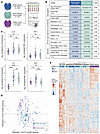 Figure 1
Shared proteomic features in pediatric patients with MODS with and without sepsis. (A) Study schematic showing patients with MODS with sepsis (n = 35) and without sepsis (n = 53), as well as participants in the healthy control (HC) group (n = 25). (B) Patient demographics and clinical outcomes, stratified by etiology (sepsis versus nonsepsis). Comparisons by χ² or Wilcoxon rank-sum test. (C) Proinflammatory cytokines are elevated in MODS relative to participants in the HC group. Box-and-whisker plots show median and IQR (whiskers 1.5 × IQR). Comparisons by Kruskal-Wallis test followed by Dunn’s post hoc test with Benjamini-Hochberg adjustment: *P < 0.05, **P < 0.01, ***P < 0.001, ****P < 0.0001. (D) PCA of 1,448 plasma proteins from 88 patients with MODS and 25 participants in the HC group demonstrates overlap between patients with MODS with and without sepsis. (E) Hierarchically clustered heatmap of 1,448 row-normalized proteins measured at MODS onset. HC samples cluster separately from MODS samples, and patients with MODS with and without sepsis exhibit overlapping proteomic profiles. Annotation bars indicate cohort membership.
Demographics and clinical outcomes were similar between our 35 patients with sepsis and 53 patients without sepsis (Figure 1B). The only organ dysfunction category that differed between groups was hematologic dysfunction, driven by thrombocytopenia at MODS onset in patients with sepsis (platelet count median [IQR]: 119 [IQR: 99–187] versus 199 [IQR: 131–278], P < 0.001; normal range 150–450 × 103/μL). Primary and secondary patient outcomes did not differ between MODS subgroups, including cumulative Pediatric Logistic Organ Dysfunction-2 (PELOD-2) organ dysfunction score, survival to pediatric intensive care unit (PICU) discharge, and duration of PICU stay.
We hypothesized that expression of proinflammatory cytokines would differ between patients with and without sepsis. While we noted the expected increased proinflammatory cytokines (IL-1β, IL-6, tumor necrosis factor (TNF), IL-18, monocyte chemoattractant protein–1 (MCP-1), IFN-γ, all P < 0.05) in patients with sepsis compared with participants in the HC group, most cytokines were also markedly elevated in patients with MODS without sepsis. We found that median IL-1β, TNF-α, and IL-6 levels did not differ between patients with and without sepsis, while IFN-γ was higher in patients with sepsis (P < 0.0001) (Figure 1C). Principal component analysis (PCA) distinguished patients with MODS from HCs, but revealed substantial overlap between sepsis and nonsepsis populations (Figure 1D). Patients with MODS also exhibited substantial heterogeneity along PC1 and PC2. These dimensions were shaped by proteins involved in biologic processes such as growth regulation (e.g., MAD1L1, HDGF), inflammatory signaling (e.g., CASP3, MAP3K5), and endothelial dysfunction (e.g., PLAUR, SRC), as illustrated by the top PCA loadings (Supplemental Table 3).
Hierarchical clustering of row-normalized protein expression similarly distinguished patients with MODS from healthy controls but failed to discriminate between sepsis and nonsepsis populations (Figure 1E). This overlap, complementary with the PCA findings, suggests that MODS may represent a final common pathway of critical illness, independent of the inciting diagnosis or infection status.
Identification of 3 severity-associated subgroups. To uncover biologic programs driving organ dysfunction in children with MODS, we first sought to define severity-associated subgroups based on plasma protein expression to enrich for mechanistic signal. Because sepsis immune responses vary by age, sex, and time (16), we used linear mixed-effects models to identify plasma proteins associated with severity of illness, defined by the PELOD-2 organ dysfunction score, after adjustment for age and sex, as fixed effects and day from MODS onset as a random effect. This yielded a set of 214 plasma proteins that were significantly associated with illness severity (Supplemental Table 4). We then employed consensus clustering (37) to identify distinct subgroups of patients based on expression of these severity-associated proteins (Figure 2A). Optimal cluster number (k=3) was identified via Monte Carlo bootstrapping and confirmed by elbow method and gap statistic (Supplemental Figure 1). Sensitivity analysis using spectral clustering confirmed the robustness of these severity-associated subgroups (adjusted Rand index = 0.73, concordance = 91%; Supplemental Figure 2). To understand the association between subgroups and clinical outcomes, we tested the effect of subgroup membership on the cumulative incidence of both mortality and survival to PICU discharge with the Fine-Gray subdistribution hazard model (38). In this survival analysis, patients in Group C have a higher cumulative incidence of death (P = 0.03) and a lower cumulative incidence of survival to PICU discharge (P = 0.04) compared with patients in Groups A and B, with separation occurring in the first week and persisting to day +28 (Figure 2B). Group B demonstrated an intermediate clinical trajectory, with survival rate and cumulative PELOD-2 organ dysfunction score falling between those of Groups A and C.
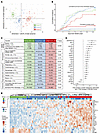 Figure 2
Three subgroups based on protein expression at MODS onset. (A) PCA of 214 severity-associated plasma proteins in 88 patients at MODS onset demonstrating separation of 3 subgroups defined by consensus clustering. (B) Cumulative incidence of survival to PICU discharge (dashed) and PICU mortality (solid) by subgroup, estimated using the Fine-Gray model. (C) Cohort demographics, etiology, and clinical outcomes, stratified by proteomic subgroup. Comparisons by χ² or Wilcoxon rank-sum test. (D) Fold-change in expression per +1 SD increase in PELOD-2 score for 24 proteins selected by elastic net regression. Points represent fold-change estimates; whiskers are 95% confidence intervals. (E) Hierarchically clustered row-normalized heatmap of the 24-protein elastic net signature in 88 patients at MODS onset. Annotation bars indicate subgroup assignment and sepsis status.
Though defined by protein expression alone, these identified subgroups also differ by clinical features and outcomes (Figure 2C). MODS etiology varies by Group, with similar proportions of patients with sepsis in Group B and C and all patients with trauma classified in Group A. Patients in Group C have higher severity of illness, more noncardiopulmonary organ failures, and increased cumulative organ dysfunction scores and mortality compared with Groups A/B. The etiology of MODS and computed subgroup for each patient are detailed in Supplemental Table 5. Preexisting immunocompromised status, defined a priori as active malignancy, hematopoietic cell transplantation, or primary immunodeficiency (39), was identified in 13% of patients with MODS (11/88), and these patients were not imbalanced across subgroups (P = 0.74), as detailed in Supplemental Table 6. Patients were exposed to corticosteroids at a similar rate across subgroups (A: 10/19 [53%], B: 13/39 [33%], C: 14/30 [47%], P = 0.31), suggesting that group assignment was not driven by treatment allocation.
Noting an ordinal increase in the number of organ failures and mortality across plasma protein–derived subgroups, we hypothesized that a reduced set of proteins could identify these subgroups and could be more suitable for translation to the clinical setting. Elastic net regularization is a common approach to generate a high-performing sparse model with good predictive accuracy (40). To define a parsimonious protein signature, we trained an ordinal elastic net model (41) with 10-fold cross validation and generated a 24-protein signature that discriminates the 3 subgroups (Supplemental Figure 3).
To evaluate the performance of this parsimonious model, we first tested the association between the elastic net proteins and severity of illness. Using a linear mixed-effects model, we estimated the fold change in protein expression associated with a 1-SD change in PELOD-2 organ dysfunction score (Figure 2D). This model demonstrates that modest changes in expression of these 24 proteins are associated with meaningful differences in illness severity. Second, we tested the discrimination of this 24-protein signature. Hierarchical clustering of elastic net proteins at MODS onset effectively separates subgroups but does not discriminate between patients with and without sepsis (Figure 2E).
To quantify performance of the elastic net protein set, we calculated the polytomous discrimination index (PDI), a measure of rank-based discrimination, for each subgroup. Category-specific PDI demonstrated excellent discrimination for each subgroup (Group A: 0.98, Group B: 0.96, Group C: 0.99), and the overall PDI for the model was 0.98. Taken together, these results suggest that this 24-protein signature reflects severity of illness and successfully discriminates subgroups at MODS onset.
Immune cell frequency and activation vary by subgroup. Having identified 3 severity-associated subgroups (Groups A/B/C) from protein expression data and built a parsimonious classification model, we next sought to define underlying mechanisms of immune dysregulation. We hypothesized that protein-derived subgroups would be associated with differences in cellular immunophenotype. For this analysis, we performed high-dimensional spectral flow cytometry on cryopreserved PBMCs obtained at MODS onset using a custom-designed 35 marker panel (Supplemental Table 2), which includes both phenotypic and functional markers. After arcsinh scaling (42) and quality control with flowAI (43), we performed FlowSOM metaclustering (44) and identified 14 immune cell populations by surface and intracellular marker expression. Supplemental Figure 4 presents a representative example of our manual gating strategy for this immune phenotyping panel, which we used to confirm the identity of the FlowSOM metaclusters, as well as representative immunophenotyping plots.
To measure the immunophenotypic differences between participants in the HC group and patients in the 3 subgroups (Figure 3A), we subsampled the data to 100,000 cells per subgroup and applied t-distributed Stochastic Neighbor Embedding with Compute Unified Device Architecture (tSNE-CUDA) dimensionality reduction (45). Differences in cell populations by subgroup are quantified in stacked bar plots (Figure 3B). The proportional abundance of central memory (CD45RA–CD27+), effector memory (CD45RA–CD27–), and Temra (CD45RA+CD27–) CD8+ T cells were markedly reduced in patients in Group C (Figure 3C), and this proportional loss of nonnaive CD8+ T cells was strongly associated with severity of illness by linear regression (P < 0.001). A similar ordinal trajectory was identified across multiple cell types (Figure 3D), including T cells, B cells, NK cells, and dendritic cells (Cuzick test of trend P < 0.001 for each cell type shown), and reduction in frequency of each of these cell types was strongly associated with severity of illness by linear regression (all P < 0.001).
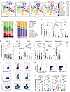 Figure 3
Immune cell abundance and activation state differ across proteomic subgroups. (A) tSNE-CUDA projection of PBMC phenotypes (14 FlowSOM-defined metaclusters) from participants in the HC group and patients with MODS. (B) Stacked bar plots showing proportional abundance of CD3+ and CD3– cell populations by subgroup. (C) Reduced frequency of CD8+ central memory, effector memory, and Temra cells in MODS, with an ordinal decrease across subgroups. (D) Proportional abundance of additional lymphoid and myeloid populations in MODS. (E) Ki67 expression in nonnaive CD4+ T cells, nonnaive CD8+ T cells, and cytotoxic NK cells is increased in Groups B and C relative to Group A (all P < 0.05), indicating increased proliferation. (F) Representative concatenated flow plots of Ki67 expression in nonnaive T cells and cytotoxic NK cells. (G) Quantification of proliferation and activation markers by patient and subgroup. Box-and-whisker plots show median and IQR (whiskers 1.5 × IQR). Comparisons by Kruskal-Wallis test followed by Dunn’s post hoc test with Benjamini-Hochberg adjustment: *P < 0.05, **P < 0.01, ***P < 0.001, ****P < 0.0001.
In addition to shifts in peripheral immune cell subset frequency, we hypothesized that subgroups would be associated with differences in immune cell activation, as measured by expression of markers of proliferation (i.e., Ki67) and activation (i.e., CD38 and HLA-DR), across subgroups. Representative bivariate plots of Ki67 expression in nonnaive CD4+ and CD8+ T cells and cytotoxic NK cells (CD56dim CD16+) demonstrate increased proliferation in cells from patients in Group B and Group C compared with cells from patients in Group A (Figure 3E). Similarly, CD38 and HLA-DR coexpression in nonnaive CD8+ T cells was markedly increased in cells from patients in Group B and Group C compared with cells from patients in Group A, but not different between Group B and Group C, indicative of CD8+ T cell activation in Groups B and C (Figure 3F). Corresponding boxplots in Figure 3G quantify differences in proliferation and activation by patient and subgroup. Despite limited pairwise separation between Groups B and C, we observed a monotonic increase in the proportion of activated cells across subgroups (Cuzick test of trend P < 0.001 for each cell type shown), indicating a graded activation trajectory across groups. Taken together, these data indicate that nonnaive CD8+ T cells in Group C exhibit the greatest degree of lymphodepletion and immune activation, and that these cellular phenotypes are associated with severity of illness.
Multiple concurrent mechanisms of immune dysregulation converge in patients in Group C. Because patients in Group C represent the most severe manifestation along a continuum of severity-associated immune dysregulation, we hypothesized that we could identify candidate mechanisms contributing to this phenotype by measuring differential expression of key canonical inflammatory pathways in patients in Group C. Using the plasma proteomics dataset, we first examined differential protein expression after adjustment for patient age, sex, severity of illness, and days since MODS onset using a linear mixed-effects model. In unadjusted analysis, 1,061 of 1,448 measured proteins were differentially expressed in Group C (Figure 4A), and in our adjusted model 1,003 of 1,061 proteins remained differentially expressed compared with Group A/B. For 98% (980/1,003) of these differentially expressed proteins, expression was upregulated in Group C.
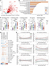 Figure 4
Patients in Group C exhibit a hyperinflammatory plasma with concurrent activation of multiple immune dysregulation pathways. (A) Volcano plot of plasma protein expression in Group C versus Groups A/B at MODS onset (FDR <0.05). (B) Ingenuity Pathway Analysis of 1,003 differentially expressed proteins after adjustment for age, sex, severity of illness, and days since MODS onset using linear mixed-effects modeling. (C) GSVA pathway enrichment scores using Hallmark gene sets. Group B and Group C showed markedly increased IL-6/JAK/STAT3 and IL-2/JAK/STAT5 signaling and an ordinal decrease in PI3K/AKT/mTOR signaling across subgroups. Box-and-whisker plots show median and IQR (whiskers 1.5 × IQR). Comparisons by Kruskal-Wallis test followed by Dunn’s post hoc test with Benjamini-Hochberg adjustment: *P < 0.05, **P < 0.01, ***P < 0.001, ****P < 0.0001. (D) Spearman correlation matrix between GSVA scores and immune cell phenotypes (FDR P < 0.05). Circle color reflects correlation coefficient (ρ; −1 to +1) and size indicates significance. (E) Longitudinal expression of canonical cytokine storm–associated proteins over 14 days (LOESS regression with 95% CI).
To compare expression of key canonical inflammatory pathways across subgroups, we performed pathway enrichment analysis of our adjusted plasma protein expression dataset using complementary group- and patient-based strategies. First, we used Ingenuity Pathway Analysis (IPA, Qiagen) (46) to identify enrichment of 22 canonical pathways in the Group C proteome in comparison with Group A/B (Figure 4B). Noting that 8 of the top 10 differentially expressed pathways identified by IPA were related to hyperinflammatory signaling, we then applied Gene Set Variation Analysis (GSVA) (47) to study enrichment of 5 canonical proinflammatory pathways on the individual patient level using Human Molecular Signatures Database (MSigDB) Hallmark gene sets (48). Using GSVA, we assigned patient-level protein module enrichment scores for each pathway of interest based on expression of proteins in the corresponding Hallmark gene set, as detailed in Methods. Module enrichment scores for each pathway are visualized in Figure 4C and demonstrate markedly increased IL-6/JAK/STAT3 and IL-2/JAK/STAT5 module enrichment in patients in Group B and Group C (each P < 0.001 vs Group A) and an ordinal decrease in PI3K/AKT/mTOR signaling across subgroups (Cuzick test of trend P < 0.001). Plasma IFN-γ response and TNF/NF-κB signaling module enrichment scores did not vary by subgroup. These data indicate that aberrant immune cell signaling is not restricted to a single subgroup but increases along a continuum of immune activation, with Group C representing the most severe manifestation of immune dysregulation within our cohort.
We then assessed the correlation between Hallmark module enrichment and immune cell subset proportional abundance and activation in patients with MODS (Figure 4D). We noted that IL-6/JAK/STAT3 module enrichment had the strongest correlation with proliferation and activation of immune cell subsets previously identified (Figure 3, E and F), including nonnaive CD4+ and CD8+ T cell proliferation (Ki67 expression) and activation (HLA-DR/CD38 coexpression and PD-1/CD39 coexpression). Classical and nonclassical monocyte HLA-DR expression was inversely correlated with IL-6/JAK/STAT3 module enrichment score, consistent with the well-established paradigm of sepsis-associated immunoparalysis, while immune regulatory cell populations (regulatory T cells, myeloid-derived suppressor cells) were positively correlated with IL-6/JAK/STAT3 module enrichment score.
Finally, we studied the longitudinal expression of cytokine storm markers (49) in patients with MODS to understand the duration of cytokinemia in patients in Group C. Normalized protein expression by day since MODS onset by subgroup is shown in Figure 4E. We noted that IL-6, MCP-1, and IL-18 expression were significantly higher in patients in Group C for the first 7 days after MODS onset (each P < 0.001 at day +7), while IL-1β and TNF-α expression remained different for only the first 4 days (P = 0.05 and P = 0.009 respectively at day +4) and IFN-γ expression did not differentiate subgroups at any timepoint, though these differences may reflect differential plasma stability or tissue consumption rather than upstream production mechanisms.
Immune dysregulation in MODS compared with patients with inborn errors of immunity that impact STAT1 and STAT3 signaling. The enrichment of STAT pathway signaling in patients with the most severe MODS and its association with increased T cell activation by flow cytometry prompted us to ask whether this endotype reflects a physiologic adaptation to critical illness or a maladaptive, pathologic form of immune dysregulation. STAT3 is a central mediator of inflammatory signaling, implicated in endothelial dysfunction, capillary leak, emergency granulopoiesis, and disrupted lymphocyte homeostasis (50). Recent translational studies have highlighted its role in human sepsis, including the identification of immature circulating CD66b+ neutrophils as markers of STAT3-driven emergency myelopoiesis in adults who are critically ill (51), and 2 recent preclinical studies have demonstrated a protective effect of selective STAT3 inhibition in mice with CLP-induced sepsis (52, 53). Informed by these studies, we hypothesized that the magnitude of STAT3 pathway activation in patients in Group C is not merely an epiphenomenon but instead reflects a pathogenic immune program that may constitute a viable target for precision immunomodulation.
To test this hypothesis, we compared plasma proteomic profiles between patients with MODS and participants with rare, monogenic inborn errors of immunity (IEI) involving amplified or impaired activation of the STAT3 and STAT1 signaling pathways, which predispose to immune dysregulation and susceptibility to infection. In this analysis, IEIs offer a unique lens through which to interpret complex immune phenotypes, serving as arbiters that reveal the immunologic consequences of discrete signaling perturbations in vivo. Patients with 4 different IEI were included in this analysis: STAT1 gain-of-function (GOF) (n = 9), STAT3 GOF (n = 5), STAT1 autosomal dominant and dominant-negative (DN) (n = 1), or STAT3 DN (n = 3). We measured protein expression using a 384-marker inflammatory proteomics panel from Olink Proteomics (Supplemental Table 7), and we analyzed pathway expression in bulk using Gene Set Enrichment Analysis (GSEA) (54) and at the individual patient level using GSVA (47).
A summary of our experimental design is shown in Figure 5A, which includes 88 patients with MODS, 18 patients with IEI, and 25 participants in the HC group. Orthogonal to our Ingenuity Pathway Analysis findings, we first confirmed population-level proteomic enrichment (normalized enrichment score 1.53, P value 0.004) of the Hallmark IL-6/JAK/STAT3 signaling pathway in Group C patients at MODS onset compared with participants in the HC group using GSEA (Figure 5B). Leading edge proteins and other enriched Hallmark pathways from this analysis are shown in Supplemental Table 8.
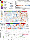 Figure 5
STAT3 pathway activation in patients in Group C meets or exceeds that observed in monogenic STAT1/STAT3 immune disorders. (A) Comparative analysis of MODS (n = 88), healthy control (HC, n = 25), and inborn error of immunity (IEI, n = 18) cohorts, including STAT1/STAT3 gain-of-function (GOF) and dominant-negative (DN) mutations. (B) GSEA of Hallmark IL-6/JAK/STAT3 pathway (Group C versus HC). (C) Row-normalized heatmap of 31 KEGG JAK/STAT target proteins in hierarchically clustered patients with MODS and IEI. Annotation bars denote IEI diagnosis and MODS subgroup. (D) Row-normalized heatmap of patient-level GSVA of 5 inflammatory Hallmark pathways. (E) Comparison of Hallmark IL-6/JAK/STAT3 GSVA scores across MODS and IEI groups. Box-and-whisker plots show median and IQR (whiskers 1.5 × IQR). Comparisons by Kruskal-Wallis test followed by Dunn’s post hoc test with Benjamini-Hochberg adjustment: *P < 0.05, **P < 0.01, ***P < 0.001, ****P < 0.0001. (F) Longitudinal IL-6/JAK/STAT3 enrichment (n = 26) in MODS patients with greater than or equal to 3 serial samples with individual patient trajectories (solid) and LOESS regression (dashed) shown within each subgroup.
To assess expression of STAT target proteins across MODS and patients with IEI, we performed unsupervised hierarchical clustering based on normalized protein expression of 31 proteins from the KEGG JAK/STAT gene set measured in our cohort. We assessed group concordance at the first bifurcation of the dendrogram, which separated the cohort into 2 clusters. As shown in Figure 5C, STAT1 GOF and STAT3 GOF patients colocalize with patients in Group C (P < 0.0001) while STAT1 DN and STAT3 DN patients colocalize with patients in Group A (P < 0.0001). This protein-level analysis also highlights the biologic heterogeneity within our severity-defined subgroups and the tested IEIs.
Having demonstrated enrichment of STAT target proteins in plasma from patients with MODS in Group C, patients with STAT1 GOF, and patients with STAT3 GOF, we next used GSVA to assess patient-level enrichment of 5 inflammatory pathways in patients with MODS and IEI. Figure 5D shows a clustered heatmap of module enrichment scores for each patient in the dataset. We noted increased IL-6/JAK/STAT3 module enrichment scores in all patients with STAT3 GOF and some with STAT1 GOF, while patients with STAT1 DN and STAT3 DN had IL-6/JAK/STAT3 module enrichment scores equivalent to participants in the HC group. Among patients with MODS, Groups B and C both showed elevated IL-6/JAK/STAT3 signaling compared with Group A (P = 0.018 and P = 0.007 respectively, Figure 5E). There was no significant difference between STAT3 GOF and patients in Group C, and many patients had enrichment scores that exceeded the median score of patients with STAT3 GOF. Collectively, these findings support the conclusion that patients in Group C exhibit sustained, high-magnitude IL-6/JAK/STAT3 pathway activation that mirrors, and, in some cases, exceeds, that observed in individuals with monogenic STAT3 gain-of-function mutations.
Finally, we examined the temporal dynamics of STAT3 pathway activation by comparing longitudinal IL-6/JAK/STAT3 module enrichment scores among the subset of 26 patients with greater than or equal to 3 serial plasma samples. Patients in Group C exhibited the highest enrichment scores at MODS onset, and these remained persistently elevated throughout the first 2 weeks of illness (Figure 5F). In contrast, enrichment scores in patients in Group B declined gradually over time, while scores in patients in Group A showed a rapid and sustained decrease. Notably, increases in IL-6/JAK/STAT3 signaling over time were rare: only 2 patients in Group C demonstrated a late rise in enrichment scores, with low initial activation followed by marked elevation approximately 1 week after MODS onset. These findings indicate that STAT3 hyperactivation in Group C is both an early and durable feature of disease, supporting its role as a driver of immune dysregulation rather than a transient host response.
STAT1 and STAT3 signaling are associated with T cell immunometabolic dysregulation in MODS. Because Group C represents the most severe phenotype in our multi-omic profiling assays, we focused our subsequent analyses on this subgroup to dissect the cellular mechanisms underlying STAT pathway hyperactivation in patients with MODS. Having demonstrated STAT3 pathway signaling in Group C patients at levels comparable to or exceeding those seen in patients with known STAT3 GOF, we next hypothesized that plasma STAT3 hyperactivation would result in immunometabolic dysregulation within lymphocytes from patients in Group C. To test this hypothesis, we performed single-cell RNA sequencing (scRNA-seq) using cryopreserved PBMCs obtained at MODS onset for patients in Group C (n = 9) and pediatric participants in the HC group (n = 3). Plasma IL-6/JAK/STAT3 module enrichment scores were significantly different between the 9 patients with MODS and 3 HC participants (0.331 versus –0.494, P < 0.0001) selected for scRNA-seq.
A schematic of our experimental approach is shown in Figure 6A. We sorted live CD45+ PBMCs and then profiled 10,000 cells per patient using the 10X Genomics 5′ single cell gene expression platform. Libraries were sequenced to a depth of approximately 30,000 reads per cell on a Novaseq S2 (Illumina). Transcripts were aligned to the GRCh38 reference genome using Cell Ranger v8.0. After quality control using SoupX (55) and DoubletFinder (56) and integration using RPCA via Seurat v5 (57), cell identities were inferred using ScType (58) and refined using Azimuth (59) and T cell phenotypes from Giles et al. (60). Using this approach, we identified 15 immune cell populations by transcriptional profile for downstream analysis. We applied UMAP (61) dimensionality reduction and subsampled 30,000 cells from each group (HC and Group C) for visualization of immunophenotypic differences (Figure 6B). Differences in cell populations between participants in the HC group and Group C are quantified in stacked bar plots (Figure 6C) and largely mirror the differences in lineages seen in our flow cytometry analysis, redemonstrating the altered immune cell composition in patients in Group C.
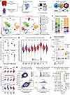 Figure 6
STAT1 and STAT3 signaling promote T cell immunometabolic dysregulation. (A) scRNA-seq experimental design (Group C, n = 9; HC, n = 3). (B) UMAP projection of lymphoid and myeloid lineages. (C) Proportional abundance of CD3+ and CD3– subsets by group. (D) Pseudobulk enrichment scores for the lymphoid protective gene module. Box-and-whisker plots show median and IQR (whiskers 1.5 × IQR). (E) Single-cell lymphoid protective module enrichment scores across lymphoid subsets. (F) Cytokine-response signatures inferred using the Immune Dictionary framework. Bubble color represents enrichment magnitude in Group C relative to HC; size indicates significance. (G) Glycolysis, oxidative phosphorylation, and mTOR signaling enrichment in CD8+ T cells. (H) Bivariate metabolic enrichment scores identify metabolically activated CD8+ T cells in Group C, and cytokine-response scores in activated versus nonactivated cells (right; mixed-effects model). (I) Ligand-receptor interaction analysis in CD4+ and CD8+ T cells. Color indicates interaction strength; size indicates significance. Comparisons by Wilcoxon rank-sum test unless otherwise noted, with Benjamini-Hochberg FDR adjustment: *P < 0.05, **P < 0.01, ***P < 10-12, ****P < 10-16.
Building on our flow-cytometry evidence of T cell depletion and activation and our plasma evidence of STAT3 hyperactivation, we first tested whether a validated transcriptomic marker of dysfunctional lymphoid immunity was similarly perturbed in patients in Group C. We applied a transcriptional framework developed by the SUBSPACE consortium (62), which identified a “lymphoid protective” gene set derived from adult patients with sepsis. Lower scores on this module denote a “lymphoid detrimental” transcriptional program associated with T cell dysfunction and poor outcomes in adult sepsis. We calculated “lymphoid protective” module enrichment scores at the single-cell level using UCell (63) and generated pseudobulk profiles for statistical analysis to control Type 1 error (64). Consistent with our protein-level findings, patients in Group C exhibited significantly lower lymphoid protective scores compared with participants in the HC group in CD4+ naive, CD8+ naive, and CD8+ nonnaive populations (Figure 6D).
To broaden our assessment of transcriptional dysregulation, we extended our analysis to include per-cell lymphoid protective module enrichment scores across 9 major lymphoid subsets (Figure 6E). Patients in Group C exhibited significantly lower module scores across all subsets, with the most pronounced deficits observed in the CD8+ T cell compartment. Together, these results indicate that lymphoid dysregulation in Group C is not restricted to numerical depletion but reflects widespread changes to immune regulatory gene programs necessary for effective immune surveillance.
To infer which cytokines may drive immune dysregulation in Group C lymphocytes, we leveraged the Immune Dictionary framework (65), which enables cell-type–specific identification of cytokine responses based on downstream gene expression patterns. As shown in Figure 6F, patients in Group C exhibited significantly increased transcriptional responses to multiple proinflammatory cytokines compared with participants in the HC group, including IL-6 (which activates STAT1 and STAT3) and IFN-γ (which predominantly activates STAT1). These findings indicate that sustained exposure to a proinflammatory cytokine milieu, particularly IL-6 and IFN-γ, may drive the transcriptional reprogramming observed in lymphocytes from patients in Group C.
Because corticosteroids modulate cytokine signaling, we performed a sensitivity analysis to determine if these transcriptional signatures were affected by treatment allocation. We compared lineage-specific IL-6/JAK/STAT3 module enrichment scores between patients in Group C who received corticosteroids (n = 4) and those who did not (n = 5). Using Cliff’s Delta (δ) to quantify effect size, corticosteroid exposure was associated with lower IL-6/JAK/STAT3 module enrichment scores in classical monocytes (δ = 0.42, medium effect), whereas effect sizes in T cell subsets were small or negligible (e.g., CD8+ central memory T cells, δ = –0.23, small effect) (Supplemental Table 9). These exploratory data, while limited by sample size, do not support corticosteroid-mediated suppression as an explanation for the lymphoid transcriptional phenotype observed in Group C.
To understand the effects of sepsis on T cell immunometabolism, we calculated KEGG pathway module enrichment scores for glycolysis, oxidative phosphorylation, and mTOR signaling pathways using UCell (63) as detailed in Methods. As shown in Figure 6G, CD8+ T cells from patients in Group C exhibited increased transcriptional activity in glycolysis and oxidative phosphorylation pathways compared to HC participants across all differentiation states. Conversely, mTOR signaling was suppressed across CD8+ T cell subsets from Group C, a pattern suggesting cytokine-driven repression of the mTOR pathway, which may affect T cell function (66).
Given that IL-6 and IFN-γ exert complex effects on immunometabolic state and mTOR signaling, we next assessed whether exposure to these signals explained the metabolic phenotype observed in Figure 6G. To test whether specific cytokines underpin this bioenergetic shift, we modeled the joint scores of single-cell Hallmark glycolysis and oxidative phosphorylation modules (Figure 6H). This revealed a distinct subset of Group C CD8+ T cells with increased glycolysis and oxidative phosphorylation transcriptional signatures (labeled as “metabolic activation”). In multivariable mixed-effects logistic regression analysis, enrichment scores for IL-6, IFN-γ, and IL-1β transcriptional responses are significantly increased in metabolically activated CD8+ T cells compared with cells within the HC baseline range (all P < 0.001), as shown in the ridge plots in Figure 6H.
Finally, to further characterize the amplified activation state of CD8+ T cells in patients in Group C, we applied CellChat (67) to the scRNA-seq dataset to identify predicted cell-to-cell communication events via curated ligand-receptor pairs and coreceptor interactions that modulate signaling. As shown in Figure 6I, patients in Group C exhibited significantly stronger inferred signaling interactions between monocyte-expressed HLA class I molecules and the CD8A coreceptor on CD8+ T cells, compared with participants in the healthy control group (P < 0.01). Increased inferred HLA-CD8A interactions are consistent with observed CD8+ T cell activation in Group C and align with our transcriptional and metabolic findings, although this modeling does not establish altered antigen presentation or a causal role of myeloid-lymphoid crosstalk in sepsis-associated immune dysregulation.
T cell activation in MODS occurs without antigen-specific clonal expansion. Having demonstrated lymphoid dysregulation and cytokine-linked metabolic reprogramming in patients in Group C, we next investigated whether these immunometabolic changes reflected antigen-specific immune responses or nonspecific bystander activation. We analyzed T cell receptor (TCR) repertoires captured alongside our scRNA-seq data using 5′ tag-based single-cell V(D)J sequencing (10X Genomics). VDJ libraries were sequenced to a depth of approximately 10,000 reads per cell on a Novaseq S2 (Illumina) and processed with Cell Ranger v8.0, aligning to the GRCh38 reference genome. TCR data were integrated with scRNA-seq analyses in Seurat using the scRepertoire package (68). We used this dataset to test the hypothesis that T cell activation in patients in Group C represented antigen-independent “bystander” activation as opposed to antigen-specific activation in response to a pathogen.
To computationally assess antigen specificity, we first quantified clonal expansion across the TCR repertoire at the individual patient level. In contrast with participants in the HC cohort who exhibited expected homeostatic clonal expansion, including the presence of hyperexpanded clones, patients in Group C were characterized by a marked absence of expanded clones (Figure 7A). No single clone in Group C patients occupied more than 1% of the repertoire, and most clones were present as single, unique sequences. This lack of clonal expansion argues against antigen-driven clonal expansion as the dominant mechanism of T cell activation in patients in Group C.
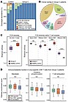 Figure 7
Polyclonal, antigen-independent T cell activation in patients in Group C. (A) Clonal abundance of CD3+ T cells from scTCR-seq data, showing predominantly single- or rare-frequency clonotypes. (B) Chord diagram of TCR clonal sharing across T cell subsets in patients in Group C. (C) Shannon diversity of TCR repertoires. Comparisons by Wilcoxon rank-sum test with Benjamini-Hochberg adjustment: *P < 0.05, **P < 0.01, ***P < 0.001, ****P < 0.0001. (D) Immunometabolic pathway enrichment of CMV-specific versus nonspecific CD8+ T cells. Comparisons by Kruskal-Wallis test with Benjamini-Hochberg adjustment. Box-and-whisker plots show median and IQR (whiskers 1.5 × IQR).
We next assessed paired TCR diversity using the Shannon index, which accounts for both the richness (number of unique clonotypes) and evenness (relative abundance) of clonotypes. As shown in Figure 7B, patients in Group C exhibited significantly higher Shannon diversity compared with participants who were healthy controls (P < 0.001), consistent with broad, nonspecific T cell activation. Subset-level analysis revealed that TCR diversity remained comparable with controls in naive and effector CD4+ T cells, but was increased in naive CD8+ T cells and decreased in effector CD8+ T cells (both P < 0.001), suggesting that alterations to the TCR repertoire are specific to CD8+ lineages.
To determine whether CD8+ T cell activation reflected antigen-specific clonal expansion, we visualized clonotype overlap across T cell subsets. As shown in Figure 7C, patients in Group C exhibited minimal overlap between naive, central memory, effector memory, and EMRA CD8+ T cell populations, again providing evidence against substantial expansion of antigen-specific clones and supporting a model of bystander activation.
Finally, we investigated whether immunometabolic pathway enrichment differed by antigen specificity predicted in silico using TReX (69) based on reference TCR sequences from VDJdb (70), McPAS-TCR (71), IEDB (72), and PIRD (73). DNA viremia is associated with immune dysregulation in pediatric sepsis (74), and we hypothesized that virus-specific CD8+ T cells could account for the immunometabolic changes noted in patients in Group C. Using cytomegalovirus (CMV) as a model of chronic viral antigen exposure, we stratified CD8+ T cells by CMV-specific, non-CMV–specific, and unannotated TCRs and compared glycolysis, oxidative phosphorylation, and T cell exhaustion gene set enrichment scores across these groups using UCell. No significant differences were observed across these groups (Figure 7D), suggesting that virus-specific CD8+ T cells do not drive the immunometabolic changes observed in Group C.
Taken together, these findings suggest that T cell activation and immunometabolic dysregulation in Group C patients occur independently of antigen-specific clonal expansion, which is typically associated with pathogen-specific responses. These data support a model of cytokine-driven, bystander T cell activation in the setting of STAT3 and STAT1 hyperactivation, rather than a conventional antigen-specific immune response.
IL-6 and IFN-γ signaling drive aberrant STAT pathway activation and T cell dysfunction in MODS. Building on our single-cell transcriptional analyses, which demonstrated antigen-independent T cell activation and marked enrichment of IL-6 and IFN-γ response modules in CD8+ T cells from patients in Group C, we next sought to test whether these cytokines drive functional changes in signaling at the protein level. To do so, we employed an optimized 13-marker T cell phosphoflow cytometry panel (Supplemental Table 10) which allows for identification of T cells and includes phospho STAT (pSTAT) antibodies that recognize pSTAT1, pSTAT3, pSTAT5, and total STAT proteins.
We first examined basal phosphorylation of STAT1 and STAT3, key transcription factors downstream of IFN-γ and IL-6 signaling, respectively, though their use as heterodimers and homodimers downstream of cytokine receptors is complex and includes many other cytokines. Compared with HC, patients in Group C at baseline exhibited higher expression of pSTAT1 (P = 0.041) and pSTAT3 (P = 0.026) in nonnaive CD8+ T cells (Figure 8A). This finding is consistent with the demonstrated elevated plasma cytokine levels, as well as our transcriptional cytokine response analysis, suggesting that IL-6 and IFN-γ exert a concurrent effect on CD8+ T cells in the patients in Group C.
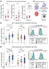 Figure 8
IL-6 and IFN-γ signaling drive aberrant STAT pathway activation and T cell dysfunction. (A) Baseline pSTAT1 and pSTAT3 expression in nonnaive CD8+ T cells. Comparisons by Wilcoxon rank-sum test with Benjamini-Hochberg adjustment. (B) Schematic of the ex vivo TCR stimulation. (C) pSTAT1 and (D) pSTAT3 upregulation in response to TCR stimulation ± IL-6, with representative histograms. Group C nonnaive CD8+ T cells exhibit blunted phosphorylation responses compared with HC. Box-and-whisker plots show median and IQR (whiskers 1.5 × IQR). Unless otherwise noted, comparisons by Kruskal-Wallis test followed by Dunn’s post hoc test with Benjamini-Hochberg adjustment: *P < 0.05, **P < 0.01, ***P < 0.001, ****P < 0.0001.
Given the elevated baseline STAT signaling in Group C, we next assessed how this cytokine exposure affects CD8+ T cell responsiveness to receptor-mediated stimulation. As illustrated in Figure 8B, PBMCs from patients in Group C were thawed and cultured for 24 hours with plate-bound αCD3 and soluble αCD28/αCD49d antibodies. Using this platform, we compared the ability of Group C CD8+ T cells to respond to TCR stimulation alone or in combination with IL-6.
As shown in Figure 8C, HC cells demonstrate robust induction of pSTAT1 following stimulation with αCD3/αCD28/αCD49d and IL-6 (P = 0.033). In contrast, Group C cells failed to increase pSTAT1 expression in response to TCR stimulation with or without IL-6 (P = 0.92). Representative pSTAT1 histograms highlight the lack of response to stimulation in CD8+ T cells from Group C patients. Analysis of pSTAT3 signaling in Figure 8D demonstrated similar findings. HC cells upregulated pSTAT3 following αCD3/αCD28/αCD49d stimulation (P = 0.005), and IL-6 further increased pSTAT3 levels (P < 0.001). In contrast, Group C cells failed to increase pSTAT3 expression in response to TCR stimulation with or without IL-6 (P = 0.46). Representative pSTAT3 histograms again highlight the lack of response to stimulation in CD8+ T cells from patients in Group C.
Together, these findings support the hypothesis that sustained cellular signaling downstream of IL-6 and IFN-γ in patients in Group C may drive chronic activation of the STAT1 and STAT3 signaling pathways, leading to a state of functional desensitization to further activation of those pathways in CD8+ T cells. While plasma IFN-γ levels were variable (Figure 4E), the presence of robust IL-6 and IFN-γ transcriptional signatures (Figure 6F) and elevated basal pSTAT1 and pSTAT3 levels (Figure 8A) suggests that these cells have integrated inflammatory signals that result in a failure to respond appropriately to subsequent TCR-mediated stimulation, indicating a state of refractory responses to cytokine signals. These data implicate STAT1 and STAT3 signaling as central mediators of CD8+ T cell dysregulation in Group C, potentially defining an endotype that drives dysregulation in this subgroup. Our multi-omic findings therefore provide a mechanistic link between systemic inflammation, transcriptional rewiring, and impaired adaptive immunity, which may be therapeutically tractable and requires further study.


Copyright © 2026 American Society for Clinical Investigation
ISSN: 0021-9738 (print), 1558-8238 (online)