
Advertisement
Clinical Research and Public HealthClinical ResearchInfectious diseaseInflammation
Open Access |  10.1172/JCI202480
Plasma chondroitin sulfate predicts the effectiveness of fluid resuscitation strategies in patients with sepsis
Kaori Oshima,1 Bailu Yan,2 Ran Tao,2,3 Gustavo Amorim,2 Chiara Di Gravio,4 Sarah A. McMurtry,5 Ryan C. Burke,6 Yunbi Nam,2 Ina Nikolli,1 Max S. Kravitz,6 Daniel Stephenson,7 Aaron Issaian,7 Kirk C. Hansen,7 Angelo D’Alessandro,7 Ivor S. Douglas,5,8 Wesley H. Self,9 Christopher J. Lindsell,10 Carolyn Leroux,11 Angelika Ringor,11 Michael A. Matthay,11 Jonathan S. Schildcrout,2 Nathan I. Shapiro,6 and Eric P. Schmidt1
1Department of Medicine, Mass General Brigham, Boston, Massachusetts, USA.
2Department of Biostatistics and
3Vanderbilt Genetics Institute, Vanderbilt University Medical Center, Nashville, Tennessee, USA.
4Department of Epidemiology and Biostatistics, School of Public Health, Imperial College London, London, United Kingdom.
5Department of Medicine, University of Colorado, Aurora, Colorado, USA.
6Department of Emergency Medicine, Beth Israel Deaconess Medical Center and Harvard Medical School, Boston, Massachusetts, USA.
7Department of Biochemistry and Molecular Genetics, University of Colorado, Aurora, Colorado, USA.
8Department of Medicine, Denver Health Medical Center, Denver, Colorado, USA.
9Vanderbilt Institute for Clinical and Translational Research and Department of Emergency Medicine, Vanderbilt University Medical Center, Nashville, Tennessee, USA.
10Department of Biostatistics and Bioinformatics and Duke Clinical Research Institute, Duke University Medical Center, Durham, North Carolina, USA.
11Department of Medicine, Cardiovascular Research Institute, University of California, San Francisco, San Francisco, California, USA.
Address correspondence to: Eric P. Schmidt, Harvard Medical School, 55 Fruit Street, Boston, Massachusetts 02114, USA. Phone: 617.643.8692; Email: eschmidt1@mgh.harvard.edu.
Authorship note: KO and BY contributed equally to this work and are co–first authors. NIS and EPS contributed equally to this work and are co–senior authors.
Find articles by Oshima, K. in: PubMed | Google Scholar
1Department of Medicine, Mass General Brigham, Boston, Massachusetts, USA.
2Department of Biostatistics and
3Vanderbilt Genetics Institute, Vanderbilt University Medical Center, Nashville, Tennessee, USA.
4Department of Epidemiology and Biostatistics, School of Public Health, Imperial College London, London, United Kingdom.
5Department of Medicine, University of Colorado, Aurora, Colorado, USA.
6Department of Emergency Medicine, Beth Israel Deaconess Medical Center and Harvard Medical School, Boston, Massachusetts, USA.
7Department of Biochemistry and Molecular Genetics, University of Colorado, Aurora, Colorado, USA.
8Department of Medicine, Denver Health Medical Center, Denver, Colorado, USA.
9Vanderbilt Institute for Clinical and Translational Research and Department of Emergency Medicine, Vanderbilt University Medical Center, Nashville, Tennessee, USA.
10Department of Biostatistics and Bioinformatics and Duke Clinical Research Institute, Duke University Medical Center, Durham, North Carolina, USA.
11Department of Medicine, Cardiovascular Research Institute, University of California, San Francisco, San Francisco, California, USA.
Address correspondence to: Eric P. Schmidt, Harvard Medical School, 55 Fruit Street, Boston, Massachusetts 02114, USA. Phone: 617.643.8692; Email: eschmidt1@mgh.harvard.edu.
Authorship note: KO and BY contributed equally to this work and are co–first authors. NIS and EPS contributed equally to this work and are co–senior authors.
Find articles by Yan, B. in: PubMed | Google Scholar
1Department of Medicine, Mass General Brigham, Boston, Massachusetts, USA.
2Department of Biostatistics and
3Vanderbilt Genetics Institute, Vanderbilt University Medical Center, Nashville, Tennessee, USA.
4Department of Epidemiology and Biostatistics, School of Public Health, Imperial College London, London, United Kingdom.
5Department of Medicine, University of Colorado, Aurora, Colorado, USA.
6Department of Emergency Medicine, Beth Israel Deaconess Medical Center and Harvard Medical School, Boston, Massachusetts, USA.
7Department of Biochemistry and Molecular Genetics, University of Colorado, Aurora, Colorado, USA.
8Department of Medicine, Denver Health Medical Center, Denver, Colorado, USA.
9Vanderbilt Institute for Clinical and Translational Research and Department of Emergency Medicine, Vanderbilt University Medical Center, Nashville, Tennessee, USA.
10Department of Biostatistics and Bioinformatics and Duke Clinical Research Institute, Duke University Medical Center, Durham, North Carolina, USA.
11Department of Medicine, Cardiovascular Research Institute, University of California, San Francisco, San Francisco, California, USA.
Address correspondence to: Eric P. Schmidt, Harvard Medical School, 55 Fruit Street, Boston, Massachusetts 02114, USA. Phone: 617.643.8692; Email: eschmidt1@mgh.harvard.edu.
Authorship note: KO and BY contributed equally to this work and are co–first authors. NIS and EPS contributed equally to this work and are co–senior authors.
Find articles by Tao, R. in: PubMed | Google Scholar
1Department of Medicine, Mass General Brigham, Boston, Massachusetts, USA.
2Department of Biostatistics and
3Vanderbilt Genetics Institute, Vanderbilt University Medical Center, Nashville, Tennessee, USA.
4Department of Epidemiology and Biostatistics, School of Public Health, Imperial College London, London, United Kingdom.
5Department of Medicine, University of Colorado, Aurora, Colorado, USA.
6Department of Emergency Medicine, Beth Israel Deaconess Medical Center and Harvard Medical School, Boston, Massachusetts, USA.
7Department of Biochemistry and Molecular Genetics, University of Colorado, Aurora, Colorado, USA.
8Department of Medicine, Denver Health Medical Center, Denver, Colorado, USA.
9Vanderbilt Institute for Clinical and Translational Research and Department of Emergency Medicine, Vanderbilt University Medical Center, Nashville, Tennessee, USA.
10Department of Biostatistics and Bioinformatics and Duke Clinical Research Institute, Duke University Medical Center, Durham, North Carolina, USA.
11Department of Medicine, Cardiovascular Research Institute, University of California, San Francisco, San Francisco, California, USA.
Address correspondence to: Eric P. Schmidt, Harvard Medical School, 55 Fruit Street, Boston, Massachusetts 02114, USA. Phone: 617.643.8692; Email: eschmidt1@mgh.harvard.edu.
Authorship note: KO and BY contributed equally to this work and are co–first authors. NIS and EPS contributed equally to this work and are co–senior authors.
Find articles by Amorim, G. in: PubMed | Google Scholar
1Department of Medicine, Mass General Brigham, Boston, Massachusetts, USA.
2Department of Biostatistics and
3Vanderbilt Genetics Institute, Vanderbilt University Medical Center, Nashville, Tennessee, USA.
4Department of Epidemiology and Biostatistics, School of Public Health, Imperial College London, London, United Kingdom.
5Department of Medicine, University of Colorado, Aurora, Colorado, USA.
6Department of Emergency Medicine, Beth Israel Deaconess Medical Center and Harvard Medical School, Boston, Massachusetts, USA.
7Department of Biochemistry and Molecular Genetics, University of Colorado, Aurora, Colorado, USA.
8Department of Medicine, Denver Health Medical Center, Denver, Colorado, USA.
9Vanderbilt Institute for Clinical and Translational Research and Department of Emergency Medicine, Vanderbilt University Medical Center, Nashville, Tennessee, USA.
10Department of Biostatistics and Bioinformatics and Duke Clinical Research Institute, Duke University Medical Center, Durham, North Carolina, USA.
11Department of Medicine, Cardiovascular Research Institute, University of California, San Francisco, San Francisco, California, USA.
Address correspondence to: Eric P. Schmidt, Harvard Medical School, 55 Fruit Street, Boston, Massachusetts 02114, USA. Phone: 617.643.8692; Email: eschmidt1@mgh.harvard.edu.
Authorship note: KO and BY contributed equally to this work and are co–first authors. NIS and EPS contributed equally to this work and are co–senior authors.
Find articles by Di Gravio, C. in: PubMed | Google Scholar
1Department of Medicine, Mass General Brigham, Boston, Massachusetts, USA.
2Department of Biostatistics and
3Vanderbilt Genetics Institute, Vanderbilt University Medical Center, Nashville, Tennessee, USA.
4Department of Epidemiology and Biostatistics, School of Public Health, Imperial College London, London, United Kingdom.
5Department of Medicine, University of Colorado, Aurora, Colorado, USA.
6Department of Emergency Medicine, Beth Israel Deaconess Medical Center and Harvard Medical School, Boston, Massachusetts, USA.
7Department of Biochemistry and Molecular Genetics, University of Colorado, Aurora, Colorado, USA.
8Department of Medicine, Denver Health Medical Center, Denver, Colorado, USA.
9Vanderbilt Institute for Clinical and Translational Research and Department of Emergency Medicine, Vanderbilt University Medical Center, Nashville, Tennessee, USA.
10Department of Biostatistics and Bioinformatics and Duke Clinical Research Institute, Duke University Medical Center, Durham, North Carolina, USA.
11Department of Medicine, Cardiovascular Research Institute, University of California, San Francisco, San Francisco, California, USA.
Address correspondence to: Eric P. Schmidt, Harvard Medical School, 55 Fruit Street, Boston, Massachusetts 02114, USA. Phone: 617.643.8692; Email: eschmidt1@mgh.harvard.edu.
Authorship note: KO and BY contributed equally to this work and are co–first authors. NIS and EPS contributed equally to this work and are co–senior authors.
Find articles by McMurtry, S. in: PubMed | Google Scholar
1Department of Medicine, Mass General Brigham, Boston, Massachusetts, USA.
2Department of Biostatistics and
3Vanderbilt Genetics Institute, Vanderbilt University Medical Center, Nashville, Tennessee, USA.
4Department of Epidemiology and Biostatistics, School of Public Health, Imperial College London, London, United Kingdom.
5Department of Medicine, University of Colorado, Aurora, Colorado, USA.
6Department of Emergency Medicine, Beth Israel Deaconess Medical Center and Harvard Medical School, Boston, Massachusetts, USA.
7Department of Biochemistry and Molecular Genetics, University of Colorado, Aurora, Colorado, USA.
8Department of Medicine, Denver Health Medical Center, Denver, Colorado, USA.
9Vanderbilt Institute for Clinical and Translational Research and Department of Emergency Medicine, Vanderbilt University Medical Center, Nashville, Tennessee, USA.
10Department of Biostatistics and Bioinformatics and Duke Clinical Research Institute, Duke University Medical Center, Durham, North Carolina, USA.
11Department of Medicine, Cardiovascular Research Institute, University of California, San Francisco, San Francisco, California, USA.
Address correspondence to: Eric P. Schmidt, Harvard Medical School, 55 Fruit Street, Boston, Massachusetts 02114, USA. Phone: 617.643.8692; Email: eschmidt1@mgh.harvard.edu.
Authorship note: KO and BY contributed equally to this work and are co–first authors. NIS and EPS contributed equally to this work and are co–senior authors.
Find articles by Burke, R. in: PubMed | Google Scholar
1Department of Medicine, Mass General Brigham, Boston, Massachusetts, USA.
2Department of Biostatistics and
3Vanderbilt Genetics Institute, Vanderbilt University Medical Center, Nashville, Tennessee, USA.
4Department of Epidemiology and Biostatistics, School of Public Health, Imperial College London, London, United Kingdom.
5Department of Medicine, University of Colorado, Aurora, Colorado, USA.
6Department of Emergency Medicine, Beth Israel Deaconess Medical Center and Harvard Medical School, Boston, Massachusetts, USA.
7Department of Biochemistry and Molecular Genetics, University of Colorado, Aurora, Colorado, USA.
8Department of Medicine, Denver Health Medical Center, Denver, Colorado, USA.
9Vanderbilt Institute for Clinical and Translational Research and Department of Emergency Medicine, Vanderbilt University Medical Center, Nashville, Tennessee, USA.
10Department of Biostatistics and Bioinformatics and Duke Clinical Research Institute, Duke University Medical Center, Durham, North Carolina, USA.
11Department of Medicine, Cardiovascular Research Institute, University of California, San Francisco, San Francisco, California, USA.
Address correspondence to: Eric P. Schmidt, Harvard Medical School, 55 Fruit Street, Boston, Massachusetts 02114, USA. Phone: 617.643.8692; Email: eschmidt1@mgh.harvard.edu.
Authorship note: KO and BY contributed equally to this work and are co–first authors. NIS and EPS contributed equally to this work and are co–senior authors.
Find articles by Nam, Y. in: PubMed | Google Scholar
1Department of Medicine, Mass General Brigham, Boston, Massachusetts, USA.
2Department of Biostatistics and
3Vanderbilt Genetics Institute, Vanderbilt University Medical Center, Nashville, Tennessee, USA.
4Department of Epidemiology and Biostatistics, School of Public Health, Imperial College London, London, United Kingdom.
5Department of Medicine, University of Colorado, Aurora, Colorado, USA.
6Department of Emergency Medicine, Beth Israel Deaconess Medical Center and Harvard Medical School, Boston, Massachusetts, USA.
7Department of Biochemistry and Molecular Genetics, University of Colorado, Aurora, Colorado, USA.
8Department of Medicine, Denver Health Medical Center, Denver, Colorado, USA.
9Vanderbilt Institute for Clinical and Translational Research and Department of Emergency Medicine, Vanderbilt University Medical Center, Nashville, Tennessee, USA.
10Department of Biostatistics and Bioinformatics and Duke Clinical Research Institute, Duke University Medical Center, Durham, North Carolina, USA.
11Department of Medicine, Cardiovascular Research Institute, University of California, San Francisco, San Francisco, California, USA.
Address correspondence to: Eric P. Schmidt, Harvard Medical School, 55 Fruit Street, Boston, Massachusetts 02114, USA. Phone: 617.643.8692; Email: eschmidt1@mgh.harvard.edu.
Authorship note: KO and BY contributed equally to this work and are co–first authors. NIS and EPS contributed equally to this work and are co–senior authors.
Find articles by Nikolli, I. in: PubMed | Google Scholar
1Department of Medicine, Mass General Brigham, Boston, Massachusetts, USA.
2Department of Biostatistics and
3Vanderbilt Genetics Institute, Vanderbilt University Medical Center, Nashville, Tennessee, USA.
4Department of Epidemiology and Biostatistics, School of Public Health, Imperial College London, London, United Kingdom.
5Department of Medicine, University of Colorado, Aurora, Colorado, USA.
6Department of Emergency Medicine, Beth Israel Deaconess Medical Center and Harvard Medical School, Boston, Massachusetts, USA.
7Department of Biochemistry and Molecular Genetics, University of Colorado, Aurora, Colorado, USA.
8Department of Medicine, Denver Health Medical Center, Denver, Colorado, USA.
9Vanderbilt Institute for Clinical and Translational Research and Department of Emergency Medicine, Vanderbilt University Medical Center, Nashville, Tennessee, USA.
10Department of Biostatistics and Bioinformatics and Duke Clinical Research Institute, Duke University Medical Center, Durham, North Carolina, USA.
11Department of Medicine, Cardiovascular Research Institute, University of California, San Francisco, San Francisco, California, USA.
Address correspondence to: Eric P. Schmidt, Harvard Medical School, 55 Fruit Street, Boston, Massachusetts 02114, USA. Phone: 617.643.8692; Email: eschmidt1@mgh.harvard.edu.
Authorship note: KO and BY contributed equally to this work and are co–first authors. NIS and EPS contributed equally to this work and are co–senior authors.
Find articles by Kravitz, M. in: PubMed | Google Scholar
1Department of Medicine, Mass General Brigham, Boston, Massachusetts, USA.
2Department of Biostatistics and
3Vanderbilt Genetics Institute, Vanderbilt University Medical Center, Nashville, Tennessee, USA.
4Department of Epidemiology and Biostatistics, School of Public Health, Imperial College London, London, United Kingdom.
5Department of Medicine, University of Colorado, Aurora, Colorado, USA.
6Department of Emergency Medicine, Beth Israel Deaconess Medical Center and Harvard Medical School, Boston, Massachusetts, USA.
7Department of Biochemistry and Molecular Genetics, University of Colorado, Aurora, Colorado, USA.
8Department of Medicine, Denver Health Medical Center, Denver, Colorado, USA.
9Vanderbilt Institute for Clinical and Translational Research and Department of Emergency Medicine, Vanderbilt University Medical Center, Nashville, Tennessee, USA.
10Department of Biostatistics and Bioinformatics and Duke Clinical Research Institute, Duke University Medical Center, Durham, North Carolina, USA.
11Department of Medicine, Cardiovascular Research Institute, University of California, San Francisco, San Francisco, California, USA.
Address correspondence to: Eric P. Schmidt, Harvard Medical School, 55 Fruit Street, Boston, Massachusetts 02114, USA. Phone: 617.643.8692; Email: eschmidt1@mgh.harvard.edu.
Authorship note: KO and BY contributed equally to this work and are co–first authors. NIS and EPS contributed equally to this work and are co–senior authors.
Find articles by Stephenson, D. in: PubMed | Google Scholar
1Department of Medicine, Mass General Brigham, Boston, Massachusetts, USA.
2Department of Biostatistics and
3Vanderbilt Genetics Institute, Vanderbilt University Medical Center, Nashville, Tennessee, USA.
4Department of Epidemiology and Biostatistics, School of Public Health, Imperial College London, London, United Kingdom.
5Department of Medicine, University of Colorado, Aurora, Colorado, USA.
6Department of Emergency Medicine, Beth Israel Deaconess Medical Center and Harvard Medical School, Boston, Massachusetts, USA.
7Department of Biochemistry and Molecular Genetics, University of Colorado, Aurora, Colorado, USA.
8Department of Medicine, Denver Health Medical Center, Denver, Colorado, USA.
9Vanderbilt Institute for Clinical and Translational Research and Department of Emergency Medicine, Vanderbilt University Medical Center, Nashville, Tennessee, USA.
10Department of Biostatistics and Bioinformatics and Duke Clinical Research Institute, Duke University Medical Center, Durham, North Carolina, USA.
11Department of Medicine, Cardiovascular Research Institute, University of California, San Francisco, San Francisco, California, USA.
Address correspondence to: Eric P. Schmidt, Harvard Medical School, 55 Fruit Street, Boston, Massachusetts 02114, USA. Phone: 617.643.8692; Email: eschmidt1@mgh.harvard.edu.
Authorship note: KO and BY contributed equally to this work and are co–first authors. NIS and EPS contributed equally to this work and are co–senior authors.
Find articles by
Issaian, A.
in:
PubMed
|
Google Scholar
|

1Department of Medicine, Mass General Brigham, Boston, Massachusetts, USA.
2Department of Biostatistics and
3Vanderbilt Genetics Institute, Vanderbilt University Medical Center, Nashville, Tennessee, USA.
4Department of Epidemiology and Biostatistics, School of Public Health, Imperial College London, London, United Kingdom.
5Department of Medicine, University of Colorado, Aurora, Colorado, USA.
6Department of Emergency Medicine, Beth Israel Deaconess Medical Center and Harvard Medical School, Boston, Massachusetts, USA.
7Department of Biochemistry and Molecular Genetics, University of Colorado, Aurora, Colorado, USA.
8Department of Medicine, Denver Health Medical Center, Denver, Colorado, USA.
9Vanderbilt Institute for Clinical and Translational Research and Department of Emergency Medicine, Vanderbilt University Medical Center, Nashville, Tennessee, USA.
10Department of Biostatistics and Bioinformatics and Duke Clinical Research Institute, Duke University Medical Center, Durham, North Carolina, USA.
11Department of Medicine, Cardiovascular Research Institute, University of California, San Francisco, San Francisco, California, USA.
Address correspondence to: Eric P. Schmidt, Harvard Medical School, 55 Fruit Street, Boston, Massachusetts 02114, USA. Phone: 617.643.8692; Email: eschmidt1@mgh.harvard.edu.
Authorship note: KO and BY contributed equally to this work and are co–first authors. NIS and EPS contributed equally to this work and are co–senior authors.
Find articles by
Hansen, K.
in:
PubMed
|
Google Scholar
|
1Department of Medicine, Mass General Brigham, Boston, Massachusetts, USA.
2Department of Biostatistics and
3Vanderbilt Genetics Institute, Vanderbilt University Medical Center, Nashville, Tennessee, USA.
4Department of Epidemiology and Biostatistics, School of Public Health, Imperial College London, London, United Kingdom.
5Department of Medicine, University of Colorado, Aurora, Colorado, USA.
6Department of Emergency Medicine, Beth Israel Deaconess Medical Center and Harvard Medical School, Boston, Massachusetts, USA.
7Department of Biochemistry and Molecular Genetics, University of Colorado, Aurora, Colorado, USA.
8Department of Medicine, Denver Health Medical Center, Denver, Colorado, USA.
9Vanderbilt Institute for Clinical and Translational Research and Department of Emergency Medicine, Vanderbilt University Medical Center, Nashville, Tennessee, USA.
10Department of Biostatistics and Bioinformatics and Duke Clinical Research Institute, Duke University Medical Center, Durham, North Carolina, USA.
11Department of Medicine, Cardiovascular Research Institute, University of California, San Francisco, San Francisco, California, USA.
Address correspondence to: Eric P. Schmidt, Harvard Medical School, 55 Fruit Street, Boston, Massachusetts 02114, USA. Phone: 617.643.8692; Email: eschmidt1@mgh.harvard.edu.
Authorship note: KO and BY contributed equally to this work and are co–first authors. NIS and EPS contributed equally to this work and are co–senior authors.
Find articles by
D’Alessandro, A.
in:
PubMed
|
Google Scholar
|
1Department of Medicine, Mass General Brigham, Boston, Massachusetts, USA.
2Department of Biostatistics and
3Vanderbilt Genetics Institute, Vanderbilt University Medical Center, Nashville, Tennessee, USA.
4Department of Epidemiology and Biostatistics, School of Public Health, Imperial College London, London, United Kingdom.
5Department of Medicine, University of Colorado, Aurora, Colorado, USA.
6Department of Emergency Medicine, Beth Israel Deaconess Medical Center and Harvard Medical School, Boston, Massachusetts, USA.
7Department of Biochemistry and Molecular Genetics, University of Colorado, Aurora, Colorado, USA.
8Department of Medicine, Denver Health Medical Center, Denver, Colorado, USA.
9Vanderbilt Institute for Clinical and Translational Research and Department of Emergency Medicine, Vanderbilt University Medical Center, Nashville, Tennessee, USA.
10Department of Biostatistics and Bioinformatics and Duke Clinical Research Institute, Duke University Medical Center, Durham, North Carolina, USA.
11Department of Medicine, Cardiovascular Research Institute, University of California, San Francisco, San Francisco, California, USA.
Address correspondence to: Eric P. Schmidt, Harvard Medical School, 55 Fruit Street, Boston, Massachusetts 02114, USA. Phone: 617.643.8692; Email: eschmidt1@mgh.harvard.edu.
Authorship note: KO and BY contributed equally to this work and are co–first authors. NIS and EPS contributed equally to this work and are co–senior authors.
Find articles by Douglas, I. in: PubMed | Google Scholar
1Department of Medicine, Mass General Brigham, Boston, Massachusetts, USA.
2Department of Biostatistics and
3Vanderbilt Genetics Institute, Vanderbilt University Medical Center, Nashville, Tennessee, USA.
4Department of Epidemiology and Biostatistics, School of Public Health, Imperial College London, London, United Kingdom.
5Department of Medicine, University of Colorado, Aurora, Colorado, USA.
6Department of Emergency Medicine, Beth Israel Deaconess Medical Center and Harvard Medical School, Boston, Massachusetts, USA.
7Department of Biochemistry and Molecular Genetics, University of Colorado, Aurora, Colorado, USA.
8Department of Medicine, Denver Health Medical Center, Denver, Colorado, USA.
9Vanderbilt Institute for Clinical and Translational Research and Department of Emergency Medicine, Vanderbilt University Medical Center, Nashville, Tennessee, USA.
10Department of Biostatistics and Bioinformatics and Duke Clinical Research Institute, Duke University Medical Center, Durham, North Carolina, USA.
11Department of Medicine, Cardiovascular Research Institute, University of California, San Francisco, San Francisco, California, USA.
Address correspondence to: Eric P. Schmidt, Harvard Medical School, 55 Fruit Street, Boston, Massachusetts 02114, USA. Phone: 617.643.8692; Email: eschmidt1@mgh.harvard.edu.
Authorship note: KO and BY contributed equally to this work and are co–first authors. NIS and EPS contributed equally to this work and are co–senior authors.
Find articles by
Self, W.
in:
PubMed
|
Google Scholar
|
1Department of Medicine, Mass General Brigham, Boston, Massachusetts, USA.
2Department of Biostatistics and
3Vanderbilt Genetics Institute, Vanderbilt University Medical Center, Nashville, Tennessee, USA.
4Department of Epidemiology and Biostatistics, School of Public Health, Imperial College London, London, United Kingdom.
5Department of Medicine, University of Colorado, Aurora, Colorado, USA.
6Department of Emergency Medicine, Beth Israel Deaconess Medical Center and Harvard Medical School, Boston, Massachusetts, USA.
7Department of Biochemistry and Molecular Genetics, University of Colorado, Aurora, Colorado, USA.
8Department of Medicine, Denver Health Medical Center, Denver, Colorado, USA.
9Vanderbilt Institute for Clinical and Translational Research and Department of Emergency Medicine, Vanderbilt University Medical Center, Nashville, Tennessee, USA.
10Department of Biostatistics and Bioinformatics and Duke Clinical Research Institute, Duke University Medical Center, Durham, North Carolina, USA.
11Department of Medicine, Cardiovascular Research Institute, University of California, San Francisco, San Francisco, California, USA.
Address correspondence to: Eric P. Schmidt, Harvard Medical School, 55 Fruit Street, Boston, Massachusetts 02114, USA. Phone: 617.643.8692; Email: eschmidt1@mgh.harvard.edu.
Authorship note: KO and BY contributed equally to this work and are co–first authors. NIS and EPS contributed equally to this work and are co–senior authors.
Find articles by Lindsell, C. in: PubMed | Google Scholar
1Department of Medicine, Mass General Brigham, Boston, Massachusetts, USA.
2Department of Biostatistics and
3Vanderbilt Genetics Institute, Vanderbilt University Medical Center, Nashville, Tennessee, USA.
4Department of Epidemiology and Biostatistics, School of Public Health, Imperial College London, London, United Kingdom.
5Department of Medicine, University of Colorado, Aurora, Colorado, USA.
6Department of Emergency Medicine, Beth Israel Deaconess Medical Center and Harvard Medical School, Boston, Massachusetts, USA.
7Department of Biochemistry and Molecular Genetics, University of Colorado, Aurora, Colorado, USA.
8Department of Medicine, Denver Health Medical Center, Denver, Colorado, USA.
9Vanderbilt Institute for Clinical and Translational Research and Department of Emergency Medicine, Vanderbilt University Medical Center, Nashville, Tennessee, USA.
10Department of Biostatistics and Bioinformatics and Duke Clinical Research Institute, Duke University Medical Center, Durham, North Carolina, USA.
11Department of Medicine, Cardiovascular Research Institute, University of California, San Francisco, San Francisco, California, USA.
Address correspondence to: Eric P. Schmidt, Harvard Medical School, 55 Fruit Street, Boston, Massachusetts 02114, USA. Phone: 617.643.8692; Email: eschmidt1@mgh.harvard.edu.
Authorship note: KO and BY contributed equally to this work and are co–first authors. NIS and EPS contributed equally to this work and are co–senior authors.
Find articles by Leroux, C. in: PubMed | Google Scholar
1Department of Medicine, Mass General Brigham, Boston, Massachusetts, USA.
2Department of Biostatistics and
3Vanderbilt Genetics Institute, Vanderbilt University Medical Center, Nashville, Tennessee, USA.
4Department of Epidemiology and Biostatistics, School of Public Health, Imperial College London, London, United Kingdom.
5Department of Medicine, University of Colorado, Aurora, Colorado, USA.
6Department of Emergency Medicine, Beth Israel Deaconess Medical Center and Harvard Medical School, Boston, Massachusetts, USA.
7Department of Biochemistry and Molecular Genetics, University of Colorado, Aurora, Colorado, USA.
8Department of Medicine, Denver Health Medical Center, Denver, Colorado, USA.
9Vanderbilt Institute for Clinical and Translational Research and Department of Emergency Medicine, Vanderbilt University Medical Center, Nashville, Tennessee, USA.
10Department of Biostatistics and Bioinformatics and Duke Clinical Research Institute, Duke University Medical Center, Durham, North Carolina, USA.
11Department of Medicine, Cardiovascular Research Institute, University of California, San Francisco, San Francisco, California, USA.
Address correspondence to: Eric P. Schmidt, Harvard Medical School, 55 Fruit Street, Boston, Massachusetts 02114, USA. Phone: 617.643.8692; Email: eschmidt1@mgh.harvard.edu.
Authorship note: KO and BY contributed equally to this work and are co–first authors. NIS and EPS contributed equally to this work and are co–senior authors.
Find articles by Ringor, A. in: PubMed | Google Scholar
1Department of Medicine, Mass General Brigham, Boston, Massachusetts, USA.
2Department of Biostatistics and
3Vanderbilt Genetics Institute, Vanderbilt University Medical Center, Nashville, Tennessee, USA.
4Department of Epidemiology and Biostatistics, School of Public Health, Imperial College London, London, United Kingdom.
5Department of Medicine, University of Colorado, Aurora, Colorado, USA.
6Department of Emergency Medicine, Beth Israel Deaconess Medical Center and Harvard Medical School, Boston, Massachusetts, USA.
7Department of Biochemistry and Molecular Genetics, University of Colorado, Aurora, Colorado, USA.
8Department of Medicine, Denver Health Medical Center, Denver, Colorado, USA.
9Vanderbilt Institute for Clinical and Translational Research and Department of Emergency Medicine, Vanderbilt University Medical Center, Nashville, Tennessee, USA.
10Department of Biostatistics and Bioinformatics and Duke Clinical Research Institute, Duke University Medical Center, Durham, North Carolina, USA.
11Department of Medicine, Cardiovascular Research Institute, University of California, San Francisco, San Francisco, California, USA.
Address correspondence to: Eric P. Schmidt, Harvard Medical School, 55 Fruit Street, Boston, Massachusetts 02114, USA. Phone: 617.643.8692; Email: eschmidt1@mgh.harvard.edu.
Authorship note: KO and BY contributed equally to this work and are co–first authors. NIS and EPS contributed equally to this work and are co–senior authors.
Find articles by Matthay, M. in: PubMed | Google Scholar
1Department of Medicine, Mass General Brigham, Boston, Massachusetts, USA.
2Department of Biostatistics and
3Vanderbilt Genetics Institute, Vanderbilt University Medical Center, Nashville, Tennessee, USA.
4Department of Epidemiology and Biostatistics, School of Public Health, Imperial College London, London, United Kingdom.
5Department of Medicine, University of Colorado, Aurora, Colorado, USA.
6Department of Emergency Medicine, Beth Israel Deaconess Medical Center and Harvard Medical School, Boston, Massachusetts, USA.
7Department of Biochemistry and Molecular Genetics, University of Colorado, Aurora, Colorado, USA.
8Department of Medicine, Denver Health Medical Center, Denver, Colorado, USA.
9Vanderbilt Institute for Clinical and Translational Research and Department of Emergency Medicine, Vanderbilt University Medical Center, Nashville, Tennessee, USA.
10Department of Biostatistics and Bioinformatics and Duke Clinical Research Institute, Duke University Medical Center, Durham, North Carolina, USA.
11Department of Medicine, Cardiovascular Research Institute, University of California, San Francisco, San Francisco, California, USA.
Address correspondence to: Eric P. Schmidt, Harvard Medical School, 55 Fruit Street, Boston, Massachusetts 02114, USA. Phone: 617.643.8692; Email: eschmidt1@mgh.harvard.edu.
Authorship note: KO and BY contributed equally to this work and are co–first authors. NIS and EPS contributed equally to this work and are co–senior authors.
Find articles by Schildcrout, J. in: PubMed | Google Scholar
1Department of Medicine, Mass General Brigham, Boston, Massachusetts, USA.
2Department of Biostatistics and
3Vanderbilt Genetics Institute, Vanderbilt University Medical Center, Nashville, Tennessee, USA.
4Department of Epidemiology and Biostatistics, School of Public Health, Imperial College London, London, United Kingdom.
5Department of Medicine, University of Colorado, Aurora, Colorado, USA.
6Department of Emergency Medicine, Beth Israel Deaconess Medical Center and Harvard Medical School, Boston, Massachusetts, USA.
7Department of Biochemistry and Molecular Genetics, University of Colorado, Aurora, Colorado, USA.
8Department of Medicine, Denver Health Medical Center, Denver, Colorado, USA.
9Vanderbilt Institute for Clinical and Translational Research and Department of Emergency Medicine, Vanderbilt University Medical Center, Nashville, Tennessee, USA.
10Department of Biostatistics and Bioinformatics and Duke Clinical Research Institute, Duke University Medical Center, Durham, North Carolina, USA.
11Department of Medicine, Cardiovascular Research Institute, University of California, San Francisco, San Francisco, California, USA.
Address correspondence to: Eric P. Schmidt, Harvard Medical School, 55 Fruit Street, Boston, Massachusetts 02114, USA. Phone: 617.643.8692; Email: eschmidt1@mgh.harvard.edu.
Authorship note: KO and BY contributed equally to this work and are co–first authors. NIS and EPS contributed equally to this work and are co–senior authors.
Find articles by
Shapiro, N.
in:
PubMed
|
Google Scholar
|
1Department of Medicine, Mass General Brigham, Boston, Massachusetts, USA.
2Department of Biostatistics and
3Vanderbilt Genetics Institute, Vanderbilt University Medical Center, Nashville, Tennessee, USA.
4Department of Epidemiology and Biostatistics, School of Public Health, Imperial College London, London, United Kingdom.
5Department of Medicine, University of Colorado, Aurora, Colorado, USA.
6Department of Emergency Medicine, Beth Israel Deaconess Medical Center and Harvard Medical School, Boston, Massachusetts, USA.
7Department of Biochemistry and Molecular Genetics, University of Colorado, Aurora, Colorado, USA.
8Department of Medicine, Denver Health Medical Center, Denver, Colorado, USA.
9Vanderbilt Institute for Clinical and Translational Research and Department of Emergency Medicine, Vanderbilt University Medical Center, Nashville, Tennessee, USA.
10Department of Biostatistics and Bioinformatics and Duke Clinical Research Institute, Duke University Medical Center, Durham, North Carolina, USA.
11Department of Medicine, Cardiovascular Research Institute, University of California, San Francisco, San Francisco, California, USA.
Address correspondence to: Eric P. Schmidt, Harvard Medical School, 55 Fruit Street, Boston, Massachusetts 02114, USA. Phone: 617.643.8692; Email: eschmidt1@mgh.harvard.edu.
Authorship note: KO and BY contributed equally to this work and are co–first authors. NIS and EPS contributed equally to this work and are co–senior authors.
Find articles by
Schmidt, E.
in:
PubMed
|
Google Scholar
|
Published February 3, 2026 - More info
J Clin Invest. 2026;136(7):e202480. https://doi.org/10.1172/JCI202480.
© 2026 Oshima et al. This work is licensed under the Creative Commons Attribution 4.0 International License. To view a copy of this license, visit http://creativecommons.org/licenses/by/4.0/.
Received: December 10, 2025; Accepted: January 27, 2026

-
Results
Study cohorts. CLOVERS inclusion criteria included patients aged >18 years with sepsis-induced hypotension, defined as systolic blood pressure <100 mmHg after 1 liter of intravenous fluid, who were early in their hospital course. The complete inclusion and exclusion criteria for the trial are available in the primary CLOVERS report (2). Plasma specimens were collected at enrollment (a median of 61 minutes after study eligibility) and 24 and 72 hours after randomization.
Since liquid chromatography–mass spectrometry quantification of chondroitin sulfate and hyaluronic acid is cost-prohibitive to perform on all CLOVERS participants, we selected a subset of 600 participants from the 1,563 participants for inclusion into the present substudy. As previously described (7), we enriched this subset with patients who died within 90 days of randomization and those who had acute respiratory distress syndrome (ARDS) at baseline or within 7 days of randomization. By enriching the subset with those who died, we observed a relatively informative sample compared with a random sample and therefore can estimate the parameters in our models with lower uncertainty (i.e., smaller standard errors) and higher power. We then randomly selected from the remaining participants to reach a total of 300 participants from each of the liberal and restrictive groups. Twenty participants did not have sufficient baseline plasma sample volume available for mass spectrometry, and 6 participants were missing baseline characteristics. Our analyses were therefore performed using samples collected from the remaining 574 participants. In those who were still alive and hospitalized, repeat measurements were made 24 hours (N = 476) and 72 hours (N = 326) after study randomization. Since the selected subsamples were not random samples from the original CLOVERS cohort, the analyses were weighted using inverse probability of selection weighting and generalized raking, so that results from our selected cohort generalize to the population represented by the entire CLOVERS cohort (Table 1 and Supplemental Table 1; supplemental material available online with this article; https://doi.org/10.1172/JCI202480DS1).
IL-6 measurements were performed as part of the trial protocol on 1,371 of the 1,563 participants enrolled in CLOVERS. There were 2 participants missing baseline characteristics, so our analyses were performed on the remaining 1,369 participants. IL-6 was measured at enrollment (N = 1,369) and 72 hours (N = 844). Like the analyses of chondroitin sulfate and hyaluronic acid, we used inverse probability of selection weighting and generalized raking estimation strategies to ensure that results from IL-6 analyses generalize to the population represented by the full CLOVERS cohort (Supplemental Table 2).
Association of chondroitin sulfate with indices of endothelial glycocalyx degradation and systemic inflammation. In each plasma sample collected for mass spectrometry analysis of circulating glycosaminoglycans, we compared concentrations of chondroitin sulfate, hyaluronic acid, and IL-6 with previously measured concentrations of heparan sulfate and syndecan-1 (7), canonical markers of endothelial glycocalyx shedding that we have previously shown to be prognostic for 90-day mortality in CLOVERS (7). We found moderate associations between circulating chondroitin sulfate and canonical glycocalyx shedding markers hyaluronic acid (r = 0.33) and heparan sulfate (r = 0.42) and the glycocalyx proteoglycan syndecan-1 (r = 0.28) in samples collected at study enrollment, confirming that circulating chondroitin sulfate is not solely a marker of endothelial glycocalyx degradation (Figure 1). Chondroitin sulfate was not well correlated with IL-6 (r = 0.07), a canonical marker of systemic inflammation in sepsis (Figure 1). Taken together, these findings indicate that plasma chondroitin sulfate reflects biological processes distinct from endothelial glycocalyx injury and/or systemic inflammation.
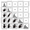 Figure 1
Correlation among chondroitin sulfate, indices of endothelial glycocalyx degradation (hyaluronic acid, heparan sulfate, and syndecan-1), and an index of systemic inflammation (IL-6) in plasma samples collected at CLOVERS enrollment. We display scatterplots of the observed joint distributions of the log-transformed biomarker concentrations at baseline in the lower triangular paired panels. The histogram and density curves of the log of biomarker concentrations are shown on the diagonals. Weighted Pearson correlation coefficients are shown in the corresponding upper triangular paired panels.
Plasma chondroitin sulfate, hyaluronic acid, and IL-6 at study enrollment are independently associated with sepsis mortality. At each time point measured, plasma concentrations of chondroitin sulfate (Figure 2A) were elevated in CLOVERS patients who died within 90 days of study randomization. Patients in the highest tertile of plasma chondroitin sulfate concentration (9,275–32,147 ng/mL) at the time of study enrollment (i.e., prior to randomization) had a significantly higher 90-day mortality compared with those in the medium (7,022–9,263 ng/mL) or low (1,272–7,017 ng/mL) plasma chondroitin sulfate tertiles (log-rank P < 0.001) (Figure 2B). The association of elevated plasma chondroitin sulfate at study enrollment with increased 90-day mortality persisted in our fitted Cox model that adjusted for severity of illness, patient demographics, randomized fluid resuscitation strategy, and chronic comorbidities (Figure 2C, entire model shown in Supplemental Figure 1). For example, the hazard ratio for mortality comparing the 10th percentile (5,303 ng/mL) to the 50th percentile (7,861 ng/mL) of baseline chondroitin sulfate was 1.06 (95% CI, 0.84–1.33), while the hazard ratio comparing the 90th percentile (12,202 ng/mL) to the 50th percentile was 1.98 (95% CI, 1.65–2.39).
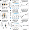 Figure 2
Plasma biomarkers as predictors of 90-day mortality in CLOVERS. (A) Plasma chondroitin sulfate, (D) hyaluronic acid, and (G) IL-6 (measured only at 72 hours) were elevated at all time points in patients who died within 90 days of CLOVERS enrollment. The number of observations at each time point and P values from the Wald test of inverse probability weighted linear regression are shown under the violin plots. (B) Kaplan-Meier plot of the unadjusted association between baseline plasma chondroitin sulfate, divided into tertiles, and survival. Tertile ranges are listed in the legend, with the table beneath the plot indicating the number of observed participants at risk at each time. Kaplan-Meier estimates and the log rank test were calculated using inverse probability of selection weights. (C) Covariate-adjusted association of baseline (log-transformed) chondroitin sulfate with mortality rates within 90 days of randomization. To address concerns about violations of the proportional hazards assumption, this Cox model and all other Cox models are stratified by sex assigned at birth and chronic heart failure. Additionally, we estimated associations between SOFA score and log-hazard of death separately for 0–4 days, 5–11 days, and 12–90 days of follow-up. Due to the stratified sampling study design, we used generalized raking when fitting the model. Identical analyses were conducted to estimate the association between baseline plasma hyaluronic acid (E and F), IL-6 (H and I), and mortality rates within 90 days of randomization. Full covariate-adjusted models are demonstrated in Supplemental Figures 1–3.
Similar to chondroitin sulfate, plasma concentrations of hyaluronic acid (a canonical endothelial glycocalyx degradation marker) and IL-6 (a canonical marker of systemic inflammation) were associated with 90-day survival. Patients who went on to die within 90 days of study enrollment had higher levels of plasma hyaluronic acid at all time points (Figure 2D) in comparison to 90-day survivors. Patients in the highest tertile of plasma hyaluronic acid concentration (370–17,812 ng/mL) at the time of study enrollment demonstrated higher 90-day mortality than those in the medium (90–368 ng/mL) and low (7–89 ng/mL) tertiles (log-rank P < 0.001) (Figure 2E). Our adjusted Cox model accordingly showed that baseline hyaluronic acid was independently associated with 90-day mortality (Figure 2F, entire model shown in Supplemental Figure 2). The hazard ratio comparing the 10th percentile (31 ng/mL) to the 50th percentile (142 ng/mL) of baseline hyaluronic acid was 0.28 (95% CI, 0.17–0.47), while the hazard ratio comparing the 90th percentile (1321 ng/mL) to the 50th percentile was 2.05 (95% CI, 1.58–2.66). Similarly, patients who went on to die within 90 days of study enrollment had higher levels of plasma IL-6 at all time points (Figure 2G) in comparison to 90-day survivors. Patients in the lowest tertile of baseline plasma IL-6 (0–29 pg/mL) at study enrollment had lower mortality than those in the highest tertile (170–34,519 pg/mL) or medium tertile (29–170 pg/mL) tertiles (log-rank P < 0.001; Figure 2H). This association of baseline IL-6 with 90-day mortality persisted in covariate-adjusted analysis (Figure 2I, entire model shown in Supplemental Figure 3). The hazard ratio comparing the 10th percentile (8 pg/mL) to the 50th percentile (63 pg/mL) of baseline IL-6 was 0.60 (95% CI, 0.44–0.81), while the hazard ratio comparing the 90th percentile (2,382 pg/mL) to the 50th percentile was 1.08 (95% CI, 0.83–1.41).
Postresuscitation plasma chondroitin sulfate, hyaluronic acid, and IL-6 are not altered by antecedent randomization to a liberal versus restrictive fluid resuscitation strategy. Previous observational studies suggested that a liberal intravenous fluid resuscitation strategy may induce iatrogenic endothelial glycocalyx degradation, leading to an increase in circulating glycocalyx fragments (16). However, such iatrogenic endothelial glycocalyx injury was not observed in randomized, prospective trials of fluid resuscitation (7). In contrast to glycocalyx-derived glycosaminoglycans, the effect of antecedent intravenous fluid resuscitation on circulating chondroitin sulfate concentrations was unstudied. We found that plasma chondroitin sulfate concentrations 24 hours after enrollment were unaffected by antecedent randomized resuscitation strategy (Figure 3A and Supplemental Figure 4), suggesting that choice of fluid resuscitation strategy did not induce changes in circulating chondroitin sulfate. Similarly, plasma hyaluronic acid (a canonical endothelial glycocalyx degradation biomarker) 24 hours after enrollment (i.e., at the completion of the study fluid protocol) was not affected by the antecedent randomization to either fluid resuscitation strategy (Figure 3B and Supplemental Figure 5), supporting previous studies’ findings that fluid resuscitation strategy does not drive glycocalyx injury.
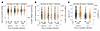 Figure 3
Plasma biomarker concentrations in CLOVERS patients randomized (after day 0 sample collection) to 24 hours of protocolized liberal or restrictive fluid resuscitation. On unadjusted analyses, treatment approach had no effect on subsequent (A) chondroitin sulfate, (B) hyaluronic acid, or (C) IL-6 (measured only at 72 hours) plasma concentrations. The number of observations at each time point and P values from the Wald test of inverse probability weighted linear regression are shown under the violin plots.
As IL-6 levels were not immediately measured after completion of the 24-hour period of protocolized fluid resuscitation, we used plasma IL-6 measured 72 hours after enrollment to determine if antecedent early fluid resuscitation strategy influenced persistent systemic inflammation. Using our adjusted linear regression model, we found that early assignment to a liberal or restrictive intravenous fluid resuscitation strategy had no association with plasma IL-6 72 hours after study enrollment, suggesting that the early (first 24 hours) resuscitation strategy had no effect on persistent indices of systemic inflammation (Figure 3C and Supplemental Figure 6).
Plasma chondroitin sulfate, but not hyaluronic acid or IL-6, modified the effectiveness of a subsequent, randomly assigned fluid resuscitation strategy on 90-day mortality. We sought to determine whether baseline plasma chondroitin sulfate concentration modified the relative effectiveness of a subsequent, randomly assigned liberal versus restrictive intravenous fluid resuscitation strategy on mortality within 90 days. Our covariate-adjusted Cox model demonstrated a differential treatment effect (Figure 4A; P = 0.022) according to chondroitin sulfate concentrations at study enrollment. As baseline plasma chondroitin sulfate concentrations increased, randomization to a restrictive (as compared with liberal) intravenous fluid resuscitation strategy was estimated to be increasingly beneficial to patients. For example, the treatment effect hazard ratio (restrictive versus liberal fluid resuscitation strategy) for mortality was estimated to be 1.49 (95% CI, 0.98–2.27), 1.30 (95% CI, 1.00–1.69), 1.09 (95% CI, 0.82–1.44), 0.88 (95% CI, 0.66–1.16), and 0.71 (95% CI, 0.52–0.97) for baseline chondroitin sulfate concentrations of 5,329 ng/mL, 6,418 ng/mL, 7,937 ng/mL, 10,112 ng/mL, and 12,779 ng/mL, respectively. These reference values of baseline chondroitin sulfate concentrations represent the 10th, 25th, 50th, 75th, and 90th percentiles of the observed distribution. Associations of chondroitin sulfate concentrations with the hazard ratios for mortality are shown separately for the liberal and restrictive resuscitation strategy arms in Figure 4B.
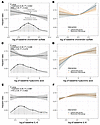 Figure 4
The effectiveness of the randomized treatment assignment is modified by (log of) baseline chondroitin sulfate but not hyaluronic acid or IL-6 plasma concentrations. Panels display results from models that added an interaction between randomized treatment assignment and baseline log-transformed plasma chondroitin sulfate (A and B), hyaluronic acid (C and D), or IL-6 (E and F) to the models shown in Figure 2, C, F, and I. Panels on the left demonstrate hazard ratios comparing restrictive versus liberal fluid resuscitation and pointwise 95% CIs across the distribution of log of baseline chondroitin sulfate (A), hyaluronic acid (C), or IL-6 (E) concentrations. The densities at the bottom of the panels refer to the distribution of baseline (log-transformed) plasma biomarker concentrations across the CLOVERS cohort, and for reference, we highlight the 10th, 25th, 50th, 75th, and 90th percentiles with dotted lines. We show the P values associated with (a) the overall treatment effect and (b) the heterogeneity of treatment effect. The heterogeneity of treatment effect test is a 2 degrees of freedom test Against the null hypothesis that the treatment effect is constant across the distribution of the biomarker values. The overall treatment effect test includes the main effect of the intervention (randomized fluid resuscitation strategy) and the 2 terms for the interaction with biomarker values. It is a test against the null hypothesis that treatment has no effect on mortality rates within 90 days of randomization against the alternative that it has any effect. Panels on the right demonstrate estimated hazard ratio and pointwise 95% CI comparing baseline log chondroitin sulfate (B), hyaluronic acid (D), or IL-6 (F) values to a reference value when the hazard ratio of the restrictive versus liberal fluid resuscitation strategies is equal to one. The associations are displayed separately for those randomized to the restrictive and liberal fluid resuscitation strategies.
In contrast to chondroitin sulfate, we observed no evidence of differential treatment effect for 90-day mortality as a function of baseline plasma hyaluronic acid (Figure 4, C and D) or IL-6 (Figure 4, E and F) and subsequent randomization to liberal or restrictive fluid resuscitation strategy.
Disaccharide analyses suggest the importance of unsulfated chondroitin sulfate in modifying response to fluid resuscitation strategy. To explore potential mechanisms underlying the predictive ability of circulating chondroitin sulfate on an individual patient’s response to a subsequent, randomly assigned fluid resuscitation strategy, we investigated the sulfation characteristics of plasma chondroitin sulfate in CLOVERS participants. Chondroitin sulfate is a linear polysaccharide, composed of repeating glucuronic acid-galactosamine disaccharides. Each disaccharide unit may be unsulfated (0S) or sulfated at either the 6-O or 4-O position of galactosamine. The relative abundance of these sulfated disaccharides within plasma chondroitin sulfate, as measured by mass spectrometry, can provide insight into the potential binding partners of circulating chondroitin sulfate during sepsis. For example, the circulating proteoglycan bikunin, a component of the endogenous antiprotease inter-α-trypsin inhibitor, is decorated with chondroitin sulfate that is enriched in 4-O sulfated disaccharides (12, 13). Interestingly, we found that the differential treatment effect of baseline plasma chondroitin sulfate on the response to a fluid resuscitation strategy was largely driven by unsulfated chondroitin sulfate (Figure 5, A and B) and not by 4-O (Figure 5, C and D) or 6-O (Figure 5, E and F) sulfated disaccharides. Therefore, the lack of predictive ability of 4-O chondroitin sulfate suggests that the observed differential response of fluid resuscitation strategy in CLOVERS might not be mediated by circulating bikunin. Patient-level factors associated with circulating concentrations of chondroitin sulfate and its subtypes are provided in Supplemental Figures 7 and 8. Intriguingly, 0S chondroitin sulfate concentrations at study entry were inversely correlated with an antecedent history of liver disease, potentially suggesting a hepatic source of this chondroitin sulfate subtype (Supplemental Figure 8A).
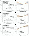 Figure 5
The effectiveness of the randomized treatment assignment is modified by (log of) baseline unsulfated chondroitin sulfate disaccharides but not 4-O or 6-O sulfated disaccharides. Panels on the left demonstrate hazard ratios comparing restrictive versus liberal fluid resuscitation and pointwise 95% CI across the distribution of log of baseline unsulfated (CS0S, A), 4-O sulfated (CS4S, C), or 6-O sulfated (CS6S, E) chondroitin sulfate concentrations, as per modeling detailed in Figure 4. The densities at the bottom of the panels refer to the distribution of baseline (log-transformed) plasma biomarker concentrations across the CLOVERS cohort, and for reference, we highlighted the 10th, 25th, 50th, 75th, and 90th percentiles with dotted lines. We show the P values associated with (a) the overall treatment effect and (b) the heterogeneity of treatment effect. Panels on the right demonstrate estimated hazard ratio and pointwise 95% CI comparing baseline log unsulfated (B), 4-O sulfated (D), or 6-O sulfated (F) chondroitin sulfate values to a reference value when the hazard ratio of the restrictive versus liberal fluid resuscitation strategies is equal to one.


Copyright © 2026 American Society for Clinical Investigation
ISSN: 0021-9738 (print), 1558-8238 (online)